The
liver is a
vital organ of
vertebrates and some other animals. In the
human it is located in the upper right
quadrant of the
abdomen, below the
diaphragm. The liver has a wide range of functions, including
detoxification of various
metabolites,
protein synthesis, and the production of
biochemicals necessary for
digestion. There is currently no way to compensate for the absence of liver function in the long term, although
liver dialysis techniques can be used in the short term.
The liver is a
gland and plays a major role in
metabolism with numerous functions in the human body, including regulation of
glycogen storage, decomposition of red blood cells,
plasma protein synthesis,
hormone production, and detoxification. It is an
accessory digestive gland and produces
bile, an
alkaline compound which aids in
digestion via the
emulsification of
lipids. The liver's highly specialized
tissue consisting of mostly
hepatocytes regulates a wide variety of high-volume biochemical reactions, including the synthesis and breakdown of small and complex molecules, many of which are necessary for normal vital functions.
[2] Estimates regarding the organ's total number of functions vary, but textbooks generally cite it being around 500.
[3]
Terminology related to the liver often starts in
hepar- or
hepat- from the
Greek word for liver,
hēpar (ἧπαρ, root hepat-, ἡπατ-).
[4][5]
§Structure
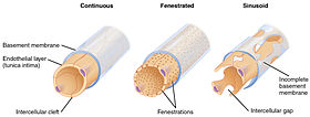
Capillaries, sinusoid on right
The liver is a reddish brown triangular organ with four
lobes of unequal size and shape. A human liver normally weighs 1.44–1.66 kg (3.2–3.7 lb).
[6] It is both the largest internal organ and the largest
gland in the human body. Located in the
right upper quadrant of the
abdominal cavity, it rests just below the
diaphragm, to the right of the stomach and overlying the
gallbladder. It is connected to two large
blood vessels, the
hepatic artery and the
portal vein. The hepatic artery carries oxygen-rich blood from the aorta, whereas the portal vein carries blood rich in digested nutrients from the entire
gastrointestinal tract and also from the
spleen and
pancreas. These blood vessels subdivide into small capillaries known as
liver sinusoids, which then lead to a
lobule. Lobules are the functional units of the liver and each lobule is made up of millions of hepatic cells (hepatocytes) which are the basic metabolic cells.
§Gross anatomy

The upper surface of the liver showing two lobes

Visceral surface showing four lobes
Gross anatomy traditionally divided the liver into two – a right and a left lobe, as viewed from the front (diaphragmatic) surface; but the underside (the
visceral surface) shows it to be divided into four lobes and includes the
caudate and
quadrate lobes.
The
falciform ligament is visible on the front of the liver and this divides the liver into a
left and a much larger
right lobe. From the visceral surface, the two additional lobes are located between the right and left lobes, one in front of the other. On the visceral surface a functional anatomy determines the organisation of the liver. A line can be
visualised running from the left of the vena cava and all the way forward to divide the liver and gallbladder into two halves. This line is called
Cantlie's line. Other anatomical landmarks exist, such as the
ligamentum venosum and the
round ligament of the liver (ligamentum teres) that further divide the left side of the liver in two sections. Now an important anatomical landmark, the transverse fissure of the liver divides this left portion of the liver into four segments which will be numbered starting at the caudate lobule as I in an anti-clock manner. From this visceral view we can see 7 segments because the 8th segment is only visible in the parietal view.
§Surfaces
On the
diaphragmatic surface, apart from a large triangular
bare area, where it connects to the
diaphragm, the liver is covered by a thin double-layered
membrane, the
peritoneum, that reduces
friction against other organs. This surface covers the convex shape of the two lobes where it accommodates the shape of the diaphragm. The
peritoneum folds back on itself to form the
falciform ligament and the
right and
left triangular ligaments.
These "
peritoneal ligaments" are not related to the true
anatomic ligaments in
joints, and the right and left triangular ligaments have no known functional importance, but they serve as recognizable surface landmarks. The falciform ligament however, does function to attach the liver to the posterior portion of the anterior body wall.
The
visceral surface or inferior surface, is uneven and concave. It is covered in peritoneum apart from where it attaches the gallbladder and the
porta hepatis.
§Impressions

Impressions on liver (posterior view)
There are several impressions on the surface of the liver which accommodate the various adjacent structures and organs. Underneath the right lobe and to the right of the gallbladder fossa, are two impressions, one behind the other and separated by a ridge. The one in front is a shallow
colic impression, formed by the
hepatic flexure and the one behind is a deeper
renal impression accommodating part of the right kidney and part of the
suprarenal gland.
The
suprarenal impression is a small triangular depressed area on the liver. It is located close to the right of the fossa between the bare area and the caudate lobe and immediately above the renal impression. The greater part of the suprarenal impression is devoid of peritoneum and it lodges the right suprarenal gland.
Medial to the renal impression is a third and slightly marked impression, lying between it and the neck of the gall-bladder. This is caused by the descending portion of the duodenum, and is known as the
duodenal impression.
The inferior surface of the left lobe of the liver presents behind and to the left the
gastric impression, moulded over the antero-superior surface of the stomach, and to the right of this a rounded eminence, the
tuber omentale, which fits into the concavity of the lesser curvature of the stomach and lies in front of the anterior layer of the
lesser omentum.
§Microscopic anatomy

Liver structure showing cells
Histology, the study of microscopic anatomy shows two major types of cells of the liver: parenchymal and non-parenchymal cells. 80% of the liver volume is occupied by
parenchymal cells commonly referred to as
hepatocytes. Non-parenchymal cells constitute 40% of the total number of liver cells but only 6.5% of its volume. The
liver sinusoids are lined with two types of cell,
sinusoidal endothelial cells, and phagocytic
Kupffer cells.
[7] Hepatic stellate cells are some of the non-parenchymal cells are external to the sinusoid in the
space of Disse.
[8]
Each of the lobes is seen to be made up of
hepatic lobules; a vein goes from the centre, which then joins to the hepatic vein to carry blood out from the liver. On the surface of the lobules, there are ducts, veins and arteries that carry fluids to and from them. A distinctive component of a lobule is the
portal triad.
§Functional anatomy
Correspondence between anatomic lobes and Couinaud segments
| Segment* |
Couinaud segments |
| Caudate |
1 |
| Lateral |
2, 3 |
| Medial |
4a, 4b |
| Right |
5, 6, 7, 8 |
* or lobe, in the case of the caudate lobe
Each number in the list corresponds to one in the table
1. Caudate
2. Superior subsegment of the lateral segment
3. Inferior subsegment of the lateral segment
4a. Superior subsegment of the medial segment
4b. Inferior subsegment of the medial segment
5. Inferior subsegment of the anterior segment
6. Inferior subsegment of the posterior segment
7. Superior subsegment of the posterior segment
8. Superior subsegment of the anterior segment
|
The central area where the
common bile duct,
hepatic portal vein, and the
hepatic artery proper enter is the
hilum known as the
porta hepatis (gateway to the liver) or the transverse fissure of the liver. The duct, vein, and artery divide into left and right branches, and the areas of the liver supplied by these branches constitute the functional left and right lobes.
The functional lobes are separated by the imaginary plane, Cantlie's line joining the gallbladder fossa to the inferior vena cava. The plane separates the liver into the true right and left lobes. The middle hepatic vein also demarcates the true right and left lobes. The right lobe is further divided into an
anterior and
posterior segment by the right hepatic vein. The left lobe is divided into the
medial and
lateral segments by the left hepatic vein. The fissure for the
round ligament of the liver (ligamentum teres) also separates the medial and lateral segments. The medial segment is also called the
quadrate lobe. In the widely used
Couinaud (or "French") system, the functional lobes are further divided into a total of eight subsegments based on a transverse plane through the bifurcation of the main portal vein. The
caudate lobe is a separate structure which receives blood flow from both the right- and left-sided vascular branches.
[9][10]
§Development
Organogenesis, the development of the organs takes place from the third to the eighth week in
human embryogenesis. The origins of the liver lie in both the ventral portion of the
foregut endoderm (endoderm being one of the 3 embryonic germ cell layers) and the constituents of the adjacent
septum transversum mesenchyme. In human
embryo, the
hepatic diverticulum is the tube of endoderm that extends out from the foregut into the surrounding mesenchyme. The mesenchyme of septum transversum induces this endoderm to proliferate, to branch, and to form the glandular epithelium of the liver. A portion of the hepatic diverticulum (that region closest to the digestive tube) continues to function as the
drainage duct of the liver, and a branch from this duct produces the
gallbladder.
[11]
Besides signals from the septum transversum mesenchyme,
fibroblast growth factor from the
developing heart also contributes to hepatic competence, along with
retinoic acid emanating from the
lateral plate mesoderm. The hepatic endodermal cells undergo a morphological transition from columnar to pseudostratified resulting in thickening into the early
liver bud. Their expansion forms a population of the bipotential hepatoblasts.
[12] Hepatic stellate cells are derived from mesenchyme.
[13]
After migration of hepatoblasts into the septum transversum mesenchyme, the hepatic architecture begins to be established, with
liver sinusoids and bile canaliculi appearing. The liver bud separates into the lobes. The left
umbilical vein becomes the
ductus venosus and the right
vitelline vein becomes the
portal vein. The expanding liver bud is colonized by
hematopoietic cells. The bipotential hepatoblasts begin differentiating into
biliary epithelial cells and
hepatocytes. The biliary epithelial cells differentiate from hepatoblasts around portal veins, first producing a monolayer, and then a bilayer of cuboidal cells. In ductal plate, focal dilations emerge at points in the bilayer, become surrounded by portal mesenchyme, and undergo tubulogenesis into intrahepatic bile ducts. Hepatoblasts not adjacent to portal veins instead differentiate into hepatocytes and arrange into cords lined by sinudoidal epithelial cells and bile canaliculi. Once hepatoblasts are specified into hepatocytes and undergo further expansion, they begin acquiring the functions of a mature hepatocyte, and eventually mature hepatocytes appear as highly polarized epithelial cells with abundant
glycogen accumulation. In the adult liver, hepatocytes are not equivalent, with position along the portocentrovenular axis within a
liver lobule dictating expression of metabolic genes involved in drug metabolism, carbohydrate metabolism, ammonia detoxification, and bile production and secretion.
WNT/β-catenin has now been identified to be playing a key role in this phenomenon.
[12]
§Fetal blood supply
In the growing fetus, a major source of blood to the liver is the
umbilical vein which supplies nutrients to the growing fetus. The umbilical vein enters the abdomen at the umbilicus, and passes upward along the free margin of the
falciform ligament of the liver to the inferior surface of the liver. There it joins with the left branch of the portal vein. The
ductus venosus carries blood from the left portal vein to the left hepatic vein and then to the
inferior vena cava, allowing placental blood to bypass the liver.
In the fetus, the liver develops throughout normal gestation, and does not perform the normal filtration of the infant liver. The liver does not perform digestive processes because the fetus does not consume meals directly, but receives nourishment from the mother via the
placenta. The fetal liver releases some blood stem cells that migrate to the fetal
thymus, so initially the
lymphocytes, called
T-cells, are created from fetal liver stem cells. Once the fetus is delivered, the formation of blood stem cells in infants shifts to the red
bone marrow.
After birth, the umbilical vein and ductus venosus are completely obliterated in two to five days; the former becomes the
ligamentum teres and the latter becomes the
ligamentum venosum. In the disease state of
cirrhosis and
portal hypertension, the umbilical vein can open up again.
§During childhood
At birth the liver comprises roughly 4% of body weight and is at average 120g. Over the course of development it will increase to 1,4–1,6 kg but will only take up 2.5–3.5% of body weight.
[14]
§Physiology
The various functions of the liver are carried out by the liver cells or
hepatocytes. Currently, there is no
artificial organ or device capable of emulating all the functions of the liver. Some functions can be emulated by
liver dialysis, an experimental treatment for
liver failure. The liver is thought to be responsible for up to 500 separate functions, usually in combination with other systems and organs.
§Blood supply
The liver gets a dual blood supply from the
hepatic portal vein and
hepatic arteries. Supplying approximately 75% of the liver's blood supply, the hepatic portal vein carries
venous blood drained from the
spleen,
gastrointestinal tract, and its associated organs. The hepatic arteries supply
arterial blood to the liver, accounting for the remainder of its
blood flow. Oxygen is provided from both sources; approximately half of the liver's oxygen demand is met by the hepatic portal vein, and half is met by the hepatic arteries.
[15]
Blood flows through the
liver sinusoids and empties into the central vein of each lobule. The
central veins coalesce into hepatic veins, which leave the liver.
-
Axial CT image showing anomalous hepatic veins coursing on the subcapsular anterior surface of the liver.
[16]
-
Maximum intensity projection (MIP) CT image as viewed anteriorly showing the anomalous hepatic veins coursing on the anterior surface of the liver
-
Lateral MIP view in the same patient
-
A CT scan in which the liver and portal vein are shown.
§Biliary flow
The term
biliary tree is derived from the branches of the bile ducts. The
bile produced in the liver is collected in
bile canaliculi, which merge to form
bile ducts. Within the liver, these ducts are called
intrahepatic (within the liver) bile ducts, and once they exit the liver they are considered
extrahepatic (outside the liver). The intrahepatic ducts eventually drain into the right and left
hepatic ducts, which merge to form the
common hepatic duct. The
cystic duct from the
gallbladder joins with the
common hepatic duct to form the
common bile duct.
Bile either drains directly into the
duodenum via the common bile duct, or is temporarily stored in the
gallbladder via the cystic duct. The common bile duct and the
pancreatic duct enter the second part of the duodenum together at the
ampulla of Vater.
§Synthesis
- A large part of amino acid synthesis
- The liver performs several roles in carbohydrate metabolism:
- The liver is responsible for the mainstay of protein metabolism, synthesis as well as degradation.
- The liver also performs several roles in lipid metabolism:
- The liver produces coagulation factors I (fibrinogen), II (prothrombin), V, VII, VIII, IX, X and XI, as well as protein C, protein S and antithrombin.
- In the first trimester fetus, the liver is the main site of red blood cell production. By the 32nd week of gestation, the bone marrow has almost completely taken over that task.
- The liver produces and excretes bile (a yellowish liquid) required for emulsifying fats and help the absorption of vitamin K from the diet. Some of the bile drains directly into the duodenum, and some is stored in the gallbladder.
- The liver also produces insulin-like growth factor 1 (IGF-1), a polypeptide protein hormone that plays an important role in childhood growth and continues to have anabolic effects in adults.
- The liver is a major site of thrombopoietin production. Thrombopoietin is a glycoprotein hormone that regulates the production of platelets by the bone marrow.
§Breakdown
§Other functions
- The liver stores a multitude of substances, including glucose (in the form of glycogen), vitamin A (1–2 years' supply), vitamin D (1–4 months' supply)[citation needed], vitamin B12 (1–3 years' supply), vitamin K, iron, and copper.
- The liver is responsible for immunological effects—the reticuloendothelial system of the liver contains many immunologically active cells, acting as a 'sieve' for antigens carried to it via the portal system.
- The liver produces albumin, the major osmolar component of blood serum.
- The liver synthesizes angiotensinogen, a hormone that is responsible for raising the blood pressure when activated by renin, an enzyme that is released when the kidney senses low blood pressure.
§Relation to medicine and pharmacology
The oxidative capacity of the liver decreases with aging and therefore any medications that require oxidation (for instance,
benzodiazepines) are more likely to accumulate to toxic levels. However, medications with shorter
half-lives, such as
lorazepam and
oxazepam, are preferred in most cases when benzodiazepines are required in regards to geriatric medicine.
§Clinical significance
§Disease
The liver supports almost every organ in the body and is vital for survival. Because of its strategic location and multidimensional functions, the liver is also prone to many diseases.
[17]
Hepatitis is a common condition of inflammation of the liver. The most usual cause of this is
viral, and the most common of these infections are
hepatitis A B C D and
E. Some of these infections are
sexually transmitted. Inflammation can also be caused by other viruses in the
Herpesviridae family such as the
herpes simplex virus. Infection with hepatitis B virus or hepatitis C virus is the main cause of
liver cancer.
Other disorders caused by excessive alcohol consumption are grouped under
alcoholic liver diseases and these include
alcoholic hepatitis,
fatty liver, and
cirrhosis. Liver damage can also be caused by drugs in particular
paracetomol and drugs used to treat cancer.
Many diseases of the liver are accompanied by
jaundice caused by increased levels of
bilirubin in the system. The bilirubin results from the breakup of the
hemoglobin of dead
red blood cells; normally, the liver removes bilirubin from the blood and excretes it through bile.
There are also many pediatric liver diseases including
biliary atresia,
alpha-1 antitrypsin deficiency,
alagille syndrome,
progressive familial intrahepatic cholestasis, and
Langerhans cell histiocytosis, to name but a few.
Diseases that interfere with liver function will lead to derangement of these processes. However, the liver has a great capacity to
regenerate and has a large reserve capacity. In most cases, the liver only produces symptoms after extensive damage.
Liver diseases may be diagnosed by
liver function tests, for example, by production of
acute phase proteins.
§Symptoms
The classic symptoms of liver damage include the following:
- Pale stools occur when stercobilin, a brown pigment, is absent from the stool. Stercobilin is derived from bilirubin metabolites produced in the liver.
- Dark urine occurs when bilirubin mixes with urine
- Jaundice (yellow skin and/or whites of the eyes) This is where bilirubin deposits in skin, causing an intense itch. Itching is the most common complaint by people who have liver failure. Often this itch cannot be relieved by drugs.
- Swelling of the abdomen, ankles and feet occurs because the liver fails to make albumin.
- Excessive fatigue occurs from a generalized loss of nutrients, minerals and vitamins.
- Bruising and easy bleeding are other features of liver disease. The liver makes substances which help prevent bleeding. When liver damage occurs, these substances are no longer present and severe bleeding can occur.[18]
§Diagnosis
The diagnosis of liver function is made by
liver function tests, groups of
blood tests, that can readily show the extent of liver damage. If
infection is suspected, then other
serological tests will be carried out. Sometimes, an
ultrasound or a
CT scan is needed to produce an image of the liver.
Physical examination of the liver can only reveal its size and any tenderness, and some form of
imaging will also be needed.
[19]
§Biopsy / scan
Damage to the liver is sometimes determined with a
biopsy, particularly when the cause of liver damage is unknown. In the 21st century they were largely replaced by high-resolution radiographic scans. The latter do not require ultrasound guidance, lab involvement, microscopic analysis, organ damage, pain, or patient sedation; and the results are available immediately on a computer screen.
In a biopsy, a needle is inserted into the skin just below the rib cage and a tissue sample obtained. The tissue is sent to the laboratory, where it is analyzed under a
microscope. Sometimes, a radiologist may assist the physician performing a
liver biopsy by providing ultrasound guidance.
[20]
§Liver regeneration
The liver is the only human internal organ capable of natural
regeneration of lost
tissue; as little as 25% of a liver can regenerate into a whole liver.
[21] This is, however, not true regeneration but rather
compensatory growth in mammals.
[22] The lobes that are removed do not regrow and the growth of the liver is a restoration of function, not original form. This contrasts with true regeneration where both original function and form are restored. In lower species such as fish, the liver undergoes true regeneration by restoring both shape and size of the organ.
[23] In liver, large areas of the tissues are formed but for the formation of new cells there must be sufficient amount of material so the circulation of the blood becomes more active.
[24]
This is predominantly due to the
hepatocytes re-entering the
cell cycle. That is, the hepatocytes go from the quiescent
G0 phase to the
G1 phase and undergo mitosis. This process is activated by the
p75 receptors.
[25] There is also some evidence of
bipotential stem cells, called hepatic oval cells or ovalocytes (not to be confused with oval red blood cells of
ovalocytosis), which are thought to reside in the
canals of Hering. These cells can differentiate into either
hepatocytes or
cholangiocytes, the latter being the cells that line the
bile ducts.
[citation needed]
Scientific and medical works about liver regeneration often refer to the Greek
Titan Prometheus who was chained to a rock in the Caucasus where, each day, his liver was devoured by an eagle, only to grow back each night. The myth suggests the
ancient Greeks knew about the liver’s remarkable capacity for self-repair, however, this claim is purely speculative.
[26]
§Liver transplantation
Human liver transplants were first performed by
Thomas Starzl in the
United States and
Roy Calne in
Cambridge,
England in 1963 and 1965, respectively.
After resection of left lobe liver tumor
Liver transplantation is the only option for those with irreversible liver failure. Most transplants are done for chronic liver diseases leading to
cirrhosis, such as chronic
hepatitis C,
alcoholism, autoimmune hepatitis, and many others. Less commonly, liver transplantation is done for
fulminant hepatic failure, in which liver failure occurs over days to weeks.
Liver
allografts for
transplant usually come from donors who have died from fatal
brain injury.
Living donor liver transplantation is a technique in which a portion of a living person's liver is removed and used to replace the entire liver of the recipient. This was first performed in 1989 for pediatric liver transplantation. Only 20 percent of an adult's liver (Couinaud segments 2 and 3) is needed to serve as a liver allograft for an infant or small child.
More recently, adult-to-adult liver transplantation has been done using the donor's right hepatic lobe, which amounts to 60 percent of the liver. Due to the ability of the liver to
regenerate, both the donor and recipient end up with normal liver function if all goes well. This procedure is more controversial, as it entails performing a much larger operation on the donor, and indeed there have been at least two donor deaths out of the first several hundred cases. A recent publication has addressed the problem of donor mortality, and at least 14 cases have been found.
[27] The risk of postoperative complications (and death) is far greater in right-sided operations than that in left-sided operations.
With the recent advances of noninvasive imaging, living liver donors usually have to undergo imaging examinations for liver anatomy to decide if the anatomy is feasible for donation. The evaluation is usually performed by multidetector row
computed tomography (MDCT) and
magnetic resonance imaging (MRI). MDCT is good in vascular anatomy and volumetry. MRI is used for biliary tree anatomy. Donors with very unusual vascular anatomy, which makes them unsuitable for donation, could be screened out to avoid unnecessary operations.
-
MDCT image. Arterial anatomy contraindicated for liver donation
-
MDCT image. Portal venous anatomy contraindicated for liver donation
-
MDCT image. 3D image created by MDCT can clearly visualize the liver, measure the liver volume, and plan the dissection plane to facilitate the liver transplantation procedure.
-
Phase contrast CT image. Contrast is perfusing the right liver but not the left due to a left portal vein thrombus.
§Society and culture
In
Greek mythology,
Prometheus was punished by the gods for revealing fire to humans, by being chained to a rock where a
vulture (or an
eagle) would peck out his liver, which would regenerate overnight. (The liver is the only human internal organ that actually can regenerate itself to a significant extent.) Many ancient peoples of the Near East and Mediterranean areas practiced a type of
divination called
haruspicy, where they tried to obtain information by examining the livers of sheep and other animals.
In Plato, and in later physiology, the liver was thought to be the seat of the darkest emotions (specifically wrath, jealousy and greed) which drive men to action.
[28] The
Talmud (tractate
Berakhot 61b) refers to the liver as the seat of
anger, with the
gallbladder counteracting this.
The
Persian,
Urdu, and
Hindi languages (جگر or जिगर or
jigar) refer to the liver in figurative speech to indicate courage and strong feelings, or "their best"; e.g., "This
Mecca has thrown to you the pieces of its liver!".
[29] The term
jan e jigar, literally "the strength (power) of my liver", is a term of endearment in Urdu. In Persian slang,
jigar is used as an adjective for any object which is desirable, especially women. In the
Zulu language, the word for liver (isibindi) is the same as the word for courage.
The legend of
Liver-Eating Johnson says that he would cut out and eat the liver of each man killed after dinner.
In the motion picture
The Message,
Hind bint Utbah is implied or portrayed eating the liver of
Hamza ibn ‘Abd al-Muttalib during the
Battle of Uhud. Although there are narrations that suggest that Hind did "taste", rather than eat, the liver of Hamza, the authenticity of these narrations have to be questioned.
§Food
The liver of mammals,
fowl, and fish are commonly eaten as
food by humans.
Domestic pig,
ox,
lamb,
calf,
chicken, and
goose livers are widely available from butchers and supermarkets.
Liver can be baked, boiled, broiled, fried,
stir-fried, or eaten raw (
asbeh nayeh or
sawda naye in
Lebanese cuisine, liver
sashimi). In many preparations, pieces of liver are combined with pieces of meat or kidneys, like in the various forms of Middle Eastern
mixed grill (e.g.
meurav Yerushalmi). Liver is often made into
spreads. Well-known examples include
liver pâté,
foie gras,
chopped liver, and
leverpastej. Liver
sausages such as
Braunschweiger and
liverwurst are also a valued meal.
Liver sausages may also be used as spreads. A traditional
South African delicacy, namely
Skilpadjies, is made of minced lamb's liver wrapped in
netvet (caul fat), and grilled over an open fire.
Animal livers are rich in iron and
vitamin A, and
cod liver oil is commonly used as a
dietary supplement. Traditionally, some fish livers were valued as food, especially the
stingray liver. It was used to prepare delicacies, such as poached skate liver on toast in England, as well as the
beignets de foie de raie and
foie de raie en croute in
French cuisine.
[30]
§Other animals
The liver is found in all
vertebrates, and is typically the largest
visceral (internal) organ. Its form varies considerably in different species, and is largely determined by the shape and arrangement of the surrounding organs. Nonetheless, in most species it is divided into right and left lobes; exceptions to this general rule include
snakes, where the shape of the body necessitates a simple cigar-like form. The internal structure of the liver is broadly similar in all vertebrates.
[31]
An organ sometimes referred to as a liver is found associated with the digestive tract of the primitive chordate
Amphioxus. However, this is an enzyme secreting gland, not a metabolic organ, and it is unclear how truly
homologous it is to the vertebrate liver.
[31]
§Additional images
-
Anterior view of the liver.
-
Inferior view of the liver.
-
Schematic posterior view of the liver.
-
View of the various organs and blood-vessels in proximity with liver.
-
Liver lifted to show gall bladder and stomach in situ.
-
Cross section showing the liver as the large brown mass in the left of the images, right of the individual.
-
Cross section of an inferior portion of the liver, showing gallbladder and various structures.
-
Human liver.Visceral surface of liver.
-
Human liver.Horizontal section to newborn


