Search This Blog

Wednesday, April 16, 2025

Cellular respiration

From Wikipedia, the free encyclopedia
https://en.wikipedia.org/wiki/Cellular_respiration
 
Typical eukaryotic cell

Cellular respiration is the process of oxidizing biological fuels using an inorganic electron acceptor, such as oxygen, to drive production of adenosine triphosphate (ATP), which stores chemical energy in a biologically accessible form. Cellular respiration may be described as a set of metabolic reactions and processes that take place in the cells of organisms to transfer chemical energy from nutrients to ATP, with the flow of electrons to an electron acceptor, and then release waste products.

If the electron acceptor is oxygen, the process is more specifically known as aerobic cellular respiration. If the electron acceptor is a molecule other than oxygen, this is anaerobic cellular respiration. Fermentation, which is also an anaerobic process, is not respiration, as no external electron acceptor is involved.

The reactions involved in respiration are catabolic reactions, which break large molecules into smaller ones, producing large amounts of energy (ATP). Respiration is one of the key ways a cell releases chemical energy to fuel cellular activity. The overall reaction occurs in a series of biochemical steps, some of which are redox reactions. Although cellular respiration is technically a combustion reaction, it is an unusual one because of the slow, controlled release of energy from the series of reactions.

Nutrients that are commonly used by animal and plant cells in respiration include sugar, amino acids and fatty acids, and the most common oxidizing agent is molecular oxygen (O2). The chemical energy stored in ATP (the bond of its third phosphate group to the rest of the molecule can be broken allowing more stable products to form, thereby releasing energy for use by the cell) can then be used to drive processes requiring energy, including biosynthesis, locomotion or transportation of molecules across cell membranes.

Aerobic respiration

Aerobic respiration requires oxygen (O2) in order to create ATP. Although carbohydrates, fats and proteins are consumed as reactants, aerobic respiration is the preferred method of pyruvate production in glycolysis, and requires pyruvate be transported by the mitochondria in order to be oxidized by the citric acid cycle. The products of this process are carbon dioxide and water, and the energy transferred is used to make bonds between ADP and a third phosphate group to form ATP (adenosine triphosphate), by substrate-level phosphorylation, NADH and FADH2.

Mass balance of the global reaction: C6H12O6 (s) + 6 O2 (g) → 6 CO2 (g) + 6 H2O (l) + energy

ΔG = −2880 kJ per mol of C6H12O6

The negative ΔG indicates that the reaction is exothermic (exergonic) and can occur spontaneously.

The potential of NADH and FADH2 is converted to more ATP through an electron transport chain with oxygen and protons (hydrogen ions) as the "terminal electron acceptors". Most of the ATP produced by aerobic cellular respiration is made by oxidative phosphorylation. The energy released is used to create a chemiosmotic potential by pumping protons across a membrane. This potential is then used to drive ATP synthase and produce ATP from ADP and a phosphate group. Biology textbooks often state that 38 ATP molecules can be made per oxidized glucose molecule during cellular respiration (2 from glycolysis, 2 from the Krebs cycle, and about 34 from the electron transport system). However, this maximum yield is never quite reached because of losses due to leaky membranes as well as the cost of moving pyruvate and ADP into the mitochondrial matrix, and current estimates range around 29 to 30 ATP per glucose.

Aerobic metabolism is up to 15 times more efficient than anaerobic metabolism (which yields 2 molecules of ATP per 1 molecule of glucose). However, some anaerobic organisms, such as methanogens are able to continue with anaerobic respiration, yielding more ATP by using inorganic molecules other than oxygen as final electron acceptors in the electron transport chain. They share the initial pathway of glycolysis but aerobic metabolism continues with the Krebs cycle and oxidative phosphorylation. The post-glycolytic reactions take place in the mitochondria in eukaryotic cells, and in the cytoplasm in prokaryotic cells.

Although plants are net consumers of carbon dioxide and producers of oxygen via photosynthesis, plant respiration accounts for about half of the CO2 generated annually by terrestrial ecosystems.

Glycolysis

Out of the cytoplasm it goes into the Krebs cycle with the acetyl CoA. It then mixes with CO2 and makes 2 ATP, NADH, and FADH. From there the NADH and FADH go into the NADH reductase, which produces the enzyme. The NADH pulls the enzyme's electrons to send through the electron transport chain. The electron transport chain pulls H+ ions through the chain. From the electron transport chain, the released hydrogen ions make ADP for an result of 32 ATP. Lastly, ATP leaves through the ATP channel and out of the mitochondria.

Glycolysis is a metabolic pathway that takes place in the cytosol of cells in all living organisms. Glycolysis can be literally translated as "sugar splitting", and occurs regardless of oxygen's presence or absence. The process converts one molecule of glucose into two molecules of pyruvate (pyruvic acid), generating energy in the form of two net molecules of ATP. Four molecules of ATP per glucose are actually produced, but two are consumed as part of the preparatory phase. The initial phosphorylation of glucose is required to increase the reactivity (decrease its stability) in order for the molecule to be cleaved into two pyruvate molecules by the enzyme aldolase. During the pay-off phase of glycolysis, four phosphate groups are transferred to four ADP by substrate-level phosphorylation to make four ATP, and two NADH are also produced during the pay-off phase. The overall reaction can be expressed this way:

Glucose + 2 NAD+ + 2 Pi + 2 ADP → 2 pyruvate + 2 NADH + 2 ATP + 2 H+ + 2 H2O + energy

Starting with glucose, 1 ATP is used to donate a phosphate to glucose to produce glucose 6-phosphate. Glycogen can be converted into glucose 6-phosphate as well with the help of glycogen phosphorylase. During energy metabolism, glucose 6-phosphate becomes fructose 6-phosphate. An additional ATP is used to phosphorylate fructose 6-phosphate into fructose 1,6-bisphosphate by the help of phosphofructokinase. Fructose 1,6-biphosphate then splits into two phosphorylated molecules with three carbon chains which later degrades into pyruvate.

Oxidative decarboxylation of pyruvate

Pyruvate is oxidized to acetyl-CoA and CO2 by the pyruvate dehydrogenase complex (PDC). The PDC contains multiple copies of three enzymes and is located in the mitochondria of eukaryotic cells and in the cytosol of prokaryotes. In the conversion of pyruvate to acetyl-CoA, one molecule of NADH and one molecule of CO2 is formed.

Citric acid cycle

The citric acid cycle is also called the Krebs cycle or the tricarboxylic acid cycle. When oxygen is present, acetyl-CoA is produced from the pyruvate molecules created from glycolysis. Once acetyl-CoA is formed, aerobic or anaerobic respiration can occur. When oxygen is present, the mitochondria will undergo aerobic respiration which leads to the Krebs cycle. However, if oxygen is not present, fermentation of the pyruvate molecule will occur. In the presence of oxygen, when acetyl-CoA is produced, the molecule then enters the citric acid cycle (Krebs cycle) inside the mitochondrial matrix, and is oxidized to CO2 while at the same time reducing NAD to NADH. NADH can be used by the electron transport chain to create further ATP as part of oxidative phosphorylation. To fully oxidize the equivalent of one glucose molecule, two acetyl-CoA must be metabolized by the Krebs cycle. Two low-energy waste products, H2O and CO2, are created during this cycle.

The citric acid cycle is an 8-step process involving 18 different enzymes and co-enzymes. During the cycle, acetyl-CoA (2 carbons) + oxaloacetate (4 carbons) yields citrate (6 carbons), which is rearranged to a more reactive form called isocitrate (6 carbons). Isocitrate is modified to become α-ketoglutarate (5 carbons), succinyl-CoA, succinate, fumarate, malate and, finally, oxaloacetate.

The net gain from one cycle is 3 NADH and 1 FADH2 as hydrogen (proton plus electron) carrying compounds and 1 high-energy GTP, which may subsequently be used to produce ATP. Thus, the total yield from 1 glucose molecule (2 pyruvate molecules) is 6 NADH, 2 FADH2, and 2 ATP.

Oxidative phosphorylation

Diagram of oxidative phosphorylation

In eukaryotes, oxidative phosphorylation occurs in the mitochondrial cristae. It comprises the electron transport chain that establishes a proton gradient (chemiosmotic potential) across the boundary of the inner membrane by oxidizing the NADH produced from the Krebs cycle. ATP is synthesized by the ATP synthase enzyme when the chemiosmotic gradient is used to drive the phosphorylation of ADP. The electrons are finally transferred to exogenous oxygen and, with the addition of two protons, water is formed.

Efficiency of ATP production

The table below describes the reactions involved when one glucose molecule is fully oxidized into carbon dioxide. It is assumed that all the reduced coenzymes are oxidized by the electron transport chain and used for oxidative phosphorylation.

Step coenzyme yield ATP yield Source of ATP
Glycolysis preparatory phase
−2 Phosphorylation of glucose and fructose 6-phosphate uses two ATP from the cytoplasm.
Glycolysis pay-off phase
4 Substrate-level phosphorylation
2 NADH 3 or 5 Oxidative phosphorylation: Each NADH produces net 1.5 ATP (instead of usual 2.5) due to NADH transport over the mitochondrial membrane
Oxidative decarboxylation of pyruvate 2 NADH 5 Oxidative phosphorylation
Krebs cycle
2 Substrate-level phosphorylation
6 NADH 15 Oxidative phosphorylation
2 FADH2 3 Oxidative phosphorylation
Total yield 30 or 32 ATP From the complete oxidation of one glucose molecule to carbon dioxide and oxidation of all the reduced coenzymes.

Although there is a theoretical yield of 38 ATP molecules per glucose during cellular respiration, such conditions are generally not realized because of losses such as the cost of moving pyruvate (from glycolysis), phosphate, and ADP (substrates for ATP synthesis) into the mitochondria. All are actively transported using carriers that utilize the stored energy in the proton electrochemical gradient.

  • Pyruvate is taken up by a specific, low Km transporter to bring it into the mitochondrial matrix for oxidation by the pyruvate dehydrogenase complex.
  • The phosphate carrier (PiC) mediates the electroneutral exchange (antiport) of phosphate (H2PO4; Pi) for OH or symport of phosphate and protons (H+) across the inner membrane, and the driving force for moving phosphate ions into the mitochondria is the proton motive force.
  • The ATP-ADP translocase (also called adenine nucleotide translocase, ANT) is an antiporter and exchanges ADP and ATP across the inner membrane. The driving force is due to the ATP (−4) having a more negative charge than the ADP (−3), and thus it dissipates some of the electrical component of the proton electrochemical gradient.

The outcome of these transport processes using the proton electrochemical gradient is that more than 3 H+ are needed to make 1 ATP. Obviously, this reduces the theoretical efficiency of the whole process and the likely maximum is closer to 28–30 ATP molecules. In practice the efficiency may be even lower because the inner membrane of the mitochondria is slightly leaky to protons. Other factors may also dissipate the proton gradient creating an apparently leaky mitochondria. An uncoupling protein known as thermogenin is expressed in some cell types and is a channel that can transport protons. When this protein is active in the inner membrane it short circuits the coupling between the electron transport chain and ATP synthesis. The potential energy from the proton gradient is not used to make ATP but generates heat. This is particularly important in brown fat thermogenesis of newborn and hibernating mammals.

Stoichiometry of aerobic respiration and most known fermentation types in eucaryotic cell.  Numbers in circles indicate counts of carbon atoms in molecules, C6 is glucose C6H12O6, C1 carbon dioxide CO2. Mitochondrial outer membrane is omitted.

According to some newer sources, the ATP yield during aerobic respiration is not 36–38, but only about 30–32 ATP molecules / 1 molecule of glucose, because:

So the final stoichiometry is
1 NADH+H+ : 10 H+ : 10/4 ATP = 1 NADH+H+ : 2.5 ATP
1 FADH2 : 6 H+ : 6/4 ATP = 1 FADH2 : 1.5 ATP
  • ATP : NADH+H+ coming from glycolysis ratio during the oxidative phosphorylation is
    • 1.5, as for FADH2, if hydrogen atoms (2H++2e) are transferred from cytosolic NADH+H+ to mitochondrial FAD by the glycerol phosphate shuttle located in the inner mitochondrial membrane.
    • 2.5 in case of malate-aspartate shuttle transferring hydrogen atoms from cytosolic NADH+H+ to mitochondrial NAD+

So finally we have, per molecule of glucose

Altogether this gives 4 + 3 (or 5) + 20 + 3 = 30 (or 32) ATP per molecule of glucose

These figures may still require further tweaking as new structural details become available. The above value of 3 H+ / ATP for the synthase assumes that the synthase translocates 9 protons, and produces 3 ATP, per rotation. The number of protons depends on the number of c subunits in the Fo c-ring, and it is now known that this is 10 in yeast Fo and 8 for vertebrates. Including one H+ for the transport reactions, this means that synthesis of one ATP requires 1 + 10/3 = 4.33 protons in yeast and 1 + 8/3 = 3.67 in vertebrates. This would imply that in human mitochondria the 10 protons from oxidizing NADH would produce 2.72 ATP (instead of 2.5) and the 6 protons from oxidizing succinate or ubiquinol would produce 1.64 ATP (instead of 1.5). This is consistent with experimental results within the margin of error described in a recent review.

The total ATP yield in ethanol or lactic acid fermentation is only 2 molecules coming from glycolysis, because pyruvate is not transferred to the mitochondrion and finally oxidized to the carbon dioxide (CO2), but reduced to ethanol or lactic acid in the cytoplasm.

Fermentation

Without oxygen, pyruvate (pyruvic acid) is not metabolized by cellular respiration but undergoes a process of fermentation. The pyruvate is not transported into the mitochondrion but remains in the cytoplasm, where it is converted to waste products that may be removed from the cell. This serves the purpose of oxidizing the electron carriers so that they can perform glycolysis again and removing the excess pyruvate. Fermentation oxidizes NADH to NAD+ so it can be re-used in glycolysis. In the absence of oxygen, fermentation prevents the buildup of NADH in the cytoplasm and provides NAD+ for glycolysis. This waste product varies depending on the organism. In skeletal muscles, the waste product is lactic acid. This type of fermentation is called lactic acid fermentation. In strenuous exercise, when energy demands exceed energy supply, the respiratory chain cannot process all of the hydrogen atoms joined by NADH. During anaerobic glycolysis, NAD+ regenerates when pairs of hydrogen combine with pyruvate to form lactate. Lactate formation is catalyzed by lactate dehydrogenase in a reversible reaction. Lactate can also be used as an indirect precursor for liver glycogen. During recovery, when oxygen becomes available, NAD+ attaches to hydrogen from lactate to form ATP. In yeast, the waste products are ethanol and carbon dioxide. This type of fermentation is known as alcoholic or ethanol fermentation. The ATP generated in this process is made by substrate-level phosphorylation, which does not require oxygen.

Fermentation is less efficient at using the energy from glucose: only 2 ATP are produced per glucose, compared to the 38 ATP per glucose nominally produced by aerobic respiration. Glycolytic ATP, however, is produced more quickly. For prokaryotes to continue a rapid growth rate when they are shifted from an aerobic environment to an anaerobic environment, they must increase the rate of the glycolytic reactions. For multicellular organisms, during short bursts of strenuous activity, muscle cells use fermentation to supplement the ATP production from the slower aerobic respiration, so fermentation may be used by a cell even before the oxygen levels are depleted, as is the case in sports that do not require athletes to pace themselves, such as sprinting.

Anaerobic respiration

Cellular respiration is the process by which biological fuels are oxidised in the presence of an inorganic electron acceptor, such as oxygen, to produce large amounts of energy and drive the bulk production of ATP.

Anaerobic respiration is used by microorganisms, either bacteria or archaea, in which neither oxygen (aerobic respiration) nor pyruvate derivatives (fermentation) is the final electron acceptor. Rather, an inorganic acceptor such as sulfate (SO2−4), nitrate (NO3), or sulfur (S) is used. Such organisms could be found in unusual places such as underwater caves or near hydrothermal vents at the bottom of the ocean., as well as in anoxic soils or sediment in wetland ecosystems.

In July 2019, a scientific study of Kidd Mine in Canada discovered sulfur-breathing organisms which live 7900 feet (2400 meters) below the surface. These organisms are also remarkable because they consume minerals such as pyrite as their food source.
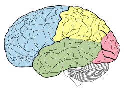
Temporal lobe

From Wikipedia, the free encyclopedia
https://en.wikipedia.org/wiki/Temporal_lobe
 
Temporal lobe
Lobes of the human brain (temporal lobe is shown in green)
Section of brain showing upper surface of temporal lobe.
Details
Part ofCerebrum
Artery
Vein
Identifiers
Latinlobus temporalis
MeSHD013702
NeuroNames125
NeuroLex IDbirnlex_1160
TA98A14.1.09.136
TA25488
FMA61825
Anatomical terms of neuroanatomy

The temporal lobe is one of the four major lobes of the cerebral cortex in the brain of mammals. The temporal lobe is located beneath the lateral fissure on both cerebral hemispheres of the mammalian brain.

The temporal lobe is involved in processing sensory input into derived meanings for the appropriate retention of visual memory, language comprehension, and emotion association. Temporal refers to the head's temples.

Structure

The temporal lobe consists of structures that are vital for declarative or long-term memory. Declarative (denotative) or explicit memory is conscious memory divided into semantic memory (facts) and episodic memory (events).

The medial temporal lobe structures are critical for long-term memory, and include the hippocampal formation, perirhinal cortex, parahippocampal, and entorhinal neocortical regions. The hippocampus is critical for memory formation, and the surrounding medial temporal cortex is currently theorized to be critical for memory storage. The prefrontal and visual cortices are also involved in explicit memory.

Research has shown that lesions in the hippocampus of monkeys results in limited impairment of function, whereas extensive lesions that include the hippocampus and the medial temporal cortex result in severe impairment.

A form of epilepsy that involves the medial lobe is usually known as mesial temporal lobe epilepsy.

Function

Visual memories

The temporal lobe communicates with the hippocampus and plays a key role in the formation of explicit long-term memory modulated by the amygdala.

Processing sensory input

Auditory
Adjacent areas in the superior, posterior, and lateral parts of the temporal lobes are involved in high-level auditory processing. The temporal lobe is involved in primary auditory perception, such as hearing, and holds the primary auditory cortex. The primary auditory cortex receives sensory information from the ears and secondary areas process the information into meaningful units such as speech and words. The superior temporal gyrus includes an area (within the lateral fissure) where auditory signals from the cochlea first reach the cerebral cortex and are processed by the primary auditory cortex in the left temporal lobe.
Visual
The areas associated with vision in the temporal lobe interpret the meaning of visual stimuliand establish object recognition. The ventral part of the temporal cortices appears to be involved in high-level visual processing of complex stimuli such as faces (fusiform gyrus) and scenes (parahippocampal gyrus). Anterior parts of this ventral stream for visual processing are involved in object perception and recognition.
Animation showing the position of the human left temporal lobe

Language recognition

In humans, temporal lobe regions are critical for accessing the semantic meaning of spoken words, printed words, and visual objects. Wernicke's area, which spans the region between temporal and parietal lobes of the dominant cerebral hemisphere (the left, in the majority of cases), plays a key role (in tandem with Broca's area in the frontal lobe) in language comprehension, whether spoken language or signed language. FMRI imaging shows these portions of the brain are activated by signed or spoken languages. These areas of the brain are active in children's language acquisition whether accessed via hearing a spoken language, watching a signed language, or via hand-over-hand tactile versions of a signed language.

The functions of the left temporal lobe are not limited to low-level perception but extend to comprehension, naming, and verbal memory.

New memories

The medial temporal lobes (near the sagittal plane) are thought to be involved in encoding declarative long term memory. The medial temporal lobes include the hippocampi, which are essential for memory storage, therefore damage to this area can result in impairment in new memory formation leading to permanent or temporary anterograde amnesia.

Clinical significance

Unilateral temporal lesion

  • Contralateral homonymous upper quadrantanopia (sector anopsia)
  • Complex hallucinations (smell, sound, vision, memory)

Dominant hemisphere

Non-dominant hemisphere

  • Impaired non-verbal memory
  • Impaired musical skills

Bitemporal lesions (additional features)

Damage

Individuals who suffer from medial temporal lobe damage have a difficult time recalling visual stimuli. This neurotransmission deficit is not due to lacking perception of visual stimuli, but rather to the inability to interpret what is perceived. The most common symptom of inferior temporal lobe damage is visual agnosia, which involves impairment in the identification of familiar objects. Another less common type of inferior temporal lobe damage is prosopagnosia which is an impairment in the recognition of faces and distinction of unique individual facial features.

Damage specifically to the anterior portion of the left temporal lobe can cause savant syndrome.

Disorders

Pick's disease, also known as frontotemporal amnesia, is caused by atrophy of the frontotemporal lobe. Emotional symptoms include mood changes, which the patient may be unaware of, including poor attention span and aggressive behavior towards themselves or others. Language symptoms include loss of speech, inability to read or write, loss of vocabulary and overall degeneration of motor ability.

Temporal lobe epilepsy is a chronic neurological condition characterized by recurrent seizures; symptoms include a variety of sensory (visual, auditory, olfactory, and gustation) hallucinations, as well as an inability to process semantic and episodic memories.

Schizophrenia is a severe psychotic disorder characterized by severe disorientation. Its most explicit symptom is the perception of external voices in the form of auditory hallucinations. The cause of such hallucinations has been attributed to deficits in the left temporal lobe, specifically within the primary auditory cortex. Decreased gray matter, among other cellular deficits, contribute to spontaneous neural activity that affects the primary auditory cortex as if it were experiencing acoustic auditory input. The misrepresentation of speech in the auditory cortex results in the perception of external voices in the form of auditory hallucinations in schizophrenic patients. Structural and functional MRI techniques have accounted for this neural activity by testing affected and non-affected individuals with external auditory stimuli.

Chromosome

From Wikipedia, the free encyclopedia
https://en.wikipedia.org/wiki/Chromosome
A chromosome and its packaged long strand of DNA unraveled. The DNA's base pairs encode genes, which provide functions. A human DNA can have up to 500 million base pairs with thousands of genes.
Condensed chromosome (purple rod) inside a bone marrow erythrokaryocyte undergoing mitosis
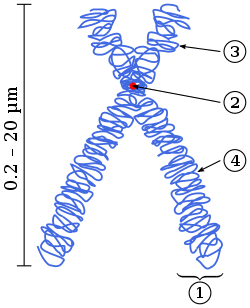
Diagram of a replicated and condensed metaphase eukaryotic chromosome:
  1. Chromatid
  2. Centromere
  3. Short arm
  4. Long arm

A chromosome is a package of DNA containing part or all of the genetic material of an organism. In most chromosomes, the very long thin DNA fibers are coated with nucleosome-forming packaging proteins; in eukaryotic cells, the most important of these proteins are the histones. Aided by chaperone proteins, the histones bind to and condense the DNA molecule to maintain its integrity. These eukaryotic chromosomes display a complex three-dimensional structure that has a significant role in transcriptional regulation.

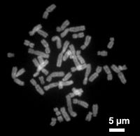
Normally, chromosomes are visible under a light microscope only during the metaphase of cell division, where all chromosomes are aligned in the center of the cell in their condensed form. Before this stage occurs, each chromosome is duplicated (S phase), and the two copies are joined by a centromere—resulting in either an X-shaped structure if the centromere is located equatorially, or a two-armed structure if the centromere is located distally; the joined copies are called 'sister chromatids'. During metaphase, the duplicated structure (called a 'metaphase chromosome') is highly condensed and thus easiest to distinguish and study. In animal cells, chromosomes reach their highest compaction level in anaphase during chromosome segregation.

Chromosomal recombination during meiosis and subsequent sexual reproduction plays a crucial role in genetic diversity. If these structures are manipulated incorrectly, through processes known as chromosomal instability and translocation, the cell may undergo mitotic catastrophe. This will usually cause the cell to initiate apoptosis, leading to its own death, but the process is occasionally hampered by cell mutations that result in the progression of cancer.

The term 'chromosome' is sometimes used in a wider sense to refer to the individualized portions of chromatin in cells, which may or may not be visible under light microscopy. In a narrower sense, 'chromosome' can be used to refer to the individualized portions of chromatin during cell division, which are visible under light microscopy due to high condensation.

Etymology

The word chromosome (/ˈkrməˌsm, -ˌzm/) comes from the Greek words χρῶμα (chroma, "colour") and σῶμα (soma, "body"), describing the strong staining produced by particular dyes. The term was coined by the German anatomist Heinrich Wilhelm Waldeyer, referring to the term 'chromatin', which was introduced by Walther Flemming.

Some of the early karyological terms have become outdated. For example, 'chromatin' (Flemming 1880) and 'chromosom' (Waldeyer 1888) both ascribe color to a non-colored state.

History of discovery

Walter Sutton (top) and Theodor Boveri (bottom) independently developed the chromosome theory of inheritance in 1902.

Otto Bütschli was the first scientist to recognize the structures now known as chromosomes.

In a series of experiments beginning in the mid-1880s, Theodor Boveri gave definitive contributions to elucidating that chromosomes are the vectors of heredity, with two notions that became known as 'chromosome continuity' and 'chromosome individuality'.

Wilhelm Roux suggested that every chromosome carries a different genetic configuration, and Boveri was able to test and confirm this hypothesis. Aided by the rediscovery at the start of the 1900s of Gregor Mendel's earlier experimental work, Boveri identified the connection between the rules of inheritance and the behaviour of the chromosomes. Two generations of American cytologists were influenced by Boveri: Edmund Beecher Wilson, Nettie Stevens, Walter Sutton and Theophilus Painter (Wilson, Stevens, and Painter actually worked with him).

In his famous textbook, The Cell in Development and Heredity, Wilson linked together the independent work of Boveri and Sutton (both around 1902) by naming the chromosome theory of inheritance the 'Boveri–Sutton chromosome theory' (sometimes known as the 'Sutton–Boveri chromosome theory'). Ernst Mayr remarks that the theory was hotly contested by some famous geneticists, including William Bateson, Wilhelm Johannsen, Richard Goldschmidt and T.H. Morgan, all of a rather dogmatic mindset. Eventually, absolute proof came from chromosome maps in Morgan's own laboratory.

The number of human chromosomes was published by Painter in 1923. By inspection through a microscope, he counted 24 pairs of chromosomes, giving 48 in total. His error was copied by others, and it was not until 1956 that the true number (46) was determined by Indonesian-born cytogeneticist Joe Hin Tjio.

Prokaryotes

The prokaryotes – bacteria and archaea – typically have a single circular chromosome. The chromosomes of most bacteria (also called genophores), can range in size from only 130,000 base pairs in the endosymbiotic bacteria Candidatus Hodgkinia cicadicola and Candidatus Tremblaya princeps, to more than 14,000,000 base pairs in the soil-dwelling bacterium Sorangium cellulosum.

Some bacteria have more than one chromosome. For instance, Spirochaetes such as Borrelia burgdorferi (causing Lyme disease), contain a single linear chromosome. Vibrios typically carry two chromosomes of very different size. Genomes of the genus Burkholderia carry one, two, or three chromosomes.

Structure in sequences

Prokaryotic chromosomes have less sequence-based structure than eukaryotes. Bacteria typically have a one-point (the origin of replication) from which replication starts, whereas some archaea contain multiple replication origins. The genes in prokaryotes are often organized in operons and do not usually contain introns, unlike eukaryotes.

DNA packaging

Prokaryotes do not possess nuclei. Instead, their DNA is organized into a structure called the nucleoid. The nucleoid is a distinct structure and occupies a defined region of the bacterial cell. This structure is, however, dynamic and is maintained and remodeled by the actions of a range of histone-like proteins, which associate with the bacterial chromosome. In archaea, the DNA in chromosomes is even more organized, with the DNA packaged within structures similar to eukaryotic nucleosomes.

Certain bacteria also contain plasmids or other extrachromosomal DNA. These are circular structures in the cytoplasm that contain cellular DNA and play a role in horizontal gene transfer. In prokaryotes and viruses, the DNA is often densely packed and organized; in the case of archaea, by homology to eukaryotic histones, and in the case of bacteria, by histone-like proteins.

Bacterial chromosomes tend to be tethered to the plasma membrane of the bacteria. In molecular biology application, this allows for its isolation from plasmid DNA by centrifugation of lysed bacteria and pelleting of the membranes (and the attached DNA).

Prokaryotic chromosomes and plasmids are, like eukaryotic DNA, generally supercoiled. The DNA must first be released into its relaxed state for access for transcription, regulation, and replication.

Eukaryotes

Organization of DNA in a eukaryotic cell

Each eukaryotic chromosome consists of a long linear DNA molecule associated with proteins, forming a compact complex of proteins and DNA called chromatin. Chromatin contains the vast majority of the DNA in an organism, but a small amount inherited maternally can be found in the mitochondria. It is present in most cells, with a few exceptions, for example, red blood cells.

Histones are responsible for the first and most basic unit of chromosome organization, the nucleosome.

Eukaryotes (cells with nuclei such as those found in plants, fungi, and animals) possess multiple large linear chromosomes contained in the cell's nucleus. Each chromosome has one centromere, with one or two arms projecting from the centromere, although, under most circumstances, these arms are not visible as such. In addition, most eukaryotes have a small circular mitochondrial genome, and some eukaryotes may have additional small circular or linear cytoplasmic chromosomes.

The major structures in DNA compaction: DNA, the nucleosome, the 10 nm "beads-on-a-string" fibre, the 30 nm fibre and the metaphase chromosome

In the nuclear chromosomes of eukaryotes, the uncondensed DNA exists in a semi-ordered structure, where it is wrapped around histones (structural proteins), forming a composite material called chromatin.

Interphase chromatin

The packaging of DNA into nucleosomes causes a 10 nanometer fibre which may further condense up to 30 nm fibres. Most of the euchromatin in interphase nuclei appears to be in the form of 30-nm fibers. Chromatin structure is the more decondensed state, i.e. the 10-nm conformation allows transcription.

Heterochromatin vs. euchromatin

During interphase (the period of the cell cycle where the cell is not dividing), two types of chromatin can be distinguished:

  • Euchromatin, which consists of DNA that is active, e.g., being expressed as protein.
  • Heterochromatin, which consists of mostly inactive DNA. It seems to serve structural purposes during the chromosomal stages. Heterochromatin can be further distinguished into two types:
    • Constitutive heterochromatin, which is never expressed. It is located around the centromere and usually contains repetitive sequences.
    • Facultative heterochromatin, which is sometimes expressed.

Metaphase chromatin and division

Human chromosomes during metaphase
Stages of early mitosis in a vertebrate cell with micrographs of chromatids

In the early stages of mitosis or meiosis (cell division), the chromatin double helix becomes more and more condensed. They cease to function as accessible genetic material (transcription stops) and become a compact transportable form. The loops of thirty-nanometer chromatin fibers are thought to fold upon themselves further to form the compact metaphase chromosomes of mitotic cells. The DNA is thus condensed about ten-thousand-fold.

The chromosome scaffold, which is made of proteins such as condensin, TOP2A and KIF4, plays an important role in holding the chromatin into compact chromosomes. Loops of thirty-nanometer structure further condense with scaffold into higher order structures.

This highly compact form makes the individual chromosomes visible, and they form the classic four-arm structure, a pair of sister chromatids attached to each other at the centromere. The shorter arms are called p arms (from the French petit, small) and the longer arms are called q arms (q follows p in the Latin alphabet; q-g "grande"; alternatively it is sometimes said q is short for queue meaning tail in French). This is the only natural context in which individual chromosomes are visible with an optical microscope.

Mitotic metaphase chromosomes are best described by a linearly organized longitudinally compressed array of consecutive chromatin loops.

During mitosis, microtubules grow from centrosomes located at opposite ends of the cell and also attach to the centromere at specialized structures called kinetochores, one of which is present on each sister chromatid. A special DNA base sequence in the region of the kinetochores provides, along with special proteins, longer-lasting attachment in this region. The microtubules then pull the chromatids apart toward the centrosomes, so that each daughter cell inherits one set of chromatids. Once the cells have divided, the chromatids are uncoiled and DNA can again be transcribed. In spite of their appearance, chromosomes are structurally highly condensed, which enables these giant DNA structures to be contained within a cell nucleus.

Human chromosomes

Chromosomes in humans can be divided into two types: autosomes (body chromosome(s)) and allosome (sex chromosome(s)). Certain genetic traits are linked to a person's sex and are passed on through the sex chromosomes. The autosomes contain the rest of the genetic hereditary information. All act in the same way during cell division. Human cells have 23 pairs of chromosomes (22 pairs of autosomes and one pair of sex chromosomes), giving a total of 46 per cell. In addition to these, human cells have many hundreds of copies of the mitochondrial genome. Sequencing of the human genome has provided a great deal of information about each of the chromosomes. Below is a table compiling statistics for the chromosomes, based on the Sanger Institute's human genome information in the Vertebrate Genome Annotation (VEGA) database. Number of genes is an estimate, as it is in part based on gene predictions. Total chromosome length is an estimate as well, based on the estimated size of unsequenced heterochromatin regions.


Chromosome Genes Total base pairs % of bases
1 2000 247,199,719 8.0
2 1300 242,751,149 7.9
3 1000 199,446,827 6.5
4 1000 191,263,063 6.2
5 900 180,837,866 5.9
6 1000 170,896,993 5.5
7 900 158,821,424 5.2
8 700 146,274,826 4.7
9 800 140,442,298 4.6
10 700 135,374,737 4.4
11 1300 134,452,384 4.4
12 1100 132,289,534 4.3
13 300 114,127,980 3.7
14 800 106,360,585 3.5
15 600 100,338,915 3.3
16 800 88,822,254 2.9
17 1200 78,654,742 2.6
18 200 76,117,153 2.5
19 1500 63,806,651 2.1
20 500 62,435,965 2.0
21 200 46,944,323 1.5
22 500 49,528,953 1.6
X (sex chromosome) 800 154,913,754 5.0
Y (sex chromosome) 200 57,741,652 1.9
Total 21,000 3,079,843,747 100.0

Based on the micrographic characteristics of size, position of the centromere and sometimes the presence of a chromosomal satellite, the human chromosomes are classified into the following groups:

Group Chromosomes Features
A 1–3 Large, metacentric or submetacentric
B 4–5 Large, submetacentric
C 6–12, X Medium-sized, submetacentric
D 13–15 Medium-sized, acrocentric, with satellite
E 16–18 Small, metacentric or submetacentric
F 19–20 Very small, metacentric
G 21–22, Y Very small, acrocentric (and 21, 22 with satellite)

Karyotype

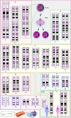
Karyogram of a human male
Schematic karyogram of a human, with annotated bands and sub-bands. It is a graphical representation of the idealized human diploid karyotype. It shows dark and white regions on G banding. Each row is vertically aligned at centromere level. It shows 22 homologous chromosomes, both the female (XX) and male (XY) versions of the sex chromosome (bottom right), as well as the mitochondrial genome (at bottom left).

In general, the karyotype is the characteristic chromosome complement of a eukaryote species. The preparation and study of karyotypes is part of cytogenetics.

Although the replication and transcription of DNA is highly standardized in eukaryotes, the same cannot be said for their karyotypes, which are often highly variable. There may be variation between species in chromosome number and in detailed organization. In some cases, there is significant variation within species. Often there is:

1. variation between the two sexes
2. variation between the germline and soma (between gametes and the rest of the body)
3. variation between members of a population, due to balanced genetic polymorphism
4. geographical variation between races
5. mosaics or otherwise abnormal individuals.

Also, variation in karyotype may occur during development from the fertilized egg.

The technique of determining the karyotype is usually called karyotyping. Cells can be locked part-way through division (in metaphase) in vitro (in a reaction vial) with colchicine. These cells are then stained, photographed, and arranged into a karyogram, with the set of chromosomes arranged, autosomes in order of length, and sex chromosomes (here X/Y) at the end.

Like many sexually reproducing species, humans have special gonosomes (sex chromosomes, in contrast to autosomes). These are XX in females and XY in males.

History and analysis techniques

Investigation into the human karyotype took many years to settle the most basic question: How many chromosomes does a normal diploid human cell contain? In 1912, Hans von Winiwarter reported 47 chromosomes in spermatogonia and 48 in oogonia, concluding an XX/XO sex determination mechanism. In 1922, Painter was not certain whether the diploid number of man is 46 or 48, at first favouring 46. He revised his opinion later from 46 to 48, and he correctly insisted on humans having an XX/XY system.

New techniques were needed to definitively solve the problem:

  1. Using cells in culture
  2. Arresting mitosis in metaphase by a solution of colchicine
  3. Pretreating cells in a hypotonic solution 0.075 M KCl, which swells them and spreads the chromosomes
  4. Squashing the preparation on the slide forcing the chromosomes into a single plane
  5. Cutting up a photomicrograph and arranging the result into an indisputable karyogram.

It took until 1954 before the human diploid number was confirmed as 46. Considering the techniques of Winiwarter and Painter, their results were quite remarkable. Chimpanzees, the closest living relatives to modern humans, have 48 chromosomes as do the other great apes: in humans two chromosomes fused to form chromosome 2.

Aberrations

In Down syndrome, there are three copies of chromosome 21.

Chromosomal aberrations are disruptions in the normal chromosomal content of a cell. They can cause genetic conditions in humans, such as Down syndrome, although most aberrations have little to no effect. Some chromosome abnormalities do not cause disease in carriers, such as translocations, or chromosomal inversions, although they may lead to a higher chance of bearing a child with a chromosome disorder. Abnormal numbers of chromosomes or chromosome sets, called aneuploidy, may be lethal or may give rise to genetic disorders. Genetic counseling is offered for families that may carry a chromosome rearrangement.

The gain or loss of DNA from chromosomes can lead to a variety of genetic disorders. Human examples include:

  • Cri du chat, caused by the deletion of part of the short arm of chromosome 5. "Cri du chat" means "cry of the cat" in French; the condition was so-named because affected babies make high-pitched cries that sound like those of a cat. Affected individuals have wide-set eyes, a small head and jaw, moderate to severe mental health problems, and are very short.
  • DiGeorge syndrome, also known as 22q11.2 deletion syndrome. Symptoms are mild learning disabilities in children, with adults having an increased risk of schizophrenia. Infections are also common in children because of problems with the immune system's T cell-mediated response due to an absence of hypoplastic thymus.
  • Down syndrome, the most common trisomy, usually caused by an extra copy of chromosome 21 (trisomy 21). Characteristics include decreased muscle tone, stockier build, asymmetrical skull, slanting eyes, and mild to moderate developmental disability.
  • Edwards syndrome, or trisomy-18, the second most common trisomy. Symptoms include motor retardation, developmental disability, and numerous congenital anomalies causing serious health problems. Ninety percent of those affected die in infancy. They have characteristic clenched hands and overlapping fingers.
  • Isodicentric 15, also called idic(15), partial tetrasomy 15q, or inverted duplication 15 (inv dup 15).
  • Jacobsen syndrome, which is very rare. It is also called the 11q terminal deletion disorder. Those affected have normal intelligence or mild developmental disability, with poor expressive language skills. Most have a bleeding disorder called Paris-Trousseau syndrome.
  • Klinefelter syndrome (XXY). Men with Klinefelter syndrome are usually sterile, and tend to be taller than their peers, with longer arms and legs. Boys with the syndrome are often shy and quiet, and have a higher incidence of speech delay and dyslexia. Without testosterone treatment, some may develop gynecomastia during puberty.
  • Patau Syndrome, also called D-Syndrome or trisomy-13. Symptoms are somewhat similar to those of trisomy-18, without the characteristic folded hand.
  • Small supernumerary marker chromosome. This means there is an extra, abnormal chromosome. Features depend on the origin of the extra genetic material. Cat-eye syndrome and isodicentric chromosome 15 syndrome (or Idic15) are both caused by a supernumerary marker chromosome, as is Pallister–Killian syndrome.
  • Triple-X syndrome (XXX). XXX girls tend to be tall and thin, and have a higher incidence of dyslexia.
  • Turner syndrome (X instead of XX or XY). In Turner syndrome, female sexual characteristics are present but underdeveloped. Females with Turner syndrome often have a short stature, low hairline, abnormal eye features and bone development, and a "caved-in" appearance to the chest.
  • Wolf–Hirschhorn syndrome, caused by partial deletion of the short arm of chromosome 4. It is characterized by growth retardation, delayed motor skills development, "Greek Helmet" facial features, and mild to profound mental health problems.
  • XYY syndrome. XYY boys are usually taller than their siblings. Like XXY boys and XXX girls, they are more likely to have learning difficulties.

Sperm aneuploidy

Exposure of males to certain lifestyle, environmental and/or occupational hazards may increase the risk of aneuploid spermatozoa. In particular, risk of aneuploidy is increased by tobacco smoking, and occupational exposure to benzene, insecticides, and perfluorinated compounds. Increased aneuploidy is often associated with increased DNA damage in spermatozoa.

Number in various organisms

In eukaryotes

The number of chromosomes in eukaryotes is highly variable. It is possible for chromosomes to fuse or break and thus evolve into novel karyotypes. Chromosomes can also be fused artificially. For example, when the 16 chromosomes of yeast were fused into one giant chromosome, it was found that the cells were still viable with only somewhat reduced growth rates.

The tables below give the total number of chromosomes (including sex chromosomes) in a cell nucleus for various eukaryotes. Most are diploid, such as humans who have 22 different types of autosomes—each present as two homologous pairs—and two sex chromosomes, giving 46 chromosomes in total. Some other organisms have more than two copies of their chromosome types, for example bread wheat which is hexaploid, having six copies of seven different chromosome types for a total of 42 chromosomes.

Chromosome numbers in some plants
Plant species #
Thale cress (diploid) 10
Rye (diploid) 14
Einkorn wheat (diploid) 14
Maize (diploid or palaeotetraploid) 20
Durum wheat (tetraploid) 28
Bread wheat (hexaploid) 42
Cultivated tobacco (tetraploid) 48
Adder's tongue fern (polyploid) approx. 1,200
Chromosome numbers (2n) in some animals
Species #
Indian muntjac 6♀, 7♂
Common fruit fly 8
Pill millipede 30
Earthworm 36
Tibetan fox 36
Domestic cat 38
Domestic pig 38
Laboratory mouse 40
Laboratory rat 42
Rabbit 44
Syrian hamster 44
Guppy 46
Human 46
Hare 48
Gorilla 48
Chimpanzee 48
Domestic sheep 54
Garden snail 54
Silkworm 56
Elephant 56
Cow 60
Donkey 62
Guinea pig 64
Horse 64
Dog 78
Hedgehog 90
Goldfish 100–104
Kingfisher 132
Chromosome numbers in other organisms
Species Large
chromosomes
Intermediate
chromosomes
Microchromosomes
Trypanosoma brucei 11 6 ≈100
Domestic pigeon 18 59–63
Chicken 8 2 sex chromosomes 60

Normal members of a particular eukaryotic species all have the same number of nuclear chromosomes. Other eukaryotic chromosomes, i.e., mitochondrial and plasmid-like small chromosomes, are much more variable in number, and there may be thousands of copies per cell.

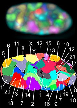
The 23 human chromosome territories during prometaphase in fibroblast cells

Asexually reproducing species have one set of chromosomes that are the same in all body cells. However, asexual species can be either haploid or diploid.

Sexually reproducing species have somatic cells (body cells) that are diploid [2n], having two sets of chromosomes (23 pairs in humans), one set from the mother and one from the father. Gametes (reproductive cells) are haploid [n], having one set of chromosomes. Gametes are produced by meiosis of a diploid germline cell, during which the matching chromosomes of father and mother can exchange small parts of themselves (crossover) and thus create new chromosomes that are not inherited solely from either parent. When a male and a female gamete merge during fertilization, a new diploid organism is formed.

Some animal and plant species are polyploid [Xn], having more than two sets of homologous chromosomes. Important crops such as tobacco or wheat are often polyploid, compared to their ancestral species. Wheat has a haploid number of seven chromosomes, still seen in some cultivars as well as the wild progenitors. The more common types of pasta and bread wheat are polyploid, having 28 (tetraploid) and 42 (hexaploid) chromosomes, compared to the 14 (diploid) chromosomes in wild wheat.

In prokaryotes

Prokaryote species generally have one copy of each major chromosome, but most cells can easily survive with multiple copies. For example, Buchnera, a symbiont of aphids has multiple copies of its chromosome, ranging from 10 to 400 copies per cell. However, in some large bacteria, such as Epulopiscium fishelsoni up to 100,000 copies of the chromosome can be present. Plasmids and plasmid-like small chromosomes are, as in eukaryotes, highly variable in copy number. The number of plasmids in the cell is almost entirely determined by the rate of division of the plasmid – fast division causes high copy number.

AllTrials

From Wikipedia, the free encyclopedia https://en.wikipedia.org/wiki/AllTrials ...