Search This Blog

Saturday, June 29, 2019

Vertebral column

From Wikipedia, the free encyclopedia

Vertebral column
Illu vertebral column.jpg
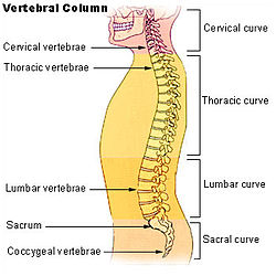
The human vertebral column and its regions
Anatomy and physiology of animals Regions of a vertebral column.jpg
Vertebral column of a goat
Details
Identifiers

The vertebral column, also known as the backbone or spine, is part of the axial skeleton. The vertebral column is the defining characteristic of a vertebrate in which the notochord (a flexible rod of uniform composition) found in all chordates has been replaced by a segmented series of bone: vertebrae separated by intervertebral discs. The vertebral column houses the spinal canal, a cavity that encloses and protects the spinal cord.

There are about 50,000 species of animals that have a vertebral column. The human vertebral column is one of the most-studied examples.

Structure

In a human's vertebral column there are normally thirty-three vertebrae; the upper twenty-four are articulating and separated from each other by intervertebral discs, and the lower nine are fused in adults, five in the sacrum and four in the coccyx or tailbone. The articulating vertebrae are named according to their region of the spine. There are seven cervical vertebrae, twelve thoracic vertebrae and five lumbar vertebrae. The number of vertebrae in a region can vary but overall the number remains the same. The number of those in the cervical region however is only rarely changed.

There are ligaments extending the length of the column at the front and the back, and in between the vertebrae joining the spinous processes, the transverse processes and the vertebral laminae.

Vertebrae

Numbering order of the vertebrae of the human spinal column
 
The vertebrae in the human vertebral column are divided into different regions, which correspond to the curves of the spinal column. The articulating vertebrae are named according to their region of the spine. Vertebrae in these regions are essentially alike, with minor variation. These regions are called the cervical spine, thoracic spine, lumbar spine, sacrum and coccyx. There are seven cervical vertebrae, twelve thoracic vertebrae and five lumbar vertebrae. The number of vertebrae in a region can vary but overall the number remains the same. The number of those in the cervical region however is only rarely changed. The vertebrae of the cervical, thoracic and lumbar spines are independent bones, and generally quite similar. The vertebrae of the sacrum and coccyx are usually fused and unable to move independently. Two special vertebrae are the atlas and axis, on which the head rests. 

Anatomy of a vertebra
 
A typical vertebra consists of two parts: the vertebral body and the vertebral arch. The vertebral arch is posterior, meaning it faces the back of a person. Together, these enclose the vertebral foramen, which contains the spinal cord. Because the spinal cord ends in the lumbar spine, and the sacrum and coccyx are fused, they do not contain a central foramen. The vertebral arch is formed by a pair of pedicles and a pair of laminae, and supports seven processes, four articular, two transverse, and one spinous, the latter also being known as the neural spine. Two transverse processes and one spinous process are posterior to (behind) the vertebral body. The spinous process comes out the back, one transverse process comes out the left, and one on the right. The spinous processes of the cervical and lumbar regions can be felt through the skin. 

Above and below each vertebra are joints called facet joints. These restrict the range of movement possible, and are joined by a thin portion of the neural arch called the pars interarticularis. In between each pair of vertebrae are two small holes called intervertebral foramina. The spinal nerves leave the spinal cord through these holes. 

Individual vertebrae are named according to their region and position. From top to bottom, the vertebrae are:

Shape

The upper cervical spine has a curve, convex forward, that begins at the axis (second cervical vertebra) at the apex of the odontoid process or dens, and ends at the middle of the second thoracic vertebra; it is the least marked of all the curves. This inward curve is known as a lordotic curve.

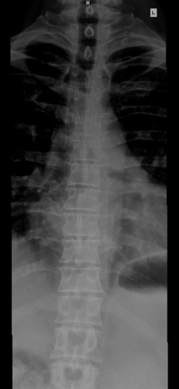
A thoracic spine X-ray of a 57-year-old male.
 
The thoracic curve, concave forward, begins at the middle of the second and ends at the middle of the twelfth thoracic vertebra. Its most prominent point behind corresponds to the spinous process of the seventh thoracic vertebra. This curve is known as a kyphotic curve. 

Lateral lumbar X-ray of a 34-year-old male.
 
The lumbar curve is more marked in the female than in the male; it begins at the middle of the last thoracic vertebra, and ends at the sacrovertebral angle. It is convex anteriorly, the convexity of the lower three vertebrae being much greater than that of the upper two. This curve is described as a lordotic curve. 

The sacral curve begins at the sacrovertebral articulation, and ends at the point of the coccyx; its concavity is directed downward and forward as a kyphotic curve. 

The thoracic and sacral kyphotic curves are termed primary curves, because they are present in the fetus. The cervical and lumbar curves are compensatory or secondary, and are developed after birth. The cervical curve forms when the infant is able to hold up its head (at three or four months) and to sit upright (at nine months). The lumbar curve forms later from twelve to eighteen months, when the child begins to walk.

Surfaces

Anterior surface
When viewed from in front, the width of the bodies of the vertebrae is seen to increase from the second cervical to the first thoracic; there is then a slight diminution in the next three vertebrae; below this there is again a gradual and progressive increase in width as low as the sacrovertebral angle. From this point there is a rapid diminution, to the apex of the coccyx.
Posterior surface
From behind, the vertebral column presents in the median line the spinous processes. In the cervical region (with the exception of the second and seventh vertebrae) these are short, horizontal and bifid. In the upper part of the thoracic region they are directed obliquely downward; in the middle they are almost vertical, and in the lower part they are nearly horizontal. In the lumbar region they are nearly horizontal. The spinous processes are separated by considerable intervals in the lumbar region, by narrower intervals in the neck, and are closely approximated in the middle of the thoracic region. Occasionally one of these processes deviates a little from the median line — which can sometimes be indicative of a fracture or a displacement of the spine. On either side of the spinous processes is the vertebral groove formed by the laminae in the cervical and lumbar regions, where it is shallow, and by the laminae and transverse processes in the thoracic region, where it is deep and broad; these grooves lodge the deep muscles of the back. Lateral to the spinous processes are the articular processes, and still more laterally the transverse processes. In the thoracic region, the transverse processes stand backward, on a plane considerably behind that of the same processes in the cervical and lumbar regions. In the cervical region, the transverse processes are placed in front of the articular processes, lateral to the pedicles and between the intervertebral foramina. In the thoracic region they are posterior to the pedicles, intervertebral foramina, and articular processes. In the lumbar region they are in front of the articular processes, but behind the intervertebral foramina.
Lateral surfaces
The sides of the vertebral column are separated from the posterior surface by the articular processes in the cervical and thoracic regions, and by the transverse processes in the lumbar region. In the thoracic region, the sides of the bodies of the vertebrae are marked in the back by the facets for articulation with the heads of the ribs. More posteriorly are the intervertebral foramina, formed by the juxtaposition of the vertebral notches, oval in shape, smallest in the cervical and upper part of the thoracic regions, and gradually increasing in size to the last lumbar. They transmit the special spinal nerves and are situated between the transverse processes in the cervical region, and in front of them in the thoracic and lumbar regions.

Ligaments

There are different ligaments involved in the holding together of the vertebrae in the column, and in the column's movement. The anterior and posterior longitudinal ligaments extend the length of the vertebral column along the front and back of the vertebral bodies. The interspinous ligaments connect the adjoining spinous processes of the vertebrae. The supraspinous ligament extends the length of the spine running along the back of the spinous processes, from the sacrum to the seventh cervical vertebra. From there it is continuous with the nuchal ligament.

Development

The striking segmented pattern of the spine is established during embryogenesis when somites are rhythmically added to the posterior of the embryo. Somite formation begins around the third week when the embryo begins gastrulation and continues until around 52 somites are formed. The somites are spheres, formed from the paraxial mesoderm that lies at the sides of the neural tube and they contain the precursors of spinal bone, the vertebrae ribs and some of the skull, as well as muscle, ligaments and skin. Somitogenesis and the subsequent distribution of somites is controlled by a clock and wavefront model acting in cells of the paraxial mesoderm. Soon after their formation, sclerotomes, which give rise to some of the bone of the skull, the vertebrae and ribs, migrate, leaving the remainder of the somite now termed a dermamyotome behind. This then splits to give the myotomes which will form the muscles and dermatomes which will form the skin of the back. Sclerotomes become subdivided into an anterior and a posterior compartment. This subdivision plays a key role in the definitive patterning of vertebrae that form when the posterior part of one somite fuses to the anterior part of the consecutive somite during a process termed resegmentation. Disruption of the somitogenesis process in humans results in diseases such as congenital scoliosis. So far, the human homologues of three genes associated to the mouse segmentation clock, (MESP2, DLL3 and LFNG), have been shown to be mutated in cases of congenital scoliosis, suggesting that the mechanisms involved in vertebral segmentation are conserved across vertebrates. In humans the first four somites are incorporated in the base of the occipital bone of the skull and the next 33 somites will form the vertebrae, ribs, muscles, ligaments and skin. The remaining posterior somites degenerate. During the fourth week of embryogenesis, the sclerotomes shift their position to surround the spinal cord and the notochord. This column of tissue has a segmented appearance, with alternating areas of dense and less dense areas. 

As the sclerotome develops, it condenses further eventually developing into the vertebral body. Development of the appropriate shapes of the vertebral bodies is regulated by HOX genes

The less dense tissue that separates the sclerotome segments develop into the intervertebral discs

The notochord disappears in the sclerotome (vertebral body) segments, but persists in the region of the intervertebral discs as the nucleus pulposus. The nucleus pulposus and the fibers of the anulus fibrosus make up the intervertebral disc. 

The primary curves (thoracic and sacral curvatures) form during fetal development. The secondary curves develop after birth. The cervical curvature forms as a result of lifting the head and the lumbar curvature forms as a result of walking.

Function

Spinal cord

The spinal cord nested in the vertebral column.

The vertebral column surrounds the spinal cord which travels within the spinal canal, formed from a central hole within each vertebra. The spinal cord is part of the central nervous system that supplies nerves and receives information from the peripheral nervous system within the body. The spinal cord consists of grey and white matter and a central cavity, the central canal. Adjacent to each vertebra emerge spinal nerves. The spinal nerves provide sympathetic nervous supply to the body, with nerves emerging forming the sympathetic trunk and the splanchnic nerves

The spinal canal follows the different curves of the column; it is large and triangular in those parts of the column which enjoy the greatest freedom of movement, such as the cervical and lumbar regions; and is small and rounded in the thoracic region, where motion is more limited.

The spinal cord terminates in the conus medullaris and cauda equina.

Clinical significance

Disease

Spina bifida is a congenital disorder in which there is a defective closure of the vertebral arch. Sometimes the spinal meninges and also the spinal cord can protrude through this, and this is called Spina bifida cystica. Where the condition does not involve this protrusion it is known as Spina bifida occulta. Sometimes all of the vertebral arches may remain incomplete. Another, though rare, congenital disease is Klippel-Feil syndrome which is the fusion of any two of the cervical vertebrae.

Spondylolisthesis is the forward displacement of a vertebra and retrolisthesis is a posterior displacement of one vertebral body with respect to the adjacent vertebra to a degree less than a dislocation. 

Spinal disc herniation, more commonly called a "slipped disc", is the result of a tear in the outer ring (anulus fibrosus) of the intervertebral disc, which lets some of the soft gel-like material, the nucleus pulposus, bulge out in a hernia

Spinal stenosis is a narrowing of the spinal canal which can occur in any region of the spine though less commonly in the thoracic region. The stenosis can constrict the spinal canal giving rise to a neurological deficit

Pain at the coccyx (tailbone) is known as coccydynia.

Spinal cord injury is damage to the spinal cord that causes changes in its function, either temporary or permanent.

Curvature

Excessive or abnormal spinal curvature is classed as a spinal disease or dorsopathy and includes the following abnormal curvatures:
  • Kyphosis is an exaggerated kyphotic (concave) curvature in the thoracic region, also called hyperkyphosis. This produces the so-called "humpback" or "dowager's hump", a condition commonly resulting from osteoporosis.
  • Lordosis as an exaggerated lordotic (convex) curvature of the lumbar region, is known as lumbar hyperlordosis and also as "swayback". Temporary lordosis is common during pregnancy.
  • Scoliosis, lateral curvature, is the most common abnormal curvature, occurring in 0.5% of the population. It is more common among females and may result from unequal growth of the two sides of one or more vertebrae, so that they do not fuse properly. It can also be caused by pulmonary atelectasis (partial or complete deflation of one or more lobes of the lungs) as observed in asthma or pneumothorax.
  • Kyphoscoliosis, a combination of kyphosis and scoliosis.

Anatomical landmarks

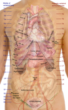
Surface projections of organs of the torso. The transpyloric line is seen at L1
 
Individual vertebrae of the human vertebral column can be felt and used as surface anatomy, with reference points are taken from the middle of the vertebral body. This provides anatomical landmarks that can be used to guide procedures such as a lumbar puncture and also as vertical reference points to describe the locations of other parts of human anatomy, such as the positions of organs.

Other animals

Variations in vertebrae

The general structure of vertebrae in other animals is largely the same as in humans. Individual vertebrae are composed of a centrum (body), arches protruding from the top and bottom of the centrum, and various processes projecting from the centrum and/or arches. An arch extending from the top of the centrum is called a neural arch, while the haemal arch or chevron is found underneath the centrum in the caudal (tail) vertebrae of fish, most reptiles, some birds, some dinosaurs and some mammals with long tails. The vertebral processes can either give the structure rigidity, help them articulate with ribs, or serve as muscle attachment points. Common types are transverse process, diapophyses, parapophyses, and zygapophyses (both the cranial zygapophyses and the caudal zygapophyses). The centrum of the vertebra can be classified based on the fusion of its elements. In temnospondyls, bones such as the spinous process, the pleurocentrum and the intercentrum are separate ossifications. Fused elements, however, classify a vertebra as having holospondyly.

A vertebra can also be described in terms of the shape of the ends of the centrum. Centra with flat ends are acoelous, like those in mammals. These flat ends of the centra are especially good at supporting and distributing compressive forces. Amphicoelous vertebra have centra with both ends concave. This shape is common in fish, where most motion is limited. Amphicoelous centra often are integrated with a full notochord. Procoelous vertebrae are anteriorly concave and posteriorly convex. They are found in frogs and modern reptiles. Opisthocoelous vertebrae are the opposite, possessing anterior convexity and posterior concavity. They are found in salamanders, and in some non-avian dinosaurs. Heterocoelous vertebrae have saddle-shaped articular surfaces. This type of configuration is seen in turtles that retract their necks, and birds, because it permits extensive lateral and vertical flexion motion without stretching the nerve cord too extensively or wringing it about its long axis.

In horses, the Arabian (breed) can have one less vertebrae and pair of ribs. This anomaly disappears in foals that are the product of an Arabian and another breed of horse.

Regional vertebrae

Vertebrae are defined by the regions of the vertebral column that they occur in, as in humans. Cervical vertebrae are those in the neck area. With the exception of the two sloth genera (Choloepus and Bradypus) and the manatee genus, (Trichechus), all mammals have seven cervical vertebrae. In other vertebrates, the number of cervical vertebrae can range from a single vertebra in amphibians, to as many as 25 in swans or 76 in the extinct plesiosaur Elasmosaurus. The dorsal vertebrae range from the bottom of the neck to the top of the pelvis. Dorsal vertebrae attached to the ribs are called thoracic vertebrae, while those without ribs are called lumbar vertebrae. The sacral vertebrae are those in the pelvic region, and range from one in amphibians, to two in most birds and modern reptiles, or up to three to five in mammals. When multiple sacral vertebrae are fused into a single structure, it is called the sacrum. The synsacrum is a similar fused structure found in birds that is composed of the sacral, lumbar, and some of the thoracic and caudal vertebra, as well as the pelvic girdle. Caudal vertebrae compose the tail, and the final few can be fused into the pygostyle in birds, or into the coccygeal or tail bone in chimpanzees (and humans).

Fish and amphibians

A vertebra (diameter 5 mm) of a small ray-finned fish

The vertebrae of lobe-finned fishes consist of three discrete bony elements. The vertebral arch surrounds the spinal cord, and is of broadly similar form to that found in most other vertebrates. Just beneath the arch lies a small plate-like pleurocentrum, which protects the upper surface of the notochord, and below that, a larger arch-shaped intercentrum to protect the lower border. Both of these structures are embedded within a single cylindrical mass of cartilage. A similar arrangement was found in the primitive Labyrinthodonts, but in the evolutionary line that led to reptiles (and hence, also to mammals and birds), the intercentrum became partially or wholly replaced by an enlarged pleurocentrum, which in turn became the bony vertebral body. In most ray-finned fishes, including all teleosts, these two structures are fused with, and embedded within, a solid piece of bone superficially resembling the vertebral body of mammals. In living amphibians, there is simply a cylindrical piece of bone below the vertebral arch, with no trace of the separate elements present in the early tetrapods.

In cartilaginous fish, such as sharks, the vertebrae consist of two cartilaginous tubes. The upper tube is formed from the vertebral arches, but also includes additional cartilaginous structures filling in the gaps between the vertebrae, and so enclosing the spinal cord in an essentially continuous sheath. The lower tube surrounds the notochord, and has a complex structure, often including multiple layers of calcification.

Lampreys have vertebral arches, but nothing resembling the vertebral bodies found in all higher vertebrates. Even the arches are discontinuous, consisting of separate pieces of arch-shaped cartilage around the spinal cord in most parts of the body, changing to long strips of cartilage above and below in the tail region. Hagfishes lack a true vertebral column, and are therefore not properly considered vertebrates, but a few tiny neural arches are present in the tail.

Other vertebrates

Vertebral anatomy of a human spine
 
The general structure of human vertebrae is fairly typical of that found in mammals, reptiles, and birds. The shape of the vertebral body does, however, vary somewhat between different groups. In mammals, such as humans, it typically has flat upper and lower surfaces, while in reptiles the anterior surface commonly has a concave socket into which the expanded convex face of the next vertebral body fits. Even these patterns are only generalisations, however, and there may be variation in form of the vertebrae along the length of the spine even within a single species. Some unusual variations include the saddle-shaped sockets between the cervical vertebrae of birds and the presence of a narrow hollow canal running down the centre of the vertebral bodies of geckos and tuataras, containing a remnant of the notochord.

Reptiles often retain the primitive intercentra, which are present as small crescent-shaped bony elements lying between the bodies of adjacent vertebrae; similar structures are often found in the caudal vertebrae of mammals. In the tail, these are attached to chevron-shaped bones called haemal arches, which attach below the base of the spine, and help to support the musculature. These latter bones are probably homologous with the ventral ribs of fish. The number of vertebrae in the spines of reptiles is highly variable, and may be several hundred in some species of snake.

In birds, there is a variable number of cervical vertebrae, which often form the only truly flexible part of the spine. The thoracic vertebrae are partially fused, providing a solid brace for the wings during flight. The sacral vertebrae are fused with the lumbar vertebrae, and some thoracic and caudal vertebrae, to form a single structure, the synsacrum, which is thus of greater relative length than the sacrum of mammals. In living birds, the remaining caudal vertebrae are fused into a further bone, the pygostyle, for attachment of the tail feathers.

Aside from the tail, the number of vertebrae in mammals is generally fairly constant. There are almost always seven cervical vertebrae (sloths and manatees are among the few exceptions), followed by around twenty or so further vertebrae, divided between the thoracic and lumbar forms, depending on the number of ribs. There are generally three to five vertebrae with the sacrum, and anything up to fifty caudal vertebrae.

Dinosaurs

The vertebral column in dinosaurs consists of the cervical (neck), dorsal (back), sacral (hips), and caudal (tail) vertebrae. Saurischian dinosaur vertebrae sometimes possess features known as pleurocoels, which are hollow depressions on the lateral portions of the vertebrae, perforated to create an entrance into the air chambers within the vertebrae, which served to decrease the weight of these bones without sacrificing strength. These pleurocoels were filled with air sacs, which would have further decreased weight. In sauropod dinosaurs, the largest known land vertebrates, pleurocoels and air sacs may have reduced the animal's weight by over a ton in some instances, a handy evolutionary adaption in animals that grew to over 30 metres in length. In many hadrosaur and theropod dinosaurs, the caudal vertebrae were reinforced by ossified tendons. The presence of three or more sacral vertebrae, in association with the hip bones, is one of the defining characteristics of dinosaurs. The occipital condyle is a structure on the posterior part of a dinosaur's skull which articulates with the first cervical vertebra.

Development of the cerebral cortex

From Wikipedia, the free encyclopedia

Corticogenesis is the process in which the cerebral cortex of the brain is formed during the development of the nervous system. The cortex is the outer layer of the brain and is composed of up to six layers. Neurons formed in the ventricular zone migrate to their final locations in one of the six layers of the cortex  The process occurs from embryonic day 10 to 17 in mice and between gestational weeks seven to 18 in humans.


Visualization of corticogenesis in the mouse. The 6 cortex layers migrate from the ventricular zone through the subplate to come to rest in the cortical plate (layers 2 through 6) or in the marginal zone (layer 1)

Cortical plates and zones

Plates

The preplate is the first stage in corticogenesis prior to the development of the cortical plate. The preplate is located between the pia and the ventricular zone. According to current knowledge, the preplate contains the first-born or pioneer neurons. These neurons are mainly thought to be Cajal-Retzius cells. The preplate also contains the predecessor to the subplate, which is sometimes referred to as a layer. As the cortical plate appears, the preplate separates into two components. The Cajal-Retzius cells go into the marginal zone, above the cortical plate, while the subplate moves to below the 6 cortical layers. It is during this transition from preplate to cortical plate when many malformations may arise. 

The cortical plate is the final plate formed in corticogenesis. It includes the cortex layers two through six.

The subplate is located beneath the cortical plate. It is named for both its location relative to the cortical plate and for the time frame in which it is created. While cortical plate matures, the cells located in the subplate establish connections with neurons that have not yet moved to their destination layer within the cortical plate. Pioneer cells are also present in the subplate and work to create fibers and synapses within the plate.

Zones

The intermediate zone is located between the ventricular zone and the cortical plate. The white matter in this area is where neurons, that are created in the ventricular zone, migrate through in order to reach the cortical plate. This zone is only present during corticogenesis and eventually transforms into adult white matter.

The ventricular and subventricular zones exist below the intermediate zone and communicate to other zones through cell signalling, also creating neurons destined to migrate to other areas in the cortex.

The marginal zone, along with the cortical zone, make up the 6 layers that form the cortex. This zone is the predecessor for layer 1 of the cortex. Astrocytes form an outer limiting membrane to interact with the pia. In humans it has been found that the cells here also form a subpial layer. Cajal-Retzius cells are also present in this zone and release reelin along the radial axis, a key to proper neuronal migration during corticogenesis.

Formation of layers

The cerebral cortex is divided into layers. Each layer is formed by radial glial cells located in the ventricular zone or subventricular zone, and then migrate to their final destination.

Layer I

Layer I, the molecular layer, is the first cortical layer produced during neurogenesis at mouse E10.5 to E12.5. Of the six layers found within the neocortex, layer I is the most superficial composed of Cajal–Retzius cells and pyramidal cells. This layer is unique in the aspect that these cells migrate to the outer edge of the cortex opposed to the migration experienced by the other 5 layers. Layer one is also characterized by expression of reelin, transcription factor T-box brain 1, and cortical migratory neuronal marker.

Layers 2 and 3

The second and third layers, or the External Granular layer and External Pyramidal layer respectively, are formed around mouse E13.5 to E16. These layers are the last to form during corticogenesis and include pyramidal neurons, astrocytes, Stellates, and radial glial cells. The pyramidal and stellate neurons express SATB2 and CUX1. SATB2 and CUX1 are DNA binding proteins involved in determining the fate of cortical cells.

Layers 4, 5 and 6

The fourth, fifth and sixth layers, or the Internal Granular layer, Internal Pyramidal layer, and Polymorphic or Multiform layer respectively, are formed during mouse E11.5 to E14.5. Included in these layers are stellates, radial glia, and pyramidal neurons. Layer six is adjacent to the ventricular zone. During the production of these layers, transcription factors TBR1 and OTX1 are expressed along with CTIP2, or corticoneuronal zinc finger protein.

Neuronal migration

Neuronal migration plays significant role in corticogenesis. Throughout the process of creating the six cortical layers, all the neurons and cells migrate from the ventricular zone, through the subplate, and come to rest at their appropriate layer of the cortex. Neuronal migration is generally subdivided into radial migration, tangential migration and multipolar migration. The migration of subcortical brain functions to the cortex is known as corticalization.

Cell signaling

Appropriate formation of the cerebral cortex relies heavily on a densely intertwined network of multiple signaling pathways and distinct signaling molecules. While the majority of the process remains to be understood, some signals and pathways have been carefully unraveled in an effort to gain full knowledge of the mechanisms that control corticogenesis.

Reelin-DAB1 pathway

The Reelin-DAB1 pathway is a well-defined pathway involved in corticogenesis. Cajal-Retzius cells located in the marginal zone secrete reelin to start the cascade. Reelin is able to interact with specific neurons in the cortical plate and direct these neurons to their proper locations. It is thought that the result downstream from this signalling could influence the cytoskeleton. Reelin is secreted only by the Cajal-Retzius cells located in the marginal zone, and its receptors are confined to the cortical plate. This segregation could be used to understand the actions of Reelin.

DAB1 is a regulator protein downstream of the reelin receptors. This protein is located inside cells residing in the ventricular zone, displaying highest concentrations in migrating pyramidal cells. When either reelin or DAB1 are inactivated in mice, the resulting phenotypes are the same. In this case, the neurons are unable to migrate properly through the cortical plate. It does not affect the proliferation of neurons and in the wild does not seem to have detrimental effects on memory or learning.

Sonic hedgehog

Knocking out the Sonic hedgehog, or Shh, has been shown to severely affect corticogenesis in the genetically modified mice. The ventral and dorsal sides of the cerebrum are affected as Shh expresses the transcription factors to Nkx2 which is important in patterning the cortex. Shh is also important to corticogenesis as it affects cell proliferation and differentiation, helping neuronal progenitor cells in fate determination.

Bmp-7

Bone morphogenetic protein 7 (Bmp-7), is an important regulator in corticogenesis, though it is not understood whether it promotes or inhibits neurogenesis. Bmp-7 can be detected in the ventricular zone and is secreted into cerebrospinal fluid (CSF). The CSF is an area to promote neurogenesis and it is believed that the synergy between Bmp-7 and other regulators promote cell division along with homeostasis.

Other bone morphogenetic proteins are also known to impact corticogenesis. Bmp2, 4, 5, and 6 are expressed during the process and can compensate for one another. For example, if Bmp-4 was absent from corticogenesis, very little would change in the cortex phenotype, due to the other Bmps helping accomplish the tasks of Bmp-4. However, Bmp-7 is the only Bmp that promotes radial glia survival and therefore considered more important.

Cdk5-p35 pathway

Cdk5 has a pathway parallel to the Reelin-DAB1. This pathway affects the neuronal positioning, and results in similar malformations when absent as the Reelin or DAB1 malformations except that migration is affected at an earlier stage on the cortical plate. Cdk5/p35 pathway is also responsible for actin and microtubule dynamics involved in neuronal migration.

Cyclin-dependent kinase inhibitor 1C, or p57, also affects corticogenesis. It has been shown the p57 induces cells to exit from the cell cycle and begin differentiation, but it is dependent on Cdks. p57 is able to induce neuronal progenitor cells to start differentiating into highly specialized neurons in the cortex. However, the mechanism by which p57 is able to affect such control is not yet known.

Other signals

Besides the ones listed above, there are several more signals that affect corticogenesis. Cnr1 is a g protein receptor that is widely expressed throughout the brain, and in interneurons. In knockout mice, the cortex exhibited decreased immunoreactivity. Nrp1, Robo1, and Robo2 have also been shown to be present and important in the development of interneurons. Cdh8 is known to be expressed in the intermediate and subventricular zone, though not in specific neurons in that area, and it is suggested to regulate fiber releasing.

Disorders

Lissencephaly

Lissencephaly, or 'smooth brain', is a disorder in which the brain does not properly form the gyri and sulci as a result from neuronal migration and cortical folding. This disorder can also result in epilepsy and cognitive impariment. Type 1 lissencephaly is due to an error in migration. LISI, also known as PAFAH1B, is expressed in both dividing and migrating cells found in the brain. When LIS1 is deleted, lissencephaly occurs.

LIS1 is thought to have several important roles in the creation of the cortex. Since LIS1 is similar to the nuclear distribution protein F (nudF), they are thought to work similarly. The nud family is known to be a factor in nuclear translocation, or moving the nuclei of daughter cells after cell division has occurred. By relation, it is thought that LIS1 is a factor in neuronal migration. LIS1 is also considered to be a factor in controlling dynein, a motor protein that affects intercellular movement such as protein sorting and the process of cell division.

Another protein that contributes to a lissencephaly disorder is DCX, or Doublecortin. DCX is a microtubule associated protein that is responsible for double cortex malformations. DCX is found in the second layer of the cortex, and in fact is still present in immature neurons of an adult cortex. It is thought that DCX affects neuronal migration through affecting the microtubule dynamics. Since DCX malformations results as a similar phenotype as with LIS1 malformations, it is thought they interact with one another on a cellular level. However, it is not know how this occurs yet.

Tsc1 knockout

TSC, or tuberous sclerosis, is an autosomal dominate disorder. TSC1 or TSC2 inactivation can cause TSC and the associated tumors in the brain. When inactivation of TSC1 is present during corticogenesis, malformations of cortical tubers, or abnormal benign tissue growth, along with white matter nodes would form in mice. This replicates the effect TSC is found to have in humans afflicted with TSC. In the mice there would be a lack of GFAP in astrocytes however astrogliosis would not occur like in the human TSC.

Recapitulation

Recapitulation of corticogenesis in both human and mouse embryos have been accomplished with a three dimensional culture using embryonic stem cells (ESC). Recapitulation is the theory in which an organism passes through embryonic development in stages similar to evolution of that organism. By carefully using embryo body intermediates and cultured in a serum free environment cortical progenitors form in a space and time related pattern similar to in vivo corticogenesis. Using immunocytochemical analysis on mouse neural stem cells derived from ESCs, after 6 days there was evidence of neuronal differentiation. The recapitulation ability only follows after the knowledge of spatial and temporal patterns have been identified, along with giving the knowledge that corticogenesis can occur without input from the brain.

Neuroscientist

From Wikipedia, the free encyclopedia

A neuroscientist (or neurobiologist) is a scientist who has specialised knowledge in the field of neuroscience, the branch of biology that deals with the physiology, biochemistry, anatomy and molecular biology of neurons and neural circuits and especially their association with behaviour and learning.

Camillo Golgi (1843–1926), Italian physician, neuroscientist, and namesake of the Golgi apparatus
 
Neuroscientists generally work as researchers within a college, university, government agency, or private industry setting. In research-oriented careers, neuroscientists typically spend their time designing and carrying out scientific experiments that contribute to the understanding of the nervous system and its function. They can engage in basic or applied research. Basic research seeks to add information to our current understanding of the nervous system, whereas applied research seeks to address a specific problem, such as developing a treatment for a neurological disorder. Biomedically-oriented neuroscientists typically engage in applied research. Neuroscientists also have a number of career opportunities outside the realm of research, including careers in industry, science writing, government program management, science advocacy, and education. These individuals most commonly hold doctorate degrees in the sciences, but may also hold a master's degree. The Neuroscientists day is celebrated on August 13th. 

Job overview

Job description

A dissected sheep brain.
 
Neuroscientists focus primarily on the study and research of the nervous system. The nervous system is composed of the brain, spinal cord and nerve cells. Studies of the nervous system may focus on the cellular level, as in studies of the ion channels, or instead may focus on a systemic level as in behavioural or cognitive studies. A significant portion of nervous system studies is devoted to understanding the diseases that affect the nervous system, like multiple sclerosis, Alzheimer's, Parkinson's, and Lou Gehrig's. Research commonly occurs in private, government and public research institutions and universities.
Some common tasks for neuroscientists are:
  • Developing experiments and leading groups of people in supporting roles
  • Conducting theoretical and computational neuronal data analysis
  • Research and development of new treatments for neurological disorders
  • Working with doctors to perform experimental studies of new drugs on willing patients
  • Following safety and sanitation procedures and guidelines
  • Dissecting experimental specimens

Salary

The overall median salary for neuroscientists in the United States was $79,940 in May 2014. Neuroscientists are usually full-time employees. Below, median salaries for common work places in the United States are shown.

Common Work Places Median Annual Pay
Colleges and universities $58,140
Hospitals $73,590
Laboratories $82,700
Research and Development $90,200
Pharmaceutical $150,000

Work environment

Neuroscientists research and study both the biological and psychological aspects of the nervous system. Once neuroscientists finish their post doctoral programs, 39% go on to perform more doctoral work, while 36% take on faculty jobs. Neuroscientists use a wide range of mathematical methods, computer programs, biochemical approaches and imaging techniques such as magnetic resonance imaging, computed tomography angiography, and diffusion tensor imaging. Imaging techniques allow scientists to observe physical changes in the brain, as signals occur. Neuroscientists can also be part of several different neuroscience organizations where they can publish and read different research topics.

Job outlook

Neuroscience is expecting a job growth of about 8% from 2014 to 2024, a considerably average job growth rate when compared to other professions. Factors leading to this growth include an aging population, new discoveries leading to new areas of research, and an increasing utilization of medications. Government funding for research will also continue to influence the demand for this specialty.

Education

Neuroscientists typically enroll in a four-year undergraduate program and then move on to a PhD program for graduate studies. Once finished with their graduate studies, neuroscientists may continue doing postdoctoral work to gain more lab experience and explore new laboratory methods. In their undergraduate years, neuroscientists typically take physical and life science courses to gain a foundation in the field of research. Typical undergraduate majors include biology, behavioral neuroscience, and cognitive neuroscience.

Many colleges and universities now have PhD training programs in the neurosciences, often with divisions between cognitive, cellular and molecular, computational and systems neuroscience.

Interdisciplinary fields

Neuroscience has a unique perspective in that it can be applied in a broad range of disciplines, and thus the fields neuroscientists work in vary. Neuroscientists may study topics from the large hemispheres of the brain to neurotransmitters and synapses occurring in neurons at a micro-level. Some fields that combine psychology and neurobiology include cognitive neuroscience, and behavioural neuroscience. Cognitive neuroscientists study human consciousness, specifically the brain, and how it can be seen through a lens of biochemical and biophysical processes. Behaviorial neuroscience encompasses the whole nervous system, environment and the brain how these areas show us aspects of motivation, learning, and motor skills along with many others.

History

Egyptian understanding and early Greek philosophers

Hieroglyphic stating the word, "brain", dated to 1700 BC. This work is considered a copy of an original writing as old as 3000 BC.
 
Some of the first writings about the brain come from the Egyptians. In about 3000 BC the first known written description of the brain also indicated that the location of brain injuries may be related to specific symptoms. This document contrasted common theory at the time. Most of the Egyptians' other writings are very spiritual, describing thought and feelings as responsibilities of the heart. This idea was widely accepted and can be found into 17th century Europe.

Plato believed that the brain was the locus of mental processes. However, Aristotle believed instead the heart to be the source of mental processes and that the brain acted as a cooling system for the cardiovascular system.

Galen

In the Middle Ages, Galen made a considerable impact on human anatomy. In terms of neuroscience, Galen described the seven cranial nerves' functions along with giving a foundational understanding of the spinal cord. When it came to the brain, he believed that sensory sensation was caused in the middle of the brain, while the motor sensations were produced in the anterior portion of the brain. Galen imparted some ideas on mental health disorders and what caused these disorders to arise. He believed that the cause was backed-up black bile, and that epilepsy was caused by phlegm. Galen's observations on neuroscience were not challenged for many years.

Medieval European beliefs and Andreas Vesalius

Medieval beliefs generally held true the proposals of Galen, including the attribution of mental processes to specific ventricles in the brain. Functions of regions of the brain were defined based on their texture and composition: memory function was attributed to the posterior ventricle, a harder region of the brain and thus a good place for memory storage.

Andreas Vesalius redirected the study of neuroscience away from the anatomical focus; he considered the attribution of functions based on location to be crude. Pushing away from the superficial proposals made by Galen and medieval beliefs, Vesalius did not believe that studying anatomy would lead to any significant advances in the understanding of thinking and the brain.

Current and developing research topics

Research in neuroscience is expanding and becoming increasingly interdisciplinary. Many current research projects involve the integration of computer programs in mapping the human nervous system. The National Institutes of Health (NIH) sponsored Human Connectome Project, launched in 2009, hopes to establish a highly detailed map of the human nervous system and its millions of connections. Detailed neural mapping could lead the way for advances in the diagnosis and treatment of neurological disorders

Neuroscientists are also at work studying epigenetics, the study of how certain factors that we face in our everyday lives not only affect us and our genes but also how they will affect our children and change their genes to adapt to the environments we faced.

Behavioral and developmental studies

Neuroscientists have been working to show how the brain is far more elastic and able to change than we once thought. They have been using work that psychologists previously reported to show how the observations work, and give a model for it. 

L-phenylalanine
 
One recent behavioral study is that of phenylketonuria (PKU), a disorder that heavily damages the brain due to toxic levels of the amino acid phenylalanine. Before neuroscientists had studied this disorder, psychologists did not have a mechanistic understanding as to how this disorder caused high levels of the amino acid and thus treatment was not well understood, and oftentimes, was inadequate. The neuroscientists that studied this disorder used the previous observations of psychologists to propose a mechanistic model that gave a better understanding of the disorder at the molecular level. This in turn led to better understanding of the disorder as a whole and greatly changed treatment that led to better lives for patients with the disorder.

Another recent study was that of mirror neurons, neurons that fire when mimicking or observing another animal or person that is making some sort of expression, movement, or gesture. This study was again one where neuroscientists used the observations of psychologists to create a model for how the observation worked. The initial observation was that newborn infants mimicked facial expressions that were expressed to them. Scientists were not certain that newborn infants were developed enough to have complex neurons that allowed them to mimic different people and there was something else that allowed them to mimic expressions. Neuroscientists then provided a model for what was occurring and concluded that infants did in fact have these neurons that fired when watching and mimicking facial expressions.

Effects of early experience on the brain

Neuroscientists have also studied the effects of "nurture" on the developing brain. Saul Schanberg and other neuroscientists did a study on how important nurturing touch is to the developing brains in rats. They found that the rats who were deprived of nurture from the mother for just one hour had reduced functions in processes like DNA synthesis and hormone secretion.

Michael Meaney and his colleagues found that the offspring of mother rats who provided significant nurture and attention tended to show less fear, responded more positively to stress, and functioned at higher levels and for longer times when fully mature. They also found that the rats who were given much attention as adolescents also gave their offspring the same amount of attention and thus showed that rats raised their offspring similar to how they were raised. These studies were also seen on a microscopic level where different genes were expressed for the rats that were given high amounts of nurture and those same genes were not expressed in the rats who received less attention.

The effects of nurture and touch were not only studied in rats, but also in newborn humans. Many neuroscientists have performed studies where the importance of touch is show in newborn humans. The same results that were shown in rats, also held true for humans. Babies that received less touch and nurture developed slower than babies that received a lot of attention and nurture. Stress levels were also lower in babies that were nurtured regularly and cognitive development was also higher due to increased touch. Human offspring, much like rat offspring, thrive off of nurture, as shown by the various studies of neuroscientists.

Famous neuroscientists

Neuroscientists awarded Nobel Prizes in physiology or medicine

Neuroscientists in popular culture

Brodmann area (brain)

From Wikipedia, the free encyclopedia

Brodmann area
Brodmann areas 3D.png
3D representation of Brodmann areas
Details
Part ofCerebrum
Identifiers
NeuroNames427
FMA68596

A Brodmann area is a region of the cerebral cortex, in the human or other primate brain, defined by its cytoarchitecture, or histological structure and organization of cells.

History

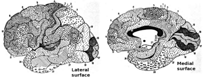
A number of important Brodmann areas have been marked out on this diagram.
 
Brodmann areas were originally defined and numbered by the German anatomist Korbinian Brodmann based on the cytoarchitectural organization of neurons he observed in the cerebral cortex using the Nissl method of cell staining. Brodmann published his maps of cortical areas in humans, monkeys, and other species in 1909, along with many other findings and observations regarding the general cell types and laminar organization of the mammalian cortex. The same Brodmann area number in different species does not necessarily indicate homologous areas. A similar, but more detailed cortical map was published by Constantin von Economo and Georg N. Koskinas in 1925.

Present importance

Brodmann areas have been discussed, debated, refined, and renamed exhaustively for nearly a century and remain the most widely known and frequently cited cytoarchitectural organization of the human cortex. 

Many of the areas Brodmann defined based solely on their neuronal organization have since been correlated closely to diverse cortical functions. For example, Brodmann areas 3, 1 and 2 are the primary somatosensory cortex; area 4 is the primary motor cortex; area 17 is the primary visual cortex; and areas 41 and 42 correspond closely to primary auditory cortex. Higher order functions of the association cortical areas are also consistently localized to the same Brodmann areas by neurophysiological, functional imaging, and other methods (e.g., the consistent localization of Broca's speech and language area to the left Brodmann areas 44 and 45). However, functional imaging can only identify the approximate localization of brain activations in terms of Brodmann areas since their actual boundaries in any individual brain requires its histological examination.

Overview

Brodmann's classification of areas of the cortex
 
Different parts of the cerebral cortex are involved in different cognitive and behavioral functions. The differences show up in a number of ways: the effects of localized brain damage, regional activity patterns exposed when the brain is examined using functional imaging techniques, connectivity with subcortical areas, and regional differences in the cellular architecture of the cortex. Neuroscientists describe most of the cortex—the part they call the neocortex—as having six layers, but not all layers are apparent in all areas, and even when a layer is present, its thickness and cellular organization may vary. Scientists have constructed maps of cortical areas on the basis of variations in the appearance of the layers as seen with a microscope. One of the most widely used schemes came from Korbinian Brodmann, who split the cortex into 52 different areas and assigned each a number (many of these Brodmann areas have since been subdivided). For example, Brodmann area 1 is the primary somatosensory cortex, Brodmann area 17 is the primary visual cortex, and Brodmann area 25 is the anterior cingulate cortex.

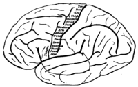
Topography of the primary motor cortex, showing which zone controls each body part
 
Many of those brain areas defined by Brodmann have their own complex internal structures. In a number of cases, brain areas are organized into topographic maps, where adjoining bits of the cortex correspond to adjoining parts of the body, or of some more abstract entity. A simple example of this type of correspondence is the primary motor cortex, a strip of tissue running along the anterior edge of the central sulcus. Motor areas innervating each part of the body arise from a distinct zone, with neighboring body parts represented by neighboring zones. Electrical stimulation of the cortex at any point causes a muscle-contraction in the represented body part. This "somatotopic" representation is not evenly distributed, however. The head, for example, is represented by a region about three times as large as the zone for the entire back and trunk. The size of any zone correlates to the precision of motor control and sensory discrimination possible. The areas for the lips, fingers, and tongue are particularly large, considering the proportional size of their represented body parts. 

In visual areas, the maps are retinotopic; this means they reflect the topography of the retina, the layer of light-activated neurons lining the back of the eye. In this case too, the representation is uneven: the fovea—the area at the center of the visual field—is greatly overrepresented compared to the periphery. The visual circuitry in the human cerebral cortex contains several dozen distinct retinotopic maps, each devoted to analyzing the visual input stream in a particular way. The primary visual cortex (Brodmann area 17), which is the main recipient of direct input from the visual part of the thalamus, contains many neurons that are most easily activated by edges with a particular orientation moving across a particular point in the visual field. Visual areas farther downstream extract features such as color, motion, and shape. 

In auditory areas, the primary map is tonotopic. Sounds are parsed according to frequency (i.e., high pitch vs. low pitch) by subcortical auditory areas, and this parsing is reflected by the primary auditory zone of the cortex. As with the visual system, there are a number of tonotopic cortical maps, each devoted to analyzing sound in a particular way. 

Within a topographic map there can sometimes be finer levels of spatial structure. In the primary visual cortex, for example, where the main organization is retinotopic and the main responses are to moving edges, cells that respond to different edge-orientations are spatially segregated from one another.

For humans and other primates

(*) Area only found in non-human primates

Some of the original Brodmann areas have been subdivided further, e.g., "23a" and "23b".

Criticism

When von Bonin and Bailey constructed a brain map for the macaque monkey they found the description of Brodmann inadequate and wrote: "Brodmann (1907), it is true, prepared a map of the human brain which has been widely reproduced, but, unfortunately, the data on which it was based was never published" They instead used the cytoarchitechtonic scheme of Constantin von Economo and Georg N. Koskinas published in 1925 which had the "only acceptable detailed description of the human cortex".

Distance education

From Wikipedia, the free encyclopedia https://en.wikipedia.org/wiki/Distance_...