Search This Blog

Sunday, March 21, 2021

Arthropod head problem

From Wikipedia, the free encyclopedia
 
Four types of arthropods showing the acron and 9 head and/or body segments. Trilobites and chelicerates are shown with 7 head segments, and crustaceans and tracheates with 5 head segments. Of these, the first head segment of chelicerates and the second head segment of trachates is lost in development. All four start with an acron at the anterior end bearing compound eyes. All have nephridia on some or all head segments, some of which are lost in development in chelicerates. All—other than chelicerates—have antennae on the first head segment, and crustaceans also have antennae on the second head segment. Only chelicerans have chelicera, on the second head segment and first body segment, and pedipalps, on the third body segment. Crustaceans have mandibles on the third head segment and maxillae on each of the fourth and fifth head segments. Trilobites and chelicerates bear legs on all remaining head segments, but crustaceans and tracheates have legs on the anterior body segments.
A
L
L
L
L
L
L
x
C
P
L
L
L
L
Ci
A
A
Mnd
Mx
Mx
L
L
L
L
L
A
x
Mnd
Mx
Mx
L
L
L
L
    = acron
    = segments contributing to the head
    = body segments
x = lost during development
    = eyes
    = nephridia
O = nephridia lost during development
L = Leg
Mnd = Mandible
Mx = Maxilla
Four types of arthropods showing the acron and 9 head and/or body segments. Trilobites and chelicerates are shown with 7 head segments, and crustaceans and tracheates with 5 head segments. Of these, the first head segment of chelicerates and the second head segment of trachates is lost in development. All four start with an acron at the anterior end bearing compound eyes. All have nephridia on some or all head segments, some of which are lost in development in chelicerates. All—other than chelicerates—have antennae on the first head segment, and crustaceans also have antennae on the second head segment. Only chelicerans have chelicera, on the second head segment and first body segment, and pedipalps, on the third body segment. Crustaceans have mandibles on the third head segment and maxillae on each of the fourth and fifth head segments. Trilobites and chelicerates bear legs on all remaining head segments, but crustaceans and tracheates have legs on the anterior body segments.

The (pan)arthropod head problem is a long-standing zoological dispute concerning the segmental composition of the heads of the various arthropod groups, and how they are evolutionarily related to each other. While the dispute has historically centered on the exact make-up of the insect head, it has been widened to include other living arthropods such as the crustaceans and chelicerates; and fossil forms, such as the many arthropods known from exceptionally preserved Cambrian faunas. While the topic has classically been based on insect embryology, in recent years a great deal of developmental molecular data has become available. Dozens of more or less distinct solutions to the problem, dating back to at least 1897, have been published, including several in the 2000s.

The arthropod head problem is popularly known as the endless dispute, the title of a famous paper on the subject by Jacob G. Rempel in 1975, referring to its seemingly intractable nature. Although some progress has been made since that time, the precise nature of especially the labrum and the pre-oral region of arthropods remain highly controversial.

Background

It would be too bad if the question of head segmentation ever should be finally settled; it has been for so long such fertile ground for theorizing that arthropodists would miss it as a field for mental exercise.

R.E. Snodgrass, 1960

Some key events in the evolution of the arthropod body resulted from changes in certain Hox genes' DNA sequences. The trunks of arthropods comprise repeated segments, which are typically associated with various structures such as a pair of appendages, apodemes for muscle attachment, ganglia and (at least embryologically) coelomic cavities. While many arthropod segments are modified to a greater or lesser extent (for example, only three of the insect thorax and abdominal segments typically bear appendages), arthropodists widely assume that all of the segments were nearly identical in the ancestral state. However, while one can usually readily see the segmental organisation of the trunks of adult arthropods, that of the head is much less obvious. Arthropod heads are typically fused capsules that bear a variety of complex structures such as the eyes, antennae and mouth parts. The challenge that the arthropod head problem has to address is to what extent the various structures of the arthropod head can be resolved into a set of hypothetical ancestral segments. Given the high compaction and complexity of adult arthropod heads, much attention has been directed towards understanding the developmental processes that give rise to them, in the hope that they will reveal their segmental organisation more clearly.

Head components

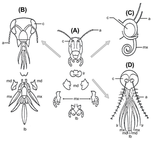
Insect head parts. Legend: a, antennae; c, compound eye; lb, labium; lr, labrum; md, mandibles; mx, maxillae.
 
Insect anatomy

A typical insect head possesses a pair of antennae; eyes; mandibles, labrum, maxillae and labium (the latter four forming the cluster of "mouth parts", no. 32. in the diagram). Lying above the oesophagus is the brain or supraesophageal ganglion, divided into three pairs of ganglia: the protocerebrum, deutocerebrum and tritocerebrum from front to back (collectively no. 5 in the diagram). Nerves from the protocerebrum lead to the large compound eyes; from the deutocerebrum to the antennae; and from the tritocerebrum to the labrum and stomatogastric nervous system. Circum-oesophageal connectives lead from the tritocerebrum around the gut to connect the brain to the ventral ganglionated nerve cord: nerves from the first three pairs of ganglia lead to the mandibles, maxillae and labium, respectively. The position of the mouth and the circum-oesophageal connectives allows a distinction to be made between pre- and post-oral structures; although it should be borne in mind that because structures can move around during development, a pre-oral position of a structure in the adult does not necessarily prove that its developmental origin is from there. The myriapod head is very similar to that of the insects.

The crustacean head is broadly similar to that of the insects, but possesses, in addition, a second pair of antennae that are innervated from the tritocerebrum. In place of the labium, crustaceans possess a second pair of maxillae.

Chelicerate head structures differ considerably from those of mandibulates (i.e. insects, crustaceans and myriapods); they possess eyes and a single pair of grasping appendages innervated from the brain, plus a labrum-like structure. Behind the mouth lies another pair of mouthparts, the pedipalps, and behind them lie the series of walking limbs. In chelicerates, the leg-bearing segments are fused with the anterior segments to form a prosoma, so that in living arthropods a distinct head only exists in mandibulates.

The acron concept

The arthropod head problem has until recently been predicated on the Articulata theory, i.e. that the arthropods and annelids are close relatives. Although arthropods are essentially direct developers that do not possess a trochophore-like larva, the annelids do. During annelid metamorphosis, segments are added close to the posterior of the body, behind the mouth; whereas the brain is derived from the episphere or region in front of the mouth. Recognition of this led to the concept of a primary, non-segmental component of the body in annelids known as the acron being developed, from which the brain is ultimately derived. Because the arthropod and annelid heads, in the light of the Articulata theory, were assumed to be structurally homologous in some way, the arthropod head was also often considered to incorporate a non-segmental acronal component. Taking the homology between annelid and arthropod heads at face value, Swedish workers such as Hanström and Holmgren assumed that a large part of the arthropod head must correspond to the acron, a view followed later by several prominent American insect workers such as Butt and Snodgrass. They proposed that all pre-oral structures in insects were non-segmental, although such a view is at odds with the preoral position of apparently bona fide appendages such as the antennae. A less extreme set of theories propose that only the protocerebrum and associated structures should be considered to be acronal.

The view that the arthropod head must contain an acronal remnant has been shaken by the relatively recent revision of protostome phylogeny, which has dismantled the Articulata and placed the arthropods together with a group of unsegmented worms often referred to as the Cycloneuralia in the so-called Ecdysozoa. All members of the Ecdysozoa are direct developers without a trochophore, and the cycloneuralians have terminal mouths. As a result, the idea of the arthropods having inherited a preoral acron from their ancestors seems less likely.

Molecular development and the arthropod head problem

Expression of Hox genes in the body segments of different groups of arthropod. The Hox genes 7, 8, and 9 correspond in these groups but are shifted (by heterochrony) by up to three segments. Segments with maxillopeds have Hox gene 7. Fossil trilobites probably had three body regions, each with a unique combination of Hox genes.

The study of how developmental genes are expressed during embryogenesis has become an important new tool in the last twenty years for understanding the structure and evolution of morphology. The arthropod head problem has been tackled in three main ways in this regard, first by using genetic segmental markers to probe the obscure region in front of the mouth, especially in insects; second by looking at Hox gene expression patterns to detect patterns of homology among different arthropods; and third, by studying gene expression in particular features (especially the labrum) to determine its appendiculate or other status. Because all arthropods have the same complement of nine Hox loci, the morphological diversification observed is caused by heterochrony, meaning that the genes are expressed at different times.

Areas of agreement

It is widely agreed that the insect, myriapod and crustacean heads are very similar. The apparent lack of a second antenna in insects and myriapods is explained by the idea that this appendage has been lost, leaving an appendage-less segment known as the intercalary segment. Modern phylogenies do not in general support an insect-myriapod relationship, suggesting that the second antenna has been lost independently in each group, perhaps as a result of a convergent adaptation to life on land. Furthermore, there is general agreement that the mandibles, first maxillae and labium/second maxillae each represent a post-oral segment; and that the first antenna represents a preoral segment.

Areas of disagreement

Areas of disagreement can be grouped into three categories: the nature of the pre-antennal region in mandibulates; the nature of the labrum; and the relationship between the chelicerate and mandibulate anterior segments.

Nature of the preoral region

The degree to which the area in front of the mouth is segmented remains one of the major controversies in the arthropod head problem. As already mentioned, earlier workers often considered the entire pre-oral region to be "acronal" and thus nonsegmental. Modern workers universally accept that at least the deuterocerebrum is segmental. However, the nature of the region in front of this is much less certain. Some molecular development studies have given limited support to the idea of an "ocular" segment corresponding to the protocerebrum; but these data are not unequivocal. The idea of the protocerebrum actually comprising two components has also received support from both molecular and embryological data.

On this view, the protocerebrum comprises a typical 'segment', the prosocerebrum, marked by the expression of engrailed at its caudal margin and a pair of appendages (in most crown euarthropods, compound eyes, which are interpreted as modified trunk appendages), and a pre-segmental region, the archicerebrum, which bore a pair of appendages that are not serial homologues of the trunk appendages; these are represented by the onychophoran antennae and the 'great appendages' of certain stem euarthropods. The archicerebrum is in some ways equivalent to the 'acron', and may be equivalent (by means of a shared equivalent structure in the common ancestor of lophotrochozoans and ecdysozoans) with the annelid prototroch; it can be recognized by the expression of the genes optix and six3 during development, whereas the prosocerebrum is associated with orthodenticle and its homologs.

(Note that the terms archicerebrum and prosocerebrum are not always used consistently; see Composition of the protocerebrum.)

The labrum

The labrum is a flap-like structure that lies immediately in front of the mouth in almost all extant euarthropods, the general exception being provided by the probable chelicerate-relatives the pycnogonids. It has proved to be by far the most controversial of all arthropod head structures. It is innervated in crustaceans and insects from the tritocerebrum, i.e. the back of the brain. However, in development it often appears at the anterior of the head, and migrates backwards towards its adult position. Furthermore, it often appears as a bilobed structure, with a set of muscles, nerves and gene expression in many ways similar to that of a trunk appendage. This evidence has been used to suggest that the labrum is in fact a highly reduced appendage.

Its innervation from the rear of the brain has suggested to some workers that, if an appendage, it is the appendage of the tritocerebral segment; a point disputed by others who argue that the presence of a well-developed appendage in at least crustaceans in this segment (i.e., the second antenna, corresponding to the intercalary segment of insects) rules this out. If the labrum is an appendage then, it seems possible that its origin is indicated by its developmentally anterior position, i.e., that it is the appendage of a segment anterior to the first antenna. The most obvious choice for this is the segment whose ganglion is the protocerebrum, which in extant euarthropods bears no appendage (apart from the eyes). Strausfeld finds support for this hypothesis in the presence of a median nerve bundle connecting the labrum to the anterior of the protocerebrum, and the expression of the gene six3 in the labrum has been taken as evidence for its homology with onychophoran antennae (frontal appendages borne from the anterior of the protocerebrum).

If the labrum is really an anterior appendage that has migrated to the posterior, then it may be homologous to the "antennae" of onychophorans, which, as discussed below, seem to be innervated from a very anterior part of the brain, i.e. in front of the eyes. It has even been suggested (e.g., by Roonwal) that the labrum belongs to an even more obscure segment that lies in front of the ocular one. Nevertheless, many workers continue to be highly skeptical about the appendiculate nature of the labrum, preferring to see it as it appears, i.e., as an outgrowth of the body wall just in front of the mouth.

Particularly in some fossil groups, such as certain trilobites, the labrum is often covered with a sclerotised plate, the hypostome. Confusion can arise where the two structures are conflated or mistaken for one another.

Mandibulate/chelicerate head homologies

Given the disagreements about the structure of the insect head, on which most effort has been spent, it is no surprise that the potential homologies between it and other arthropods, notably the chelicerates, are also very controversial. From after the Second World War to the 1980s a commonly accepted model of arthropod evolution was that the extant euarthropods were polyphyletic, i.e. the main lineages had evolved independently from soft-bodied, annelid-like ancestors, following the work of Tiegs and especially Sidnie Manton. In this view, most of the head structures would also be convergent, and thus there was no point looking for specific homologies between major groups. However, the monophyletic theory of arthropod origins has since decisively gained the upper hand, which raises the problem of head homology once more.

The classical view was that the chelicerae were homologous to the second antennae of crustaceans (i.e., they are innervated from the tritocerebrum), a view based partly on the fact that the chelicerae were innervated from the same ganglion that innervates the labrum, which is the tritocerebrum in crustaceans and insects. Given that there are apparently no appendages in front of the chelicerae, the implication was that the deuterocerebrum had been lost in chelicerates (the protocerebrum innervates the eyes in both groups in this view). While this view still has its defenders (notably Colette and Jacques Bitsch), the alternative view that the chelicerae are innervated from the deuterocerebrum has gained ground, based on molecular development in mites and spiders, and neuroanatomy in Limulus. If this is the case, then chelicerates simply have no tritocerebrum, i.e. there is no third supraoesophageal ganglion of the brain; the segment corresponding to it would be the suboesophageal pedipalp one. Such a theory does not, however, immediately account for the same ganglionic innervation of the chelicerae and labrum, although one solution is simply to claim that the labrum itself is not homologous between mandibulates and chelicerates (the view, for example, of Dieter Waloszek and colleagues).

The head of onychophorans

The brains of onychophorans (velvet worms) have been recently re-investigated and have been shown to possess two unusual features. First, although the mouth is ventral, as is the case in euarthropods, it is innervated from three different places; the sides, the posterior, and by a nerve that originates dorsally, and passes anteriorly down to curve back to the front of the mouth. This set of innervation makes sense if the mouth of onychophorans was originally terminal and has been bent downwards. Second, the antennae of the onychophorans appear to be innervated from in front of the eyes; which in euarthropod terms implies a protocerebral (or potentially even more anterior) innervation. This is supported by gene expression data, which show that the jaws too are derived from a protocerebral or deuterocerebral segment. As all euarthropod antennae are deuterocerebral or tritocerebral, this implies that the onychophoran antennae are not homologous to any euarthropod ones.

The tritocerebrum in arthropods is the first segment to express Hox genes; on this basis, it can be recognised as homologous to the third head segment in onychophora, which bears the slime glands (a pair of highly modified appendages).

The head of pentastomids

The parasitic pentastomida hatch with four head segments and three trunk segments, with two more body segments being added during postembryonic development. This number of segments then remains constant for the rest of their life. There are no antenna, and their mouth has no piercing, biting or sucking extremities. On each side of the mouth there is a pair of retractable hooks, and the mouth itself is sustained by a chitinous buccal ring. They feed through a pumping mechanism located in the pharynx, consisting of two rigid chitinous plates that is connected to several associated muscles.

The brain of tardigrades

Tardigrades bear a circumoral nerve ring which has been homologised with the nerve ring of the ancestral ecdysozoan, and the arthropod (=Euarthropoda + Onychophora) protocerebrum, suggesting that the protocerebrum is homologous with the ancestral ecdysozoan brain.

On this view, the stylet apparatus is homologous with the euarthropod labrum / onychophoran antennae, and the first pairs of walking legs correspond to the deutocerebral and tritocerebral appendages.

Fossil evidence

The Cambrian fossil record, above all the various lagerstätten such as the Burgess Shale, Sirius Passet, Chengjiang and Orsten faunas, has yielded a very rich record of well-preserved arthropods, including the well-known trilobites.

Many Cambrian arthropods, including the trilobites themselves, possess a single pair of slender antennae, which have been equated with either the first or second antennae of the crustaceans; and either the chelicerae or the missing appendages of the supposedly reduced deuterocerebrum in chelicerates. However, another group of arthropods, the so-called "great appendage" arthropods, including Yohoia, Leanchoilia and Alalcomenaeus, do not possess simple antennae, but rather have a robust, branched structure, which was called the "great appendage" by Harry B. Whittington in his restudy of these taxa. Yet another group of arthropods may possess two differentiated head appendages, of which the most important and controversial is the Chengjiang form Fuxianhuia. Fuxianhuia was claimed to possess a pair of short antennae anterior, followed by a robust pair of "sub-chelate" appendages. However, this assessment has been both disputed by Waloszek and colleagues, who consider that the sub-chelate appendages are in fact gut diverticulae; and supported by Graham Budd. Thus, its nature remains controversial at present. Other taxa have also been claimed to have a somewhat similar anterior appendage arrangement (e.g. Fortiforceps) but, with the exception of the well-preserved Branchiocaris from the Burgess Shale, most of them are highly equivocal.

In almost all Cambrian arthropods, the post-oral limbs show very little differentiation compared to the trunk limbs; the heads posterior to the mouth shows a considerable degree of variability, however, in the number of segments incorporated into the head.

Trilobites, in particular, possess a ventral sclerotised plate in the head called the hypostome. Whether this is homologous to the labrum or not is debated; although Waloszek and others have argued that as the phosphatocopines (upper stem-group crustaceans) seem to possess both, it cannot be.

Theories of Cambrian arthropod head segmentation

There are at least four main theories to account for anterior head appendages in Cambrian arthropods:

Scholtz and Edgecombe

Gerhard Scholtz and Greg Edgecombe accept that the antennae of onychophorans are protocerebral, and call them "primary" antennae to distinguish them from the "secondary" antennae of groups such as the insects and crustaceans. They also accept that taxa such as Fuxianhuia possess both antennae and "great appendages". Because in Fuxianhuia the antennae lie anterior to the great appendages, they suggest that these antennae are the inherited primitive "primary" antennae; and that the great appendages are thus equivalent to the first antennae of crustaceans. Because the secondary antennae are not present in stem group arthropods such as Fuxianhuia, nor in the extant chelicerates, they propose that arthropods, such as the trilobites, that possess secondary antennae, belong in a monophyletic group that also includes the mandibulates, called the Antennata. The trilobites are thus, in their view, not stem-group chelicerates, a commonly held view, but rather, stem-group mandibulates. The status of the labrum is not resolved by this theory, but they argue that it the evidence for it being appendiculate is not compelling; thus it does not have to correspond to a well-developed appendage of any Cambrian arthropod.

The 2014 description of Lyrarapax poses a challenge for this theorem: assuming that its nervous tissue is correctly identified as such, the great appendages of this radiodont are innervated into the front of the protocerebrum, undermining the suggestion that the great appendages are deuterocerebral.

Budd

Graham Budd's theory agrees with that of Scholtz and Edgecombe in accepting the protocerebral nature of the onychophoran antennae, and the two preoral appendages of Fuxianhuia. However, he traces the origin of the "great appendages" in the differentiated frontal appendages of Cambrian lobopods such as Aysheaia and Kerygmachela, neither of which possess convincing antennae. Thus, in Budd's view, the order of the two anterior appendages of taxa such as Fuxianhuia are reversed: the antennae are the first antennae (deutocerebral) of the mandibulates; and the great appendages correspond to the primary antennae of the onychophorans and Cambrian lobopods. Following previous work by Dewel and colleagues, Budd accounts for their reversal by arguing that the mouth in basal lobopods was terminal, and that as it rotated backwards and downwards, it brought the anterior appendage backwards with it. Given this transformation, it is likely, under this theory, that the remnant of the great appendage/primary antenna is the labrum of extant arthropods. Because in this view Fuxianhuia possesses both a hypostome and a great appendage, the hypostome cannot be straightforwardly homologous with the labrum.

Pycnogonids and the great appendage theory

Maxmen and others recently published a morphologically-based paper that claimed the enigmatic chelifores of extant pycnogonids (sea spiders) are innervated from the protocerebrum, and not from the trito- or deutocerebrum as previously claimed. This would suggest that pycnogonids had uniquely retained a "great appendage" homologue as an appendage, unlike all other euarthropods in which it had been transformed into the labrum (pycnogonids lack a labrum). However, expression data of Hox genes that were published shortly afterwards suggested that the chelifores were deuterocerebral and thus most likely to be homologous to the chelicerae. The pycnogonids are thus neutral with regard to the great appendage theory.

Waloszek

Dieter Waloszek and colleagues have offered a rather different account of Cambrian arthropod head structure. They do not necessarily accept the primary antenna theory of the onychophoran antennae; and they reject the idea that Fuxianhuia or any of its close relatives possessed a great appendage. Rather, they place the "great appendage" arthropods in the stem-group of the chelicerates, arguing that the great appendage is homologous to the chelicerae of chelicerates, and the first antennae of crustaceans.

Cotton and Braddy

Trevor Cotton and Simon Braddy, in a comprehensive cladistic analysis of Cambrian arthropods, also proposed that the great appendage arthropods were stem-group chelicerates; accepting that Fuxianhuia and relatives possessed two preoral appendages, they defended the classical view that the great appendage and the chelicerae were tritocerebral in origin; i.e. that the antennae of Fuxianhuia were deuterocerebral.

Assessment

The number and nature of the post-oral segments in the insect head have rarely been questioned. A much more difficult area, however, has been the nature of the preoral region. The obvious contradiction between a theory that no-preoral structures are segmental, and evidence, such as for the first antennae of crustaceans, that some such structures clearly are, led workers as long ago as Lankester to posit that there has been forward migration of segments in front of the mouth. Indeed, such a process can be seen in ontogeny of the tritocerebrum, which can be seen to migrate forward as the brain develops; furthermore, although in most insects and crustaceans its ganglia are part of the brain, its commissures still loop behind it, suggesting derivation from a more posterior position.

Nevertheless, even allowing for this possibility, the complexity of the anterior part of the brain, which even if the acron concept is incorrect may still have been inherited from very basal animals; untangling the new characters evolved by the earliest arthropods from those inherited from their ancestors therefore still stands centrally in the arthropod head problem.

Human evolutionary developmental biology

From Wikipedia, the free encyclopedia

Human evolutionary developmental biology or informally human evo-devo is the human-specific subset of evolutionary developmental biology. Evolutionary developmental biology is the study of the evolution of developmental processes across different organisms. It is utilized within multiple disciplines, primarily evolutionary biology and anthropology. Groundwork for the theory that "evolutionary modifications in primate development might have led to … modern humans" was laid by Geoffroy Saint-Hilaire, Ernst Haeckel, Louis Bolk, and Adolph Schultz. Evolutionary developmental biology is primarily concerned with the ways in which evolution affects development, and seeks to unravel the causes of evolutionary innovations.

The approach is relatively new, but has roots in Schultz's The physical distinctions of man, from the 1940s. Shultz urged broad comparative studies to identify uniquely human traits.

History

Brian Hall traces the roots of evolutionary developmental biology in his 2012 paper on its past present and future. He begins with Darwinian evolution and Mendel's genetics, noting the tendency of the followers of both men in the early 20th century to follow separate paths and to set aside and ignore apparently inexplicable problems. Greater understanding of genotypic and phenotypic structures from the 1940s enabled the unification of evolution and genetics in the modern synthesis. Molecular biology then enabled researchers to explore the mechanisms and evolution of embryonic development in molecular detail, including in humans.

Human and primate development

Many of the human evolutionary developmental biology studies have been modeled after primate studies and consider the two together in a comparative model. Brain ontogeny and human life history evolution were looked at by Leigh, in a 2006 paper. He compares brain growth patterns for Homo erectus and Homo sapiens to get at the evolution of brain size and weight. Leigh found three different patterns, all of which pointed to the growth rate of H. erectus either matching or exceeding H. erectus. He makes the case that this finding had wide application and relevance t the overall study of human evolution. It is pertinent specifically to the connections between energy expenditure and brain development. These finding are of specific utility in studies on maternal energy expenditure. Comparative study of nonhuman primates, fossils and modern humans to study patterns of brain growth to correlate human life history and brain growth.

Jeremy De Silva and Julie Lesnik examined chimpanzee neonatal brain size to identify implications for brain growth in Homo erectus. This changed the understanding of differences and similarities of post-natal brain growth in humans and chimpanzees. The study found that there was a distinction necessary between growth time and growth rate. The times of growth were strikingly similar, but the rates were not. The paper further advocates the use of fossils to assess brain size in general and in relation to cranial capacity.

Utilization of endocranial volume as a measure for brain size has been a popular methodology with the fossil record since Darwin in the mid 1800s. This measure has been used to access the metabolic requirements for brain growth and the subsequent trade-offs.

Neoteny

Some of the work on human evolutionary developmental biology has centered around the neotenous features that present in humans, but are not shared across the primate spectrum. Steven J. Gould discussed the presentation of neoteny with "terminal additions" in humans. Neoteny is defined as the delayed or slowed development in humans when compared with their non-human primate counterparts. The "terminal additions" were extensions or reductions in the rate and scope of stages of development and growth. Gould hypothesized that this process and production of neoteny in humans might be the key feature that ultimately lead to the emotional and communicative nature of humans. He credits this factor as an integral facet of human evolution. However, there have also been cautions against the application of this aspect to group ranking during it inappropriate as a measure of evolutionary achievement.

Fossil record

Early comparative and human studies examined the fossil record to measure features like cranial sizes and capacities so as to infer brain size, growth rate, total growth and potential implications for energy expenditure. Helpful as this is, the static nature of individual fossils presents its own challenge. The phylogenic fossil line is itself a hypothesis, so anything based upon it is equally hypothetical.

Using the fossil record of Neanderthals, modern humans, and chimpanzees, Gunz et al. examined that patterns of endocranial development. They found that there are common features shared between the three, and that modern humans diverge from these common patterns in the first year of life. They concluded that even though much of the developmental results are similar insofar as brain size, the trajectories by which they arrived are not shared. Most of the differences between the two arise post-natally, in the first year, with cognitive development.

There have been a number of studies that not only take incomplete fossil records into consideration, but have attempt to specifically identify the barriers presented by this condition. For example, Kieran McNulty covers the potential utilities and constraints of using incomplete fossil taxa to examine longitudinal development in Australopithecus africanis.

Many studies on development have been human-specific. In his 2011 paper, Bernard Crespi focused on adaptation and genomic conflict in childhood diseases. He considers the evolution of childhood diseases and their risk levels, and finds that both risk and disease have evolved.

Hotchberg and Belsky incorporate a life-history perspective, looking at adolescence. Substantial variation in phenotypic paths and presentations suggest significant environmental influence. They focus on plasticity between stages of development and the factors that shape it. Rate of maturation, fecundity, and fertility were all impacted by environmental circumstances. They argue that early maturation can be positive, reflecting opportunistic actions within specific conditions.

Genetic and epigenetic basis

Technological advances that have allowed better and better access to the growth of the human form in utero have proven particularly formative in studies involving focus on genetic and epigenetic development. Bakker et al. look at the interconnected nature of developmental processes and attempt to use fetal vertebral abnormalities as an indicator for other malformations. They found that the origin of the cells was not nearly as highly correlated as the observed developmental signals. In utero development and malformations were correlated in severity.

Freiston and Galis look at the development of ribs, digits, and mammalian asymmetry. They argue that this construction is relevant for the study of disease, the consistency in evolution of body plans, and understanding of developmental constraints. Sexual dimorphism in prenatal digit ratio was found as early as 14 weeks and was maintained whether or not the fleshy finger part was included.

Language and cognitive studies

Languages and cognitive function have also been subjects of evolutionary studies. Insofar as language and evolutionary developmental biology, there is tension from the gate. Much of this contention has centered around whether to view and study language as an adaptation in and of its self, or as a by-product of other adaptations. Jackendoff and Pinker have argued for language as an adaptation owing to the interdependent social nature of humans. To support these claims, he points to things like the bi-directionality in language usage and comprehension. This is a counter to the claims by theorists like Noam Chomsky, who argued against language as a human specific adaptation.

Adaptation and adaptive theory has been argued even separate from its utility in the study of language. Gould and Lewontin engage with what they saw as flaws in adaptive theory using the analogy of the spandrels of San Marco. Among the issues identified is the lack of distinction between what trait developed and how it is used, and the underlying reasons or forces that created the novel trait initially. This is particularly difficult to access in intangible language and cognition.

This debate has continued over decades and most often presents in the form of a response and published dialogue between theorists. This continued debate has prompted efforts to marry the two perspectives in a useful way. Fitch argues that these two approaches can be rectified with the study of "neutral computation and mammalian brain development". It may be more useful to consider specific components of neural computation and development, what has been selected for, and to what end.

Ploeger and Galis tackled modular evolvability and developmental constraints in human and other primate evolutionary trajectories. They argue that these should be treated with an interdisciplinary approach across the cognitive sciences. They frame this in the context of:

  1. Modularity — the ability of a system to organize individuals for the benefit of the whole
  2. Evolvability — ability of organism or organisms to adapt through evolution
  3. Developmental constraints — those things that act as barriers to evolutionary adaptations.

Evolutionary developmental biology

Homologous hox genes in such different animals as insects and vertebrates control embryonic development and hence the form of adult bodies. These genes have been highly conserved through hundreds of millions of years of evolution.

Evolutionary developmental biology (informally, evo-devo) is a field of biological research that compares the developmental processes of different organisms to infer the ancestral relationships between them and how developmental processes evolved.

The field grew from 19th-century beginnings, where embryology faced a mystery: zoologists did not know how embryonic development was controlled at the molecular level. Charles Darwin noted that having similar embryos implied common ancestry, but little progress was made until the 1970s. Then, recombinant DNA technology at last brought embryology together with molecular genetics. A key early discovery was of homeotic genes that regulate development in a wide range of eukaryotes.

The field is characterised by some key concepts which took evolutionary biologists by surprise. One is deep homology, the finding that dissimilar organs such as the eyes of insects, vertebrates and cephalopod molluscs, long thought to have evolved separately, are controlled by similar genes such as pax-6, from the evo-devo gene toolkit. These genes are ancient, being highly conserved among phyla; they generate the patterns in time and space which shape the embryo, and ultimately form the body plan of the organism. Another is that species do not differ much in their structural genes, such as those coding for enzymes; what does differ is the way that gene expression is regulated by the toolkit genes. These genes are reused, unchanged, many times in different parts of the embryo and at different stages of development, forming a complex cascade of control, switching other regulatory genes as well as structural genes on and off in a precise pattern. This multiple pleiotropic reuse explains why these genes are highly conserved, as any change would have many adverse consequences which natural selection would oppose.

New morphological features and ultimately new species are produced by variations in the toolkit, either when genes are expressed in a new pattern, or when toolkit genes acquire additional functions. Another possibility is the Neo-Lamarckian theory that epigenetic changes are later consolidated at gene level, something that may have been important early in the history of multicellular life.

History

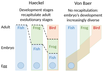
Embryology theories of Ernst Haeckel, who argued for recapitulation of evolutionary development in the embryo, and Karl Ernst von Baer's epigenesis

Recapitulation

A recapitulation theory of evolutionary development was proposed by Étienne Serres in 1824–26, echoing the 1808 ideas of Johann Friedrich Meckel. They argued that the embryos of 'higher' animals went through or recapitulated a series of stages, each of which resembled an animal lower down the great chain of being. For example, the brain of a human embryo looked first like that of a fish, then in turn like that of a reptile, bird, and mammal before becoming clearly human. The embryologist Karl Ernst von Baer opposed this, arguing in 1828 that there was no linear sequence as in the great chain of being, based on a single body plan, but a process of epigenesis in which structures differentiate. Von Baer instead recognised four distinct animal body plans: radiate, like starfish; molluscan, like clams; articulate, like lobsters; and vertebrate, like fish. Zoologists then largely abandoned recapitulation, though Ernst Haeckel revived it in 1866.

Evolutionary morphology

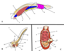
A. Lancelet (a chordate), B. Larval tunicate, C. Adult tunicate. Kowalevsky saw that the notochord (1) and gill slit (5) are shared by tunicates and vertebrates.

From the early 19th century through most of the 20th century, embryology faced a mystery. Animals were seen to develop into adults of widely differing body plan, often through similar stages, from the egg, but zoologists knew almost nothing about how embryonic development was controlled at the molecular level, and therefore equally little about how developmental processes had evolved. Charles Darwin argued that a shared embryonic structure implied a common ancestor. As an example of this, Darwin cited in his 1859 book On the Origin of Species the shrimp-like larva of the barnacle, whose sessile adults looked nothing like other arthropods; Linnaeus and Cuvier had classified them as molluscs. Darwin also noted Alexander Kowalevsky's finding that the tunicate, too, was not a mollusc, but in its larval stage had a notochord and pharyngeal slits which developed from the same germ layers as the equivalent structures in vertebrates, and should therefore be grouped with them as chordates. 19th century zoology thus converted embryology into an evolutionary science, connecting phylogeny with homologies between the germ layers of embryos. Zoologists including Fritz Müller proposed the use of embryology to discover phylogenetic relationships between taxa. Müller demonstrated that crustaceans shared the Nauplius larva, identifying several parasitic species that had not been recognised as crustaceans. Müller also recognised that natural selection must act on larvae, just as it does on adults, giving the lie to recapitulation, which would require larval forms to be shielded from natural selection. Two of Haeckel's other ideas about the evolution of development have fared better than recapitulation: he argued in the 1870s that changes in the timing (heterochrony) and changes in the positioning within the body (heterotopy) of aspects of embryonic development would drive evolution by changing the shape of a descendant's body compared to an ancestor's. It took a century before these ideas were shown to be correct. In 1917, D'Arcy Thompson wrote a book on the shapes of animals, showing with simple mathematics how small changes to parameters, such as the angles of a gastropod's spiral shell, can radically alter an animal's form, though he preferred mechanical to evolutionary explanation. But for the next century, without molecular evidence, progress stalled.

The modern synthesis of the early 20th century

In the so-called modern synthesis of the early 20th century, Ronald Fisher brought together Darwin's theory of evolution, with its insistence on natural selection, heredity, and variation, and Gregor Mendel's laws of genetics into a coherent structure for evolutionary biology. Biologists assumed that an organism was a straightforward reflection of its component genes: the genes coded for proteins, which built the organism's body. Biochemical pathways (and, they supposed, new species) evolved through mutations in these genes. It was a simple, clear and nearly comprehensive picture: but it did not explain embryology.

The evolutionary embryologist Gavin de Beer anticipated evolutionary developmental biology in his 1930 book Embryos and Ancestors, by showing that evolution could occur by heterochrony, such as in the retention of juvenile features in the adult. This, de Beer argued, could cause apparently sudden changes in the fossil record, since embryos fossilise poorly. As the gaps in the fossil record had been used as an argument against Darwin's gradualist evolution, de Beer's explanation supported the Darwinian position. However, despite de Beer, the modern synthesis largely ignored embryonic development to explain the form of organisms, since population genetics appeared to be an adequate explanation of how forms evolved.

The lac operon

The lac operon. Top:Repressed, Bottom:Active
1: RNA Polymerase, 2: Repressor, 3: Promoter, 4: Operator, 5: Lactose, 6–8: protein-encoding genes, controlled by the switch, that cause lactose to be digested

In 1961, Jacques Monod, Jean-Pierre Changeux and François Jacob discovered the lac operon in the bacterium Escherichia coli. It was a cluster of genes, arranged in a feedback control loop so that its products would only be made when "switched on" by an environmental stimulus. One of these products was an enzyme that splits a sugar, lactose; and lactose itself was the stimulus that switched the genes on. This was a revelation, as it showed for the first time that genes, even in an organism as small as a bacterium, were subject to fine-grained control. The implication was that many other genes were also elaborately regulated.

The birth of evo-devo and a second synthesis

In 1977, a revolution in thinking about evolution and developmental biology began, with the arrival of recombinant DNA technology in genetics, and the works Ontogeny and Phylogeny by Stephen J. Gould and Evolution by Tinkering by François Jacob. Gould laid to rest Haeckel's interpretation of evolutionary embryology, while Jacob set out an alternative theory. This led to a second synthesis, at last including embryology as well as molecular genetics, phylogeny, and evolutionary biology to form evo-devo. In 1978, Edward B. Lewis discovered homeotic genes that regulate embryonic development in Drosophila fruit flies, which like all insects are arthropods, one of the major phyla of invertebrate animals. Bill McGinnis quickly discovered homeotic gene sequences, homeoboxes, in animals in other phyla, in vertebrates such as frogs, birds, and mammals; they were later also found in fungi such as yeasts, and in plants. There were evidently strong similarities in the genes that controlled development across all the eukaryotes. In 1980, Christiane Nüsslein-Volhard and Eric Wieschaus described gap genes which help to create the segmentation pattern in fruit fly embryos; they and Lewis won a Nobel Prize for their work in 1995.

Later, more specific similarities were discovered: for example, the Distal-less gene was found in 1989 to be involved in the development of appendages or limbs in fruit flies, the fins of fish, the wings of chickens, the parapodia of marine annelid worms, the ampullae and siphons of tunicates, and the tube feet of sea urchins. It was evident that the gene must be ancient, dating back to the last common ancestor of bilateral animals (before the Ediacaran Period, which began some 635 million years ago). Evo-devo had started to uncover the ways that all animal bodies were built during development.

The control of body structure

Deep homology

Roughly spherical eggs of different animals give rise to extremely different bodies, from jellyfish to lobsters, butterflies to elephants. Many of these organisms share the same structural genes for body-building proteins like collagen and enzymes, but biologists had expected that each group of animals would have its own rules of development. The surprise of evo-devo is that the shaping of bodies is controlled by a rather small percentage of genes, and that these regulatory genes are ancient, shared by all animals. The giraffe does not have a gene for a long neck, any more than the elephant has a gene for a big body. Their bodies are patterned by a system of switching which causes development of different features to begin earlier or later, to occur in this or that part of the embryo, and to continue for more or less time.

The puzzle of how embryonic development was controlled began to be solved using the fruit fly Drosophila melanogaster as a model organism. The step-by-step control of its embryogenesis was visualized by attaching fluorescent dyes of different colours to specific types of protein made by genes expressed in the embryo. A dye such as green fluorescent protein, originally from a jellyfish, was typically attached to an antibody specific to a fruit fly protein, forming a precise indicator of where and when that protein appeared in the living embryo.

The pax-6 gene controls development of eyes of different types across the animal kingdom.

Using such a technique, in 1994 Walter Gehring found that the pax-6 gene, vital for forming the eyes of fruit flies, exactly matches an eye-forming gene in mice and humans. The same gene was quickly found in many other groups of animals, such as squid, a cephalopod mollusc. Biologists including Ernst Mayr had believed that eyes had arisen in the animal kingdom at least 40 times, as the anatomy of different types of eye varies widely. For example, the fruit fly's compound eye is made of hundreds of small lensed structures (ommatidia); the human eye has a blind spot where the optic nerve enters the eye, and the nerve fibres run over the surface of the retina, so light has to pass through a layer of nerve fibres before reaching the detector cells in the retina, so the structure is effectively "upside-down"; in contrast, the cephalopod eye has the retina, then a layer of nerve fibres, then the wall of the eye "the right way around". The evidence of pax-6, however, was that the same genes controlled the development of the eyes of all these animals, suggesting that they all evolved from a common ancestor. Ancient genes had been conserved through millions of years of evolution to create dissimilar structures for similar functions, demonstrating deep homology between structures once thought to be purely analogous. This notion was later extended to the evolution of embryogenesis and has caused a radical revision of the meaning of homology in evolutionary biology.

Gene toolkit

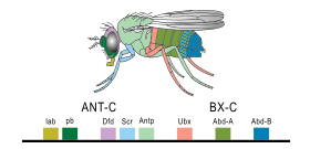
Expression of homeobox (Hox) genes in the fruit fly

A small fraction of the genes in an organism's genome control the organism's development. These genes are called the developmental-genetic toolkit. They are highly conserved among phyla, meaning that they are ancient and very similar in widely separated groups of animals. Differences in deployment of toolkit genes affect the body plan and the number, identity, and pattern of body parts. Most toolkit genes are parts of signalling pathways: they encode transcription factors, cell adhesion proteins, cell surface receptor proteins and signalling ligands that bind to them, and secreted morphogens that diffuse through the embryo. All of these help to define the fate of undifferentiated cells in the embryo. Together, they generate the patterns in time and space which shape the embryo, and ultimately form the body plan of the organism. Among the most important toolkit genes are the Hox genes. These transcription factors contain the homeobox protein-binding DNA motif, also found in other toolkit genes, and create the basic pattern of the body along its front-to-back axis. Hox genes determine where repeating parts, such as the many vertebrae of snakes, will grow in a developing embryo or larva. Pax-6, already mentioned, is a classic toolkit gene. Although other toolkit genes are involved in establishing the plant bodyplan, homeobox genes are also found in plants, implying they are common to all eukaryotes.

The embryo's regulatory networks

The protein products of the regulatory toolkit are reused not by duplication and modification, but by a complex mosaic of pleiotropy, being applied unchanged in many independent developmental processes, giving pattern to many dissimilar body structures. The loci of these pleiotropic toolkit genes have large, complicated and modular cis-regulatory elements. For example, while a non-pleiotropic rhodopsin gene in the fruit fly has a cis-regulatory element just a few hundred base pairs long, the pleiotropic eyeless cis-regulatory region contains 6 cis-regulatory elements in over 7000 base pairs. The regulatory networks involved are often very large. Each regulatory protein controls "scores to hundreds" of cis-regulatory elements. For instance, 67 fruit fly transcription factors controlled on average 124 target genes each. All this complexity enables genes involved in the development of the embryo to be switched on and off at exactly the right times and in exactly the right places. Some of these genes are structural, directly forming enzymes, tissues and organs of the embryo. But many others are themselves regulatory genes, so what is switched on is often a precisely-timed cascade of switching, involving turning on one developmental process after another in the developing embryo.

Gene product distributions along the long axis of the early embryo of a fruit fly

Such a cascading regulatory network has been studied in detail in the development of the fruit fly embryo. The young embryo is oval in shape, like a rugby ball. A small number of genes produce messenger RNAs that set up concentration gradients along the long axis of the embryo. In the early embryo, the bicoid and hunchback genes are at high concentration near the anterior end, and give pattern to the future head and thorax; the caudal and nanos genes are at high concentration near the posterior end, and give pattern to the hindmost abdominal segments. The effects of these genes interact; for instance, the Bicoid protein blocks the translation of caudal's messenger RNA, so the Caudal protein concentration becomes low at the anterior end. Caudal later switches on genes which create the fly's hindmost segments, but only at the posterior end where it is most concentrated.

Gap genes in the fruit fly are switched on by genes such as bicoid, setting up stripes across the embryo which start to pattern the body's segments.

The Bicoid, Hunchback and Caudal proteins in turn regulate the transcription of gap genes such as giant, knirps, Krüppel, and tailless in a striped pattern, creating the first level of structures that will become segments. The proteins from these in turn control the pair-rule genes, which in the next stage set up 7 bands across the embryo's long axis. Finally, the segment polarity genes such as engrailed split each of the 7 bands into two, creating 14 future segments.

This process explains the accurate conservation of toolkit gene sequences, which has resulted in deep homology and functional equivalence of toolkit proteins in dissimilar animals (seen, for example, when a mouse protein controls fruit fly development). The interactions of transcription factors and cis-regulatory elements, or of signalling proteins and receptors, become locked in through multiple usages, making almost any mutation deleterious and hence eliminated by natural selection.

The origins of novelty

Among the more surprising and, perhaps, counterintuitive (from a neo-Darwinian viewpoint) results of recent research in evolutionary developmental biology is that the diversity of body plans and morphology in organisms across many phyla are not necessarily reflected in diversity at the level of the sequences of genes, including those of the developmental genetic toolkit and other genes involved in development. Indeed, as John Gerhart and Marc Kirschner have noted, there is an apparent paradox: "where we most expect to find variation, we find conservation, a lack of change". So, if the observed morphological novelty between different clades does not come from changes in gene sequences (such as by mutation), where does it come from? Novelty may arise by mutation-driven changes in gene regulation.

Variations in the toolkit

Different species of Heliconius butterfly have independently evolved similar patterns, apparently both facilitated and constrained by the available developmental-genetic toolkit genes controlling wing pattern formation.

Variations in the toolkit may have produced a large part of the morphological evolution of animals. The toolkit can drive evolution in two ways. A toolkit gene can be expressed in a different pattern, as when the beak of Darwin's large ground-finch was enlarged by the BMP gene, or when snakes lost their legs as distal-less became under-expressed or not expressed at all in the places where other reptiles continued to form their limbs. Or, a toolkit gene can acquire a new function, as seen in the many functions of that same gene, distal-less, which controls such diverse structures as the mandible in vertebrates, legs and antennae in the fruit fly, and eyespot pattern in butterfly wings. Given that small changes in toolbox genes can cause significant changes in body structures, they have often enabled the same function convergently or in parallel. distal-less generates wing patterns in the butterflies Heliconius erato and Heliconius melpomene, which are Müllerian mimics. In so-called facilitated variation, their wing patterns arose in different evolutionary events, but are controlled by the same genes. Developmental changes can contribute directly to speciation.

Consolidation of epigenetic changes

Evolutionary innovation may sometimes begin in Lamarckian style with epigenetic alterations of gene regulation or phenotype generation, subsequently consolidated by changes at the gene level. Epigenetic changes include modification of DNA by reversible methylation, as well as nonprogrammed remoulding of the organism by physical and other environmental effects due to the inherent plasticity of developmental mechanisms. The biologists Stuart A. Newman and Gerd B. Müller have suggested that organisms early in the history of multicellular life were more susceptible to this second category of epigenetic determination than are modern organisms, providing a basis for early macroevolutionary changes.

Developmental bias

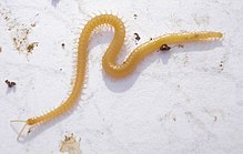
Among the centipedes, all members of the Geophilomorpha are constrained by a developmental bias to have an odd number of segments, whether as few as 27 or as many as 191.

Development in specific lineages can be biased either positively, towards a given trajectory or phenotype, or negatively, away from producing certain types of change; either may be absolute (the change is always or never produced) or relative. Evidence for any such direction in evolution is however hard to acquire and can also result from developmental constraints that limit diversification. For example, in the gastropods, the snail-type shell is always built as a tube that grows both in length and in diameter; selection has created a wide variety of shell shapes such as flat spirals, cowries and tall turret spirals within these constraints. Among the centipedes, the Lithobiomorpha always have 15 trunk segments as adults, probably the result of a developmental bias towards an odd number of trunk segments. Another centipede order, the Geophilomorpha, the number of segments varies in different species between 27 and 191, but the number is always odd, making this an absolute constraint; almost all the odd numbers in that range are occupied by one or another species.

Ecological evolutionary developmental biology

Ecological evolutionary developmental biology (eco-evo-devo) integrates research from developmental biology and ecology to examine their relationship with evolutionary theory. Researchers study concepts and mechanisms such as developmental plasticity, epigenetic inheritance, genetic assimilation, niche construction and symbiosis.

Operator (computer programming)

From Wikipedia, the free encyclopedia https://en.wikipedia.org/wiki/Operator_(computer_programmin...