Search This Blog

Wednesday, December 15, 2021

Transgene

From Wikipedia, the free encyclopedia
https://en.wikipedia.org/wiki/Transgene

A transgene is a gene that has been transferred naturally, or by any of a number of genetic engineering techniques from one organism to another. The introduction of a transgene, in a process known as transgenesis, has the potential to change the phenotype of an organism. Transgene describes a segment of DNA containing a gene sequence that has been isolated from one organism and is introduced into a different organism. Transgenes alter the genome by blocking the function of a host gene; they can either replace the host gene with one that codes for a different protein, or introduce an additional gene. In general, the DNA is incorporated into the organism's germ line. For example, in higher vertebrates this can be accomplished by injecting the foreign DNA into the nucleus of a fertilized ovum. This technique is routinely used to introduce human disease genes or other genes of interest into strains of laboratory mice to study the function or pathology involved with that particular gene.

The construction of a transgene requires the assembly of a few main parts. The transgene must contain a promoter, which is a regulatory sequence that will determine where and when the transgene is active, an exon, a protein coding sequence (usually derived from the cDNA for the protein of interest), and a stop sequence. These are typically combined in a bacterial plasmid and the coding sequences are typically chosen from transgenes with previously known functions.

Transgenic or genetically modified organisms, be they bacteria, viruses or fungi, serve many research purposes. Transgenic plants, insects, fish and mammals (including humans) have been bred. Transgenic plants such as corn and soybean have replaced wild strains in agriculture in some countries (e.g. the United States). Transgene escape has been documented for GMO crops since 2001 with persistence and invasiveness. Transgenetic organisms pose ethical questions and may cause biosafety problems.

History

The idea of shaping an organism to fit a specific need isn't a new science. However, until the late 1900s farmers and scientist could breed new strains of a plant or organism only from closely related species, because the DNA had to be compatible for offspring to be able to reproduce another generation.

In the 1970 and 1980s, scientists passed this hurdle by inventing procedures for combining the DNA of two vastly different species with genetic engineering. The organisms produced by these procedures were termed transgenic. Transgenesis is the same as gene therapy in the sense that they both transform cells for a specific purpose. However, they are completely different in their purposes, as gene therapy aims to cure a defect in cells, and transgenesis seeks to produce a genetically modified organism by incorporating the specific transgene into every cell and changing the genome. Transgenesis will therefore change the germ cells, not only the somatic cells, in order to ensure that the transgenes are passed down to the offspring when the organisms reproduce. Transgenes alter the genome by blocking the function of a host gene; they can either replace the host gene with one that codes for a different protein, or introduce an additional gene.

The first transgenic organism was created in 1974 when Annie Chang and Stanley Cohen expressed Staphylococcus aureus genes in Escherichia coli. In 1978, yeast cells were the first eukaryotic organisms to undergo gene transfer. Mouse cells were first transformed in 1979, followed by mouse embryos in 1980. Most of the very first transmutations were performed by microinjection of DNA directly into cells. Scientists were able to develop other methods to perform the transformations, such as incorporating transgenes into retroviruses and then infecting cells, using electroinfusion which takes advantage of an electric current to pass foreign DNA through the cell wall, biolistics which is the procedure of shooting DNA bullets into cells, and also delivering DNA into the egg that has just been fertilized.

The first transgenic animals were only intended for genetic research to study the specific function of a gene, and by 2003, thousands of genes had been studied.

Use in plants

A variety of transgenic plants have been designed for agriculture to produce genetically modified crops, such as corn, soybean, rapeseed oil, cotton, rice and more. As of 2012, these GMO crops were planted on 170 million hectares globally.

Golden rice

One example of a transgenic plant species is golden rice. In 1997, five million children developed xerophthalmia, a medical condition caused by vitamin A deficiency, in Southeast Asia alone. Of those children, a quarter million went blind. To combat this, scientists used biolistics to insert the daffodil phytoene synthase gene into Asia indigenous rice cultivars. The daffodil insertion increased the production of β-carotene. The product was a transgenic rice species rich in vitamin A, called golden rice. Little is known about the impact of golden rice on xerophthalmia because anti-GMO campaigns have prevented the full commercial release of golden rice into agricultural systems in need.

Transgene escape

The escape of genetically-engineered plant genes via hybridization with wild relatives was first discussed and examined in Mexico and Europe in the mid-1990s. There is agreement that escape of transgenes is inevitable, even "some proof that it is happening". Up until 2008 there were few documented cases.

Corn

Corn sampled in 2000 from the Sierra Juarez, Oaxaca, Mexico contained a transgenic 35S promoter, while a large sample taken by a different method from the same region in 2003 and 2004 did not. A sample from another region from 2002 also did not, but directed samples taken in 2004 did, suggesting transgene persistence or re-introduction. A 2009 study found recombinant proteins in 3.1% and 1.8% of samples, most commonly in southeast Mexico. Seed and grain import from the United States could explain the frequency and distribution of transgenes in west-central Mexico, but not in the southeast. Also, 5.0% of corn seed lots in Mexican corn stocks expressed recombinant proteins despite the moratorium on GM crops.

Cotton

In 2011, transgenic cotton was found in Mexico among wild cotton, after 15 years of GMO cotton cultivation.

Rapeseed (canola)

Transgenic rapeseed Brassicus napus, hybridized with a native Japanese species Brassica rapa, was found in Japan in 2011 after they had been identified 2006 in Québec, Canada. They were persistent over a 6-year study period, without herbicide selection pressure and despite hybridization with the wild form. This was the first report of the introgression—the stable incorporation of genes from one gene pool into another—of an herbicide resistance transgene from Brassica napus into the wild form gene pool.

Creeping bentgrass

Transgenic creeping bentgrass, engineered to be glyphosate-tolerant as "one of the first wind-pollinated, perennial, and highly outcrossing transgenic crops", was planted in 2003 as part of a large (about 160 ha) field trial in central Oregon near Madras, Oregon. In 2004, its pollen was found to have reached wild growing bentgrass populations up to 14 kilometres away. Cross-pollinating Agrostis gigantea was even found at a distance of 21 kilometres. The grower, Scotts Company could not remove all genetically engineered plants, and in 2007, the U.S. Department of Agriculture fined Scotts $500,000 for noncompliance with regulations.

Risk assessment

The long-term monitoring and controlling of a particular transgene has been shown not to be feasible. The European Food Safety Authority published a guidance for risk assessment in 2010.

Use in mice

Genetically modified mice are the most common animal model for transgenic research. Transgenic mice are currently being used to study a variety of diseases including cancer, obesity, heart disease, arthritis, anxiety, and Parkinson’s disease. The two most common types of genetically modified mice are knockout mice and oncomice. Knockout mice are a type of mouse model that uses transgenic insertion to disrupt an existing gene’s expression. In order to create knockout mice, a transgene with the desired sequence is inserted into an isolated mouse blastocyst using electroporation. Then, homologous recombination occurs naturally within some cells, replacing the gene of interest with the designed transgene. Through this process, researchers were able to demonstrate that a transgene can be integrated into the genome of an animal, serve a specific function within the cell, and be passed down to future generations.

Oncomice are another genetically modified mouse species created by inserting transgenes that increase the animal’s vulnerability to cancer. Cancer researchers utilize oncomice to study the profiles of different cancers in order to apply this knowledge to human studies.

Use in Drosophila

Multiple studies have been conducted concerning transgenesis in Drosophila melanogaster, the fruit fly. This organism has been a helpful genetic model for over 100 years, due to its well-understood developmental pattern. The transfer of transgenes into the Drosophila genome has been performed using various techniques, including P element, Cre-loxP, and ΦC31 insertion. The most practiced method used thus far to insert transgenes into the Drosophila genome utilizes P elements. The transposable P elements, also known as transposons, are segments of bacterial DNA that are translocated into the genome, without the presence of a complementary sequence in the host’s genome. P elements are administered in pairs of two, which flank the DNA insertion region of interest. Additionally, P elements often consist of two plasmid components, one known as the P element transposase and the other, the P transposon backbone. The transposase plasmid portion drives the transposition of the P transposon backbone, containing the transgene of interest and often a marker, between the two terminal sites of the transposon. Success of this insertion results in the nonreversible addition of the transgene of interest into the genome. While this method has been proven effective, the insertion sites of the P elements are often uncontrollable, resulting in an unfavorable, random insertion of the transgene into the Drosophila genome.

To improve the location and precision of the transgenic process, an enzyme known as Cre has been introduced. Cre has proven to be a key element in a process known as recombination-mediated cassette exchange (RMCE). While it has shown to have a lower efficiency of transgenic transformation than the P element transposases, Cre greatly lessens the labor-intensive abundance of balancing random P insertions. Cre aids in the targeted transgenesis of the DNA gene segment of interest, as it supports the mapping of the transgene insertion sites, known as loxP sites. These sites, unlike P elements, can be specifically inserted to flank a chromosomal segment of interest, aiding in targeted transgenesis. The Cre transposase is important in the catalytic cleavage of the base pairs present at the carefully positioned loxP sites, permitting more specific insertions of the transgenic donor plasmid of interest.

To overcome the limitations and low yields that transposon-mediated and Cre-loxP transformation methods produce, the bacteriophage ΦC31 has recently been utilized. Recent breakthrough studies involve the microinjection of the bacteriophage ΦC31 integrase, which shows improved transgene insertion of large DNA fragments that are unable to be transposed by P elements alone. This method involves the recombination between an attachment (attP) site in the phage and an attachment site in the bacterial host genome (attB). Compared to usual P element transgene insertion methods, ΦC31 integrates the entire transgene vector, including bacterial sequences and antibiotic resistance genes. Unfortunately, the presence of these additional insertions has been found to affect the level and reproducibility of transgene expression.

Use in livestock and aquaculture

One agricultural application is to selectively breed animals for particular traits: Transgenic cattle with an increased muscle phenotype has been produced by overexpressing a short hairpin RNA with homology to the myostatin mRNA using RNA interference. Transgenes are being used to produce milk with high levels of proteins or silk from the milk of goats. Another agricultural application is to selectively breed animals, which are resistant to diseases or animals for biopharmaceutical production.

Future potential

The application of transgenes is a rapidly growing area of molecular biology. As of 2005 it was predicted that in the next two decades, 300,000 lines of transgenic mice will be generated. Researchers have identified many applications for transgenes, particularly in the medical field. Scientists are focusing on the use of transgenes to study the function of the human genome in order to better understand disease, adapting animal organs for transplantation into humans, and the production of pharmaceutical products such as insulin, growth hormone, and blood anti-clotting factors from the milk of transgenic cows.

As of 2004 there were five thousand known genetic diseases, and the potential to treat these diseases using transgenic animals is, perhaps, one of the most promising applications of transgenes. There is a potential to use human gene therapy to replace a mutated gene with an unmutated copy of a transgene in order to treat the genetic disorder. This can be done through the use of Cre-Lox or knockout. Moreover, genetic disorders are being studied through the use of transgenic mice, pigs, rabbits, and rats. Transgenic rabbits have been created to study inherited cardiac arrhythmias, as the rabbit´s heart markedly better resembles the human heart as compared to the mouse. More recently, scientists have also begun using transgenic goats to study genetic disorders related to fertility.

Transgenes may be used for xenotransplantation from pig organs. Through the study of xeno-organ rejection, it was found that an acute rejection of the transplanted organ occurs upon the organ's contact with blood from the recipient due to the recognition of foreign antibodies on endothelial cells of the transplanted organ. Scientists have identified the antigen in pigs that causes this reaction, and therefore are able to transplant the organ without immediate rejection by removal of the antigen. However, the antigen begins to be expressed later on, and rejection occurs. Therefore, further research is being conducted. Transgenic microorganisms capable of producing catalytic proteins or enzymes which increase the rate of industrial reactions.

Ethical controversy

Transgene use in humans is currently fraught with issues. Transformation of genes into human cells has not been perfected yet. The most famous example of this involved certain patients developing T-cell leukemia after being treated for X-linked severe combined immunodeficiency (X-SCID). This was attributed to the close proximity of the inserted gene to the LMO2 promoter, which controls the transcription of the LMO2 proto-oncogene.


Molecular cloning

From Wikipedia, the free encyclopedia
 
Diagram of molecular cloning using bacteria and plasmids.

Molecular cloning is a set of experimental methods in molecular biology that are used to assemble recombinant DNA molecules and to direct their replication within host organisms. The use of the word cloning refers to the fact that the method involves the replication of one molecule to produce a population of cells with identical DNA molecules. Molecular cloning generally uses DNA sequences from two different organisms: the species that is the source of the DNA to be cloned, and the species that will serve as the living host for replication of the recombinant DNA. Molecular cloning methods are central to many contemporary areas of modern biology and medicine.

In a conventional molecular cloning experiment, the DNA to be cloned is obtained from an organism of interest, then treated with enzymes in the test tube to generate smaller DNA fragments. Subsequently, these fragments are then combined with vector DNA to generate recombinant DNA molecules. The recombinant DNA is then introduced into a host organism (typically an easy-to-grow, benign, laboratory strain of E. coli bacteria). This will generate a population of organisms in which recombinant DNA molecules are replicated along with the host DNA. Because they contain foreign DNA fragments, these are transgenic or genetically modified microorganisms (GMO). This process takes advantage of the fact that a single bacterial cell can be induced to take up and replicate a single recombinant DNA molecule. This single cell can then be expanded exponentially to generate a large amount of bacteria, each of which contain copies of the original recombinant molecule. Thus, both the resulting bacterial population, and the recombinant DNA molecule, are commonly referred to as "clones". Strictly speaking, recombinant DNA refers to DNA molecules, while molecular cloning refers to the experimental methods used to assemble them. The idea arose that different DNA sequences could be inserted into a plasmid and that these foreign sequences would be carried into bacteria and digested as part of the plasmid. That is, these plasmids could serve as cloning vectors to carry genes.

Virtually any DNA sequence can be cloned and amplified, but there are some factors that might limit the success of the process. Examples of the DNA sequences that are difficult to clone are inverted repeats, origins of replication, centromeres and telomeres. There is also a lower chance of success when inserting large-sized DNA sequences. Inserts larger than 10kbp have very limited success, but bacteriophages such as bacteriophage λ can be modified to successfully insert a sequence up to 40 kbp.

History

Prior to the 1970s, the understanding of genetics and molecular biology was severely hampered by an inability to isolate and study individual genes from complex organisms. This changed dramatically with the advent of molecular cloning methods. Microbiologists, seeking to understand the molecular mechanisms through which bacteria restricted the growth of bacteriophage, isolated restriction endonucleases, enzymes that could cleave DNA molecules only when specific DNA sequences were encountered. They showed that restriction enzymes cleaved chromosome-length DNA molecules at specific locations, and that specific sections of the larger molecule could be purified by size fractionation. Using a second enzyme, DNA ligase, fragments generated by restriction enzymes could be joined in new combinations, termed recombinant DNA. By recombining DNA segments of interest with vector DNA, such as bacteriophage or plasmids, which naturally replicate inside bacteria, large quantities of purified recombinant DNA molecules could be produced in bacterial cultures. The first recombinant DNA molecules were generated and studied in 1972.

Overview

Molecular cloning takes advantage of the fact that the chemical structure of DNA is fundamentally the same in all living organisms. Therefore, if any segment of DNA from any organism is inserted into a DNA segment containing the molecular sequences required for DNA replication, and the resulting recombinant DNA is introduced into the organism from which the replication sequences were obtained, then the foreign DNA will be replicated along with the host cell's DNA in the transgenic organism.

Molecular cloning is similar to polymerase chain reaction (PCR) in that it permits the replication of DNA sequence. The fundamental difference between the two methods is that molecular cloning involves replication of the DNA in a living microorganism, while PCR replicates DNA in an in vitro solution, free of living cells.

In silico cloning and simulations

Before actual cloning experiments are performed in the lab, most cloning experiments are planned in a computer, using specialized software. Although the detailed planning of the cloning can be done in any text editor, together with online utilities for e.g. PCR primer design, dedicated software exist for the purpose. Software for the purpose include for example ApE  (open source), DNAStrider  (open source), Serial Cloner  (gratis), Collagene  (open source), and SnapGene (commercial). These programs allow to simulate PCR reactions, restriction digests, ligations, etc., that is, all the steps described below.

Steps

The overall goal of molecular cloning is to take a gene of interest from one plasmid and insert it into another plasmid This is done by performing PCR, digestive reaction, ligation reaction, and transformation.

In standard molecular cloning experiments, the cloning of any DNA fragment essentially involves seven steps: (1) Choice of host organism and cloning vector, (2) Preparation of vector DNA, (3) Preparation of DNA to be cloned, (4) Creation of recombinant DNA, (5) Introduction of recombinant DNA into host organism, (6) Selection of organisms containing recombinant DNA, (7) Screening for clones with desired DNA inserts and biological properties.

Notably, the growing capacity and fidelity of DNA synthesis platforms allows for increasingly intricate designs in molecular engineering. These projects may include very long strands of novel DNA sequence and/or test entire libraries simultaneously, as opposed to of individual sequences. These shifts introduce complexity that require design to move away from the flat nucleotide-based representation and towards a higher level of abstraction. Examples of such tools are GenoCAD, Teselagen (free for academia) or GeneticConstructor (free for academics).

Choice of host organism and cloning vector

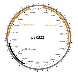
Diagram of a commonly used cloning plasmid; pBR322. It's a circular piece of DNA 4361 bases long. Two antibiotic resistance genes are present, conferring resistance to ampicillin and tetracycline, and an origin of replication that the host uses to replicate the DNA.

Although a very large number of host organisms and molecular cloning vectors are in use, the great majority of molecular cloning experiments begin with a laboratory strain of the bacterium E. coli (Escherichia coli) and a plasmid cloning vector. E. coli and plasmid vectors are in common use because they are technically sophisticated, versatile, widely available, and offer rapid growth of recombinant organisms with minimal equipment. If the DNA to be cloned is exceptionally large (hundreds of thousands to millions of base pairs), then a bacterial artificial chromosome or yeast artificial chromosome vector is often chosen.

Specialized applications may call for specialized host-vector systems. For example, if the experimentalists wish to harvest a particular protein from the recombinant organism, then an expression vector is chosen that contains appropriate signals for transcription and translation in the desired host organism. Alternatively, if replication of the DNA in different species is desired (for example, transfer of DNA from bacteria to plants), then a multiple host range vector (also termed shuttle vector) may be selected. In practice, however, specialized molecular cloning experiments usually begin with cloning into a bacterial plasmid, followed by subcloning into a specialized vector.

Whatever combination of host and vector are used, the vector almost always contains four DNA segments that are critically important to its function and experimental utility:

  • DNA replication origin is necessary for the vector (and its linked recombinant sequences) to replicate inside the host organism
  • one or more unique restriction endonuclease recognition sites to serves as sites where foreign DNA may be introduced
  • a selectable genetic marker gene that can be used to enable the survival of cells that have taken up vector sequences
  • a tag gene that can be used to screen for cells containing the foreign DNA
Cleavage of a DNA sequence containing the BamHI restriction site. The DNA is cleaved at the palindromic sequence to produce 'sticky ends'.

Preparation of vector DNA

The cloning vector is treated with a restriction endonuclease to cleave the DNA at the site where foreign DNA will be inserted. The restriction enzyme is chosen to generate a configuration at the cleavage site that is compatible with the ends of the foreign DNA (see DNA end). Typically, this is done by cleaving the vector DNA and foreign DNA with the same restriction enzyme, for example EcoRI. Most modern vectors contain a variety of convenient cleavage sites that are unique within the vector molecule (so that the vector can only be cleaved at a single site) and are located within a gene (frequently beta-galactosidase) whose inactivation can be used to distinguish recombinant from non-recombinant organisms at a later step in the process. To improve the ratio of recombinant to non-recombinant organisms, the cleaved vector may be treated with an enzyme (alkaline phosphatase) that dephosphorylates the vector ends. Vector molecules with dephosphorylated ends are unable to replicate, and replication can only be restored if foreign DNA is integrated into the cleavage site.

Preparation of DNA to be cloned

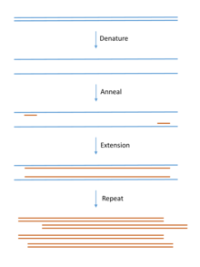
DNA for cloning is most commonly produced using PCR. Template DNA is mixed with bases (the building blocks of DNA), primers (short pieces of complementary single stranded DNA) and a DNA polymerase enzyme that builds the DNA chain. The mix goes through cycles of heating and cooling to produce large quantities of copied DNA.

For cloning of genomic DNA, the DNA to be cloned is extracted from the organism of interest. Virtually any tissue source can be used (even tissues from extinct animals), as long as the DNA is not extensively degraded. The DNA is then purified using simple methods to remove contaminating proteins (extraction with phenol), RNA (ribonuclease) and smaller molecules (precipitation and/or chromatography). Polymerase chain reaction (PCR) methods are often used for amplification of specific DNA or RNA (RT-PCR) sequences prior to molecular cloning.

DNA for cloning experiments may also be obtained from RNA using reverse transcriptase (complementary DNA or cDNA cloning), or in the form of synthetic DNA (artificial gene synthesis). cDNA cloning is usually used to obtain clones representative of the mRNA population of the cells of interest, while synthetic DNA is used to obtain any precise sequence defined by the designer. Such a designed sequence may be required when moving genes across genetic codes (for example, from the mitochrondria to the nucleus) or simply for increasing expression via codon optimization.

The purified DNA is then treated with a restriction enzyme to generate fragments with ends capable of being linked to those of the vector. If necessary, short double-stranded segments of DNA (linkers) containing desired restriction sites may be added to create end structures that are compatible with the vector.

Creation of recombinant DNA with DNA ligase

The creation of recombinant DNA is in many ways the simplest step of the molecular cloning process. DNA prepared from the vector and foreign source are simply mixed together at appropriate concentrations and exposed to an enzyme (DNA ligase) that covalently links the ends together. This joining reaction is often termed ligation. The resulting DNA mixture containing randomly joined ends is then ready for introduction into the host organism.

DNA ligase only recognizes and acts on the ends of linear DNA molecules, usually resulting in a complex mixture of DNA molecules with randomly joined ends. The desired products (vector DNA covalently linked to foreign DNA) will be present, but other sequences (e.g. foreign DNA linked to itself, vector DNA linked to itself and higher-order combinations of vector and foreign DNA) are also usually present. This complex mixture is sorted out in subsequent steps of the cloning process, after the DNA mixture is introduced into cells.

Introduction of recombinant DNA into host organism

The DNA mixture, previously manipulated in vitro, is moved back into a living cell, referred to as the host organism. The methods used to get DNA into cells are varied, and the name applied to this step in the molecular cloning process will often depend upon the experimental method that is chosen (e.g. transformation, transduction, transfection, electroporation).

When microorganisms are able to take up and replicate DNA from their local environment, the process is termed transformation, and cells that are in a physiological state such that they can take up DNA are said to be competent. In mammalian cell culture, the analogous process of introducing DNA into cells is commonly termed transfection. Both transformation and transfection usually require preparation of the cells through a special growth regime and chemical treatment process that will vary with the specific species and cell types that are used.

Electroporation uses high voltage electrical pulses to translocate DNA across the cell membrane (and cell wall, if present). In contrast, transduction involves the packaging of DNA into virus-derived particles, and using these virus-like particles to introduce the encapsulated DNA into the cell through a process resembling viral infection. Although electroporation and transduction are highly specialized methods, they may be the most efficient methods to move DNA into cells.

Selection of organisms containing vector sequences

Whichever method is used, the introduction of recombinant DNA into the chosen host organism is usually a low efficiency process; that is, only a small fraction of the cells will actually take up DNA. Experimental scientists deal with this issue through a step of artificial genetic selection, in which cells that have not taken up DNA are selectively killed, and only those cells that can actively replicate DNA containing the selectable marker gene encoded by the vector are able to survive.

When bacterial cells are used as host organisms, the selectable marker is usually a gene that confers resistance to an antibiotic that would otherwise kill the cells, typically ampicillin. Cells harboring the plasmid will survive when exposed to the antibiotic, while those that have failed to take up plasmid sequences will die. When mammalian cells (e.g. human or mouse cells) are used, a similar strategy is used, except that the marker gene (in this case typically encoded as part of the kanMX cassette) confers resistance to the antibiotic Geneticin.

Screening for clones with desired DNA inserts and biological properties

Modern bacterial cloning vectors (e.g. pUC19 and later derivatives including the pGEM vectors) use the blue-white screening system to distinguish colonies (clones) of transgenic cells from those that contain the parental vector (i.e. vector DNA with no recombinant sequence inserted). In these vectors, foreign DNA is inserted into a sequence that encodes an essential part of beta-galactosidase, an enzyme whose activity results in formation of a blue-colored colony on the culture medium that is used for this work. Insertion of the foreign DNA into the beta-galactosidase coding sequence disables the function of the enzyme so that colonies containing transformed DNA remain colorless (white). Therefore, experimentalists are easily able to identify and conduct further studies on transgenic bacterial clones, while ignoring those that do not contain recombinant DNA.

The total population of individual clones obtained in a molecular cloning experiment is often termed a DNA library. Libraries may be highly complex (as when cloning complete genomic DNA from an organism) or relatively simple (as when moving a previously cloned DNA fragment into a different plasmid), but it is almost always necessary to examine a number of different clones to be sure that the desired DNA construct is obtained. This may be accomplished through a very wide range of experimental methods, including the use of nucleic acid hybridizations, antibody probes, polymerase chain reaction, restriction fragment analysis and/or DNA sequencing.

Applications

Molecular cloning provides scientists with an essentially unlimited quantity of any individual DNA segments derived from any genome. This material can be used for a wide range of purposes, including those in both basic and applied biological science. A few of the more important applications are summarized here.

Genome organization and gene expression

Molecular cloning has led directly to the elucidation of the complete DNA sequence of the genomes of a very large number of species and to an exploration of genetic diversity within individual species, work that has been done mostly by determining the DNA sequence of large numbers of randomly cloned fragments of the genome, and assembling the overlapping sequences.

At the level of individual genes, molecular clones are used to generate probes that are used for examining how genes are expressed, and how that expression is related to other processes in biology, including the metabolic environment, extracellular signals, development, learning, senescence and cell death. Cloned genes can also provide tools to examine the biological function and importance of individual genes, by allowing investigators to inactivate the genes, or make more subtle mutations using regional mutagenesis or site-directed mutagenesis. Genes cloned into expression vectors for functional cloning provide a means to screen for genes on the basis of the expressed protein's function.

Production of recombinant proteins

Obtaining the molecular clone of a gene can lead to the development of organisms that produce the protein product of the cloned genes, termed a recombinant protein. In practice, it is frequently more difficult to develop an organism that produces an active form of the recombinant protein in desirable quantities than it is to clone the gene. This is because the molecular signals for gene expression are complex and variable, and because protein folding, stability and transport can be very challenging.

Many useful proteins are currently available as recombinant products. These include--(1) medically useful proteins whose administration can correct a defective or poorly expressed gene (e.g. recombinant factor VIII, a blood-clotting factor deficient in some forms of hemophilia, and recombinant insulin, used to treat some forms of diabetes), (2) proteins that can be administered to assist in a life-threatening emergency (e.g. tissue plasminogen activator, used to treat strokes), (3) recombinant subunit vaccines, in which a purified protein can be used to immunize patients against infectious diseases, without exposing them to the infectious agent itself (e.g. hepatitis B vaccine), and (4) recombinant proteins as standard material for diagnostic laboratory tests.

Transgenic organisms

Once characterized and manipulated to provide signals for appropriate expression, cloned genes may be inserted into organisms, generating transgenic organisms, also termed genetically modified organisms (GMOs). Although most GMOs are generated for purposes of basic biological research (see for example, transgenic mouse), a number of GMOs have been developed for commercial use, ranging from animals and plants that produce pharmaceuticals or other compounds (pharming), herbicide-resistant crop plants, and fluorescent tropical fish (GloFish) for home entertainment.

Gene therapy

Gene therapy involves supplying a functional gene to cells lacking that function, with the aim of correcting a genetic disorder or acquired disease. Gene therapy can be broadly divided into two categories. The first is alteration of germ cells, that is, sperm or eggs, which results in a permanent genetic change for the whole organism and subsequent generations. This “germ line gene therapy” is considered by many to be unethical in human beings. The second type of gene therapy, “somatic cell gene therapy”, is analogous to an organ transplant. In this case, one or more specific tissues are targeted by direct treatment or by removal of the tissue, addition of the therapeutic gene or genes in the laboratory, and return of the treated cells to the patient. Clinical trials of somatic cell gene therapy began in the late 1990s, mostly for the treatment of cancers and blood, liver, and lung disorders.

Despite a great deal of publicity and promises, the history of human gene therapy has been characterized by relatively limited success. The effect of introducing a gene into cells often promotes only partial and/or transient relief from the symptoms of the disease being treated. Some gene therapy trial patients have suffered adverse consequences of the treatment itself, including deaths. In some cases, the adverse effects result from disruption of essential genes within the patient's genome by insertional inactivation. In others, viral vectors used for gene therapy have been contaminated with infectious virus. Nevertheless, gene therapy is still held to be a promising future area of medicine, and is an area where there is a significant level of research and development activity.


Assisted reproductive technology

From Wikipedia, the free encyclopedia
 
Assisted reproductive technology
Blausen 0060 AssistedReproductiveTechnology.png
Illustration depicting intracytoplasmic sperm injection (ICSI), an example of assisted reproductive technology.
Other namesART
MeSHD027724

Assisted reproductive technology (ART) includes medical procedures used primarily to address infertility. This subject involves procedures such as in vitro fertilization (IVF), intracytoplasmic sperm injection (ICSI), cryopreservation of gametes or embryos, and/or the use of fertility medication. When used to address infertility, ART may also be referred to as fertility treatment. ART mainly belongs to the field of reproductive endocrinology and infertility. Some forms of ART may be used with regard to fertile couples for genetic purpose (see preimplantation genetic diagnosis). ART may also be used in surrogacy arrangements, although not all surrogacy arrangements involve ART.

Procedures

General

With ART, the process of sexual intercourse is bypassed and fertilization of the oocytes occurs in the laboratory environment (i.e., in vitro fertilization).

In the US, the Centers for Disease Control and Prevention (CDC) defines ART to include "all fertility treatments in which both eggs and sperm are handled. In general, ART procedures involve surgically removing eggs from a woman's ovaries, combining them with sperm in the laboratory, and returning them to the woman's body or donating them to another woman." According to CDC, "they do not include treatments in which only sperm are handled (i.e., intrauterine—or artificial—insemination) or procedures in which a woman takes medicine only to stimulate egg production without the intention of having eggs retrieved."

In Europe, ART also excludes artificial insemination and includes only procedures where oocytes are handled.

The WHO, or World Health Organization, also defines ART this way.

Ovulation induction

Ovulation induction is usually used in the sense of stimulation of the development of ovarian follicles by fertility medication to reverse anovulation or oligoovulation. These medications are given by injection for 8 to 14 days. A health care provider closely monitors the development of the eggs using transvaginal ultrasound and blood tests to assess follicle growth and estrogen production by the ovaries. When follicles have reached an adequate size and the eggs are mature enough, an injection of the hormone hCG initiates the ovulation process. Egg retrieval should occur from 34 to 36 hours after the hCG injection.

In vitro fertilization

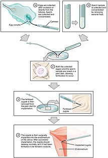
Steps of IVF Treatment

In vitro fertilization is the technique of letting fertilization of the male and female gametes (sperm and egg) occur outside the female body.

Techniques usually used in in vitro fertilization include:

  • Transvaginal ovum retrieval (OVR) is the process whereby a small needle is inserted through the back of the vagina and guided via ultrasound into the ovarian follicles to collect the fluid that contains the eggs.
  • Embryo transfer is the step in the process whereby one or several embryos are placed into the uterus of the female with the intent to establish a pregnancy.

Less commonly used techniques in in vitro fertilization are:

  • Assisted zona hatching (AZH) is performed shortly before the embryo is transferred to the uterus. A small opening is made in the outer layer surrounding the egg in order to help the embryo hatch out and aid in the implantation process of the growing embryo.
  • Intracytoplasmic sperm injection (ICSI)
    Intracytoplasmic sperm injection (ICSI) is beneficial in the case of male factor infertility where sperm counts are very low or failed fertilization occurred with previous IVF attempt(s). The ICSI procedure involves a single sperm carefully injected into the center of an egg using a microneedle. With ICSI, only one sperm per egg is needed. Without ICSI, you need between 50,000 and 100,000. This method is also sometimes employed when donor sperm is used.
  • Autologous endometrial coculture is a possible treatment for patients who have failed previous IVF attempts or who have poor embryo quality. The patient's fertilized eggs are placed on top of a layer of cells from the patient's own uterine lining, creating a more natural environment for embryo development.
  • In zygote intrafallopian transfer (ZIFT), egg cells are removed from the woman's ovaries and fertilized in the laboratory; the resulting zygote is then placed into the fallopian tube.
  • Cytoplasmic transfer is the technique in which the contents of a fertile egg from a donor are injected into the infertile egg of the patient along with the sperm.
  • Egg donors are resources for women with no eggs due to surgery, chemotherapy, or genetic causes; or with poor egg quality, previously unsuccessful IVF cycles or advanced maternal age. In the egg donor process, eggs are retrieved from a donor's ovaries, fertilized in the laboratory with the sperm from the recipient's partner, and the resulting healthy embryos are returned to the recipient's uterus.
  • Sperm donation may provide the source for the sperm used in IVF procedures where the male partner produces no sperm or has an inheritable disease, or where the woman being treated has no male partner.
  • Preimplantation genetic diagnosis (PGD) involves the use of genetic screening mechanisms such as fluorescent in-situ hybridization (FISH) or comparative genomic hybridization (CGH) to help identify genetically abnormal embryos and improve healthy outcomes.
  • Embryo splitting can be used for twinning to increase the number of available embryos.

Pre-implantation genetic diagnosis

A pre-implantation genetic diagnosis procedure may be conducted on embryos prior to implantation (as a form of embryo profiling), and sometimes even of oocytes prior to fertilization. PGD is considered in a similar fashion to prenatal diagnosis. PGD is an adjunct to ART procedures, and requires in vitro fertilization to obtain oocytes or embryos for evaluation. Embryos are generally obtained through blastomere or blastocyst biopsy. The latter technique has proved to be less deleterious for the embryo, therefore it is advisable to perform the biopsy around day 5 or 6 of development. Sex selection is the attempt to control the sex of offspring to achieve a desired sex in case of X chromosome linked diseases. It can be accomplished in several ways, both pre- and post-implantation of an embryo, as well as at birth. Pre-implantation techniques include PGD, but also sperm sorting.

Others

Other assisted reproduction techniques include:

Risks

The majority of IVF-conceived infants do not have birth defects. However, some studies have suggested that assisted reproductive technology is associated with an increased risk of birth defects. Artificial reproductive technology is becoming more available. Early studies suggest that there could be an increased risk for medical complications with both the mother and baby. Some of these include low birth weight, placental insufficiency, chromosomal disorders, preterm deliveries, gestational diabetes, and pre-eclampsia (Aiken and Brockelsby).

In the largest U.S. study, which used data from a statewide registry of birth defects, 6.2% of IVF-conceived children had major defects, as compared with 4.4% of naturally conceived children matched for maternal age and other factors (odds ratio, 1.3; 95% confidence interval, 1.00 to 1.67). ART carries with it a risk for heterotopic pregnancy (simultaneous intrauterine and extrauterine pregnancy). The main risks are:

Sperm donation is an exception, with a birth defect rate of almost a fifth compared to the general population. It may be explained by that sperm banks accept only people with high sperm count.

Germ cells of the mouse normally have a frequency of spontaneous point mutations that is 5 to 10-fold lower than that in somatic cells from the same individual. This low frequency in the germline leads to embryos that have a low frequency of point mutations in the next generation. No significant differences were observed in the frequency or specturm of mutations between naturally conceived fetuses and assisted-conception fetuses. This suggests that with respect to the maintenance of genetic integrity assisted conception is safe.

Current data indicate little or no increased risk for postpartum depression among women who use ART.

Usage of assisted reproductive technology including ovarian stimulation and in vitro fertilization have been associated with an increased overall risk of childhood cancer in the offspring, which may be caused by the same original disease or condition that caused the infertility or subfertility in the mother or father.

That said, In a landmark paper by Jacques Balayla et al. it was determined that infants born after ART have similar neurodevelopment than infants born after natural conception.

Usage

As a result of the 1992 Fertility Clinic Success Rate and Certification Act, the CDC is required to publish the annual ART success rates at U.S. fertility clinics. Assisted reproductive technology procedures performed in the U.S. has over than doubled over the last 10 years, with 140,000 procedures in 2006, resulting in 55,000 births.

In Australia, 3.1% of births are a result of ART.

The most common reasons for discontinuation of fertility treatment have been estimated to be: postponement of treatment (39%), physical and psychological burden (19%, psychological burden 14%, physical burden 6.32%), relational and personal problems (17%, personal reasons 9%, relational problems 9%), treatment rejection (13%) and organizational (12%) and clinic (8%) problems.

By country

United States

Many Americans do not have insurance coverage for fertility investigations and treatments. Many states are starting to mandate coverage, and the rate of use is 278% higher in states with complete coverage.

There are some health insurance companies that cover diagnosis of infertility, but frequently once diagnosed will not cover any treatment costs.

Approximate treatment/diagnosis costs in the United States, with inflation, as of 2020 (US$):

Another way to look at costs is to determine the expected cost of establishing a pregnancy. Thus, if a clomiphene treatment has a chance to establish a pregnancy in 8% of cycles and costs $660, the expected cost is $8,000 to establish a pregnancy, compared to an IVF cycle (cycle fecundity 40%) with a corresponding expected cost of $39,800 ($15,900 × 40%).

For the community as a whole, the cost of IVF on average pays back by 700% by tax from future employment by the conceived human being.

European Union

Number of assisted reproductive technology cycles in Europe between 1997 and 2014.

In Europe, 157,500 children were born using assisted reproductive technology in 2015, according to the European Society of Human Reproduction and Embryology (ESHRE). But there are major differences in legislation across the Old Continent. A European directive fixes standards concerning the use of human tissue and cells, but all ethical and legal questions on ART remain the prerogative of EU member states.

Conditions of assisted reproductive technology in different European countries.
  ART authorized for lesbian couples
  ART authorized for single women
  ART authorized for single women and lesbian couples
  ART prohibited for single women and lesbian couples

Across Europe, the legal criteria per availability vary somewhat. In 11 countries all women may benefit; in 8 others only heterosexual couples are concerned; in 7 only single women; and in 2 (Austria and Germany) only lesbian couples. Spain was the first European country to open ART to all women, in 1977, the year the first sperm bank was opened there. In France, the right to ART is accorded to all women since 2019. In the last 15 years, legislation has evolved quickly. For example, Portugal made ART available in 2006 with conditions very similar to those in France, before amending the law in 2016 to allow lesbian couples and single women to benefit. Italy clarified its uncertain legal situation in 2004 by adopting Europe’s strictest laws: ART is only available to heterosexual couples, married or otherwise, and sperm donation is prohibited.

Today, 21 countries provide partial public funding for ART treatment. The seven others, which do not, are Ireland, Cyprus, Estonia, Latvia, Luxembourg, Malta, and Romania. Such subsidies are subject to conditions, however. In Belgium, a fixed payment of €1,073 is made for each full cycle of the IVF process. The woman must be aged under 43 and may not carry out more than six cycles of ART. There is also a limit on the number of transferable embryos, which varies according to age and the number of cycles completed. In France, ART is subsidized in full by national health insurance for women up to age 43, with limits of 4 attempts at IVF and 6 at artificial insemination. Germany tightened its conditions for public funding in 2004, which caused a sharp drop in the number of ART cycles carried out, from more than 102,000 in 2003 to fewer than 57,000 the following year. Since then the figure has remained stable.

17 countries limit access to ART according to the age of the woman. 10 countries have established an upper age limit, varying from 40 (Finland, Netherlands) to 50 (including Spain, Greece and Estonia). Since 1994, France is one of a number of countries (including Germany, Spain, and the UK) which use the somewhat vague notion of “natural age of procreation”. In 2017, the steering council of France’s Agency of Biomedicine established an age limit of 43 for women using ART. 10 countries have no age limit for ART. These include Austria, Hungary, Italy and Poland.

Most European countries allow donations of gametes by third parties. But the situations vary depending on whether sperm or eggs are concerned. Sperm donations are authorized in 20 EU member states; in 11 of them anonymity is allowed. Egg donations are possible in 17 states, including 8 under anonymous conditions. On 12 April, the Council of Europe adopted a recommendation which encourages an end to anonymity. In the UK, anonymous sperm donations ended in 2005 and children have access to the identity of the donor when they reach adulthood. In France, the principle of anonymous donations of sperm or embryos is maintained in the law of bioethics of 2011, but a new bill under discussion may change the situation.

United Kingdom

In the United Kingdom, all patients have the right to preliminary testing, provided free of charge by the National Health Service (NHS). However, treatment is not widely available on the NHS and there can be long waiting lists. Many patients therefore pay for immediate treatment within the NHS or seek help from private clinics.

In 2013, the National Institute for Health and Care Excellence (NICE) published new guidelines about who should have access to IVF treatment on the NHS in England and Wales.

The guidelines say women aged between 40 and 42 should be offered one cycle of IVF on the NHS if they have never had IVF treatment before, have no evidence of low ovarian reserve (this is when eggs in the ovary are low in number, or low in quality), and have been informed of the additional implications of IVF and pregnancy at this age. However, if tests show IVF is the only treatment likely to help them get pregnant, women should be referred for IVF straight away.

This policy is often modified by local Clinical Commissioning Groups, in a fairly blatant breach of the NHS Constitution for England which provides that patients have the right to drugs and treatments that have been recommended by NICE for use in the NHS. For example, the Cheshire, Merseyside and West Lancashire Clinical Commissioning Group insists on additional conditions:

  • The person undergoing treatment must have commenced treatment before her 40th birthday;
  • The person undergoing treatment must have a BMI of between 19 and 29;
  • Neither partner must have any living children, from either the current or previous relationships. This includes adopted as well as biological children; and,
  • Sub-fertility must not be the direct result of a sterilisation procedure in either partner (this does not include conditions where sterilisation occurs as a result of another medical problem). Couples who have undertaken a reversal of their sterilisation procedure are not eligible for treatment.

Canada

Some treatments are covered by OHIP (public health insurance) in Ontario and others are not. Women with bilaterally blocked fallopian tubes and are under the age of 40 have treatment covered but are still required to pay test fees (around CA$3,000–4,000). Coverage varies in other provinces. Most other patients are required to pay for treatments themselves.

Israel

Israel's national health insurance, which is mandatory for all Israeli citizens, covers nearly all fertility treatments. IVF costs are fully subsidized up to the birth of two children for all Israeli women, including single women and lesbian couples. Embryo transfers for purposes of gestational surrogacy are also covered.

Germany

On 27 January 2009, the Federal Constitutional Court ruled that it is unconstitutional, that the health insurance companies have to bear only 50% of the cost for IVF. On 2 March 2012, the Federal Council has approved a draft law of some federal states, which provides that the federal government provides a subsidy of 25% to the cost. Thus, the share of costs borne for the pair would drop to just 25%. Since July 2017, assisted reproductive technology is also allowed for married lesbian couples, as German parliament allowed same-sex marriages in Germany.

France

In July 2020, the French Parliament allowed assisted reproductive technology also for lesbian couples and single women.

Cuba

Cuban sources mention that assisted reproduction is completely legal and free in the country.

Society and culture

Ethics

Some couples may find it difficult to stop treatment despite very bad prognosis, resulting in futile therapies. This has the potential to give ART providers a difficult decision of whether to continue or refuse treatment.

Some assisted reproductive technologies have the potential to be harmful to both the mother and child, posing a psychological and/or physical health risk, which may impact the ongoing use of these treatments.

Fictional representation

Films and other fiction depicting emotional struggles of assisted reproductive technology have had an upswing in the latter part of the 2000s decade, although the techniques have been available for decades. As ART becomes more utilized, the number of people that can relate to it by personal experience in one way or another is growing.

For specific examples, refer to the fiction sections in individual subarticles, e.g. surrogacy, sperm donation and fertility clinic.

In addition, reproduction and pregnancy in speculative fiction has been present for many decades.

Historical facts

25 July 1978, Louise Brown was born; this was the first successful birth of a child after IVF treatment. The procedure took place at Dr Kershaw's Cottage Hospital (now Dr Kershaw's Hospice) in Royton, Oldham, England. Patrick Steptoe (gynaecologist) and Robert Edwards (physiologist) worked together to develop the IVF technique. Steptoe described a new method of egg extraction and Edwards were carrying out a way to fertilise eggs in the lab. Robert G. Edwards was awarded the Nobel Prize in Physiology or Medicine in 2010, but not Steptoe because the Nobel Prize is not awarded posthumously.

The first successful birth by ICSI (Intracytoplasmic sperm injection) took place on 14 January 1992. The technique was developed by Gianpiero D. Palermo at the Vrije Universiteit Brussel, in the Center for Reproductive Medicine in Brussels. Actually, the discovery was made by a mistake when a spermatozoid was put into the cytoplasm.

Inequality (mathematics)

From Wikipedia, the free encyclopedia https://en.wikipedia.org/wiki/Inequality...