Search This Blog

Friday, June 23, 2023

Fusion protein

From Wikipedia, the free encyclopedia
A chimeric protein including two subunits and a linker protein synthesized via recombinant fusion technology

Fusion proteins or chimeric (kī-ˈmir-ik) proteins (literally, made of parts from different sources) are proteins created through the joining of two or more genes that originally coded for separate proteins. Translation of this fusion gene results in a single or multiple polypeptides with functional properties derived from each of the original proteins. Recombinant fusion proteins are created artificially by recombinant DNA technology for use in biological research or therapeutics. Chimeric or chimera usually designate hybrid proteins made of polypeptides having different functions or physico-chemical patterns. Chimeric mutant proteins occur naturally when a complex mutation, such as a chromosomal translocation, tandem duplication, or retrotransposition creates a novel coding sequence containing parts of the coding sequences from two different genes. Naturally occurring fusion proteins are commonly found in cancer cells, where they may function as oncoproteins. The bcr-abl fusion protein is a well-known example of an oncogenic fusion protein, and is considered to be the primary oncogenic driver of chronic myelogenous leukemia.

Functions

Some fusion proteins combine whole peptides and therefore contain all functional domains of the original proteins. However, other fusion proteins, especially those that occur naturally, combine only portions of coding sequences and therefore do not maintain the original functions of the parental genes that formed them.

Many whole gene fusions are fully functional and can still act to replace the original peptides. Some, however, experience interactions between the two proteins that can modify their functions. Beyond these effects, some gene fusions may cause regulatory changes that alter when and where these genes act. For partial gene fusions, the shuffling of different active sites and binding domains have the potential to result in new proteins with novel functions.

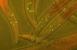
Green fluorescent protein (GFP) inserted into the neurons of Caenorhabditis elegans worms to track neuronal development

Fluorescent protein tags

The fusion of fluorescent tags to proteins in a host cell is a widely popular technique used in experimental cell and biology research in order to track protein interactions in real time. The first fluorescent tag, green fluorescent protein (GFP), was isolated from Aequorea victoria and is still used frequently in modern research. More recent derivations include photoconvertible fluorescent proteins (PCFPs), which were first isolated from Anthozoa. The most commonly used PCFP is the Kaede fluorescent tag, but the development of Kikume green-red (KikGR) in 2005 offers a brighter signal and more efficient photoconversion. The advantage of using PCFP fluorescent tags is the ability to track the interaction of overlapping biochemical pathways in real time. The tag will change color from green to red once the protein reaches a point of interest in the pathway, and the alternate colored protein can be monitored through the duration of pathway. This technique is especially useful when studying G-protein coupled receptor (GPCR) recycling pathways. The fates of recycled G-protein receptors may either be sent to the plasma membrane to be recycled, marked by a green fluorescent tag, or may be sent to a lysosome for degradation, marked by a red fluorescent tag.

Chimeric protein drugs

Sketches of mouse (top-left), chimeric (top-right) and humanized (bottom-left) monoclonal antibodies. Human parts are shown in brown, and non-human parts in blue.

The purpose of creating fusion proteins in drug development is to impart properties from each of the "parent" proteins to the resulting chimeric protein. Several chimeric protein drugs are currently available for medical use.

Many chimeric protein drugs are monoclonal antibodies whose specificity for a target molecule was developed using mice and hence were initially "mouse" antibodies. As non-human proteins, mouse antibodies tend to evoke an immune reaction if administered to humans. The chimerization process involves engineering the replacement of segments of the antibody molecule that distinguish it from a human antibody. For example, human constant domains can be introduced, thereby eliminating most of the potentially immunogenic portions of the drug without altering its specificity for the intended therapeutic target. Antibody nomenclature indicates this type of modification by inserting -xi- into the non-proprietary name (e.g., abci-xi-mab). If parts of the variable domains are also replaced by human portions, humanized antibodies are obtained. Although not conceptually distinct from chimeras, this type is indicated using -zu- such as in dacli-zu-mab. See the list of monoclonal antibodies for more examples.

In addition to chimeric and humanized antibodies, there are other pharmaceutical purposes for the creation of chimeric constructs. Etanercept, for example, is a TNFα blocker created through the combination of a tumor necrosis factor receptor (TNFR) with the immunoglobulin G1 Fc segment. TNFR provides specificity for the drug target and the antibody Fc segment is believed to add stability and deliverability of the drug. Additional chimeric proteins used for therapeutic applications include:

Recombinant technology

Fusion of two genes (BCR-ABL) to encode a recombinant oncogenic protein

A recombinant fusion protein is a protein created through genetic engineering of a fusion gene. This typically involves removing the stop codon from a cDNA sequence coding for the first protein, then appending the cDNA sequence of the second protein in frame through ligation or overlap extension PCR. That DNA sequence will then be expressed by a cell as a single protein. The protein can be engineered to include the full sequence of both original proteins, or only a portion of either.

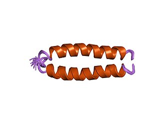
If the two entities are proteins, often linker (or "spacer") peptides are also added, which make it more likely that the proteins fold independently and behave as expected. Especially in the case where the linkers enable protein purification, linkers in protein or peptide fusions are sometimes engineered with cleavage sites for proteases or chemical agents that enable the liberation of the two separate proteins. This technique is often used for identification and purification of proteins, by fusing a GST protein, FLAG peptide, or a hexa-his peptide (6xHis-tag), which can be isolated using affinity chromatography with nickel or cobalt resins. Di- or multimeric chimeric proteins can be manufactured through genetic engineering by fusion to the original proteins of peptide domains that induce artificial protein di- or multimerization (e.g., streptavidin or leucine zippers). Fusion proteins can also be manufactured with toxins or antibodies attached to them in order to study disease development. Hydrogenase promoter, PSH, was studied constructing a PSH promoter-gfp fusion by using green fluorescent protein (gfp) reporter gene.

Recombinant functionality

Novel recombinant technologies have made it possible to improve fusion protein design for use in fields as diverse as biodetection, paper and food industries, and biopharmaceuticals. Recent improvements have involved the fusion of single peptides or protein fragments to regions of existing proteins, such as N and C termini, and are known to increase the following properties:

  • Catalytic efficiency: Fusion of certain peptides allow for greater catalytic efficiency by altering the tertiary and quaternary structure of the target protein.
  • Solubility: A common challenge in fusion protein design is the issue of insolubility of newly synthesized fusion proteins in the recombinant host, leading to an over-aggregation of the target protein in the cell. Molecular chaperones that are able to aid in protein folding may be added, thereby better segregating hydrophobic and hydrophilic interactions in the solute to increase protein solubility.
  • Thermostability: Singular peptides or protein fragments are typically added to reduce flexibility of either the N or C terminus of the target protein, which reinforces thermostability and stabilizes pH range.
  • Enzyme activity: Fusion that involves the introduction of hydrogen bonds may be used to expand overall enzyme activity.
  • Expression levels: Addition of numerous fusion fragments, such as maltose binding protein (MBP) or small ubiquitin-like molecule (SUMO), serve to enhance enzyme expression and secretion of the target protein.
  • Immobilization: PHA synthase, an enzyme that allows for the immobilization of proteins of interest, is an important fusion tag in industrial research.
  • Crystal quality: Crystal quality can be improved by adding covalent links between proteins, aiding in structure determination techniques.

Recombinant protein design

The earliest applications of recombinant protein design can be documented in the use of single peptide tags for purification of proteins in affinity chromatography. Since then, a variety of fusion protein design techniques have been developed for applications as diverse as fluorescent protein tags to recombinant fusion protein drugs. Three commonly used design techniques include tandem fusion, domain insertion, and post-translational conjugation.

Tandem fusion

The proteins of interest are simply connected end-to-end via fusion of N or C termini between the proteins. This provides a flexible bridge structure allowing enough space between fusion partners to ensure proper folding. However, the N or C termini of the peptide are often crucial components in obtaining the desired folding pattern for the recombinant protein, making simple end-to-end conjoining of domains ineffective in this case. For this reason, a protein linker is often needed to maintain the functionality of the protein domains of interest.

Domain insertion

This technique involves the fusion of consecutive protein domains by encoding desired structures into a single polypeptide chain, but sometimes may require insertion of a domain within another domain. This technique is typically regarding as more difficult to carry out than tandem fusion, due to difficulty finding an appropriate ligation site in the gene of interest.

Post-translational conjugation

This technique fuses protein domains following ribosomal translation of the proteins of interest, in contrast to genetic fusion prior to translation used in other recombinant technologies.

Protein linkers

A protein used as a linker in fusion protein design

Protein linkers aid fusion protein design by providing appropriate spacing between domains, supporting correct protein folding in the case that N or C termini interactions are crucial to folding. Commonly, protein linkers permit important domain interactions, reinforce stability, and reduce steric hindrance, making them preferred for use in fusion protein design even when N and C termini can be fused. Three major types of linkers are flexible, rigid, and in vivo cleavable.

  • Flexible linkers may consist of many small glycine residues, giving them the ability curl into a dynamic, adaptable shape.
  • Rigid linkers may be formed of large, cyclic proline residues, which can be helpful when highly specific spacing between domains must be maintained.
  • In vivo cleavable linkers are unique in that they are designed to allow the release of one or more fused domains under certain reaction conditions, such as a specific pH gradient, or when coming in contact with another biomolecule in the cell.

Natural occurrence

Naturally occurring fusion genes are most commonly created when a chromosomal translocation replaces the terminal exons of one gene with intact exons from a second gene. This creates a single gene that can be transcribed, spliced, and translated to produce a functional fusion protein. Many important cancer-promoting oncogenes are fusion genes produced in this way.

Examples include:

Antibodies are fusion proteins produced by V(D)J recombination.

There are also rare examples of naturally occurring polypeptides that appear to be a fusion of two clearly defined modules, in which each module displays its characteristic activity or function, independent of the other. Two major examples are: double PP2C chimera in Plasmodium falciparum (the malaria parasite), in which each PP2C module exhibits protein phosphatase 2C enzymatic activity, and the dual-family immunophilins that occur in a number of unicellular organisms (such as protozoan parasites and Flavobacteria) and contain full-length cyclophilin and FKBP chaperone modules. The evolutionary origin of such chimera remains unclear.
Polymer stabilizers

From Wikipedia, the free encyclopedia
https://en.wikipedia.org/wiki/Polymer_stabilizers

Polymer stabilizers (British: polymer stabilisers) are chemical additives which may be added to polymeric materials, such as plastics and rubbers, to inhibit or retard their degradation. Common polymer degradation processes include oxidation, UV-damage, thermal degradation, ozonolysis, combinations thereof such as photo-oxidation, as well as reactions with catalyst residues, dyes, or impurities. All of these degrade the polymer at a chemical level, via chain scission, uncontrolled recombination and cross-linking, which adversely affects many key properties such as strength, malleability, appearance and colour.

Stabilizers are used at all stages of the polymer life-cycle. They allow plastic items to be produced faster and with fewer defects, extend their useful lifespan, and facilitate their recycling. However they also continue to stabilise waste plastic, causing it to remain in the environment for longer. Many different types of plastic exist and each may be vulnerable to several types of degradation, which usually results in several different stabilisers being used in combination. Even for objects made from the same type of plastic, different applications may have different stabilisation requirements. Regulatory considerations, such as food contact approval are also present. A wide range of stabilizers is therefore needed.

The market for antioxidant stabilisers was estimated at US$1.69 billion for 2017, with the total market for all stabilizers expected to reach US$5.5 billion by 2025.

Antioxidants

Antioxidants inhibit autoxidation that occurs when polymers reacts with atmospheric oxygen. Aerobic degradation occurs gradually at room temperature, but almost all polymers are at risk of thermal-oxidation when they are processed at high temperatures. The molding or casting of plastics (e.g. injection molding) require them to be above their melting point or glass transition temperature (~200-300 °C). Under these conditions reactions with oxygen occur much more rapidly. Once initiated, autoxidation can be autocatalytic. As such, even though efforts are usually made to reduce oxygen levels, total exclusion is often not achievable and even exceedingly low concentrations of oxygen can be sufficient to initiate degradation. Sensitivity to oxidation varies significantly depending on the polymer in question; without stabilizers polypropylene and unsaturated polymers such as rubber will slowly degrade at room temperature where as polystyrene can be stable even at high temperatures. Antioxidants are of great importance during the process stage, with long-term stability at ambient temperature increasingly being supplied by hindered amine light stabilizers (HALs). Antioxidants are often referred to as being primary or secondary depending on their mechanism of action.

Primary antioxidants (radical scavengers)

Pentaerythritol tetrakis(3,5-di-tert-butyl-4-hydroxyhydrocinnamate): A primary antioxidant consisting of sterically hindered phenols with para-propionate groups.

Primary antioxidants (also known as chain-breaking antioxidants) act as radical scavengers and remove peroxy radicals (ROO•), as well as to a lesser extent alkoxy radicals (RO•), hydroxyl radicals (HO•) and alkyl radicals (R•). Oxidation begins with the formation of alkyl radicals, which are formed when the high temperatures and high shear stress experienced during processing snaps the polymer chains in a homolytic manner. These alkyl radicals react very rapidly with molecular oxygen (rate constants ≈ 107–109 mol–1 s–1) to give peroxy radicals, which in turn abstract hydrogen from a fresh section of polymer in a chain propagation step to give new alkyl radicals. The overall process is exceedingly complex and will vary between polymers but the first few steps are shown below in general:

R-R → 2 R•
R• + O2 → ROO•
ROO• + RH → ROOH + R•

Due to its rapid reaction with oxygen the scavenging of the initial alkyl radical (R•) is difficult and can only be achieved using specialised antioxidants the majority of primary antioxidants react instead with the longer lasting peroxy radicals (ROO•). Hydrogen abstraction is usually the rate determining step in the polymer degradation and the peroxy radicals can be scavenged by hydrogen donation from an alternative source, namely the primary antioxidant. This converts them into an organic hydroperoxide (ROOH). The most important commercial stabilizers for this are hindered phenols such as BHT or analogues thereof and secondary aromatic amines such as alkylated-diphenylamine. Amines are typically more effective, but cause pronounced discoloration, which is often undesirable (i.e., in food packaging, clothing). The overall reaction with phenols is shown below:

ROO• + ArOH → ROOH + ArO•
ArO• → nonradical products

The end products of these reactions are typically quinone methides, which may also impart unwanted colour. Modern phenolic antioxidants have complex molecular structures, often including a propionate-group at the para position of the phenol (i.e. they are ortho-alkylated analogues of phloretic acid). The quinone methides of these can rearrange once to give a hydroxycinnamate, regenerating the phenolic antioxidant group and allowing further radicals to be scavenged. Ultimately however, primary antioxidants are sacrificial and once they are fully consumed the polymer will begin to degrade.

Secondary antioxidants (hydroperoxides scavengers)

Tris(2,4-di-tert-butylphenyl)phosphite, a phosphite widely used as a secondary antioxidant in polymers.

Secondary antioxidants act to remove organic hydroperoxides (ROOH) formed by the action of primary antioxidants. Hydroperoxides are less reactive than radical species but can initiate fresh radical reactions:

ROOH + RH → RO• + R• + H2O

As they are less chemically active they require a more reactive antioxidant. The most commonly employed class are phosphite esters, often of hindered phenols e.g. Tris(2,4-di-tert-butylphenyl)phosphite. These will convert polymer hydroperoxides to alcohols, becoming oxidized to organophosphates in the process:

ROOH + P(OR')3 → OP(OR')3 + ROH

Transesterification can then take place, in which the hydroxylated polymer is exchanged for a phenol:

ROH + OP(OR')3 → R'OH + OP(OR')2OR

This exchange further stabilizes the polymer by releasing a primary antioxidant, because of this phosphites are sometimes considered multi-functional antioxidants as they can combine both types of activity. Organosulfur compounds are also efficient hydroperoxide decomposers, with thioethers being particularly effective against long-term thermal aging, they are ultimately oxidise up to sulfoxides and sulfones.

Antiozonant

N-Isopropyl-N'-phenyl-1,4-phenylenediamine (IPPD) a p-phenylenediamine based antiozonant
 

Antiozonants prevent or slow down the degradation of material caused by ozone. This is naturally present in the air at very low concentrations but is exceedingly reactive, particularly towards unsaturated polymers such as rubber, where it causes ozone cracking. The mechanism of ozonolysis is different from other forms of oxidation and hence requires its own class of antioxidant stabilizers. These are primarily based on p-phenylenediamine and work by reacting with ozone faster than it can react with vulnerable functional groups in the polymer (typically alkene groups). They achieve this by having a low ionization energy which allows them to react with ozone via electron transfer, this converts them into radical cations that are stabilized by aromaticity. Such species remain reactive and will react further, giving products such as 1,4-benzoquinone, phenylenediamine-dimers and aminoxyl radicals. Some of these products can then be scavenged by antioxidants.

Light stabilizers

Bisoctrizole: A benzotriazole-phenol based UV absorber
 

Light stabilizer are used to inhibit polymer photo-oxidation, which is the combined result of the action of light and oxygen. Like autoxidation this is a free radical process, hence the antioxidants described above are effective inhibiting agents, however additional classes of additives are also beneficial, such as UV absorbers, quenchers of excited states and HALS.

UV absorbers

UV susceptibility varies significantly between different polymers. Certain polycarbonates, polyesters and polyurethanes are highly susceptible, degrading via a Photo-Fries rearrangement. UV stabilisers absorb and dissipate the energy from UV rays as heat, typically by reversible intramolecular proton transfer. This reduces the absorption of UV rays by the polymer matrix and hence reduces the rate of weathering. Phenolic benzotriazoles (e.g. UV-360, UV-328) and hydroxyphenyl-triazines (e.g. Bemotrizinol) are used to stabilise polycarbonates and acrylics, oxanilides are used for polyamides and polyurethanes, while benzophenones are used for PVC.

Strongly light-absorbing PPS is difficult to stabilize. Even antioxidants fail in this electron-rich polymer. The acids or bases in the PPS matrix can disrupt the performance of the conventional UV absorbers such as HPBT. PTHPBT, which is a modification of HPBT are shown to be effective, even in these conditions.

Quenchers

A nickel-phenoxide quencher. CAS No 014516-71-3

Photo-oxidation can begin with the absorption of light by a chromophore within the polymer (which may be a dye or impurity) causing it to enter an excited state. This can then react with ambient oxygen, converting it into highly reactive singlet oxygen. Quenchers are able to absorb energy from excited molecules via a Förster mechanism and then dissipate it harmlessly as either heat or lower frequency fluorescent light. Singlet oxygen can be quenched by metal chelates, with nickel phenolates being a common example.

Hindered amine light stabilizers

Example structure of a HAL

The ability of hindered amine light stabilizers (HALS or HAS) to scavenge radicals produced by weathering, may be explained by the formation of aminoxyl radicals through a process known as the Denisov Cycle. The aminoxyl radical (N-O•) combines with free radicals in polymers:

N-O• + R• → N-O-R

Although they are traditionally considered as light stabilizers, they can also stabilize thermal degradation.

Even though HALS are extremely effective in polyolefins, polyethylene and polyurethane, they are ineffective in polyvinyl chloride (PVC). It is thought that their ability to form nitroxyl radicals is disrupted. HALS act as a base and become neutralized by hydrochloric acid (HCl) that is released by photooxidation of PVC. The exception is the recently developed NOR HALS, which is not a strong base and is not deactivated by HCl.

Other Classes

Polymers are susceptible to degradation by a variety of pathways beyond oxygen and light.

Acid Scavengers

Acid scavengers, also referred to as antacids, neutralize acidic impurities, especially those that release HCl. PVC is susceptible to acid-catalyzed degradation, the HCl being derived from the polymer itself. Ziegler–Natta catalysts and halogenated flame retardants also serve as sources of acids. Common acid scavengers include metallic soaps, such as calcium stearate and zinc stearate, mineral agents, such as hydrotalcite and hydrocalumite, and basic metal oxides, such as calcium oxide, zinc oxide or magnesium oxide.

Metal deactivators

Metal ions, such as those of Ti, Al and Cu, can accelerate the degradation of polymers. This is of particular concern where polymers are in direct contact with metal, such as in wiring and cable. More generally, the metal catalysts used to form the polymer may simply become encapsulated within it during production, this is typically true of Ziegler-Natta catalysts in polypropylene. In these instances metal deactivators may be added to improve stability. Deactivators work by chelation to form an inactive coordination complex with the metal ion. Salen-type compounds are common.

Heat stabilizers

Heat (or thermal) stabilizers are mostly used for PVC, as unstabilized material is particularly prone to thermal degradation. These agents minimize loss of HCl, a degradation process that starts above 70 °C. Once dehydrochlorination starts, it is autocatalytic. Many diverse agents have been used including, traditionally, derivatives of heavy metals (lead, cadmium). Increasingly, metallic soaps (metal "salts" of fatty acids) are favored, species such as calcium stearate. Addition levels vary typically from 2% to 4%. The choice of the best heat stabilizer depends on its cost effectiveness in the end use application, performance specification requirements, processing technology and regulatory approvals.

Flame retardants

Flame retardants are a broad range of compounds that improve fire resistance of polymers. Examples include brominated compounds along with aluminium hydroxide, antimony trioxide, and various organophosphates. Flame retardants are known to reduce the effectiveness of antioxidants.

Biocides

Degradation resulting from microorganisms (biodegradation) involves its own class of special bio-stabilizers and biocides (e.g. isothiazolinones).

Cell division

From Wikipedia, the free encyclopedia
Cell division in prokaryotes (binary fission) and eukaryotes (mitosis and meiosis). The thick lines are chromosomes, and the thin blue lines are fibers pulling on the chromosomes and pushing the ends of the cell apart.
 
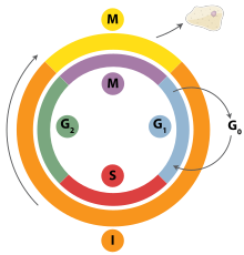
The cell cycle in eukaryotes: I = Interphase, M = Mitosis, G0 = Gap 0, G1 = Gap 1, G2 = Gap 2, S = Synthesis, G3 = Gap 3.

Cell division is the process by which a parent cell divides into two daughter cells. Cell division usually occurs as part of a larger cell cycle in which the cell grows and replicates its chromosome(s) before dividing. In eukaryotes, there are two distinct types of cell division: a vegetative division (mitosis), producing daughter cells genetically identical to the parent cell, and a cell division that produces haploid gametes for sexual reproduction (meiosis), reducing the number of chromosomes from two of each type in the diploid parent cell to one of each type in the daughter cells. In cell biology, mitosis (/maɪˈtoʊsɪs/) is a part of the cell cycle, in which, replicated chromosomes are separated into two new nuclei. Cell division gives rise to genetically identical cells in which the total number of chromosomes is maintained. In general, mitosis (division of the nucleus) is preceded by the S stage of interphase (during which the DNA replication occurs) and is often followed by telophase and cytokinesis; which divides the cytoplasm, organelles, and cell membrane of one cell into two new cells containing roughly equal shares of these cellular components. The different stages of mitosis all together define the mitotic (M) phase of animal cell cycle—the division of the mother cell into two genetically identical daughter cells. Meiosis results in four haploid daughter cells by undergoing one round of DNA replication followed by two divisions. Homologous chromosomes are separated in the first division, and sister chromatids are separated in the second division. Both of these cell division cycles are used in the process of sexual reproduction at some point in their life cycle. Both are believed to be present in the last eukaryotic common ancestor.

Prokaryotes (bacteria and archaea) usually undergo a vegetative cell division known as binary fission, where their genetic material is segregated equally into two daughter cells, but there are alternative manners of division, such as budding, that have been observed. All cell divisions, regardless of organism, are preceded by a single round of DNA replication.

For simple unicellular microorganisms such as the amoeba, one cell division is equivalent to reproduction – an entire new organism is created. On a larger scale, mitotic cell division can create progeny from multicellular organisms, such as plants that grow from cuttings. Mitotic cell division enables sexually reproducing organisms to develop from the one-celled zygote, which itself is produced by fusion of two gametes, each having been produced by meiotic cell division. After growth from the zygote to the adult, cell division by mitosis allows for continual construction and repair of the organism. The human body experiences about 10 quadrillion cell divisions in a lifetime.

The primary concern of cell division is the maintenance of the original cell's genome. Before division can occur, the genomic information that is stored in chromosomes must be replicated, and the duplicated genome must be cleanly divided between progeny cells. A great deal of cellular infrastructure is involved in ensuring consistency of genomic information among generations.

In bacteria

Divisome and elongasome complexes responsible for peptidoglycan synthesis during lateral cell-wall growth and division.

Bacterial cell division happens through binary fission or sometimes through budding. The divisome is a protein complex in bacteria that is responsible for cell division, constriction of inner and outer membranes during division, and remodeling of the peptidoglycan cell wall at the division site. A tubulin-like protein, FtsZ plays a critical role in formation of a contractile ring for the cell division.

In eukaryotes

Cell division in eukaryotes is more complicated than in prokaryotes. If the chromosomal number is reduced, eukaryotic cell division is classified as meiosis (reductional division). If the chromosomal number is not reduced, eukaryotic cell division is classified as mitosis (equational division). A primitive form of cell division, called amitosis, also exists. The amitotic or mitotic cell divisions are more atypical and diverse among the various groups of organisms, such as protists (namely diatoms, dinoflagellates, etc.) and fungi.

In the mitotic metaphase (see below), typically the chromosomes (each containing 2 sister chromatids that developed during replication in the S phase of interphase) align themselves on the metaphase plate. Then, the sister chromatids split and are distributed between two daughter cells.

In meiosis I, the homologous chromosomes are paired before being separated and distributed between two daughter cells. On the other hand, meiosis II is similar to mitosis. The chromatids are separated and distributed in the same way. In humans, other higher animals, and many other organisms, the process of meiosis is called gametic meiosis, during which meiosis produces four gametes. Whereas, in several other groups of organisms, especially in plants (observable during meiosis in lower plants, but during the vestigial stage in higher plants), meiosis gives rise to spores that germinate into the haploid vegetative phase (gametophyte). This kind of meiosis is called sporic meiosis.

Phases of eukaryotic cell division

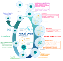
The phases (ordered counter-clockwise) of cell division (mitosis) and the cell cycle in animal cells.

Interphase

Interphase is the process through which a cell must go before mitosis, meiosis, and cytokinesis. Interphase consists of three main phases: G1, S, and G2. G1 is a time of growth for the cell where specialized cellular functions occur in order to prepare the cell for DNA replication. There are checkpoints during interphase that allow the cell to either advance or halt further development. One of the checkpoint is between G1 and S, the purpose for this checkpoint is to check for appropriate cell size and any DNA damage . The second check point is in the G2 phase, this checkpoint also checks for cell size but also the DNA replication. The last check point is located at the site of metaphase, where it checks that the chromosomes are correctly connected to the mitotic spindles. In S phase, the chromosomes are replicated in order for the genetic content to be maintained. During G2, the cell undergoes the final stages of growth before it enters the M phase, where spindles are synthesized. The M phase can be either mitosis or meiosis depending on the type of cell. Germ cells, or gametes, undergo meiosis, while somatic cells will undergo mitosis. After the cell proceeds successfully through the M phase, it may then undergo cell division through cytokinesis. The control of each checkpoint is controlled by cyclin and cyclin-dependent kinases. The progression of interphase is the result of the increased amount of cyclin. As the amount of cyclin increases, more and more cyclin dependent kinases attach to cyclin signaling the cell further into interphase. At the peak of the cyclin, attached to the cyclin dependent kinases this system pushes the cell out of interphase and into the M phase, where mitosis, meiosis, and cytokinesis occur. There are three transition checkpoints the cell has to go through before entering the M phase. The most important being the G1-S transition checkpoint. If the cell does not pass this checkpoint, it results in the cell exiting the cell cycle.

Prophase

Prophase is the first stage of division. The nuclear envelope is broken down in this stage, long strands of chromatin condense to form shorter more visible strands called chromosomes, the nucleolus disappears, and microtubules attach to the chromosomes at the disc-shaped kinetochores present in the centromere. Microtubules associated with the alignment and separation of chromosomes are referred to as the spindle and spindle fibers. Chromosomes will also be visible under a microscope and will be connected at the centromere. During this condensation and alignment period in meiosis, the homologous chromosomes undergo a break in their double-stranded DNA at the same locations, followed by a recombination of the now fragmented parental DNA strands into non-parental combinations, known as crossing over. This process is evidenced to be caused in a large part by the highly conserved Spo11 protein through a mechanism similar to that seen with toposomerase in DNA replication and transcription.

Metaphase

In metaphase, the centromeres of the chromosomes convene themselves on the metaphase plate (or equatorial plate), an imaginary line that is at equal distances from the two centrosome poles and held together by complexes known as cohesins. Chromosomes line up in the middle of the cell by microtubule organizing centers (MTOCs) pushing and pulling on centromeres of both chromatids thereby causing the chromosome to move to the center. At this point the chromosomes are still condensing and are currently one step away from being the most coiled and condensed they will be, and the spindle fibers have already connected to the kinetochores. During this phase all the microtubules, with the exception of the kinetochores, are in a state of instability promoting their progression toward anaphase. At this point, the chromosomes are ready to split into opposite poles of the cell toward the spindle to which they are connected.

Anaphase

Anaphase is a very short stage of the cell cycle and it occurs after the chromosomes align at the mitotic plate. Kinetochores emit anaphase-inhibition signals until their attachment to the mitotic spindle. Once the final chromosome is properly aligned and attached the final signal dissipates and triggers the abrupt shift to anaphase. This abrupt shift is caused by the activation of the anaphase-promoting complex and its function of tagging degradation of proteins important toward the metaphase-anaphase transition. One of these proteins that is broken down is securin which through its breakdown releases the enzyme separase that cleaves the cohesin rings holding together the sister chromatids thereby leading to the chromosomes separating. After the chromosomes line up in the middle of the cell, the spindle fibers will pull them apart. The chromosomes are split apart while the sister chromatids move to opposite sides of the cell. As the sister chromatids are being pulled apart, the cell and plasma are elongated by non-kinetochore microtubules.

Telophase

Telophase is the last stage of the cell cycle in which a cleavage furrow splits the cells cytoplasm (cytokinesis) and chromatin. This occurs through the synthesis of a new nuclear envelope that forms around the chromatin gathered at each pole. The nucleolus reforms as the chromatin reverts back to the loose state it possessed during interphase. The division of the cellular contents is not always equal and can vary by cell type as seen with oocyte formation where one of the four daughter cells possess the majority of the duckling.

Cytokinesis

The last stage of the cell division process is cytokinesis. In this stage there is a cytoplasmic division that occurs at the end of either mitosis or meiosis. At this stage there is a resulting irreversible separation leading to two daughter cells. Cell division plays an important role in determining the fate of the cell. This is due to there being the possibility of an asymmetric division. This as a result leads to cytokinesis producing unequal daughter cells containing completely different amounts or concentrations of fate-determining molecules.

In animals the cytokinesis ends with formation of a contractile ring and thereafter a cleavage. But in plants it happen differently. At first a cell plate is formed and then a cell wall develops between the two daughter cells.

In Fission yeast (S. pombe) the cytokinesis happens in G1 phase 

Variants

Image of the mitotic spindle in a human cell showing microtubules in green, chromosomes (DNA) in blue, and kinetochores in red

Cells are broadly classified into two main categories: simple non-nucleated prokaryotic cells and complex nucleated eukaryotic cells. Due to their structural differences, eukaryotic and prokaryotic cells do not divide in the same way. Also, the pattern of cell division that transforms eukaryotic stem cells into gametes (sperm cells in males or egg cells in females), termed meiosis, is different from that of the division of somatic cells in the body. Image of the mitotic spindle in a human cell showing microtubules in green, chromosomes (DNA) in blue, and kinetochores in red.

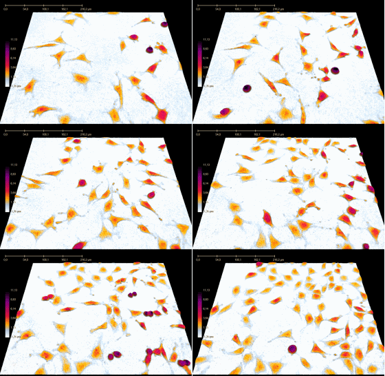
Cell division over 42. The cells were directly imaged in the cell culture vessel, using non-invasive quantitative phase contrast time-lapse microscopy.

In 2022, scientists discovered a new type of cell division called asynthetic fission found in the squamous epithelial cells in the epidermis of juvenile zebrafish. When juvenile zebrafish are growing, skin cells must quickly cover the rapidly increasing surface area of the zebrafish. These skin cells divide without duplicating their DNA (the S phase of mitosis) causing up to 50% of the cells to have a reduced genome size. These cells are later replaced by cells with a standard amount of DNA. Scientists expect to find this type of division in other vertebrates.

Degradation

Multicellular organisms replace worn-out cells through cell division. In some animals, however, cell division eventually halts. In humans this occurs, on average, after 52 divisions, known as the Hayflick limit. The cell is then referred to as senescent. With each division the cells telomeres, protective sequences of DNA on the end of a chromosome that prevent degradation of the chromosomal DNA, shorten. This shortening has been correlated to negative effects such as age-related diseases and shortened lifespans in humans. Cancer cells, on the other hand, are not thought to degrade in this way, if at all. An enzyme complex called telomerase, present in large quantities in cancerous cells, rebuilds the telomeres through synthesis of telomeric DNA repeats, allowing division to continue indefinitely.

History

Kurt Michel with his phase-contrast microscope

A cell division under microscope was first discovered by German botanist Hugo von Mohl in 1835 as he worked over the green alga Cladophora glomerata.

In 1943, cell division was filmed for the first time by Kurt Michel using a phase-contrast microscope.

Disulfide

From Wikipedia, the free encyclopedia
https://en.wikipedia.org/wiki/Disulfide

In biochemistry, a disulfide (or disulphide in British English) refers to a functional group with the structure R−S−S−R′. The linkage is also called an SS-bond or sometimes a disulfide bridge and is usually derived by the coupling of two thiol groups. In biology, disulfide bridges formed between thiol groups in two cysteine residues are an important component of the secondary and tertiary structure of proteins. Persulfide usually refers to R−S−S−H compounds.

In inorganic chemistry disulfide usually refers to the corresponding anion S2−
2
(S−S).

Organic disulfides

A selection of disulfides:
 
Pyrite, FeS2, "fool's gold"
 
Disulfur dichloride, S2Cl2, a common industrial chemical
 
cystine, crosslinker in many proteins
lipoic acid, a vitamin
Diphenyl disulfide, Ph2S2, a common organic disulfide

Symmetrical disulfides are compounds of the formula R2S2. Most disulfides encountered in organo sulfur chemistry are symmetrical disulfides. Unsymmetrical disulfides (also called heterodisulfides) are compounds of the formula RSSR'. They are less common in organic chemistry, but most disulfides in nature are unsymmetrical.

Properties

The disulfide bonds are strong, with a typical bond dissociation energy of 60 kcal/mol (251 kJ mol−1). However, being about 40% weaker than C−C and C−H bonds, the disulfide bond is often the "weak link" in many molecules. Furthermore, reflecting the polarizability of divalent sulfur, the S−S bond is susceptible to scission by polar reagents, both electrophiles and especially nucleophiles (Nu):

The disulfide bond is about 2.05 Å in length, about 0.5 Å longer than a C−C bond. Rotation about the S−S axis is subject to a low barrier. Disulfides show a distinct preference for dihedral angles approaching 90°. When the angle approaches 0° or 180°, then the disulfide is a significantly better oxidant.

Disulfides where the two R groups are the same are called symmetric, examples being diphenyl disulfide and dimethyl disulfide. When the two R groups are not identical, the compound is said to be an asymmetric or mixed disulfide.

Although the hydrogenation of disulfides is usually not practical, the equilibrium constant for the reaction provides a measure of the standard redox potential for disulfides:

This value is about −250 mV versus the standard hydrogen electrode (pH = 7). By comparison, the standard reduction potential for ferrodoxins is about −430 mV.

Synthesis

Disulfide bonds are usually formed from the oxidation of sulfhydryl (−SH) groups, especially in biological contexts. The transformation is depicted as follows:

A variety of oxidants participate in this reaction including oxygen and hydrogen peroxide. Such reactions are thought to proceed via sulfenic acid intermediates. In the laboratory, iodine in the presence of base is commonly employed to oxidize thiols to disulfides. Several metals, such as copper(II) and iron(III) complexes affect this reaction. Alternatively, disulfide bonds in proteins often formed by thiol-disulfide exchange:

Such reactions are mediated by enzymes in some cases and in other cases are under equilibrium control, especially in the presence of a catalytic amount of base.

The alkylation of alkali metal di- and polysulfides gives disulfides. "Thiokol" polymers arise when sodium polysulfide is treated with an alkyl dihalide. In the converse reaction, carbanionic reagents react with elemental sulfur to afford mixtures of the thioether, disulfide, and higher polysulfides. These reactions are often unselective but can be optimized for specific applications.

Synthesis of unsymmetrical disulfides (heterodisulfides)

Many specialized methods have been developed for forming unsymmetrical disulfides. Reagents that deliver the equivalent of "RS+" react with thiols to give asymmetrical disulfides:

where R″2N is the phthalimido group. Bunte salts, derivatives of the type RSSO3Na+are also used to generate unsymmetrical disulfides:

Reactions

The most important aspect of disulfide bonds is their cleavage, which occurs via reduction. A variety of reductants can be used. In biochemistry, thiols such as β-mercaptoethanol (β-ME) or dithiothreitol (DTT) serve as reductants; the thiol reagents are used in excess to drive the equilibrium to the right:

The reductant tris(2-carboxyethyl)phosphine (TCEP) is useful, beside being odorless compared to β-ME and DTT, because it is selective, working at both alkaline and acidic conditions (unlike DTT), is more hydrophilic and more resistant to oxidation in air. Furthermore, it is often not needed to remove TCEP before modification of protein thiols.

In organic synthesis, hydride agents are typically employed for scission of disulfides, such as sodium borohydride. More aggressive, alkali metals will effect this reaction:

These reactions are often followed by protonation of the resulting metal thiolate:

Thiol–disulfide exchange is a chemical reaction in which a thiolate group −S attacks a sulfur atom of a disulfide bond −S−S−. The original disulfide bond is broken, and its other sulfur atom is released as a new thiolate, carrying away the negative charge. Meanwhile, a new disulfide bond forms between the attacking thiolate and the original sulfur atom.

Thiol–disulfide exchange showing the linear intermediate in which the charge is shared among the three sulfur atoms. The thiolate group (shown in red) attacks a sulfur atom (shown in blue) of the disulfide bond, displacing the other sulfur atom (shown in green) and forming a new disulfide bond.

Thiolates, not thiols, attack disulfide bonds. Hence, thiol–disulfide exchange is inhibited at low pH (typically, below 8) where the protonated thiol form is favored relative to the deprotonated thiolate form. (The pKa of a typical thiol group is roughly 8.3, but can vary due to its environment.)

Thiol–disulfide exchange is the principal reaction by which disulfide bonds are formed and rearranged in a protein. The rearrangement of disulfide bonds within a protein generally occurs via intra-protein thiol–disulfide exchange reactions; a thiolate group of a cysteine residue attacks one of the protein's own disulfide bonds. This process of disulfide rearrangement (known as disulfide shuffling) does not change the number of disulfide bonds within a protein, merely their location (i.e., which cysteines are bonded). Disulfide reshuffling is generally much faster than oxidation/reduction reactions, which change the number of disulfide bonds within a protein. The oxidation and reduction of protein disulfide bonds in vitro also generally occurs via thiol–disulfide exchange reactions. Typically, the thiolate of a redox reagent such as glutathione or dithiothreitol attacks the disulfide bond on a protein forming a mixed disulfide bond between the protein and the reagent. This mixed disulfide bond when attacked by another thiolate from the reagent, leaves the cysteine oxidized. In effect, the disulfide bond is transferred from the protein to the reagent in two steps, both thiol–disulfide exchange reactions.

The in vivo oxidation and reduction of protein disulfide bonds by thiol–disulfide exchange is facilitated by a protein called thioredoxin. This small protein, essential in all known organisms, contains two cysteine amino acid residues in a vicinal arrangement (i.e., next to each other), which allows it to form an internal disulfide bond, or disulfide bonds with other proteins. As such, it can be used as a repository of reduced or oxidized disulfide bond moieties.

Many specialized organic reactions have been developed for disulfides, again mainly associated with the scission of the S−S bond, which is usually the weakest bond in a molecule. In the Zincke disulfide cleavage reactions, disulfides are cleaved by halogens. This reaction gives a sulfenyl halide:

Occurrence in biology

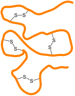
Schematic of disulfide bonds crosslinking regions of a protein

Occurrence in proteins

Disulfide bonds can be formed under oxidising conditions and play an important role in the folding and stability of some proteins, usually proteins secreted to the extracellular medium. Since most cellular compartments are reducing environments, in general, disulfide bonds are unstable in the cytosol, with some exceptions as noted below, unless a sulfhydryl oxidase is present.

Cystine is composed of two cysteines linked by a disulfide bond (shown here in its neutral form).

Disulfide bonds in proteins are formed between the thiol groups of cysteine residues by the process of oxidative folding. The other sulfur-containing amino acid, methionine, cannot form disulfide bonds. A disulfide bond is typically denoted by hyphenating the abbreviations for cysteine, e.g., when referring to ribonuclease A the "Cys26–Cys84 disulfide bond", or the "26–84 disulfide bond", or most simply as "C26–C84" where the disulfide bond is understood and does not need to be mentioned. The prototype of a protein disulfide bond is the two-amino-acid peptide cystine, which is composed of two cysteine amino acids joined by a disulfide bond. The structure of a disulfide bond can be described by its χss dihedral angle between the Cβ−Sγ−Sγ−Cβ atoms, which is usually close to ±90°.

The disulfide bond stabilizes the folded form of a protein in several ways:

  1. It holds two portions of the protein together, biasing the protein towards the folded topology. That is, the disulfide bond destabilizes the unfolded form of the protein by lowering its entropy.
  2. The disulfide bond may form the nucleus of a hydrophobic core of the folded protein, i.e., local hydrophobic residues may condense around the disulfide bond and onto each other through hydrophobic interactions.
  3. Related to 1 and 2, the disulfide bond links two segments of the protein chain, increases the effective local concentration of protein residues, and lowers the effective local concentration of water molecules. Since water molecules attack amide-amide hydrogen bonds and break up secondary structure, a disulfide bond stabilizes secondary structure in its vicinity. For example, researchers have identified several pairs of peptides that are unstructured in isolation, but adopt stable secondary and tertiary structure upon formation of a disulfide bond between them.

A disulfide species is a particular pairing of cysteines in a disulfide-bonded protein and is usually depicted by listing the disulfide bonds in parentheses, e.g., the "(26–84, 58–110) disulfide species". A disulfide ensemble is a grouping of all disulfide species with the same number of disulfide bonds, and is usually denoted as the 1S ensemble, the 2S ensemble, etc. for disulfide species having one, two, etc. disulfide bonds. Thus, the (26–84) disulfide species belongs to the 1S ensemble, whereas the (26–84, 58–110) species belongs to the 2S ensemble. The single species with no disulfide bonds is usually denoted as R for "fully reduced". Under typical conditions, disulfide reshuffling is much faster than the formation of new disulfide bonds or their reduction; hence, the disulfide species within an ensemble equilibrate more quickly than between ensembles.

The native form of a protein is usually a single disulfide species, although some proteins may cycle between a few disulfide states as part of their function, e.g., thioredoxin. In proteins with more than two cysteines, non-native disulfide species may be formed, which are almost always misfolded. As the number of cysteines increases, the number of nonnative species increases factorially.

In bacteria and archaea

Disulfide bonds play an important protective role for bacteria as a reversible switch that turns a protein on or off when bacterial cells are exposed to oxidation reactions. Hydrogen peroxide (H2O2) in particular could severely damage DNA and kill the bacterium at low concentrations if not for the protective action of the SS-bond. Archaea typically have fewer disulfides than higher organisms.

In eukaryotes

In eukaryotic cells, in general, stable disulfide bonds are formed in the lumen of the RER (rough endoplasmic reticulum) and the mitochondrial intermembrane space but not in the cytosol. This is due to the more oxidizing environment of the aforementioned compartments and more reducing environment of the cytosol (see glutathione). Thus disulfide bonds are mostly found in secretory proteins, lysosomal proteins, and the exoplasmic domains of membrane proteins.

There are notable exceptions to this rule. For example, many nuclear and cytosolic proteins can become disulfide-crosslinked during necrotic cell death. Similarly, a number of cytosolic proteins which have cysteine residues in proximity to each other that function as oxidation sensors or redox catalysts; when the reductive potential of the cell fails, they oxidize and trigger cellular response mechanisms. The virus Vaccinia also produces cytosolic proteins and peptides that have many disulfide bonds; although the reason for this is unknown presumably they have protective effects against intracellular proteolysis machinery.

Disulfide bonds are also formed within and between protamines in the sperm chromatin of many mammalian species.

Disulfides in regulatory proteins

As disulfide bonds can be reversibly reduced and re-oxidized, the redox state of these bonds has evolved into a signaling element. In chloroplasts, for example, the enzymatic reduction of disulfide bonds has been linked to the control of numerous metabolic pathways as well as gene expression. The reductive signaling activity has been shown, thus far, to be carried by the ferredoxin-thioredoxin system, channeling electrons from the light reactions of photosystem I to catalytically reduce disulfides in regulated proteins in a light dependent manner. In this way chloroplasts adjust the activity of key processes such as the Calvin–Benson cycle, starch degradation, ATP production and gene expression according to light intensity. Additionally, It has been reported that disulfides plays a significant role on redox state regulation of Two-component systems (TCSs), which could be found in certain bacteria including photogenic strain. A unique intramolecular cysteine disulfide bonds in the ATP-binding domain of SrrAB TCs found in Staphylococcus aureus is a good example of disulfides in regulatory proteins, which the redox state of SrrB molecule is controlled by cysteine disulfide bonds, leading to the modification of SrrA activity including gene regulation.

In hair and feathers

Over 90% of the dry weight of hair comprises proteins called keratins, which have a high disulfide content, from the amino acid cysteine. The robustness conferred in part by disulfide linkages is illustrated by the recovery of virtually intact hair from ancient Egyptian tombs. Feathers have similar keratins and are extremely resistant to protein digestive enzymes. The stiffness of hair and feather is determined by the disulfide content. Manipulating disulfide bonds in hair is the basis for the permanent wave in hairstyling. Reagents that affect the making and breaking of S−S bonds are key, e.g., ammonium thioglycolate. The high disulfide content of feathers dictates the high sulfur content of bird eggs. The high sulfur content of hair and feathers contributes to the disagreeable odor that results when they are burned.

Inorganic disulfides

The disulfide anion is S2−
2
, or S−S. In disulfide, sulfur exists in the reduced state with oxidation number −1. Its electron configuration then resembles that of a chlorine atom. It thus tends to form a covalent bond with another S center to form S2−
2
group, similar to elemental chlorine existing as the diatomic Cl2. Oxygen may also behave similarly, e.g. in peroxides such as H2O2. Examples:

Related compounds

CS2
 
MoS2

Thiosulfoxides are orthogonally isomeric with disulfides, having the second sulfur branching from the first and not partaking in a continuous chain, i.e. >S=S rather than −S−S−.

Disulfide bonds are analogous but more common than related peroxide, thioselenide, and diselenide bonds. Intermediate compounds of these also exist, for example thioperoxides (also known as oxasulfides) such as hydrogen thioperoxide, have the formula R1OSR2 (equivalently R2SOR1). These are isomeric to sulfoxides in a similar manner to the above; i.e. >S=O rather than −S−O−.

Thiuram disulfides, with the formula (R2NCSS)2, are disulfides but they behave distinctly because of the thiocarbonyl group.

Compounds with three sulfur atoms, such as CH3S−S−SCH3, are called trisulfides, or trisulfide bonds.

Misnomers

Disulfide is also used to refer to compounds that contain two sulfide (S2−) centers. The compound carbon disulfide, CS2 is described with the structural formula i.e. S=C=S. This molecule is not a disulfide in the sense that it lacks a S-S bond. Similarly, molybdenum disulfide, MoS2, is not a disulfide in the sense again that its sulfur atoms are not linked.

Applications

Rubber manufacturing

The vulcanization of rubber results in crosslinking groups which consist of disulfide (and polysulfide) bonds; in analogy to the role of disulfides in proteins, the S−S linkages in rubber strongly affect the stability and rheology of the material. Although the exact mechanism underlying the vulcanization process is not entirely understood (as multiple reaction pathways are present but the predominant one is unknown), it has been extensively shown that the extent to which the process is allowed to proceed determines the physical properties of the resulting rubber- namely, a greater degree of crosslinking corresponds to a stronger and more rigid material. The current conventional methods of rubber manufacturing are typically irreversible, as the unregulated reaction mechanisms can result in complex networks of sulfide linkages; as such, rubber is considered to be a thermoset material.

Covalent adaptable networks

Due to their relatively weak bond dissociation energy (in comparison to C−C bonds and the like), disulfides have been employed in covalent adaptable network (CAN) systems in order to allow for dynamic breakage and reformation of crosslinks. By incorporating disulfide functional groups as crosslinks between polymer chains, materials can be produced which are stable at room temperature while also allowing for reversible crosslink dissociation upon application of elevated temperature. The mechanism behind this reaction can be attributed to the cleavage of disulfide linkages (RS−SR) into thiyl radicals (2 RS•) which can subsequently reassociate into new bonds, resulting in reprocessability and self-healing characteristics for the bulk material. However, since the bond dissociation energy of the disulfide bond is still fairly high, it is typically necessary to augment the bond with adjacent chemistry that can stabilize the unpaired electron of the intermediate state. As such, studies usually employ aromatic disulfides or disulfidediamine (RNS−SNR) functional groups to encourage the dynamic dissociation of the S−S bond; these chemistries can result in the bond dissociation energy being reduced to half (or even less) of its prior magnitude.

In practical terms, disulfide-containing CANs can be used to impart recyclability to polymeric materials while still exhibiting physical properties similar to that of thermosets. Typically, recyclability is restricted to thermoplastic materials, as said materials consist of polymer chains which are not bonded to each other at the molecular level; as a result, they can be melted down and reformed (as the addition of thermal energy allows the chains to untangle, move past each other, and adopt new configurations), but this comes at the expense of their physical robustness. Meanwhile, conventional thermosets contain permanent crosslinks which bolster their strength, toughness, creep resistance, and the like (as the bonding between chains provides resistance to deformation at the macroscopic level), but due to the permanence of said crosslinks, these materials cannot be reprocessed akin to thermoplastics. However, due to the dynamic nature of the crosslinks in disulfide CANs, they can be designed to exhibit the best attributes of both of the aforementioned material types. Studies have shown that disulfide CANs can be reprocessed multiple times with negligible degradation in performance while also exhibiting creep resistance, glass transition, and dynamic modulus values comparable to those observed in similar conventional thermoset systems.

Operator (computer programming)

From Wikipedia, the free encyclopedia https://en.wikipedia.org/wiki/Operator_(computer_programmin...