Search This Blog

Sunday, June 10, 2018

Neurostimulation

From Wikipedia, the free encyclopedia
 
Neurostimulation
OPS-301 code 8-631

Neurostimulation is the purposeful modulation of the nervous system's activity using invasive (e.g. microelectrodes) or non-invasive means (e.g. transcranial magnetic stimulation or transcranial electric stimulation, tES, such as tDCS or transcranial alternating current stimulation, tACS). Neurostimulation usually refers to the electromagnetic approaches to neuromodulation.

Neurostimulation technology can improve the life quality of those who are severely paralyzed or suffering from profound losses to various sense organs, as well as for permanent reduction of severe, chronic pain which would otherwise require constant (around-the-clock), high-dose opioid therapy (such as neuropathic pain and spinal-cord injury). It serves as the key part of neural prosthetics for hearing aids, artificial vision, artificial limbs, and brain-machine interfaces. In the case of neural stimulation, mostly an electrical stimulation is utilized and charge-balanced biphasic constant current waveforms or capacitively coupled charge injection approaches are adopted. Alternatively, transcranial magnetic stimulation and transcranial electric stimulation have been proposed as non-invasive methods in which either a magnetic field or transcranially applied electric currents cause neurostimulation.[1][2]

Brain stimulation

Brain stimulation has potentials to treat some disorders such as epilepsy. In this method, scheduled stimulation is applied to specific cortical or subcortical targets. There are available commercial devices[3] that can deliver an electrical pulse at scheduled time intervals. Scheduled stimulation is hypothesized to alter the intrinsic neurophysiologic properties of epileptic networks. The most explored targets for scheduled stimulation are the anterior nucleus of the thalamus and the hippocampus. The anterior nucleus of the thalamus has been studied, which has shown a significant seizure reduction with the stimulator on versus off during several months after stimulator implantation.[4] Moreover, the cluster headache (CH) can be treated by using a temporary stimulating electrode at sphenopalatine ganglion (SPG). Pain relief is reported within several minutes of stimulation in this method.[5] To avoid use of implanted electrodes, researchers have engineered ways to inscribe a "window" made of zirconia that has been modified to be transparent and implanted in mice skulls, to allow optical waves to penetrate more deeply, as in optogenetics, to stimulate or inhibit individual neurons.[6]

Deep brain stimulation

Deep brain stimulation (DBS) has shown benefits for movement disorders such as Parkinson's disease, tremor and dystonia and affective disorders such as depression, obsessive-compulsive disorder, Tourette syndrome, chronic pain and cluster headache. Since DBS can directly change the brain activity in a controlled manner, it is used to map fundamental mechanisms of brain functions along with neuroimaging methods. A simple DBS system consists of two different parts. First, tiny microelectrodes are implanted in the brain to deliver stimulation pulses to the tissue. Second, an electrical pulse generator (PG) generates stimulation pulses, which is connected to the electrodes via microwires. Physiological properties of the brain tissue, which may change with disease state, stimulation parameters, which include amplitude and temporal characteristics, and the geometric configuration of the electrode and the surrounding tissue are all parameters on which DBS of both the normal and the diseased brain depend on. In spite of a huge amount of studies on DBS, its mechanism of action is still not well understood. Developing DBS microelectrodes is still challenging.[7]

Non-invasive brain stimulation

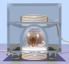
rTMS in a rodent. From Oscar Arias-CarriĆ³n, 2008

Transcranial magnetic stimulation

Compared to electrical stimulation that utilizes brief, high-voltage electric shock to activate neurons, which can potentially activate pain fibers, transcranial magnetic stimulation (TMS) was developed by Baker in 1985. TMS uses a magnetic wire above the scalp, which carries a sharp and high current pulse. A time variant magnetic field is induced perpendicular to the coil due to the applied pulse which consequently generates an electric field based on Maxwell's law. The electric field provides the necessary current for a non-invasive and much less painful stimulation. There are two TMS devices called single pulse TMS and repetitive pulse TMS (rTMS) while the latter has greater effect but potential to cause seizure. TMS can be used for therapy particularly in psychiatry, as a tool to measure central motor conduction and a research tool to study different aspects of human brain physiology such as motor function, vision, and language. The rTMS method has been used to treat epilepsy with rates of 8–25 Hz for 10 seconds. The other therapeutic uses of rTMS include parkinson diseases, dystonia and mood diseases. Also, TMS can be used to determine the contribution of cortical networks to specific cognitive functions by disrupting activity in the focal brain region.[1] Early, inconclusive, results have been obtained in recovery from coma (persistent vegetative state) by Pape et al. (2009).[8]
 
Transcranial electrical stimulation techniques. While tDCS uses constant current intensity, tRNS and tACS use oscillating current. The vertical axis represents the current intensity in milliamp (mA), while the horizontal axis illustrates the time-course.

Transcranial electrical stimulation

Spinal cord stimulation

Spinal cord stimulation (SCS) is an effective therapy for the treatment of chronic and intractable pain including diabetic neuropathy, failed back surgery syndrome, complex regional pain syndrome, phantom limb pain, ischemic limb pain, refractory unilateral limb pain syndrome, postherpetic neuralgia and acute herpes zoster pain. Another pain condition that is a potential candidate for SCS treatment is Charcot-Marie-Tooth (CMT) disease, which is associated with moderate to severe chronic extremity pain.[9] SCS therapy consists of the electrical stimulation of the spinal cord to 'mask' pain. The gate theory proposed in 1965 by Melzack and Wall[10] provided a theoretical construct to attempt SCS as a clinical treatment for chronic pain. This theory postulates that activation of large diameter, myelinated primary afferent fibers suppresses the response of dorsal horn neurons to input from small, unmyelinated primary afferents. A simple SCS system consists of three different parts. First, microelectrodes are implanted in the epidural space to deliver stimulation pulses to the tissue. Second, an electrical pulse generator implanted in the lower abdominal area or gluteal region while is connected to the electrodes via wires, and third a remote control to adjust the stimulus parameters such as pulse width and pulse rate in the PG. Improvements have been made in both the clinical aspects of SCS such as transition from subdural placement of contacts to epidural placement, which reduces the risk and morbidity of SCS implantation, and also technical aspects of SCS such as improving percutaneous leads, and fully implantable multi-channel stimulators. However, there are many parameters that need to be optimized including number of implanted contacts, contact size and spacing, and electrical sources for stimulation. The stimulus pulse width and pulse rate are important parameters that need to be adjusted in SCS, which are typically 400 us and 8–200 Hz respectively.[11]

Transcutaneous supraorbital nerve stimulation

Tentative evidence supports transcutaneous supraorbital nerve stimulation.[12] Side effects are few.[13]

Cochlear implants

Cochlear implant

Cochlear implants have provided partial hearing to more than 120,000 persons worldwide as of 2008. The electrical stimulation is used in a cochlear implant to provide functional hearing in totally deafened persons. Cochlear implants include several subsystem components from the external speech processor and radio frequency (RF) transmission link to the internal receiver, stimulator, and electrode arrays. Modern cochlear implant research started in the 1960s and 1970s. In 1961, a crude single electrode device was implanted in two deaf patients and useful hearing with electric stimulation was reported. The first FDA approved complete single channel device was released in 1984.[14] In cochlear implants, the sound is picked up by a microphone and transmitted to the behind-the-ear external processor to be converted to the digital data. The digitized data is then modulated on a radio frequency signal and transmitted to an antenna inside a headpiece. The data and power carrier are transmitted through a pair of coupled coils to the hermetically sealed internal unit. By extracting the power and demodulating the data, electric current commands are sent to the cochlea to stimulate the auditory nerve through microelectrodes.[15] The key point is that the internal unit does not have a battery and it should be able to extract the required energy. Also to reduce the infection, data is transmitted wirelessly along with power. Inductively coupled coils are the best candidate for power and data telemetry. Parameters needed by the internal unit include the pulse amplitude, pulse duration, pulse gap, active electrode, and return electrode that are used to define a biphasic pulse and the stimulation mode. An example of the commercial devices include Nucleus 22 device that utilized a carrier frequency of 2.5 MHz and later in the newer revision called Nucleus 24 device, the carrier frequency was increased to 5 MHz.[16] The internal unit in the cochlear implants is an ASIC (application-specific integrated circuit) chip that is responsible to ensure safe and reliable electric stimulation. Inside the ASIC chip, there is a forward pathway, a backward pathway, and control units. The forward pathway recovers digital information from the RF signal which includes stimulation parameters and some handshaking bits to reduce the communication error. The backward pathway usually includes a back telemetry voltage sampler that reads the voltage over a period of time on the recording electrode. The stimulator block is responsible to deliver predetermined current by external unit to the microelectrodes. This block includes a reference current and a digital to analog converter to transform digital commands to an analog current.[17]

Visual prosthesis

Visual cortical implant designed by Mohamad Sawan
The Visual Cortical Implant

Theoretical and experimental clinical evidences suggest that direct electrical stimulation of the retina might be able to provide some vision to subjects who have lost the photoreceptive elements of their retina.[18] Therefore, visual prostheses are developed to restore vision for the blind by using the stimulation. Depending upon which visual pathway location is targeted for neural stimulation, different approaches have been considered. Visual pathway consists mainly of the eye, optic nerve, lateral geniculate nucleus (LGN), and visual cortex. Therefore, retinal, optic nerve and visual cortex stimulation are the three different methods used in visual prostheses.[19] Retinal degenerative diseases, such as retinitis pigmentosa (RP) and age-related macular degeneration (AMD), are two likely candidate diseases in which retinal stimulation may be helpful. Three approaches called intraocular epiretinal, subretinal and extraocular transretinal stimulation are pursued in retinal devices that stimulate remaining retinal neural cells to bypass lost photoreceptors and allow the visual signal to reach the brain via the normal visual pathway. In epiretinal approach, electrodes are placed on the top side of the retina near ganglion cells,[20] whereas the electrodes are placed under the retina in subretinal approaches.[21] Finally, the posterior scleral surface of the eye is the place in which extraocular approach electrodes are positioned. Second Sight and the Humayun group at USC are the most active groups in the design of intraocular retinal prostheses. The ArgusTM 16 retinal implant is an intraocular retinal prosthesis utilizing video processing technologies. Regarding to the visual cortex stimulation, Brindley, and Dobelle were the first ones who did the experiments and demonstrated that by stimulating the top side of the visual cortex most of the electrodes can produce visual percept.[11] More recently Sawan built a complete implant for intracortical stimulation and validated the operation in rats[22]

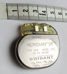
A pacemaker, scale in centimeters

LGN, which is located in the midbrain to relay signals from the retina to the visual cortex, is another potential area that can be used for stimulation. But this area has limited access due to surgical difficulty. The recent success of deep brain stimulation techniques targeting the midbrain has encouraged research to pursue the approach of LGN stimulation for a visual prosthesis.[23]

Cardiac electrostimulation devices

Implantable pacemakers were proposed for the first time in 1959 and became more sophisticated since then. The therapeutic application of pacemakers consists of numerous rhythm disturbances including some forms of tachycardia (too fast a heart beat), heart failure, and even stroke. Early implantable pacemakers worked only a short time and needed periodic recharging by an inductive link. These implantable pacemakers needed a pulse generator to stimulate heart muscles with a certain rate in addition to electrodes.[24] Today, modern pulse generators are programmed non-invasively by sophisticated computerized machines using RF, obtaining information about the patient's and device's status by telemetry. Also they use a single hermetically sealed lithium iodide (LiI) cell as the battery. The pacemaker circuitry includes sense amplifiers to detect the heart's intrinsic electrical signals, which are used to track heart activity, rate adaptive circuitry, which determine the need for increased or reduced pacing rate, a microprocessor, memory to store the parameters, telemetry control for communication protocol and power supplies to provide regulated voltage.[25]

Stimulation microelectrode technologies

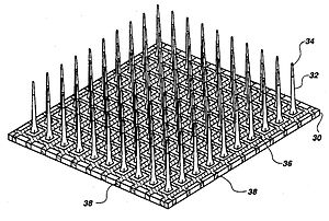
Utah microelectrode array

Microelectrodes are one of the key components of the neurostimulation, which deliver the current to neurons. Typical microelectrodes have three main components: a substrate (the carrier), a conductive metal layer, and an insulation material. In cochlear implants, microelectrodes are formed from platinum-iridium alloy. State-of-the-art electrodes include deeper insertion to better match the tonotopic place of stimulation to the frequency band assigned to each electrode channel, improving efficiency of stimulation, and reducing insertion related trauma. These cochlear implant electrodes are either straight or spiral such as Med El Combi 40+ and Advanced Bionics Helix microelectrodes respectively. In visual implants, there are two types of electrode arrays called planar type or three dimensional needle or pillar type, where needle type array such as Utah array is mostly used for cortical and optic nerve stimulations and rarely used in retinal implants due to the possible damage of retina. However, a pillar-shaped gold electrode array on thin-film polyimide has been used in an extraocular implant. On the other hand, planar electrode arrays are formed from flexible polymers, such as silicone, polyimide, and parylene as candidates for retinal implants. Regarding to DBS microelectrodes an array, which can be controlled independently, distributed throughout the target nucleus would permit precise control of the spatial distribution of the stimulation, and thus, allow better personalized DBS. There are several requirements for DBS microelectrodes that include long lifetime without injury to the tissue or degradation of the electrodes, customized for different brain sites, long-term biocompatibility of the material, mechanically durable in order to reach the target without being damaged during handling by the implant surgeon, and finally uniformity of performance across the microelectrodes in a particular array. Tungsten microwire, iridium microwires, and sputtered or electrodeposited[26] Platinum-iridium alloy microelectrodes are the examples of microelectrode used in DBS.[11] Silicon carbide is a potential interesting material for realizing biocompatible semiconductor devices.[27]

History

The primary findings about neurostimulation originated from the idea to stimulate nerves for therapeutic purposes. The 1st recorded use of electrical stimulation for pain relief goes back to 46 AD, when Scribonius Largus used torpedo fish (electric ray) for relieving headaches.[28] In the late 18th century, Luigi Galvani discovered that the muscles of dead frog legs twitched when struck by direct current on the nervous system.[29] The modulation of the brain activity by electrical stimulation of the motor cortex in dogs was shown in 1870 that resulted in limb movement.[30] From the late 18th century to today many milestones have been developed. Nowadays, sensory prosthetic devices, such as visual implants, cochlear implants, auditory midbrain implants, and spinal cord stimulators and also motor prosthetic devices, such as deep brain stimulators, Bion microstimulators, the brain control and sensing interface, and cardiac electro-stimulation devices are widely used.[11]

In 2013 the British pharmaceutical company GlaxoSmithKline (GSK) coined the term "electroceutical" to broadly encompass medical devices that use electrical, mechanical, or light stimulation to affect electrical signaling in relevant tissue types.[31][32] Clinical neural implants such as cochlear implants to restore hearing, retinal implants to restore sight, spinal cord stimulators for pain relief or cardiac pacemakers and implantable defibrillators are proposed examples of electroceuticals.[31] GSK formed a venture fund and said it would host a conference in 2013 to lay out a research agenda for the field.[33] A 2016 review of research on interactions between the nervous and immune systems in autoimmune disorders and mentioned "electroceuticals" in passing and quotation marks, referring to neurostimulation devices in development for conditions like arthritis.[34]

Research

In addition to the enormous usage of neurostimulation for clinical applications, it is also used widely in laboratories started dates back to 1920s by people link Delgado who used stimulation as an experimental manipulation to study basics of how the brain works. The primary works were on the reward center of the brain in which stimulation of those structures led to pleasure that requested more stimulation. Another most recent example is the electrical stimulation of the MT area of primary visual cortex to bias perception. In particular, the directionality of motion is represented in a regular way in the MT area. They presented monkeys with moving images on screen and monkey throughput was to determine what the direction is. They found that by systematically introducing some errors to the monkey's responses, by stimulating the MT area which is responsible for perceiving the motion in another direction, the monkey responded to somewhere in between the actual motion and the stimulated one. This was an elegant use of stimulation to show that MT area is essential in the actual perception of motion. Within the memory field, stimulation is used very frequently to test the strength of the connection between one bundle of cells to another by applying a small current in one cell which results in the release of neurotransmitters and measuring the postsynaptic potential.

Generally, a short but high-frequency current in the range of 100 Hz helps strengthening the connection known as long-term potentiation. However, longer but low-frequency current tends to weaken the connections known as long-term depression.[35]
Paul Davies

From Wikipedia, the free encyclopedia

Paul Davies
Paul Davies 2016.jpg
Davies in 2016
Born Paul Charles William Davies
22 April 1946 (age 72)
London, England
Nationality British
Alma mater University College London
Known for Fulling–Davies–Unruh effect
Bunch–Davies vacuum state
Awards Templeton Prize (1995)
Kelvin Medal (2001)
Faraday Prize (2002)
Klumpke-Roberts Award (2011)
Scientific career
Fields Physicist
Institutions Arizona State University
University of Cambridge
University of Adelaide
Macquarie University
University of Newcastle
Thesis Contributions to theoretical physics: (i) Radiation damping in the optical continuum; (ii) A quantum theory of Wheeler–Feynman electrodynamics (1970)
Doctoral advisor Michael J. Seaton[1]
Sigurd Zienau
Other academic advisors Fred Hoyle (postdoc advisor)
Website http://cosmos.asu.edu/

Paul Charles William Davies, AM (born 22 April 1946) is an English physicist, writer and broadcaster, a professor at Arizona State University as well as the Director of BEYOND: Center for Fundamental Concepts in Science. He is affiliated with the Institute for Quantum Studies at Chapman University in California. He has held previous academic appointments at the University of Cambridge, University College London, University of Newcastle upon Tyne, University of Adelaide and Macquarie University. His research interests are in the fields of cosmology, quantum field theory, and astrobiology. He has proposed that a one-way trip to Mars could be a viable option.

In 2005, he took up the chair of the SETI: Post-Detection Science and Technology Taskgroup of the International Academy of Astronautics. He is also an adviser to the Microbes Mind Forum.

Education

Davies was brought up in Finchley, London. He attended Woodhouse Grammar School and then studied physics at University College London, gaining a first class Bachelor of Science degree in 1967.

In 1970, he completed his PhD under the supervision of Michael J. Seaton and Sigurd Zienau at University College London.[1][2] He then carried out postdoctoral research under Fred Hoyle at the University of Cambridge.

Scientific research

Davies' inquiries have included theoretical physics, cosmology, and astrobiology; his research has been mainly in the area of quantum field theory in curved spacetime. His notable contributions are the so-called Fulling–Davies–Unruh effect, according to which an observer accelerating through empty space will be subject to a bath of induced thermal radiation, and the Bunch–Davies vacuum state, often used as the basis for explaining the fluctuations in the cosmic background radiation left over from the big bang. A paper co-authored with Stephen Fulling and William Unruh was the first to suggest that black holes evaporating via the Hawking effect lose mass as a result of a flux of negative energy streaming into the hole from the surrounding space. Davies has had a longstanding association with the problem of time's arrow, and was also an early proponent of the theory that life on Earth may have come from Mars cocooned in rocks ejected by asteroid and comet impacts. During his time in Australia he helped establish the Australian Centre for Astrobiology.

Davies was a co-author of Felisa Wolfe-Simon on the Science article "A Bacterium That Can Grow by Using Arsenic Instead of Phosphorus".[3] Reports refuting the most significant aspects of the original results were published in the same journal in 2012, including by researchers from the University of British Columbia and Princeton University.[4]

Davies is Principal Investigator at Arizona State University's Center for Convergence of Physical Science and Cancer Biology. This is part of a program set up by the National Institutes of Health's National Cancer Institute to involve physicists in cancer research which has set up a network of 12 Physical Sciences-Oncology Centers.[5][6][7]

Awards

Davies' talent as a communicator of science has been recognized in Australia by an Advance Australia Award and two Eureka Prizes, and in the UK by the 2001 Kelvin Medal and Prize by the Institute of Physics, and the 2002 Faraday Prize by The Royal Society. Davies received the Templeton Prize in 1995.

Davies was made a member of the Order of Australia in the 2007 Queen's birthday honours list.

The asteroid 6870 Pauldavies is named after him.

Media work

Davies writes and comments on scientific and philosophical issues. He made a documentary series for BBC Radio 3, and two Australian television series, The Big Questions and More Big Questions. His BBC documentary The Cradle of Life featured the subject of his Faraday Prize lecture. He writes regularly for newspapers and magazines worldwide. He has been guest on numerous radio and television programmes including the children's podcast programme Ask A Biologist.

A 2007 opinion piece "Taking Science on Faith" in the New York Times,[8] generated controversy over its exploration of the role of faith in scientific inquiry. Davies argued that the faith scientists have in the immutability of physical laws has origins in Christian theology, and that the claim that science is "free of faith" is "manifestly bogus."[8] The Edge Foundation presented a criticism of Davies' article written by Jerry Coyne, Nathan Myhrvold, Lawrence Krauss, Scott Atran, Sean Carroll, Jeremy Bernstein, PZ Myers, Lee Smolin, John Horgan, Alan Sokal and a response by Davies beginning I was dismayed at how many of my detractors completely misunderstood what I had written. Indeed, their responses bore the hallmarks of a superficial knee-jerk reaction to the sight of the words "science" and "faith" juxtaposed.[9] While atheists Richard Dawkins[10] and Victor J. Stenger[11] have criticised Davies' public stance on science and religion, others, including the John Templeton Foundation, have praised his work.

Davies wrote an article in the Wall Street Journal describing the background to the December 2010 arsenic bacteria press conference and stating that he supported the finding of Felisa Wolfe-Simon that arsenic can replace phosphorus because "I had the advantage of being unencumbered by knowledge. I dropped chemistry at the age of 16, and all I knew about arsenic came from Agatha Christie novels."[12] He also made the statement, "Well, I would be astonished if this was the only arsenic-based organism on Earth and Felisa just happened to scrape it up from the bottom of Mono Lake on the first try, It's quite clear that it is the tip of an iceberg. I think it's a window into a whole new world of microbiology. And as a matter of fact, she already has 20 or so candidate other organisms that we're very anxious to take a look at. I think we're going to see a whole new domain of life here." [13] It was later independently demonstrated that the organism's DNA contained no arsenic at all.[14][15][16][17] In a similar vein, a 2013 article in The Guardian by Davies suggested that the origin of life will be uncovered through information theory rather than chemistry.[18] Concerns have been raised about his responsibility as one of Wolfe-Simon's co-authors.[19]

In popular culture

  • The novel Naive, Super, by Norwegian writer Erlend Loe (translated by Tor Ketil Solberg), published in 1996, refers to Davies frequently.
  • Numbers (season 5, episode 12) refers to Paul Davies' Cosmic Think Tank at Arizona State.
  • Lawrence Leung's Unbelievable (season 1, episode 3), Leung interviews Paul Davies on Alien abduction, where Paul admits to having experienced sleep paralysis.
  • The novel The Extinction Machine, by American writer Jonathan Maberry, published in 2013, refers to Paul Davies.
  • Paul Davies' book "How to Build a Time Machine" was the primary influence on the song Time Machine Fix by the independent rock band Blue Eyed Infidels. Davies is mentioned by name in the song as someone to consult about fixing the past using the knowledge of time travel.

Works

Popular books

Technical books

  • 1974 The Physics of Time Asymmetry, University of California Press, Berkeley California,
  • 1982 (with N. D. Birrell) Quantum Fields in Curved Space, Series: Cambridge Monographs on Mathematical Physics, Cambridge University Press.[20]
  • 1984 Quantum Mechanics, (with David S. Betts), 2nd edition, CRC Press, 1994.

Essays and papers

Neural engineering

From Wikipedia, the free encyclopedia

Neural engineering (also known as neuroengineering) is a discipline within biomedical engineering that uses engineering techniques to understand, repair, replace, enhance, or otherwise exploit the properties of neural systems. Neural engineers are uniquely qualified to solve design problems at the interface of living neural tissue and non-living constructs (Hetling, 2008).

Overview

The field of neural engineering draws on the fields of computational neuroscience, experimental neuroscience, clinical neurology, electrical engineering and signal processing of living neural tissue, and encompasses elements from robotics, cybernetics, computer engineering, neural tissue engineering, materials science, and nanotechnology.

Prominent goals in the field include restoration and augmentation of human function via direct interactions between the nervous system and artificial devices.

Much current research is focused on understanding the coding and processing of information in the sensory and motor systems, quantifying how this processing is altered in the pathological state, and how it can be manipulated through interactions with artificial devices including brain-computer interfaces and neuroprosthetics.

Other research concentrates more on investigation by experimentation, including the use of neural implants connected with external technology.

Neurohydrodynamics is a division of neural engineering that focuses on hydrodynamics of the neurological system.

History

As neural engineering is a relatively new field, information and research relating to it is comparatively limited, although this is changing rapidly. The first journals specifically devoted to neural engineering, The Journal of Neural Engineering and The Journal of NeuroEngineering and Rehabilitation both emerged in 2004. International conferences on neural engineering have been held by the IEEE since 2003, from 29 April until 2 May 2009 in Antalya, Turkey 4th Conference on Neural Engineering,[1] the 5th International IEEE EMBS Conference on Neural Engineering in April/May 2011 in CancĆŗn, Mexico, and the 6th conference in San Diego, California in November 2013. The 7th conference was held in April 2015 in Montpellier. The 8th conference was held in May 2017 in Shanghai.

Fundamentals

The fundamentals behind neuroengineering involve the relationship of neurons, neural networks, and nervous system functions to quantifiable models to aid the development of devices that could interpret and control signals and produce purposeful responses.

Neuroscience

Messages that the body uses to influence thoughts, senses, movements, and survival are directed by nerve impulses transmitted across brain tissue and to the rest of the body. Neurons are the basic functional unit of the nervous system and are highly specialized cells that are capable of sending these signals that operate high and low level functions needed for survival and quality of life. Neurons have special electro-chemical properties that allow them to process information and then transmit that information to other cells. Neuronal activity is dependent upon neural membrane potential and the changes that occur along and across it. A constant voltage, known as the membrane potential, is normally maintained by certain concentrations of specific ions across neuronal membranes. Disruptions or variations in this voltage create an imbalance, or polarization, across the membrane. Depolarization of the membrane past its threshold potential generates an action potential, which is the main source of signal transmission, known as neurotransmission of the nervous system. An action potential results in a cascade of ion flux down and across an axonal membrane, creating an effective voltage spike train or "electrical signal" which can transmit further electrical changes in other cells. Signals can be generated by electrical, chemical, magnetic, optical, and other forms of stimuli that influence the flow of charges, and thus voltage levels across neural membranes (He 2005).

Engineering

Engineers employ quantitative tools that can be used for understanding and interacting with complex neural systems. Methods of studying and generating chemical, electrical, magnetic, and optical signals responsible for extracellular field potentials and synaptic transmission in neural tissue aid researchers in the modulation of neural system activity (Babb et al. 2008). To understand properties of neural system activity, engineers use signal processing techniques and computational modeling (Eliasmith & Anderson 2003). To process these signals, neural engineers must translate the voltages across neural membranes into corresponding code, a process known as neural coding. Neural coding uses studies on how the brain encodes simple commands in the form of central pattern generators (CPGs), movement vectors, the cerebellar internal model, and somatotopic maps to understand movement and sensory phenomena. Decoding of these signals in the realm of neuroscience is the process by which neurons understand the voltages that have been transmitted to them. Transformations involve the mechanisms that signals of a certain form get interpreted and then translated into another form. Engineers look to mathematically model these transformations (Eliasmith & Anderson 2003). There are a variety of methods being used to record these voltage signals. These can be intracellular or extracellular. Extracellular methods involve single-unit recordings, extracellular field potentials, and amperometry; more recently, multielectrode arrays have been used to record and mimic signals.

Scope

Neuromechanics

Neuromechanics is the coupling of neurobiology, biomechanics, sensation and perception, and robotics (Edwards 2010). Researchers are using advanced techniques and models to study the mechanical properties of neural tissues and their effects on the tissues' ability to withstand and generate force and movements as well as their vulnerability to traumatic loading (Laplaca & Prado 2010). This area of research focuses on translating the transformations of information among the neuromuscular and skeletal systems to develop functions and governing rules relating to operation and organization of these systems (Nishikawa et al. 2007). Neuromechanics can be simulated by connecting computational models of neural circuits to models of animal bodies situated in virtual physical worlds (Edwards 2010). Experimental analysis of biomechanics including the kinematics and dynamics of movements, the process and patterns of motor and sensory feedback during movement processes, and the circuit and synaptic organization of the brain responsible for motor control are all currently being researched to understand the complexity of animal movement. Dr. Michelle LaPlaca's lab at Georgia Institute of Technology is involved in the study of mechanical stretch of cell cultures, shear deformation of planar cell cultures, and shear deformation of 3D cell containing matrices. Understanding of these processes is followed by development of functioning models capable of characterizing these systems under closed loop conditions with specially defined parameters. The study of neuromechanics is aimed at improving treatments for physiological health problems which includes optimization of prostheses design, restoration of movement post injury, and design and control of mobile robots. By studying structures in 3D hydrogels, researchers can identify new models of nerve cell mechanoproperties. For example, LaPlaca et al. developed a new model showing that strain may play a role in cell culture (LaPlaca et al. 2005).

Neuromodulation

Neuromodulation aims to treat disease or injury by employing medical device technologies that would enhance or suppress activity of the nervous system with the delivery of pharmaceutical agents, electrical signals, or other forms of energy stimulus to re-establish balance in impaired regions of the brain. Researchers in this field face the challenge of linking advances in understanding neural signals to advancements in technologies delivering and analyzing these signals with increased sensitivity, biocompatibility, and viability in closed loops schemes in the brain such that new treatments and clinical applications can be created to treat those suffering from neural damage of various kinds.[2] Neuromodulator devices can correct nervous system dysfunction related to Parkinson's disease, dystonia, tremor, Tourette's, chronic pain, OCD, severe depression, and eventually epilepsy.[2] Neuromodulation is appealing as treatment for varying defects because it focuses in on treating highly specific regions of the brain only, contrasting that of systemic treatments that can have side effects on the body. Neuromodulator stimulators such as microelectrode arrays can stimulate and record brain function and with further improvements are meant to become adjustable and responsive delivery devices for drugs and other stimuli.[3]

Neural regrowth and repair

Neural engineering and rehabilitation applies neuroscience and engineering to investigating peripheral and central nervous system function and to finding clinical solutions to problems created by brain damage or malfunction. Engineering applied to neuroregeneration focuses on engineering devices and materials that facilitate the growth of neurons for specific applications such as the regeneration of peripheral nerve injury, the regeneration of the spinal cord tissue for spinal cord injury, and the regeneration of retinal tissue. Genetic engineering and tissue engineering are areas developing scaffolds for spinal cord to regrow across thus helping neurological problems (Schmidt & Leach 2003).[2]

Research and applications

Research focused on neural engineering utilizes devices to study how the nervous system functions and malfunctions (Schmidt & Leach 2003).

Neural imaging

Neuroimaging techniques are used to investigate the activity of neural networks, as well as the structure and function of the brain. Neuroimaging technologies include functional magnetic resonance imaging (fMRI), magnetic resonance imaging (MRI), positron emission tomography (PET) and computed axial tomography (CAT) scans. Functional neuroimaging studies are interested in which areas of the brain perform specific tasks. fMRI measures hemodynamic activity that is closely linked to neural activity. It probes the brain by tuning the brain scanner to a certain wavelength to see which part of the brain are activated doing different tasks by seeing what lights up doing different things. PET, CT scanners, and electroencephalography (EEG) are currently being improved and used for similar purposes.[2]

Neural networks

Scientists can use experimental observations of neuronal systems and theoretical and computational models of these systems to create Neural networks with the hopes of modeling neural systems in as realistic a manner as possible. Neural networks can be used for analyses to help design further neurotechnological devices. Specifically, researchers handle analytical or finite element modeling to determine nervous system control of movements and apply these techniques to help patients with brain injuries or disorders. Artificial neural networks can be built from theoretical and computational models and implemented on computers from theoretically devices equations or experimental results of observed behavior of neuronal systems. Models might represent ion concentration dynamics, channel kinetics, synaptic transmission, single neuron computation, oxygen metabolism, or application of dynamic system theory (LaPlaca et al. 2005). Liquid-based template assembly was used to engineer 3D neural networks from neuron-seeded microcarrier beads.[4]

Neural interfaces

Neural interfaces are a major element used for studying neural systems and enhancing or replacing neuronal function with engineered devices. Engineers are challenged with developing electrodes that can selectively record from associated electronic circuits to collect information about the nervous system activity and to stimulate specified regions of neural tissue to restore function or sensation of that tissue (Cullen et al. 2011). The materials used for these devices must match the mechanical properties of neural tissue in which they are placed and the damage must be assessed. Neural interfacing involves temporary regeneration of biomaterial scaffolds or chronic electrodes and must manage the body's response to foreign materials. Microelectrode arrays are recent advances that can be used to study neural networks (Cullen & Pfister 2011). Optical neural interfaces involve optical recordings and optogenetics stimulation that makes brain cells light sensitive. Fiber optics can be implanted in the brain to stimulate and record this photon activity instead of electrodes. Two-photon excitation microscopy can study living neuronal networks and the communicatory events among neurons.[2]

Brain computer interfaces

Brain computer interfaces seek to directly communicate with human nervous system to monitor and stimulate neural circuits as well as diagnose and treat intrinsic neurological dysfunction. Deep brain stimulation is a significant advance in this field that is especially effective in treating movement disorders such as Parkinson's disease with high frequency stimulation of neural tissue to suppress tremors (Lega et al. 2011).

Microsystems

Neural microsystems can be developed to interpret and deliver electrical, chemical, magnetic, and optical signals to neural tissue. They can detect variations in membrane potential and measure electrical properties such as spike population, amplitude, or rate by using electrodes, or by assessment of chemical concentrations, fluorescence light intensity, or magnetic field potential. The goal of these systems is to deliver signals that would influence neuronal tissue potential and thus stimulate the brain tissue to evoke a desired response (He 2005).[citation needed]
Microelectrode arrays
Microelectrode arrays are specific tools used to detect the sharp changes in voltage in the extracellular environments that occur from propagation of an action potential down an axon. Dr. Mark Allen and Dr. LaPlaca have microfabricated 3D electrodes out of cytocompatible materials such as SU-8 and SLA polymers which have led to the development of in vitro and in vivo microelectrode systems with the characteristics of high compliance and flexibility to minimize tissue disruption.

Neural prostheses

Neuroprosthetics are devices capable of supplementing or replacing missing functions of the nervous system by stimulating the nervous system and recording its activity. Electrodes that measure firing of nerves can integrate with prosthetic devices and signal them to perform the function intended by the transmitted signal. Sensory prostheses use artificial sensors to replace neural input that might be missing from biological sources (He 2005). Engineers researching these devices are charged with providing a chronic, safe, artificial interface with neuronal tissue. Perhaps the most successful of these sensory prostheses is the cochlear implant which has restored hearing abilities to the deaf.  Visual prosthesis for restoring visual capabilities of blind persons is still in more elementary stages of development. Motor prosthesics are devices involved with electrical stimulation of biological neural muscular system that can substitute for control mechanisms of the brain or spinal cord. Smart prostheses can be designed to replace missing limbs controlled by neural signals by transplanting nerves from the stump of an amputee to muscles. Electrodes placed on the skin can interpret signals and then control the prosthetic limb. These prosthetics have been very successful. Functional electrical stimulation (FES) is a system aimed at restoring motor processes such as standing, walking, and hand grasp.[2]

Neurorobotics

Neurorobotics is the study of how neural systems can be embodied and movements emulated in mechanical machines. Neurorobots are typically used to study motor control and locomotion, learning and memory selection, and value systems and action selection. By studying neurorobots in real-world environments, they are more easily observed and assessed to describe heuristics of robot function in terms of its embedded neural systems and the reactions of these systems to its environment (Krichmar 2008).[5] For instance, making use of a computational model of epilectic spike-wave dynamics, it has been already proven the effectiveness of a method to simulate seizure abatement through a pseudospectral protocol. The computational model emulates the brain connectivity by using a magnetic imaging resonance from a patient suffering of idiopathic generalized epilepsy. The method was able to generate stimuli able to lessen the seizures.

Neural tissue regeneration

Neural tissue regeneration, or neuroregeneration looks to restore function to those neurons that have been damaged in small injuries and larger injuries like those caused by traumatic brain injury. Functional restoration of damaged nerves involves re-establishment of a continuous pathway for regenerating axons to the site of innervation. Researchers like Dr. LaPlaca at Georgia Institute of Technology are looking to help find treatment for repair and regeneration after traumatic brain injury and spinal cord injuries by applying tissue engineering strategies. Dr. LaPlaca is looking into methods combining neural stem cells with an extracellular matrix protein based scaffold for minimally invasive delivery into the irregular shaped lesions that form after a traumatic insult. By studying the neural stem cells in vitro and exploring alternative cell sources, engineering novel biopolymers that could be utilized in a scaffold, and investigating cell or tissue engineered construct transplants in vivo in models of traumatic brain and spinal cord injury, Dr. LaPlaca's lab aims to identify optimal strategies for nerve regeneration post injury.

Current approaches to clinical treatment

End to end surgical suture of damaged nerve ends can repair small gaps with autologous nerve grafts. For larger injuries, an autologous nerve graft that has been harvested from another site in the body might be used, though this process is time consuming, costly and requires two surgeries (Schmidt & Leach 2003). Clinical treatment for CNS is minimally available and focuses mostly on reducing collateral damage caused by bone fragments near the site of injury or inflammation. After swelling surrounding injury lessens, patients undergo rehabilitation so that remaining nerves can be trained to compensate for the lack of nerve function in injured nerves. No treatment currently exists to restore nerve function of CNS nerves that have been damaged (Schmidt & Leach 2003).

Engineering strategies for repair

Engineering strategies for the repair of spinal cord injury are focused on creating a friendly environment for nerve regeneration. Only PNS nerve damage has been clinically possible so far, but advances in research of genetic techniques and biomaterials demonstrate the potential for SC nerves to regenerate in permissible environments.
Grafts
Advantages of autologous tissue grafts are that they come from natural materials which have a high likelihood of biocompatibility while providing structural support to nerves that encourage cell adhesion and migration (Schmidt & Leach 2003). Nonautologous tissue, acellular grafts, and extracellular matrix based materials are all options that may also provide ideal scaffolding for nerve regeneration. Some come from allogenic or xenogenic tissues that must be combined with immunosuppressants. while others include small intestinal submucosa and amniotic tissue grafts (Schmidt & Leach 2003). Synthetic materials are attractive options because their physical and chemical properties can typically be controlled. A challenge that remains with synthetic materials is biocompatibility (Schmidt & Leach 2003). Methylcellulose-based constructs have been shown to be a biocompatible option serving this purpose (Tate et al. 2001). AxoGen uses a cell graft technology AVANCE to mimic a human nerve. It has been shown to achieve meaningful recovery in 87 percent of patients with peripheral nerve injuries.[6]
Nerve guidance channels
Nerve guidance channels, Nerve guidance conduit are innovative strategies focusing on larger defects that provide a conduit for sprouting axons directing growth and reducing growth inhibition from scar tissue. Nerve guidance channels must be readily formed into a conduit with the desired dimensions, sterilizable, tear resistant, and easy to handle and suture (Schmidt & Leach 2003). Ideally they would degrade over time with nerve regeneration, be pliable, semipermeable, maintain their shape, and have a smooth inner wall that mimics that of a real nerve (Schmidt & Leach 2003).
Biomolecular therapies
Highly controlled delivery systems are needed to promote neural regeneration. Neurotrophic factors can influence development, survival, outgrowth, and branching. Neurotrophins include nerve growth factor (NGF), brain derived neurotrophic factor (BDNF), neurotrophin-3 (NT-3) and neurotrophin-4/5 (NT-4/5). Other factors are ciliary neurotrophic factor (CNTF), glial cell line-derived growth factor (GDNF) and acidic and basic fibroblast growth factor (aFGF, bFGF) that promote a range of neural responses.(Schmidt & Leach 2003) Fibronectin has also been shown to support nerve regeneration following TBI in rats (Tate et al. 2002). Other therapies are looking into regeneration of nerves by upregulating regeneration associated genes (RAGs), neuronal cytoskeletal components, and antiapoptosis factors. RAGs include GAP-43 and Cap-23, adhesion molecules such as L1 family, NCAM, and N-cadherin (Schmidt & Leach 2003). There is also the potential for blocking inhibitory biomolecules in the CNS due to glial scarring. Some currently being studied are treatments with chondroitinase ABC and blocking NgR, ADP-ribose (Schmidt & Leach 2003).
Delivery techniques
Delivery devices must be biocompatible and stable in vivo. Some examples include osmotic pumps, silicone reservoirs, polymer matrices, and microspheres. Gene therapy techniques have also been studied to provide long-term production of growth factors and could be delivered with viral or non-viral vectors such as lipoplexes. Cells are also effective delivery vehicles for ECM components, neurotrophic factors and cell adhesion molecules. Olfactory ensheathing cells (OECs) and stem cells as well as genetically modified cells have been used as transplants to support nerve regeneration (LaPlaca et al. 2005, Schmidt & Leach 2003, Tate et al. 2002).
Advanced therapies
Advanced therapies combine complex guidance channels and multiple stimuli that focus on internal structures that mimic the nerve architecture containing internal matrices of longitudinally aligned fibers or channels. Fabrication of these structures can use a number of technologies: magnetic polymer fiber alignment, injection molding, phase separation, solid free-form fabrication, and ink jet polymer printing (Schmidt & Leach 2003).

Neural enhancement

Augmentation of human neural systems, or human enhancement using engineering techniques is another possible application of neuroengineering. Deep brain stimulation has already been shown to enhance memory recall as noted by patients currently using this treatment for neurological disorders. Brain stimulation techniques are postulated to be able to sculpt emotions and personalities as well as enhance motivation, reduce inhibitions, etc. as requested by the individual. Ethical issues with this sort of human augmentation are a new set of questions that neural engineers have to grapple with as these studies develop.[2]

Neurorobotics

From Wikipedia, the free encyclopedia

Neurorobotics, a combined study of neuroscience, robotics, and artificial intelligence, is the science and technology of embodied autonomous neural systems. Neural systems include brain-inspired algorithms (e.g. connectionist networks), computational models of biological neural networks (e.g. artificial spiking neural networks, large-scale simulations of neural microcircuits) and actual biological systems (e.g. in vivo and in vitro neural nets). Such neural systems can be embodied in machines with mechanic or any other forms of physical actuation. This includes robots, prosthetic or wearable systems but also, at smaller scale, micro-machines and, at the larger scales, furniture and infrastructures.

Neurorobotics is that branch of neuroscience with robotics, which deals with the study and application of science and technology of embodied autonomous neural systems like brain-inspired algorithms. At its core, neurorobotics is based on the idea that the brain is embodied and the body is embedded in the environment. Therefore, most neurorobots are required to function in the real world, as opposed to a simulated environment.[1]

Beyond brain-inspired algorithms for robots neurorobotics may also involve the design of brain-controlled robot systems.[2][3][4]

Introduction

Neurorobotics represents the two-front approach to the study of intelligence. Neuroscience attempts to discern what intelligence consists of and how it works by investigating intelligent biological systems, while the study of artificial intelligence attempts to recreate intelligence through non-biological, or artificial means. Neurorobotics is the overlap of the two, where biologically inspired theories are tested in a grounded environment, with a physical implementation of said model. The successes and failures of a neurorobot and the model it is built from can provide evidence to refute or support that theory, and give insight for future study.

Major classes of neurorobotic models

Neurorobots can be divided into various major classes based on the robot's purpose. Each class is designed to implement a specific mechanism of interest for study. Common types of neurorobots are those used to study motor control, memory, action selection, and perception.

Locomotion and motor control

Neurorobots are often used to study motor feedback and control systems, and have proved their merit in developing controllers for robots. Locomotion is modeled by a number of neurologically inspired theories on the action of motor systems. Locomotion control has been mimicked using models or central pattern generators, clumps of neurons capable of driving repetitive behavior, to make four-legged walking robots.[5] Other groups have expanded the idea of combining rudimentary control systems into a hierarchical set of simple autonomous systems. These systems can formulate complex movements from a combination of these rudimentary subsets.[6] This theory of motor action is based on the organization of cortical columns, which progressively integrate from simple sensory input into a complex afferent signals, or from complex motor programs to simple controls for each muscle fiber in efferent signals, forming a similar hierarchical structure.

Another method for motor control uses learned error correction and predictive controls to form a sort of simulated muscle memory. In this model, awkward, random, and error-prone movements are corrected for using error feedback to produce smooth and accurate movements over time. The controller learns to create the correct control signal by predicting the error. Using these ideas, robots have been designed which can learn to produce adaptive arm movements[7] or to avoid obstacles in a course.

Learning and memory systems

Robots designed to test theories of animal memory systems. Many studies currently examine the memory system of rats, particularly the rat hippocampus, dealing with place cells, which fire for a specific location that has been learned.[8][9] Systems modeled after the rat hippocampus are generally able to learn mental maps of the environment, including recognizing landmarks and associating behaviors with them, allowing them to predict the upcoming obstacles and landmarks.[9]

Another study has produced a robot based on the proposed learning paradigm of barn owls for orientation and localization based on primarily auditory, but also visual stimuli. The hypothesized method involves synaptic plasticity and neuromodulation,[10] a mostly chemical effect in which reward neurotransmitters such as dopamine or serotonin affect the firing sensitivity of a neuron to be sharper.[11] The robot used in the study adequately matched the behavior of barn owls.[12] Furthermore, the close interaction between motor output and auditory feedback proved to be vital in the learning process, supporting active sensing theories that are involved in many of the learning models.[10]

Neurorobots in these studies are presented with simple mazes or patterns to learn. Some of the problems presented to the neurorobot include recognition of symbols, colors, or other patterns and execute simple actions based on the pattern. In the case of the barn owl simulation, the robot had to determine its location and direction to navigate in its environment.

Action selection and value systems

Action selection studies deal with negative or positive weighting to an action and its outcome. Neurorobots can and have been used to study *simple* ethical interactions, such as the classical thought experiment where there are more people than a life raft can hold, and someone must leave the boat to save the rest. However, more neurorobots used in the study of action selection contend with much simpler persuasions such as self-preservation or perpetuation of the population of robots in the study. These neurorobots are modeled after the neuromodulation of synapses to encourage circuits with positive results.[11][13] In biological systems, neurotransmitters such as dopamine or acetylcholine positively reinforce neural signals that are beneficial. One study of such interaction involved the robot Darwin VII, which used visual, auditory, and a simulated taste input to "eat" conductive metal blocks. The arbitrarily chosen good blocks had a striped pattern on them while the bad blocks had a circular shape on them. The taste sense was simulated by conductivity of the blocks. The robot had positive and negative feedbacks to the taste based on its level of conductivity. The researchers observed the robot to see how it learned its action selection behaviors based on the inputs it had.[14] Other studies have used herds of small robots which feed on batteries strewn about the room, and communicate its findings to other robots.[15]

Sensory perception

Neurorobots have also been used to study sensory perception, particularly vision. These are primarily systems that result from embedding neural models of sensory pathways in automatas. This approach gives exposure to the sensory signals that occur during behavior and also enables a more realistic assessment of the degree of robustness of the neural model. It is well known that changes in the sensory signals produced by motor activity provide useful perceptual cues that are used extensively by organisms. For example, researchers have used the depth information that emerges during replication of human head and eye movements to establish robust representations of the visual scene.[16] [17]

Biological robots

Biological robots are not officially neurorobots in that they are not neurologically inspired AI systems, but actual neuron tissue wired to a robot. This employs the use of cultured neural networks to study brain development or neural interactions. These typically consist of a neural culture raised on a multielectrode array (MEA), which is capable of both recording the neural activity and stimulating the tissue. In some cases, the MEA is connected to a computer which presents a simulated environment to the brain tissue and translates brain activity into actions in the simulation, as well as providing sensory feedback.[18] The ability to record neural activity gives researchers a window into a brain, albeit simple, which they can use to learn about a number of the same issues neurorobots are used for.

An area of concern with the biological robots is ethics. Many questions are raised about how to treat such experiments. Seemingly the most important question is that of consciousness and whether or not the rat brain experiences it. This discussion boils down to the many theories of what consciousness is.[19]

Implications for neuroscience

Neuroscientists benefit from neurorobotics because it provides a blank slate to test various possible methods of brain function in a controlled and testable environment. Furthermore, while the robots are more simplified versions of the systems they emulate, they are more specific, allowing more direct testing of the issue at hand.[10] They also have the benefit of being accessible at all times, while it is much more difficult to monitor even large portions of a brain while the animal is active, let alone individual neurons.

With subject of neuroscience growing as it has, numerous neural treatments have emerged, from pharmaceuticals to neural rehabilitation.[20] Progress is dependent on an intricate understanding of the brain and how exactly it functions. It is very difficult to study the brain, especially in humans due to the danger associated with cranial surgeries. Therefore, the use of technology to fill the void of testable subjects is vital. Neurorobots accomplish exactly this, improving the range of tests and experiments that can be performed in the study of neural processes.

Operator (computer programming)

From Wikipedia, the free encyclopedia https://en.wikipedia.org/wiki/Operator_(computer_programmin...