Search This Blog

Wednesday, August 1, 2018

Grief

From Wikipedia, the free encyclopedia

Grief is a multifaceted response to loss, particularly to the loss of someone or something that has died, to which a bond or affection was formed. Although conventionally focused on the emotional response to loss, it also has physical, cognitive, behavioral, social, cultural, spiritual and philosophical dimensions. While the terms are often used interchangeably, bereavement refers to the state of loss, and grief is the reaction to that loss.

Grief is a natural response to loss. It is the emotional suffering one feels when something or someone the individual loves is taken away. The grief associated with death is familiar to most people, but individuals grieve in connection with a variety of losses throughout their lives, such as unemployment, ill health or the end of a relationship.[1] Loss can be categorized as either physical or abstract,[2] the physical loss being related to something that the individual can touch or measure, such as losing a spouse through death, while other types of loss are abstract, and relate to aspects of a person’s social interactions.[3]

Grieving process

A grief-stricken soldier is comforted by a fellow soldier after a friend is killed in action during the Korean War.
 
A family mourns during a funeral at the Lion's cemetery during the Siege of Sarajevo in 1992.

Recently[when?], there has been a high level of skepticism about the universal and predictable “emotional pathway” that leads from distress to “recovery” with an appreciation that grief is a more complex process of adapting to loss than stage and phase models have previously suggested. The Two-Track Model of Bereavement, created by Simon Shimshon Rubin in 1981, is a grief theory that provided deeper focus on the grieving process. The model examines the long-term effects of bereavement by measuring how well the person is adapting to the loss of a significant person in their life. The main objective of the Two-Track Model of Bereavement is for the individual to “manage and live in reality in which the deceased is absent” as well as returning to normal biological functioning. (Malkinson, 2006)

Track One is focused on the biopsychosocial functioning of grief. This focuses on the anxiety, depression, somatic concerns, traumatic responses, familial relationships, interpersonal relationships, self-esteem, meaning structure, work, and investment in life tasks. Rubin (2010) Points out, “Track 1, the range of aspects of the individuals functioning across affective, interpersonal, somatic and classical psychiatric indicators is considered”(Shimshon 686). All of the terms listed above are noted for the importance they have in relation to people’s responses to grief and loss.

The significance of the closeness between the bereaved and the deceased is important to Track 1 because this could determine the severity of the mourning and grief the bereaved will endure. This first track is the response to the extremely stressful life events and requires adaption along with change and integration.The second track focuses on the ongoing relationship you have with the deceased. Track two mainly focuses on how the bereaved was connected to the deceased and on what level of closeness was shared. The stronger the relationship to the deceased is will lead to a greater evaluation of the relationship with heightened shock. Track two brings up both the positive and negative memories that you shared with the deceased and the degree of emotional involvement you shared causing reflection.

Any memory could be a trigger for the bereaved, the way the bereaved chose to remember their loved one, and how the bereaved integrate the memory of their deceased into their daily lives.

Ten main attributes to this track include; imagery/memory, emotional distance, positive effect, negative effect, preoccupation with the loss, conflict, idealization, memorialization/transformation of the loss, impact on self-perception and loss process (shock, searching, disorganized) (Rubin, 1999). An outcome of this track is being able to recognize how transformation has occurred beyond grief and mourning (Rubin, 1999). By outlining the main aspects of the bereavement process into two interactive tracks, individuals can examine and understand how grief has affected their life following loss and begin to adapt to this post-loss life.The Model offers a better understanding with the duration of time in the wake of one's loss and the outcomes that evolve from death. By using this model, researchers can effectively examine the response to an individual’s loss by assessing the behavioral-psychological functioning and the relationship with the deceased. [4]

The authors from Whats Your Grief?, Litza Williams and Eleanor Haley, state in their understanding of the clinical and therapeutic uses of the model:
“in terms of functioning, this model can help the bereaved identify which areas of his/her life has been impacted by the grief in a negative way as well as areas that the bereaved has already begun to adapt to after the loss. If the bereaved is unable to return to their normal functioning as in before loss occurred, it is likely they will find difficulty in the process of working through the loss as well as their separation from the deceased. Along the relational aspect, the bereaved can become aware of their relationship with the deceased and how it has changed or may change in the future” (Williams & Haley, 2017).[5]
“The Two-Track Model of Bereavement can help specify areas of mutuality (how people respond affectivity to trauma and change) and also difference (how bereaved people may be preoccupied with the deceased following loss compared to how they may be preoccupied with trauma following the exposure to it)” (Rubin, S.S, 1999).[6]

Reactions

Crying is a normal and natural part of grieving. It has also been found, however, that crying and talking about the loss is not the only healthy response and, if forced or excessive, can be harmful.[7][8] Responses or actions in the affected person, called "coping ugly" by researcher George Bonanno, may seem counter-intuitive or even appear dysfunctional, e.g., celebratory responses, laughter, or self-serving bias in interpreting events.[9] Lack of crying is also a natural, healthy reaction, potentially protective of the individual, and may also be seen as a sign of resilience.[7][8][10] Science has found that some healthy people who are grieving do not spontaneously talk about the loss. Pressing people to cry or retell the experience of a loss can be harmful.[8] Genuine laughter is healthy.[7][10]

Five identities of grievers

Berger identifies five ways of grieving, as exemplified by:[11]
  1. Nomads: Nomads have not yet resolved their grief and do not seem to understand the loss that has affected their lives.
  2. Memorialists: This identity is committed to preserving the memory of the loved one that they have lost.
  3. Normalizers: This identity is committed to re-creating a sense of family and community.
  4. Activists: This identity focuses on helping other people who are dealing with the same disease or with the same issues that caused their loved one's death.
  5. Seekers: This identity will adopt religious, philosophical, or spiritual beliefs to create meaning in their lives.

Bereavement science

Grief can be caused by the loss of one's home and possessions, as occurs with refugees.

Bonanno's four trajectories of grief

George Bonanno, a professor of clinical psychology at Columbia University, conducted more than two decades of scientific studies on grief and trauma, which have been published in several papers in the most respected peer-reviewed journals in the field of psychology, such as Psychological Science and The Journal of Abnormal Psychology. Subjects of his studies number in the several thousand and include people who have suffered losses in the U.S. and cross-cultural studies in various countries around the world, such as Israel, Bosnia-Herzegovina, and China. His subjects suffered losses through war, terrorism, deaths of children, premature deaths of spouses, sexual abuse, childhood diagnoses of AIDS, and other potentially devastating loss events or potential trauma events.
In Bonanno's book, The Other Side of Sadness: What the New Science of Bereavement Tells Us About Life After a Loss (ISBN 978-0-465-01360-9), he summarizes his research. His findings include that a natural resilience is the main component of grief and trauma reactions.[7] The first researcher to use pre-loss data, he outlined four trajectories of grief.[7] Bonanno's work has also demonstrated that absence of grief or trauma symptoms is a healthy outcome, rather than something to be feared as has been the thought and practice until his research.[9] Because grief responses can take many forms, including laughter, celebration, and bawdiness, in addition to sadness,[10][12] Bonanno coined the phrase "coping ugly" to describe the idea that some forms of coping may seem counter intuitive.[9] Bonanno has found that resilience is natural to humans, suggesting that it cannot be "taught" through specialized programs[9] and that there is virtually no existing research with which to design resilience training, nor is there existing research to support major investment in such things as military resilience training programs.[9]

The four trajectories are as follows:
  • Resilience: "The ability of adults in otherwise normal circumstances who are exposed to an isolated and potentially highly disruptive event, such as the death of a close relation or a violent or life-threatening situation, to maintain relatively stable, healthy levels of psychological and physical functioning" as well as "the capacity for generative experiences and positive emotions."
  • Recovery: When "normal functioning temporarily gives way to threshold or sub-threshold psychopathology (e.g., symptoms of depression or Posttraumatic stress disorder, or PTSD), usually for a period of at least several months, and then gradually returns to pre-event levels."
  • Chronic dysfunction: Prolonged suffering and inability to function, usually lasting several years or longer.
  • Delayed grief or trauma: When adjustment seems normal but then distress and symptoms increase months later. Researchers have not found evidence of delayed grief, but delayed trauma appears to be a genuine phenomenon.

Five stages theory

The Kübler-Ross model, commonly known as the five stages of grief, is a theory first introduced by Elisabeth Kübler-Ross in her 1969 book, On Death and Dying.[13] Kübler-Ross actually applied the stages to persons who were dying, not persons who were grieving. Her studies involved her work with the terminally ill and it was not until much later in her career did she give into the notion that it could be applied to those grieving. She once remarked that it is hard to deny that a loved one has died, but easier to deny that you, in fact, are terminally ill. The popular but empirically unsupported model describes in five distinct stages how people deal with grief and tragedy. Such events might include being diagnosed with a terminal illness or enduring a catastrophic loss.
The five stages are:
  1. denial
  2. anger
  3. bargaining
  4. depression
  5. acceptance
The theory holds that the stages are a part of the framework that helps people learn to live without what they lost. Lay people and practitioners consider the stages as tools to help frame and identify what a person who's suffered a loss may be feeling. The theory holds that the stages are not stops on a linear time line of grief. The theory also states that not everyone goes through all of the stages, nor in a prescribed order. In addition to the five-stages theory, Kübler-Ross has been credited with bringing mainstream awareness to the sensitivity required for better treatment of people who are dealing with a fatal disease.[14]

The stages model, which came about in the 1960s, is a theory based on observation of people who are dying, not people who experienced the death of a loved one. This model found empirical support in a study by Maciejewski et al.[15] The research of George Bonanno, however, is acknowledged as inadvertently debunking the five stages of grief because his large body of peer-reviewed studies show that the vast majority of people who have experienced a loss do not grieve, but are resilient. The logic is that if there is no grief, there are no stages to pass through.[8]

Physiological and neurological processes

"Pietà" by El Greco, 1571–1576. Philadelphia Museum of Art

Studies of fMRI scans of women from whom grief was elicited about the death of a mother or a sister in the past 5 years resulted in the conclusion that grief produced a local inflammation response as measured by salivary concentrations of pro-inflammatory cytokines. These responses were correlated with activation in the anterior cingulate cortex and orbitofrontal cortex. This activation also correlated with the free recall of grief-related word stimuli. This suggests that grief can cause stress, and that this reaction is linked to the emotional processing parts of the frontal lobe.[16] Activation of the anterior cingulate cortex and vagus nerve is similarly implicated in the experience of heartbreak whether due to social rejection or bereavement.

Among those persons who have been bereaved within the previous three months of a given report, those who report many intrusive thoughts about the deceased show ventral amygdala and rostral anterior cingulate cortex hyperactivity to reminders of their loss. In the case of the amygdala, this links to their sadness intensity. In those individuals who avoid such thoughts, there is a related opposite type of pattern in which there is a decrease in the activation of the dorsal amgydala and the dorsolateral prefrontal cortex.

In those not so emotionally affected by reminders of their loss, studies of fMRI scans have been used to conclude that there is a high functional connectivity between the dorsolateral prefrontal cortex and amygdala activity, suggesting that the former regulates activity in the latter. In those people who had greater intensity of sadness, there was a low functional connection between the rostal anterior cingulate cortex and amygdala activity, suggesting a lack of regulation of the former part of the brain upon the latter.[17]

Evolutionary theories

From an evolutionary perspective, grief is perplexing because it appears costly, and it is not clear what benefits it provides the sufferer. Several researchers have proposed functional explanations for grief, attempting to solve this puzzle. Sigmund Freud argued that grief is a process of libidinal reinvestment. The griever must, Freud argued, disinvest from the deceased, which is a painful process.[18] But this disinvestment allows the griever to use libidinal energies on other, possibly new attachments, so it provides a valuable function. John Archer, approaching grief from an attachment theory perspective, argued that grief is a byproduct of the human attachment system.[19] Generally, a grief-type response is adaptive because it compels a social organism to search for a lost individual (e.g., a mother or a child). However, in the case of death, the response is maladaptive because the individual is not simply lost and the griever cannot reunite with the deceased. Grief, from this perspective, is a painful cost of the human capacity to form commitments.

Other researchers such as Randolph Nesse have proposed that grief is a kind of psychological pain that orients the sufferer to a new existence without the deceased and creates a painful but instructive memory.[20] If, for example, leaving an offspring alone at a watering hole led to the offspring’s death, grief creates an intensively painful memory of the event, dissuading a parent from ever again leaving an offspring alone at a watering hole. More recently, Winegard, Reynolds, Winegard, Baumeister, and Maner argued that grief might be a socially selected signal of an individual’s propensity for forming strong, committed relationships.[21] From this social signaling perspective, grief targets old and new social partners, informing them that the griever is capable of forming strong social commitments. That is, because grief signals a person's capacity to form strong and faithful social bonds, those who displayed prolonged grief responses were preferentially chosen by alliance partners. The authors argue that throughout human evolution, grief was therefore shaped and elaborated by the social decisions of selective alliance partners.

Risks

Bereavement, while a normal part of life, carries a degree of risk when severe. Severe reactions affect approximately 10% to 15% of people.[7] Severe reactions mainly occur in people with depression present before the loss event.[7] Severe grief reactions may carry over into family relations. Some researchers have found an increased risk of marital breakup following the death of a child, for example. Others have found no increase. John James, author of the Grief Recovery Handbook and founder of the Grief Recovery Institute, reported that his marriage broke up after the death of his infant son.

Many studies have looked at the bereaved in terms of increased risks for stress-related illnesses. Colin Murray Parkes in the 1960s and 1970s in England noted increased doctor visits, with symptoms such as abdominal pain, breathing difficulties, and so forth in the first six months following a death. Others have noted increased mortality rates (Ward, A.W. 1976) and Bunch et al. found a five times greater risk of suicide in teens following the death of a parent.[22]

Complicated grief

Prolonged grief disorder (PGD), formerly known as complicated grief disorder (CGD), is a pathological reaction to loss representing a cluster of empirically derived symptoms that have been associated with long-term physical and psycho-social dysfunction. Individuals with PGD experience severe grief symptoms for at least six months and are stuck in a maladaptive state.[23] An attempt is being made to create a diagnosis category for complicated grief in the DSM-5.[24][25] It is currently an "area for further study" in the DSM, under the name Persistent Complex Bereavement Disorder. Critics of including the diagnosis of complicated grief in the DSM-5 say that doing so will constitute characterizing a natural response as a pathology, and will result in wholesale medicating of people who are essentially normal.[24][26]

Shear and colleagues found an effective treatment for complicated grief, by treating the reactions in the same way as trauma reactions.[27][28]

Complicated grief is not synonymous with grief. Complicated grief is characterised by an extended grieving period and other criteria, including mental and physical impairments.[29] An important part of understanding complicated grief is understanding how the symptoms differ from normal grief. The Mayo Clinic states that with normal grief the feelings of loss are evident. When the reaction turns into complicated grief, however, the feelings of loss become incapacitating and continue even though time passes.[30] The signs and symptoms characteristic of complicated grief are listed as "extreme focus on the loss and reminders of the loved one, intense longing or pining for the deceased, problems accepting the death, numbness or detachment… bitterness about your loss, inability to enjoy life, depression or deep sadness, trouble carrying out normal routines, withdrawing from social activities, feeling that life holds no meaning or purpose, irritability or agitation, lack of trust in others."[30] The symptoms seen in complicated grief are specific because the symptoms seem to be a combination of the symptoms found in separation as well as traumatic distress. They are also considered to be complicated because, unlike normal grief, these symptoms will continue regardless of the amount of time that has passed and despite treatment given from tricyclic antidepressants.[31]

In the study "Bereavement and Late-Life Depression: Grief and its Complications in the Elderly" six subjects with symptoms of complicated grief were given a dose of Paroxetine, a selective serotonin re-uptake inhibitor, and showed a 50% decrease in their symptoms within a three-month period. The Mental Health Clinical Research team theorizes that the symptoms of complicated grief in bereaved elderly are an alternative of post-traumatic stress. These symptoms were correlated with cancer, hypertension, anxiety, depression, suicidal ideation, increased smoking, and sleep impairments at around six months after spousal death.[31]

A treatment that has been found beneficial in dealing with the symptoms associated with complicated grief is the use of serotonin specific reuptake inhibitors such as Paroxetine. These inhibitors have been found to reduce intrusive thoughts, avoidant behaviors, and hyperarousal that are associated with complicated grief. In addition psychotherapy techniques are in the process of being developed.[31]

Examples of bereavement

Death of a child

Death of a child can take the form of a loss in infancy such as miscarriage or stillbirth[32] or neonatal death, SIDS, or the death of an older child. In most cases, parents find the grief almost unbearably devastating, and it tends to hold greater risk factors than any other loss. This loss also bears a lifelong process: one does not get 'over' the death but instead must assimilate and live with it.[33] Intervention and comforting support can make all the difference to the survival of a parent in this type of grief but the risk factors are great and may include family breakup or suicide.[34]

Feelings of guilt, whether legitimate or not, are pervasive, and the dependent nature of the relationship disposes parents to a variety of problems as they seek to cope with this great loss. Parents who suffer miscarriage or a regretful or coerced abortion may experience resentment towards others who experience successful pregnancies.

Suicide

Suicide rates are growing worldwide and over the last thirty years there has been international research trying to curb this phenomenon and gather knowledge about who is "at-risk". When a parent loses their child through suicide it is traumatic, sudden and affects all loved ones impacted by this child. Suicide leaves many unanswered questions and leaves most parents feeling hurt, angry and deeply saddened by such a loss. Parents may feel they can't openly discuss their grief and feel their emotions because of how their child died and how the people around them may perceive the situation. Parents, family members and service providers have all confirmed the unique nature of suicide-related bereavement following the loss of a child. They report a wall of silence that goes up around them and how people interact towards them. One of the best ways to grieve and move on from this type of loss is to find ways to keep that child as an active part of their lives. It might be privately at first but as parents move away from the silence they can move into a more proactive healing time.[35]

Death of a spouse

The death of a spouse is usually a particularly powerful loss. A spouse often becomes part of the other in a unique way: many widows and widowers describe losing 'half' of themselves. The days, months and years after the loss of a spouse will never be the same and learning to live without them may be harder than one would expect. The grief experience is unique to each person. Sharing and building a life with another human being, then learning to live singularly, can be an adjustment that is more complex than a person could ever expect.

After a long marriage, at older ages, the elderly may find it a very difficult assimilation to begin anew; but at younger ages as well, a marriage relationship was often a profound one for the survivor.

A factor is the manner in which the spouse died. The survivor of a spouse who died of an illness has a different experience of such loss than a survivor of a spouse who died by an act of violence. The grief, in all events, however, can always be of the most profound sort to the widow and the widower. Emotional unsteadiness, bouts of crying, helplessness and hopelessness are just a small sample of what a widow or widower can expect to face. Depression and loneliness are very common. Feeling bitter and resentful are normal feelings for the spouse who is "left behind". Oftentimes, the widow/widower may feel it necessary to seek professional help in dealing with their new life.

Furthermore, most couples have a division of 'tasks' or 'labor', e.g., the husband mows the yard, the wife pays the bills, etc. which, in addition to dealing with great grief and life changes, means added responsibilities for the bereaved. Immediately after the death of a spouse, there are tasks that must be completed. Planning and financing a funeral can be very difficult if pre-planning was not completed. Changes in insurance, bank accounts, claiming of life insurance, securing childcare are just some of the issues that can be intimidating to someone who is grieving. Social isolation may also become imminent, as many groups composed of couples find it difficult to adjust to the new identity of the bereaved, and the bereaved themselves have great challenges in reconnecting with others. Widows of many cultures, for instance, wear black for the rest of their lives to signify the loss of their spouse and their grief. Only in more recent decades has this tradition been reduced to a period of two years, while some religions such as Christian Orthodox many widows will still continue to wear black for the remainder of their lives.[36]

Death of a parent

For a child, the death of a parent, without support to manage the effects of the grief, may result in long-term psychological harm. This is more likely if the adult carers are struggling with their own grief and are psychologically unavailable to the child. There is a critical role of the surviving parent or caregiver in helping the children adapt to a parent's death. Studies have shown that losing a parent at a young age did not just lead to negative outcomes; there are some positive effects. Some children had an increased maturity, better coping skills and improved communication. Adolescents valued other people more than those who have not experienced such a close loss.[37]

When an adult child loses a parent in later adulthood, it is considered to be "timely" and to be a normative life course event. This allows the adult children to feel a permitted level of grief. However, research shows that the death of a parent in an adult's midlife is not a normative event by any measure, but is a major life transition causing an evaluation of one's own life or mortality. Others may shut out friends and family in processing the loss of someone with whom they have had the longest relationship.[38]

An adult may be expected to cope with the death of a parent in a less emotional way; however, the loss can still invoke extremely powerful emotions. This is especially true when the death occurs at an important or difficult period of life, such as when becoming a parent, at graduation, or at other times of emotional stress. It is important to recognize the effects that the loss of a parent can cause, and to address these effects. For an adult, the willingness to be open to grief is often diminished. A failure to accept and deal with loss will only result in further pain and suffering. “Mourning is the open expression of your thoughts and feelings about the death. It is an essential part of healing.”[39]

Death of a sibling

The loss of a sibling can be a devastating life event. Despite this, sibling grief is often the most disenfranchised or overlooked of the four main forms of grief, especially with regard to adult siblings. Grieving siblings are often referred to as the 'forgotten mourners' who are made to feel as if their grief is not as severe as their parents grief (N.a., 2015).[40] However, the sibling relationship tends to be the longest significant relationship of the lifespan and siblings who have been part of each other's lives since birth, such as twins, help form and sustain each other's identities; with the death of one sibling comes the loss of that part of the survivor's identity because “your identity is based on having them there.”[41]

The sibling relationship is a unique one, as they share a special bond and a common history from birth, have a certain role and place in the family, often complement each other, and share genetic traits. Siblings who enjoy a close relationship participate in each other's daily lives and special events, confide in each other, share joys, spend leisure time together (whether they are children or adults), and have a relationship that not only exists in the present but often looks toward a future together (even into retirement). Surviving siblings lose this “companionship and a future” with their deceased siblings.[42]

Siblings who play a major part in each other's lives are essential to each other. Adult siblings eventually expect the loss of aging parents, the only other people who have been an integral part of their lives since birth, but they do not expect to lose their siblings early; as a result, when a sibling dies, the surviving sibling may experience a longer period of shock and disbelief.[citation needed]

Overall, with the loss of a sibling, a substantial part of the surviving sibling's past, present, and future is also lost. If siblings were not on good terms or close with each other, then intense feelings of guilt may ensue on the part of the surviving sibling (guilt may also ensue for having survived, not being able to prevent the death, having argued with their sibling, etc.)[43]

Loss during childhood

When a parent or caregiver dies or leaves, children may have symptoms of psychopathology, but they are less severe than in children with major depression.[44] The loss of a parent, grandparent or sibling can be very troubling in childhood, but even in childhood there are age differences in relation to the loss. A very young child, under one or two, may be found to have no reaction if a carer dies, but other children may be affected by the loss.

At a time when trust and dependency are formed, a break even of no more than separation can cause problems in well-being; this is especially true if the loss is around critical periods such as 8–12 months, when attachment and separation are at their height information, and even a brief separation from a parent or other person who cares for the child can cause distress.[45]

Even as a child grows older, death is still difficult to fathom and this affects how a child responds. For example, younger children see death more as a separation, and may believe death is curable or temporary. Reactions can manifest themselves in "acting out" behaviors: a return to earlier behaviors such as sucking thumbs, clinging to a toy or angry behavior; though they do not have the maturity to mourn as an adult, they feel the same intensity.[citation needed] As children enter pre-teen and teen years, there is a more mature understanding.

Adolescents may respond by delinquency, or oppositely become "over-achievers": repetitive actions are not uncommon such as washing a car repeatedly or taking up repetitive tasks such as sewing, computer games, etc. It is an effort to stay above the grief.[citation needed] Childhood loss as mentioned before can predispose a child not only to physical illness but to emotional problems and an increased risk for suicide, especially in the adolescent period.[citation needed]

Children can experience grief as a result of losses due to causes other than death. For example, children who have been physically, psychologically or sexually abused often grieve over the damage to or the loss of their ability to trust. Since such children usually have no support or acknowledgement from any source outside the family unit, this is likely to be experienced as disenfranchised grief.[citation needed]

Relocations can cause children significant grief particularly if they are combined with other difficult circumstances such as neglectful or abusive parental behaviors, other significant losses, etc.[46][47]

Loss of a friend or classmate

Children may experience the death of a friend or a classmate through illness, accidents, suicide, or violence. Initial support involves reassuring children that their emotional and physical feelings are normal. Schools are advised to plan for these possibilities in advance.[48]

Survivor guilt (or survivor's guilt; also called survivor syndrome or survivor's syndrome) is a mental condition that occurs when a person perceives themselves to have done wrong by surviving a traumatic event when others did not. It may be found among survivors of combat, natural disasters, epidemics, among the friends and family of those who have died by suicide, and in non-mortal situations such as among those whose colleagues are laid off.

Other losses

People who become unemployed, such as these California workers, may face grief from the loss of their job

Parents may grieve due to loss of children through means other than death, for example through loss of custody in divorce proceedings; legal termination of parental rights by the government, such as in cases of child abuse; through kidnapping; because the child voluntarily left home (either as a runaway or, for overage children, by leaving home legally); or because an adult refuses or is unable to have contact with a parent. This loss differs from the death of a child in that the grief process is prolonged or denied because of hope that the relationship will be restored.[citation needed]

Grief may occur after the loss of a romantic relationship (i.e. divorce or break up), a vocation, a pet (animal loss), a home, children leaving home (empty nest syndrome), sibling(s) leaving home, a friend, a faith in one's religion, etc. A person who strongly identifies with their occupation may feel a sense of grief if they have to stop their job due to retirement, being laid off, injury, or loss of certification. Those who have experienced a loss of trust will often also experience some form of grief.[49]

Gradual bereavement

Many of the above examples of bereavement happen abruptly, but there are also cases of being gradually bereft of something or someone. For example, the gradual loss of a loved one by Alzheimer's produces a “gradual grief.” [50]

The author Kara Tippetts described her dying of cancer, as dying “by degrees”: her “body failing” and her “abilities vanishing.”[51] Milton Crum, writing about gradual bereavement says that “every degree of death, every death of a person’s characteristics, every death of a person’s abilities, is a bereavement.”[52]

The Macklin Intergenerational Institute's Xtreme Aging program has an exercise to simulate gradual bereavement. Lay out three sets of five pieces of note paper on a table. On set #1, write your five most enjoyed activities; on set #2, write your five most valued possessions; on set #3, write your five most loved people. Then “lose” them one by one, trying to feel each loss, until you have lost them all.[53]

Support

Professional support

Many people who grieve do not need professional help.[54] Some, however, may seek additional support from licensed psychologists or psychiatrists. And support resources available to the bereaved may include grief counseling, professional support-groups or educational classes, and peer-led support groups. In the United States of America, local hospice agencies may provide a first contact for those seeking bereavement support.[55]

It is important to recognize when grief has turned into something more serious, thus mandating contacting a medical professional. Grief can result in depression or alcohol- and drug-abuse and, if left untreated, it can become severe enough to impact daily living.[56] It recommends contacting a medical professional if "you can’t deal with grief, you are using excessive amounts of drugs or alcohol, you become very depressed, or you have prolonged depression that interferes with your daily life."[56] Other reasons to seek medical attention may include: "Can focus on little else but your loved one’s death, have persistent pining or longing for the deceased person, have thoughts of guilt or self-blame, believe that you did something wrong or could have prevented the death, feel as if life isn’t worth living, have lost your sense of purpose in life, wish you had died along with your loved one."[30]

Professionals can use multiple ways to help someone cope and move through their grief. Hypnosis is sometimes used as an adjunct therapy in helping patients experiencing grief.[57] Hypnosis enhances and facilitates mourning and helps patients to resolve traumatic grief.[58]

Lichtenthal and Cruess (2010) studied how bereavement-specific written disclosure had benefits in helping adjust to loss, and in helping improve the effects of post-traumatic stress disorder (PTSD), prolonged grief disorder, and depression. Directed writing helped many of the individuals who had experienced a loss of a significant relationship. It involved individuals trying to make meaning out of the loss through sense making, (making sense of what happened and the cause of the death), or through benefit finding (consideration of the global significance of the loss of one's goals, and helping the family develop a greater appreciation of life). This meaning-making can come naturally for some, but many need direct intervention to "move on".[59]

Support groups

  • Our House is a non-profit Grief Support Center located in Southern California that specifically helps children heal from the loss of a parent, sibling, or close relative. It hosts several programs, support groups, and camps to give individuals the space needed to grieve. Camp Erin takes place bi-annually and volunteers do crafts with children. This is also an opportunity for children to meet other children in similar situations.[60]
  • Griefshare support Group - Griefshare is a Bible-based support group sponsored by many churches across the country for the loss of a loved one. It is a 13-week support group that covers topics such as What is normal, Challenges of grief, Relationships, Why?, Complicating factors, Stuck in grief, what do I live for now?. This is a very powerful support group that gives people the tools to move through their grief in a healthy manner. The 3 components of Griefshare are 1- video's, 2- group discussion time, 3- and workbook. To find local church's in your area that have this support group
  • available contact Griefshare.org.
  • The Compassionate Friends – support group for bereaved parents, siblings and grandparents. National organisations in most English-speaking countries, with comprehensive system of local groups.
  • Stillbirth and Neonatal Death Society (SANDS) - runs a UK-wide network of local support groups.

Cultural diversity in grieving

Each culture specifies manners such as rituals, styles of dress, or other habits, as well as attitudes, in which the bereaved are encouraged or expected to take part. An analysis of non-Western cultures suggests that beliefs about continuing ties with the deceased varies. In Japan, maintenance of ties with the deceased is accepted and carried out through religious rituals. In the Hopi of Arizona, the deceased are quickly forgotten and life continues on.[citation needed]
Different cultures grieve in different ways, but all have ways that are vital in healthy coping with the death of a loved one.[61] The American family's approach to grieving was depicted in "The Grief Committee", by T. Glen Coughlin. The short story gives an inside look at how the American culture has learned to cope with the tribulations and difficulties of grief. (The story is taught in the course, The Politics of Mourning: Grief Management in a Cross-Cultural Fiction. Columbia University)[62]

In those with cognitive impairment

Some believe that those who have a high degree of cognitive impairment, such as an intellectual disability, are unable to process the loss of those around them, but this is untrue, those with cognitive impairments such as an intellectual disability are able to process grief in a similar manner to those without cognitive impairment.[63] One of the main differences between those with an intellectual disability and those without, is typically the ability to verbalize their feelings about the loss, which is why non-verbal cues and changes in behavior become so important, because these are usually signs of distress and expression of grief among this population.[64] It is important when working with individuals with these such impairments that caregivers and family members meet them where their level of functioning is and allow them to process the loss and grief with assistance given where needed, and not to ignore the grief that these individuals undergo.[65] An important aspect of treatment of grief for those with an intellectual disability is family involvement where possible, this can be a biological family or a family created in a group home or clinical setting. By having the family involved in an open and supporting dialogue with the individual it helps them to process. However, if the family is not properly educated on how these individuals handle loss, their involvement may not be as beneficial than those who are educated. The importance of the family unit is very crucial in a soci-cognitive approach to bereavement counseling. In this approach the individual with intellectual disability has the opportunity to see how those around them handle the loss and have the opportunity to act accordingly by modeling behavior. This approach also helps the individual know that their emotions are ok and normal.[66]

In animals

In August Friedrich Schenck's 1878 painting Anguish, held at the National Gallery of Victoria, a grieving ewe mourns the death of her lamb.

Previously it was believed that grief was only a human emotion, but studies have shown that other animals have shown grief or grief-like states during the death of another animal. This can occur between bonded animals which are animals that attempt to survive together (i.e. a pack of wolves or mated prairie voles).

Mammals

Mammals have demonstrated grief-like states, especially between a mother and her offspring. She will often stay close to her dead offspring for short periods of time and may investigate the reasons for the baby's non-response. For example, some deer will often sniff, poke, and look at its lifeless fawn before realising it is dead and leaving it to rejoin the herd shortly afterwards. Other animals, such as a lioness, will pick up its cub in its mouth and place it somewhere else before abandoning it.

When a baby chimpanzee or gorilla dies, the mother will carry the body around for several days before it may finally be able to move on without it; this behavior has been observed in other primates, as well. Jane Goodall has described chimpanzees as exhibiting mournful behavior toward the loss of a group member with silence and by showing more attention to it. And they will often continue grooming it and stay close to the carcass until the group must move on without it. Another notable example is Koko, a gorilla that uses sign language, who expressed sadness and even described sadness about the death of her pet cat, All Ball.

Elephants, have shown unusual behavior upon encountering the remains of another deceased elephant. They will often investigate it by touching and grabbing it with their trunks and have the whole herd stand around it for long periods of time until they must leave it behind. It is unknown whether they are mourning over it and showing sympathy, or are just curious and investigating the dead body. Elephants are thought to be able to discern relatives even from their remains. An episode of the acclaimed BBC Documentary Life on Earth shows this in detail - The elephants, upon finding a dead herd member, pause for several minutes at a time, and carefully touch and hold the dead creature's bones.

Birds

Some birds seem to lack the perception of grief or quickly accept it- for example, Mallard hens, although shocked for a moment when losing one of their young to a predator, will soon return to doing what they were doing before the predator attacked. However, some other waterbirds, such as Mute swans, are known to grieve for the loss of a partner or cygnet, and are known to engage in pining for days, weeks or even months at a time.[67]

Monogamous animals

Another form of grief in animals is when an individual loses its mate; this can be especially brutal when the species is monogamous. So when a pair bonding species, such as a black-backed jackal, loses its mate it can be very difficult for it to detach itself from its dead mate.

Neuroscientists restore vegetative-state patient’s consciousness with vagus nerve stimulation

September 25, 2017
Original link:  http://www.kurzweilai.net/neuroscientists-restore-vegetative-state-patients-consciousness-with-vagus-nerve-stimulation
Information sharing increases after vagus nerve stimulation over centroposterior regions of the brain. (Left) Coronal view of weighted symbolic mutual information (wSMI) shared by all channels pre- and post-vagus nerve stimulation (VNS) (top and bottom, respectively). For visual clarity, only links with wSMI higher than 0.025 are shown. (Right) Topographies of the median wSMI that each EEG channel shares with all the other channels pre- and post-VNS (top and bottom, respectively). The bar graph represents the median wSMI over right centroposterior electrodes (darker dots) which significantly increases post-VNS. (credit: Martina Corazzol et al./Current Biology)

A 35-year-old man who had been in a vegetative state for 15 years after a car accident has shown signs of consciousness after neurosurgeons in France implanted a vagus nerve stimulator into his chest — challenging the general belief that disorders of consciousness that persist for longer than 12 months are irreversible.

In a 2007 Weill Cornell Medical College study reported in Nature, neurologists found temporary improvements in patients in a state of minimal consciousness while being treated with bilateral deep brain electrical stimulation (DBS) of the central thalamus. Aiming instead to achieve permanent results, the French researchers proposed use of vagus nerve stimulation* (VNS) to activate the thalamo-cortical network, based on the “hypothesis that vagus nerve stimulation functionally reorganizes the thalamo-cortical network.”
A vagus neural stimulation therapy system. The vagus nerve connects the brain to many other parts of the body, including the gut. It’s known to be important in waking, alertness, and many other essential functions. (credit: Cyberonics, Inc./LivaNova)

After one month of VNS — a treatment currently used for epilepsy and depression — the patient’s attention, movements, and brain activity significantly improved and he began responding to simple orders that were impossible before, the researchers report today (Sept. 25, 2017) in an open-access paper in Current Biology.

For example, he could follow an object with his eyes and turn his head upon request, and when the examiner’s head suddenly approached the patient’s face, he reacted with surprise by opening his eyes wide.

Evidence from brain-activity recordings

PET images acquired during baseline (left: pre-VNS) and 3 months post vagus nerve stimulation (right: post-VNS). After vagus nerve stimulation, the metabolism increased in the right parieto-occipital cortex, thalamus and striatum. (credit: Corazzol et al.)

“After one month of stimulation, when [electrical current] intensity reached 1 mA, clinical examination revealed reproducible and consistent improvements in general arousal, sustained attention, body motility, and visual pursuit,” the researchers note.

Brain-activity recordings in the new study revealed major changes. A theta EEG signal (important for distinguishing between a vegetative and minimally conscious state) increased significantly in those areas of the brain involved in movement, sensation, and awareness. The brain’s functional connectivity also increased. And a PET scan showed increases in metabolic activity in both cortical and subcortical regions of the brain.

The researchers also speculate that “since the vagus nerve has bidirectional control over the brain and the body, reactivation of sensory/visceral afferences might have enhanced brain activity within a body/brain closed loop process.”

The team is now planning a large collaborative study to confirm and extend the therapeutic potential of VNS for patients in a vegetative or minimally conscious state.

However, “some physicians and brain injury specialists remain skeptical about whether the treatment truly worked as described,” according to an article today in Science. “The surgery to implant the electrical stimulator, the frequent behavioral observations, and the moving in and out of brain scanners all could have contributed to the patient’s improved state, says Andrew Cole, a neurologist at Harvard Medical School in Boston who studies consciousness. ‘I’m not saying their claim is untrue,’ he says. ‘I’m just saying it’s hard to interpret based on the results as presented.’”

The study was supported by CNRS, ANR, and a grant from the University of Lyon

* “The vagus nerve carries somatic and visceral efferents and afferents distributed throughout the central nervous system, either monosynaptically or via the nucleus of the solitary tract (NTS). The vagus directly modulates activity in the brainstem and via the NTS it reaches the dorsal raphe nuclei, the thalamus, the amygdala, and the hippocampus. In humans, vagus nerve stimulation increases metabolism in the forebrain, thalamus and reticular formation. It also enhances neuronal firing in the locus coeruleus which leads to massive release of norepinephrine in the thalamus and hippocampus, a noradrenergic pathway important for arousal, alertness and the fight-or-flight response.” — Corazzol and Lio et al./Current Biology


Abstract of Restoring consciousness with vagus nerve stimulation

Patients lying in a vegetative state present severe impairments of consciousness [1] caused by lesions in the cortex, the brainstem, the thalamus and the white matter [2]. There is agreement that this condition may involve disconnections in long-range cortico–cortical and thalamo-cortical pathways [3]. Hence, in the vegetative state cortical activity is ‘deafferented’ from subcortical modulation and/or principally disrupted between fronto-parietal regions. Some patients in a vegetative state recover while others persistently remain in such a state. The neural signature of spontaneous recovery is linked to increased thalamo-cortical activity and improved fronto-parietal functional connectivity [3]. The likelihood of consciousness recovery depends on the extent of brain damage and patients’ etiology, but after one year of unresponsive behavior, chances become low [1]. There is thus a need to explore novel ways of repairing lost consciousness. Here we report beneficial effects of vagus nerve stimulation on consciousness level of a single patient in a vegetative state, including improved behavioral responsiveness and enhanced brain connectivity patterns.

Synapse

From Wikipedia, the free encyclopedia

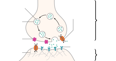
Structure of a typical chemical synapse

In the nervous system, a synapse is a structure that permits a neuron (or nerve cell) to pass an electrical or chemical signal to another neuron or to the target efferent cell.

Santiago Ramón y Cajal proposed that neurons are not continuous throughout the body, yet still communicate with each other, an idea known as the neuron doctrine. The word "synapse" – from the Greek synapsis (συνάψις), meaning "conjunction", in turn from συνάπτεὶν (συν ("together") and ἅπτειν ("to fasten")) – was introduced in 1897 by the English neurophysiologist Charles Sherrington in Michael Foster's Textbook of Physiology. Sherrington struggled to find a good term that emphasized a union between two separate elements, and the actual term "synapse" was suggested by the English classical scholar Arthur Woollgar Verrall, a friend of Michael Foster. Some authors generalize the concept of the synapse to include the communication from a neuron to any other cell type, such as to a motor cell, although such non-neuronal contacts may be referred to as junctions (a historically older term).

Synapses are essential to neuronal function: neurons are cells that are specialized to pass signals to individual target cells, and synapses are the means by which they do so. At a synapse, the plasma membrane of the signal-passing neuron (the presynaptic neuron) comes into close apposition with the membrane of the target (postsynaptic) cell. Both the presynaptic and postsynaptic sites contain extensive arrays of a molecular machinery that link the two membranes together and carry out the signaling process. In many synapses, the presynaptic part is located on an axon and the postsynaptic part is located on a dendrite or soma. Astrocytes also exchange information with the synaptic neurons, responding to synaptic activity and, in turn, regulating neurotransmission.[6] Synapses (at least chemical synapses) are stabilized in position by synaptic adhesion molecules (SAMs) projecting from both the pre- and post-synaptic neuron and sticking together where they overlap; SAMs may also assist in the generation and functioning of synapses.[7]

Chemical or electrical

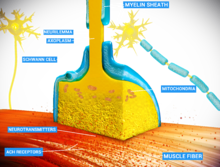
An example of chemical synapse by the release of neurotransmitters like acetylcholine or glutamic acid.

There are two fundamentally different types of synapses:
  • In a chemical synapse, electrical activity in the presynaptic neuron is converted (via the activation of voltage-gated calcium channels) into the release of a chemical called a neurotransmitter that binds to receptors located in the plasma membrane of the postsynaptic cell. The neurotransmitter may initiate an electrical response or a secondary messenger pathway that may either excite or inhibit the postsynaptic neuron. Chemical synapses can be classified according to the neurotransmitter released: glutamatergic (often excitatory), GABAergic (often inhibitory), cholinergic (e.g. vertebrate neuromuscular junction), and adrenergic (releasing norepinephrine). Because of the complexity of receptor signal transduction, chemical synapses can have complex effects on the postsynaptic cell.
  • In an electrical synapse, the presynaptic and postsynaptic cell membranes are connected by special channels called gap junctions or synaptic cleft that are capable of passing an electric current, causing voltage changes in the presynaptic cell to induce voltage changes in the postsynaptic cell. The main advantage of an electrical synapse is the rapid transfer of signals from one cell to the next.[8]
Synaptic communication is distinct from an ephaptic coupling, in which communication between neurons occurs via indirect electric fields.

An autapse is a chemical or electrical synapse that forms when the axon of one neuron synapses onto dendrites of the same neuron.

Types of interfaces

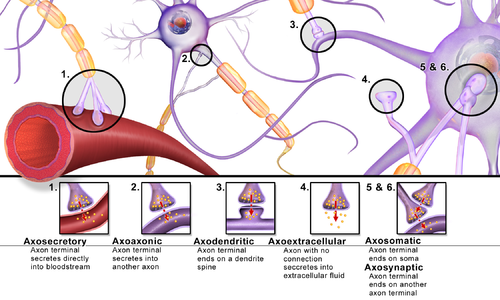
Synapses can be classified by the type of cellular structures serving as the pre- and post-synaptic components. The vast majority of synapses in the mammalian nervous system are classical axo-dendritic synapses (axon synapsing upon a dendrite), however, a variety of other arrangements exist. These include but are not limited to axo-axonic, dendro-dendritic, axo-secretory, somato-dendritic, dendro-somatic, and somato-somatic synapses.

The axon can synapse onto a dendrite, onto a cell body, or onto another axon or axon terminal, as well as into the bloodstream or diffusely into the adjacent nervous tissue.

Different types of synapses

Role in memory

It is widely accepted that the synapse plays a role in the formation of memory. As neurotransmitters activate receptors across the synaptic cleft, the connection between the two neurons is strengthened when both neurons are active at the same time, as a result of the receptor's signaling mechanisms. The strength of two connected neural pathways is thought to result in the storage of information, resulting in memory. This process of synaptic strengthening is known as long-term potentiation.[9]

By altering the release of neurotransmitters, the plasticity of synapses can be controlled in the presynaptic cell. The postsynaptic cell can be regulated by altering the function and number of its receptors. Changes in postsynaptic signaling are most commonly associated with a N-methyl-d-aspartic acid receptor (NMDAR)-dependent long-term potentiation (LTP) and long-term depression (LTD) due to the influx of calcium into the post-synaptic cell, which are the most analyzed forms of plasticity at excitatory synapses.[10]

Study models

For technical reasons, synaptic structure and function have been historically studied at unusually large model synapses, for example:

Synaptic polarization

The function of neurons depends upon cell polarity. The distinctive structure of nerve cells allows action potentials to travel directionally (from dendrites to cell body down the axon), and for these signals to then be received and carried on by post-synaptic neurons or received by effector cells. Nerve cells have long been used as models for cellular polarization, and of particular interest are the mechanisms underlying the polarized localization of synaptic molecules. PIP2 signaling regulated by IMPase plays an integral role in synaptic polarity.

Phosphoinositides (PIP, PIP2, and PIP3) are molecules that have been shown to affect neuronal polarity.[12] A gene (ttx-7) was identified in Caenorhabditis elegans that encodes myo-inositol monophosphatase (IMPase), an enzyme that produces inositol by dephosphorylating inositol phosphate. Organisms with mutant ttx-7 genes demonstrated behavioral and localization defects, which were rescued by expression of IMPase. This led to the conclusion that IMPase is required for the correct localization of synaptic protein components.[13][14] The egl-8 gene encodes a homolog of phospholipase Cβ (PLCβ), an enzyme that cleaves PIP2. When ttx-7 mutants also had a mutant egl-8 gene, the defects caused by the faulty ttx-7 gene were largely reversed. These results suggest that PIP2 signaling establishes polarized localization of synaptic components in living neurons.[13]

Additional images

Human vs. deep-neural-network performance in object recognition

September 27, 2017
Original link:  http://www.kurzweilai.net/human-vs-deep-neural-network-performance-in-object-recognition
(credit: UC Santa Barbara)

Before you read this: look for toothbrushes in the photo above.

Did you notice the huge toothbrush on the left? Probably not. That’s because when humans search through scenes for a particular object, we often miss objects whose size is inconsistent with the rest of the scene, according to scientists in the Department of Psychological & Brain Sciences at UC Santa Barbara.

The scientists are investigating this phenomenon in an effort to better understand how humans and computers compare in doing visual searches. Their findings are published in the journal Current Biology.

Hiding in plain sight

“When something appears at the wrong scale, you will miss it more often because your brain automatically ignores it,” said UCSB professor Miguel Eckstein, who specializes in computational human vision, visual attention, and search.

The experiment used scenes of ordinary objects featured in computer-generated images that varied in color, viewing angle, and size, mixed with “target-absent” scenes. The researchers asked 60 viewers to search for these objects (e.g., toothbrush, parking meter, computer mouse) while eye-tracking software monitored the paths of their gaze.

The researchers found that people tended to miss the target more often when it was mis-scaled (too large or too small) — even when looking directly at the target object.
Computer vision, by contrast, doesn’t have this issue, the scientists reported. However, in the experiments, the researchers found that the most advanced form of computer vision — deep neural networks — had its own limitations.

Human search strategies that could improve computer vision

Red rectangle marks incorrect image identification as a cell phone by a deep-learning algorithm (credit: UC Santa Barbara)

For example, a CNN deep-learning neural net incorrectly identified a computer keyboard as a cell phone, based on similarity in shape and the location of the object in spatial proximity to a human hand (as would be expected of a cell phone). But for humans, the object’s size (compared to the nearby hands) is clearly seen as inconsistent with a cell phone.

“This strategy allows humans to reduce false positives when making fast decisions,” the researchers note in the paper.

“The idea is when you first see a scene, your brain rapidly processes it within a few hundred milliseconds or less, and then you use that information to guide your search towards likely locations where the object typically appears,” Eckstein said. “Also, you focus your attention on objects that are actually at the size that is consistent with the object that you’re looking for.”

That is, human brains use the relationships between objects and their context within the scene to guide their eyes — a useful strategy to process scenes rapidly, eliminate distractors, and reduce false positives.

This finding might suggest ways to improve computer vision by implementing some of the tricks the brain utilizes to reduce false positives, according to the researchers.

Future research

“There are some theories that suggest that people with autism spectrum disorder focus more on local scene information and less on global structure,” says Eckstein, who is contemplating a follow-up study. “So there is a possibility that people with autism spectrum disorder might miss the mis-scaled objects less often, but we won’t know that until we do the study.”

In the more immediate future, the team’s research will look into the brain activity that occurs when we view mis-scaled objects.

“Many studies have identified brain regions that process scenes and objects, and now researchers are trying to understand which particular properties of scenes and objects are represented in these regions,” said postdoctoral researcher Lauren Welbourne, whose current research concentrates on how objects are represented in the cortex, and how scene context influences the perception of objects.

“So what we’re trying to do is find out how these brain areas respond to objects that are either correctly or incorrectly scaled within a scene. This may help us determine which regions are responsible for making it more difficult for us to find objects if they are mis-scaled.”


Abstract of Humans, but Not Deep Neural Networks, Often Miss Giant Targets in Scenes

Even with great advances in machine vision, animals are still unmatched in their ability to visually search complex scenes. Animals from bees [ 1, 2 ] to birds [ 3 ] to humans [ 4–12 ] learn about the statistical relations in visual environments to guide and aid their search for targets. Here, we investigate a novel manner in which humans utilize rapidly acquired information about scenes by guiding search toward likely target sizes. We show that humans often miss targets when their size is inconsistent with the rest of the scene, even when the targets were made larger and more salient and observers fixated the target. In contrast, we show that state-of-the-art deep neural networks do not exhibit such deficits in finding mis-scaled targets but, unlike humans, can be fooled by target-shaped distractors that are inconsistent with the expected target’s size within the scene. Thus, it is not a human deficiency to miss targets when they are inconsistent in size with the scene; instead, it is a byproduct of a useful strategy that the brain has implemented to rapidly discount potential distractors.

Magnetoencephalography

From Wikipedia, the free encyclopedia
 
Magnetoencephalography
Medical diagnostics
NIMH MEG.jpg
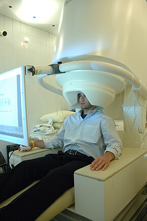
Person undergoing an MEG
MeSH D015225

Magnetoencephalography (MEG) is a functional neuroimaging technique for mapping brain activity by recording magnetic fields produced by electrical currents occurring naturally in the brain, using very sensitive magnetometers. Arrays of SQUIDs (superconducting quantum interference devices) are currently the most common magnetometer, while the SERF (spin exchange relaxation-free) magnetometer is being investigated for future machines. Applications of MEG include basic research into perceptual and cognitive brain processes, localizing regions affected by pathology before surgical removal, determining the function of various parts of the brain, and neurofeedback. This can be applied in a clinical setting to find locations of abnormalities as well as in an experimental setting to simply measure brain activity.

History

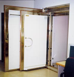
Dr. Cohen's shielded room at MIT, in which first MEG was measured with a SQUID
 
First MEG measured with SQUID, in Dr. Cohen's room at MIT

MEG signals were first measured by University of Illinois physicist David Cohen in 1968,[4] before the availability of the SQUID, using a copper induction coil as the detector. To reduce the magnetic background noise, the measurements were made in a magnetically shielded room. The coil detector was barely sensitive enough, resulting in poor, noisy MEG measurements that were difficult to use. Later, Cohen built a much better shielded room at MIT, and used one of the first SQUID detectors, just developed by James E. Zimmerman, a researcher at Ford Motor Company,[5] to again measure MEG signals.[6] This time the signals were almost as clear as those of EEG. This stimulated the interest of physicists who had been looking for uses of SQUIDs. Subsequent to this, various types of spontaneous and evoked MEGs began to be measured.

At first, a single SQUID detector was used to successively measure the magnetic field at a number of points around the subject's head. This was cumbersome, and, in the 1980s, MEG manufacturers began to arrange multiple sensors into arrays to cover a larger area of the head. Present-day MEG arrays are set in a helmet-shaped vacuum flask that typically contain 300 sensors, covering most of the head. In this way, MEGs of a subject or patient can now be accumulated rapidly and efficiently.

Recent developments attempt to increase portability of MEG scanners by using spin exchange relaxation-free (SERF) magnetometers. SERF magnetometers are relatively small, as they do not require bulky cooling systems to operate. At the same time, they feature sensitivity equivalent to that of SQUIDs. In 2012, it was demonstrated that MEG could work with a chip-scale atomic magnetometer (CSAM, type of SERF).[7] More recently, in 2017, researchers built a working prototype that uses SERF magnetometers installed into portable individually 3D-printed helmets [2].

The basis of the MEG signal

Synchronized neuronal currents induce weak magnetic fields. The brain's magnetic field, measuring at 10 femtotesla (fT) for cortical activity and 103 fT for the human alpha rhythm, is considerably smaller than the ambient magnetic noise in an urban environment, which is on the order of 108 fT or 0.1 µT. The essential problem of biomagnetism is, thus, the weakness of the signal relative to the sensitivity of the detectors, and to the competing environmental noise.

Origin of the brain's magnetic field. The electric current also produces the EEG signal.

The MEG (and EEG) signals derive from the net effect of ionic currents flowing in the dendrites of neurons during synaptic transmission. In accordance with Maxwell's equations, any electrical current will produce a magnetic field, and it is this field that is measured. The net currents can be thought of as current dipoles[citation needed], i.e. currents with a position, orientation, and magnitude, but no spatial extent[dubious ]. According to the right-hand rule, a current dipole gives rise to a magnetic field that points around the axis of its vector component.

To generate a signal that is detectable, approximately 50,000 active neurons are needed.[8] Since current dipoles must have similar orientations to generate magnetic fields that reinforce each other, it is often the layer of pyramidal cells, which are situated perpendicular to the cortical surface, that gives rise to measurable magnetic fields. Bundles of these neurons that are orientated tangentially to the scalp surface project measurable portions of their magnetic fields outside of the head, and these bundles are typically located in the sulci. Researchers are experimenting with various signal processing methods in the search for methods that detect deep brain (i.e., non-cortical) signal, but no clinically useful method is currently available.

It is worth noting that action potentials do not usually produce an observable field, mainly because the currents associated with action potentials flow in opposite directions and the magnetic fields cancel out. However, action fields have been measured from peripheral nerves.

Magnetic shielding

Since the magnetic signals emitted by the brain are on the order of a few femtoteslas, shielding from external magnetic signals, including the Earth's magnetic field, is necessary. Appropriate magnetic shielding can be obtained by constructing rooms made of aluminium and mu-metal for reducing high-frequency and low-frequency noise, respectively.

Entrance to MSR, showing the separate shielding layers

Magnetically shielded room (MSR)

A magnetically shielded room (MSR) model consists of three nested main layers. Each of these layers is made of a pure aluminium layer plus a high-permeability ferromagnetic layer, similar in composition to molybdenum permalloy. The ferromagnetic layer is supplied as 1 mm sheets, while the innermost layer is composed of four sheets in close contact, and the outer two layers are composed of three sheets each. Magnetic continuity is maintained by overlay strips. Insulating washers are used in the screw assemblies to ensure that each main layer is electrically isolated. This helps eliminate radio frequency radiation, which would degrade SQUID performance. Electrical continuity of the aluminium is also maintained by aluminium overlay strips to ensure AC eddy current shielding, which is important at frequencies greater than 1 Hz. The junctions of the inner layer are often electroplated with silver or gold to improve conductivity of the aluminium layers.[9]

Active shielding system

Active systems are designed for three-dimensional noise cancellation. To implement an active system, low-noise fluxgate magnetometers are mounted at the center of each surface and oriented orthogonally to it. This negatively feeds a DC amplifier through a low-pass network with a slow falloff to minimize positive feedback and oscillation. Built into the system are shaking and degaussing wires. Shaking wires increase the magnetic permeability, while the permanent degaussing wires are applied to all surfaces of the inner main layer to degauss the surfaces.[4] Moreover, noise cancellation algorithms can reduce both low-frequency and high-frequency noise. Modern systems have a noise floor of around 2–3 fT/Hz0.5 above 1 Hz.

Source localization

The inverse problem

The challenge posed by MEG is to determine the location of electric activity within the brain from the induced magnetic fields outside the head. Problems such as this, where model parameters (the location of the activity) have to be estimated from measured data (the SQUID signals) are referred to as inverse problems (in contrast to forward problems[10] where the model parameters (e.g. source location) are known and the data (e.g. the field at a given distance) is to be estimated.) The primary difficulty is that the inverse problem does not have a unique solution (i.e., there are infinite possible "correct" answers), and the problem of defining the "best" solution is itself the subject of intensive research.[11] Possible solutions can be derived using models involving prior knowledge of brain activity.
The source models can be either over-determined or under-determined. An over-determined model may consist of a few point-like sources ("equivalent dipoles"), whose locations are then estimated from the data. Under-determined models may be used in cases where many different distributed areas are activated ("distributed source solutions"): there are infinitely many possible current distributions explaining the measurement results, but the most likely is selected. Localization algorithms make use of given source and head models to find a likely location for an underlying focal field generator.

One type of localization algorithm for overdetermined models operates by expectation-maximization: the system is initialized with a first guess. A loop is started, in which a forward model is used to simulate the magnetic field that would result from the current guess. The guess is adjusted to reduce the discrepancy between the simulated field and the measured field. This process is iterated until convergence.

Another common technique is beamforming, wherein a theoretical model of the magnetic field produced by a given current dipole is used as a prior, along with second-order statistics of the data in the form of a covariance matrix, to calculate a linear weighting of the sensor array (the beamformer) via the Backus-Gilbert inverse. This is also known as a linearly constrained minimum variance (LCMV) beamformer. When the beamformer is applied to the data, it produces an estimate of the power in a "virtual channel" at the source location.

The extent to which the constraint-free MEG inverse problem is ill-posed cannot be overemphasized. If one's goal is to estimate the current density within the human brain with say a 5mm resolution then it is well established that the vast majority of the information needed to perform a unique inversion must come not from the magnetic field measurement but rather from the constraints applied to the problem. Furthermore, even when a unique inversion is possible in the presence of such constraints said inversion can be unstable. These conclusions are easily deduced from published works.[12]

Magnetic source imaging

The source locations can be combined with magnetic resonance imaging (MRI) images to create magnetic source images (MSI). The two sets of data are combined by measuring the location of a common set of fiducial points marked during MRI with lipid markers and marked during MEG with electrified coils of wire that give off magnetic fields. The locations of the fiducial points in each data set are then used to define a common coordinate system so that superimposing the functional MEG data onto the structural MRI data ("coregistration") is possible.

A criticism of the use of this technique in clinical practice is that it produces colored areas with definite boundaries superimposed upon an MRI scan: the untrained viewer may not realize that the colors do not represent a physiological certainty, because of the relatively low spatial resolution of MEG, but rather a probability cloud derived from statistical processes. However, when the magnetic source image corroborates other data, it can be of clinical utility.

Dipole model source localization

A widely accepted source-modeling technique for MEG involves calculating a set of equivalent current dipoles (ECDs), which assumes the underlying neuronal sources to be focal. This dipole fitting procedure is non-linear and over-determined, since the number of unknown dipole parameters is smaller than the number of MEG measurements.[13] Automated multiple dipole model algorithms such as multiple signal classification (MUSIC) and MSST (MultiStart Spatial and Temporal) modeling are applied to the analysis of MEG responses. The limitations of dipole models for characterizing neuronal responses are (1) difficulties in localizing extended sources with ECDs, (2) problems with accurately estimating the total number of dipoles in advance, and (3) dependency on dipole location, especially depth in the brain.

Distributed source models

Unlike multiple-dipole modeling, distributed source models divide the source space into a grid containing a large number of dipoles. The inverse problem is to obtain the dipole moments for the grid nodes.[14] As the number of unknown dipole moments is much greater than the number of MEG sensors, the inverse solution is highly underdetermined, so additional constraints are needed to reduce ambiguity of the solution. The primary advantage of this approach is that no prior specification of the source model is necessary. However, the resulting distributions may be difficult to interpret, because they only reflect a "blurred" (or even distorted) image of the true neuronal source distribution. The matter is complicated by the fact that spatial resolution depends strongly on several parameters such as brain area, depth, orientation, number of sensors etc.[15]

Independent component analysis (ICA)

Independent component analysis (ICA) is another signal processing solution that separates different signals that are statistically independent in time. It is primarily used to remove artifacts such as blinking, eye muscle movement, facial muscle artifacts, cardiac artifacts, etc. from MEG and EEG signals that may be contaminated with outside noise.[16] However, ICA has poor resolution of highly correlated brain sources.

Use in the field

In research, MEG's primary use is the measurement of time courses of activity. MEG can resolve events with a precision of 10 milliseconds or faster, while functional MRI (fMRI), which depends on changes in blood flow, can at best resolve events with a precision of several hundred milliseconds. MEG also accurately pinpoints sources in primary auditory, somatosensory, and motor areas. For creating functional maps of human cortex during more complex cognitive tasks, MEG is most often combined with fMRI, as the methods complement each other. Neuronal (MEG) and hemodynamic (fMRI) data do not necessarily agree, in spite of the tight relationship between local field potentials (LFP) and blood oxygenation level-dependent (BOLD) signals. MEG and BOLD signals may originate from the same source (though the BOLD signals are filtered through the hemodynamic response).

MEG is also being used to better localize responses in the brain. The openness of the MEG setup allows external auditory and visual stimuli to be easily introduced. Some movement by the subject is also possible as long as it does not jar the subject's head. The responses in the brain before, during, and after the introduction of such stimuli/movement can then be mapped with greater spatial resolution than was previously possible with EEG.[17] Psychologists are also taking advantage of MEG neuroimaging to better understand relationships between brain function and behavior. For example, a number of studies have been done comparing the MEG responses of patients with psychological troubles to control patients. There has been great success isolating unique responses in patients with schizophrenia, such as auditory gating deficits to human voices.[18] MEG is also being used to correlate standard psychological responses, such as the emotional dependence of language comprehension.[19]

Recent studies have reported successful classification of patients with multiple sclerosis, Alzheimer's disease, schizophrenia, Sjögren's syndrome, chronic alcoholism, and facial pain. MEG can be used to distinguish these patients from healthy control subjects, suggesting a future role of MEG in diagnostics.[20][21]

Focal epilepsy

The clinical uses of MEG are in detecting and localizing pathological activity in patients with epilepsy, and in localizing eloquent cortex for surgical planning in patients with brain tumors or intractable epilepsy. The goal of epilepsy surgery is to remove the epileptogenic tissue while sparing healthy brain areas.[22] Knowing the exact position of essential brain regions (such as the primary motor cortex and primary sensory cortex, visual cortex, and areas involved in speech production and comprehension) helps to avoid surgically induced neurological deficits. Direct cortical stimulation and somatosensory evoked potentials recorded on ECoG are considered the gold standard for localizing essential brain regions. These procedures can be performed either intraoperatively or from chronically indwelling subdural grid electrodes. Both are invasive.

Noninvasive MEG localizations of the central sulcus obtained from somatosensory evoked magnetic fields show strong agreement with these invasive recordings.[23][24][25] MEG studies assist in clarification of the functional organization of primary somatosensory cortex and to delineate the spatial extent of hand somatosensory cortex by stimulation of the individual digits. This agreement between invasive localization of cortical tissue and MEG recordings shows the effectiveness of MEG analysis and indicates that MEG may substitute invasive procedures in the future.

Fetal

MEG has been used to study cognitive processes such as vision, audition, and language processing in fetuses and newborns.[26]

Comparison with related techniques

MEG has been in development since the 1960s but has been greatly aided by recent advances in computing algorithms and hardware, and promises improved spatial resolution coupled with extremely high temporal resolution (better than 1 ms). Since the MEG signal is a direct measure of neuronal activity, its temporal resolution is comparable with that of intracranial electrodes.
MEG complements other brain activity measurement techniques such as electroencephalography (EEG), positron emission tomography (PET), and fMRI. Its strengths consist in independence of head geometry compared to EEG (unless ferromagnetic implants are present), non-invasiveness, use of no ionizing radiation, as opposed to PET and high temporal resolution as opposed to fMRI.

Vs. EEG

Although EEG and MEG signals originate from the same neurophysiological processes, there are important differences.[27] Magnetic fields are less distorted than electric fields by the skull and scalp, which results in a better spatial resolution of the MEG. Whereas scalp EEG is sensitive to both tangential and radial components of a current source in a spherical volume conductor, MEG detects only its tangential components. Scalp EEG can, therefore, detect activity both in the sulci and at the top of the cortical gyri, whereas MEG is most sensitive to activity originating in sulci. EEG is, therefore, sensitive to activity in more brain areas, but activity that is visible in MEG can also be localized with more accuracy.

Scalp EEG is sensitive to extracellular volume currents produced by postsynaptic potentials. MEG detects intracellular currents associated primarily with these synaptic potentials because the field components generated by volume currents tend to cancel out in a spherical volume conductor[28] The decay of magnetic fields as a function of distance is more pronounced than for electric fields. Therefore, MEG is more sensitive to superficial cortical activity, which makes it useful for the study of neocortical epilepsy. Finally, MEG is reference-free, while scalp EEG relies on a reference that, when active, makes interpretation of the data difficult.

Fast-moving spinning magnetized nanoparticles could lead to ultra-high-speed, high-density data storage

May help solve data-storage problems in the zettabyte era 
 
 
Artist’s impression of skyrmion data storage (credit: Moritz Eisebitt)

An international team led by MIT associate professor of materials science and engineering Geoffrey Beach has demonstrated a practical way to use “skyrmions” to create a radical new high-speed, high-density data-storage method that could one day replace disk drives — and even replace high-speed RAM memory.

Rather than reading and writing data one bit at a time by changing the orientation of magnetized nanoparticles on a surface, Skyrmions could store data using only a tiny area of a magnetic surface — perhaps just a few atoms across — and for long periods of time, without the need for further energy input (unlike disk drives and RAM).

Beach and associates conceive skyrmions as little sub-nanosecond spin-generating eddies of magnetism controlled by electric fields — replacing the magnetic-disk system of reading and writing data one bit at a time. In experiments, skyrmions have been generated on a thin metallic film sandwiched with non-magnetic heavy metals and transition-metal ferromagnetic layers — exploiting a defect, such as a constriction in the magnetic track.*

Skyrmions are also highly stable to external magnetic and mechanical perturbations, unlike the individual magnetic poles in a conventional magnetic storage device — allowing for vastly more data to be written onto a surface of a given size.

A practical data-storage system

Google data center (credit: Google Inc.)

Beach has recently collaborated with researchers at MIT and others in Germany** to demonstrate experimentally for the first time that it’s possible to create skyrmions in specific locations, which is needed for a data-storage system. The new findings were reported October 2, 2017 in the journal Nature Nanotechnology.

Conventional magnetic systems are now reaching speed and density limits set by the basic physics of their existing materials. The new system, once perfected, could provide a way to continue that progress toward ever-denser data storage, Beach says.

However, the researchers note that to create a commercialized system will require an efficient, reliable way to create skyrmions when and where they were needed, along with a way to read out the data (which now requires sophisticated, expensive X-ray magnetic spectroscopy). The team is now pursuing possible strategies to accomplish that.***

* The system focuses on the boundary region between atoms whose magnetic poles are pointing in one direction and those with poles pointing the other way. This boundary region can move back and forth within the magnetic material, Beach says. What he and his team found four years ago was that these boundary regions could be controlled by placing a second sheet of nonmagnetic heavy metal very close to the magnetic layer. The nonmagnetic layer can then influence the magnetic one, with electric fields in the nonmagnetic layer pushing around the magnetic domains in the magnetic layer. Skyrmions are little swirls of magnetic orientation within these layers. The key to being able to create skyrmions at will in particular locations lays in material defects. By introducing a particular kind of defect in the magnetic layer, the skyrmions become pinned to specific locations on the surface, the team found. Those surfaces with intentional defects can then be used as a controllable writing surface for data encoded in the skyrmions.

** The team also includes researchers at the Max Born Institute and the Institute of Optics and Atomic Physics, both in Berlin; the Institute for Laser Technologies in Medicine and Metrology at the University of Ulm, in Germany; and the Deutches Elektroniken-Syncrotron (DESY), in Hamburg. The work was supported by the U.S. Department of Energy and the German Science Foundation.

*** The researchers believe an alternative way of reading the data is possible, using an additional metal layer added to the other layers. By creating a particular texture on this added layer, it may be possible to detect differences in the layer’s electrical resistance depending on whether a skyrmion is present or not in the adjacent layer.


Abstract of Field-free deterministic ultrafast creation of magnetic skyrmions by spin–orbit torques

Magnetic skyrmions are stabilized by a combination of external magnetic fields, stray field energies, higher-order exchange interactions and the Dzyaloshinskii–Moriya interaction (DMI). The last favours homochiral skyrmions, whose motion is driven by spin–orbit torques and is deterministic, which makes systems with a large DMI relevant for applications. Asymmetric multilayers of non-magnetic heavy metals with strong spin–orbit interactions and transition-metal ferromagnetic layers provide a large and tunable DMI. Also, the non-magnetic heavy metal layer can inject a vertical spin current with transverse spin polarization into the ferromagnetic layer via the spin Hall effect. This leads to torques that can be used to switch the magnetization completely in out-of-plane magnetized ferromagnetic elements, but the switching is deterministic only in the presence of a symmetry-breaking in-plane field. Although spin–orbit torques led to domain nucleation in continuous films and to stochastic nucleation of skyrmions in magnetic tracks, no practical means to create individual skyrmions controllably in an integrated device design at a selected position has been reported yet. Here we demonstrate that sub-nanosecond spin–orbit torque pulses can generate single skyrmions at custom-defined positions in a magnetic racetrack deterministically using the same current path as used for the shifting operation. The effect of the DMI implies that no external in-plane magnetic fields are needed for this aim. This implementation exploits a defect, such as a constriction in the magnetic track, that can serve as a skyrmion generator. The concept is applicable to any track geometry, including three-dimensional designs.

Walking DNA nanorobot could deliver a drug to a precise location in your body

Future uses could include creating programmable drugs or delivering them when a specific signal is received in the bloodstream or cells
September 15, 2017
Original link:  http://www.kurzweilai.net/walking-dna-nanorobot-could-deliver-a-drug-to-a-precise-location-in-your-body
DNA nanorobot cargo carrier (artist’s impression) (credit: Ella Maru Studio)

Caltech scientists have developed a “cargo sorting” DNA nanorobot programmed to autonomously “walk” around a surface, pick up certain molecules, and drop them off in designated locations.
The research is described in a paper in the Friday, September 15, 2017 issue of Science.
The major advance in this study is “their methodology for designing simple DNA devices that work in parallel to solve nontrivial tasks,” notes Duke University computer scientist John H. Reif in an article in the same issue of Science.

Such tasks could include synthesizing a drug in a molecular factory or delivering a drug only when a specific signal is present in bloodstreams, say the researchers. “So far, the development of DNA robots has been limited to simple functions,” the researchers note.

Walking nanobots that work in parallel

Conceptual illustration of two DNA nanorobots collectively performing a cargo-sorting task on a DNA origami surface: transporting fluorescent molecules with different colors from initially random locations to ordered destinations. (credit: Demin Liu)

The DNA nanorobot, intended as a proof of concept, has a “leg” with two “feet” for walking, and an “arm” and “hand” for picking up cargo. It also has a segment that can recognize a specific drop-off point and signal to the hand to release its cargo. Each of these building blocks are made of just a few nucleotides (molecules that form DNA) within a single strand of DNA.*

As the robot encounters cargo molecules tethered to pegs, it grabs them with its “hand” components and carries them around (with a 6-nm step size) until it detects the signal of the drop-off point.

Multiple DNA nanorobots independently execute three operations in parallel: [1] cargo pickup, [2] random movement to adjacent stepping stones, and [3] cargo drop-off at ordered locations. (credit: C. Bickel/Science)
 
In experiments, the nanorobots successfully sorted six randomly scattered molecules into their correct places in 24 hours. The process is slow, but adding more robots to the surface shortened the time it took to complete the task. The very simple robot design utilizes very little chemical energy, according to the researchers.**
“The same system design can be generalized to work with dozens of types of cargos at any arbitrary initial location on the surface,” says lead author Anupama Thubagere. “One could also have multiple robots performing diverse sorting tasks in parallel,” [programmed] like macroscopic robots.”

Future applications

“We don’t develop DNA robots for any specific applications. Our lab focuses on discovering the engineering principles that enable the development of general-purpose DNA robots,” explains Lulu Qian, assistant professor of bioengineering.

“However, it is my hope that other researchers could use these principles for exciting applications, such as synthesizing a therapeutic chemical from its constituent parts in an artificial molecular factory, or sorting molecular components in trash for recycling. Just like electromechanical robots are sent off to faraway places, like Mars, we would like to send molecular robots to minuscule places where humans can’t go, such as the bloodstream.”

Funding was provided by Caltech Summer Undergraduate Research Fellowships, the National Science Foundation, and the Burroughs Wellcome Fund.

* The key to designing DNA machines is the fact that DNA has unique chemical and physical properties that are known and programmable. A single strand of DNA is made up of four different molecules called nucleotides—abbreviated A, G, C, and T—and arranged in a string called a sequence. These nucleotides bond in specific pairs: A with T, and G with C. When a single strand encounters a “reverse complementary strand” — for example, CGATT meets AATCG —the two strands zip together in the classic double-helix shape.

** Using these chemical and physical principles, researchers can also design “playgrounds,” such as molecular pegboards, to test them on, according to the researchers. In the current work, the DNA robot moves around on a 58-nanometer-by-58-nanometer pegboard on which the pegs are made of single strands of DNA complementary to the robot’s leg and foot. The robot binds to a peg with its leg and one of its feet — the other foot floats freely. When random molecular fluctuations cause this free foot to encounter a nearby peg, it pulls the robot to the new peg and its other foot is freed. This process continues with the robot moving in a random direction at each step.


Abstract of A cargo-sorting DNA robot

Two critical challenges in the design and synthesis of molecular robots are modularity and
algorithm simplicity.We demonstrate three modular building blocks for a DNA robot that
performs cargo sorting at themolecular level. A simple algorithm encoding recognition between
cargos and their destinations allows for a simple robot design: a single-stranded DNA with
one leg and two foot domains for walking, and one arm and one hand domain for picking up and
dropping off cargos.The robot explores a two-dimensional testing ground on the surface of
DNA origami, picks up multiple cargos of two types that are initially at unordered locations, and
delivers them to specified destinations until all molecules are sorted into two distinct piles.
The robot is designed to perform a random walk without any energy supply. Exploiting this
feature, a single robot can repeatedly sort multiple cargos. Localization on DNA origami allows
for distinct cargo-sorting tasks to take place simultaneously in one test tube or for multiple
robots to collectively perform the same task.

Operator (computer programming)

From Wikipedia, the free encyclopedia https://en.wikipedia.org/wiki/Operator_(computer_programmin...