Search This Blog

Friday, March 22, 2019

Development of the nervous system

From Wikipedia, the free encyclopedia
 
The development of the nervous system refers to the processes that generate, shape, and reshape the nervous system of animals, from the earliest stages of embryonic development to adulthood. The field of neural development draws on both neuroscience and developmental biology to describe and provide insight into the cellular and molecular mechanisms by which complex nervous systems develop, from the nematode and fruit fly to mammals. Defects in neural development can lead to malformations and a wide variety of sensory, motor, and cognitive impairments, including holoprosencephaly and other neurological disorders in the human such as Rett syndrome, Down syndrome and intellectual disability.

Overview of brain development

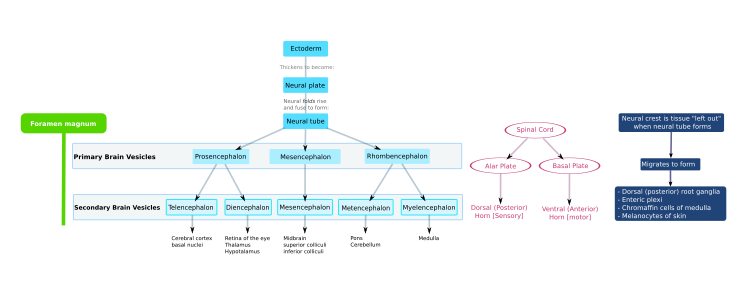
The mammalian central nervous system (CNS) is derived from the ectoderm—the outermost tissue layer—of the embryo. In the third week of human development the neuroectoderm appears and forms the neural plate along the dorsal side of the embryo. The neural plate is the source of the majority of neurons and glial cells of the CNS. A groove forms along the long axis of the neural plate and, by week four of development, the neural plate wraps in on itself to give rise to the neural tube, which is filled with cerebrospinal fluid (CSF). As the embryo develops, the anterior part of the neural tube forms three brain vesicles, which become the primary anatomical regions of the brain: the forebrain (prosencephalon), midbrain (mesencephalon), and hindbrain (rhombencephalon). These simple, early vesicles enlarge and further divide into the telencephalon (future cerebral cortex and basal ganglia), diencephalon (future thalamus and hypothalamus), mesencephalon (future colliculi), metencephalon (future pons and cerebellum), and myelencephalon (future medulla). The CSF-filled central chamber is continuous from the telencephalon to the spinal cord, and constitutes the developing ventricular system of the CNS. Because the neural tube gives rise to the brain and spinal cord any mutations at this stage in development can lead to fatal deformities like anencephaly or lifelong disabilities like spina bifida. During this time, the walls of the neural tube contain neural stem cells, which drive brain growth as they divide many times. Gradually some of the cells stop dividing and differentiate into neurons and glial cells, which are the main cellular components of the CNS. The newly generated neurons migrate to different parts of the developing brain to self-organize into different brain structures. Once the neurons have reached their regional positions, they extend axons and dendrites, which allow them to communicate with other neurons via synapses. Synaptic communication between neurons leads to the establishment of functional neural circuits that mediate sensory and motor processing, and underlie behavior.

Flowchart of human brain development.

Aspects

Some landmarks of neural development include the birth and differentiation of neurons from stem cell precursors, the migration of immature neurons from their birthplaces in the embryo to their final positions, outgrowth of axons and dendrites from neurons, guidance of the motile growth cone through the embryo towards postsynaptic partners, the generation of synapses between these axons and their postsynaptic partners, and finally the lifelong changes in synapses, which are thought to underlie learning and memory. 

Typically, these neurodevelopmental processes can be broadly divided into two classes: activity-independent mechanisms and activity-dependent mechanisms. Activity-independent mechanisms are generally believed to occur as hardwired processes determined by genetic programs played out within individual neurons. These include differentiation, migration and axon guidance to their initial target areas. These processes are thought of as being independent of neural activity and sensory experience. Once axons reach their target areas, activity-dependent mechanisms come into play. Although synapse formation is an activity-independent event, modification of synapses and synapse elimination requires neural activity. 

Developmental neuroscience uses a variety of animal models including mice Mus musculus, the fruit fly Drosophila melanogaster, the zebrafish Danio rerio, Xenopus laevis tadpoles and the worm Caenorhabditis elegans, among others. 

Myelination, formation of the lipid myelin bilayer around neuronal axons, is a process that is essential for normal brain function. The myelin sheath provides insulation for the nerve impulse when communicating between neural systems. Without it, the impulse would be disrupted and the signal would not reach its target, thus impairing normal functioning. Because so much of brain development occurs in the prenatal stage and infancy, it is crucial that myelination, along with cortical development occur properly. Magnetic resonance imaging (MRI) is a non-invasive technique used to investigate myelination and cortical maturation (the cortex is the outer layer of the brain composed of gray matter). Rather than showing the actual myelin, the MRI picks up on the myelin water fraction (MWF), a measure of myelin content. Multicomponent relaxometry (MCR) allow visualization and quantification of myelin content. MCR is also useful for tracking white matter maturation, which plays an important role in cognitive development. It has been discovered that in infancy, myelination occurs in a posterior-to-anterior pattern. Because there is little evidence of a relationship between myelination and cortical thickness, it was revealed that cortical thickness is independent of white matter MWF. This allows various aspects of the brain to grow simultaneously, leading to a more fully developed brain.

Neural induction

During early embryonic development the ectoderm becomes specified to give rise to the epidermis (skin) and the neural plate. The conversion of undifferentiated ectoderm to neuro-ectoderm requires signals from the mesoderm. At the onset of gastrulation presumptive mesodermal cells move through the dorsal blastopore lip and form a layer in between the endoderm and the ectoderm. These mesodermal cells that migrate along the dorsal midline give rise to a structure called the notochord. Ectodermal cells overlying the notochord develop into the neural plate in response to a diffusible signal produced by the notochord. The remainder of the ectoderm gives rise to the epidermis (skin). The ability of the mesoderm to convert the overlying ectoderm into neural tissue is called neural induction

In the human, the neural plate folds outwards during the third week of gestation to form the neural groove. Beginning in the future neck region, the neural folds of this groove close to create the neural tube. The formation of the neural tube from the ectoderm is called neurulation. The ventral part of the neural tube is called the basal plate; the dorsal part is called the alar plate. The hollow interior is called the neural canal. By the end of the fourth week of gestation, the open ends of the neural tube, called the neuropores, close off.

A transplanted blastopore lip can convert ectoderm into neural tissue and is said to have an inductive effect. Neural inducers are molecules that can induce the expression of neural genes in ectoderm explants without inducing mesodermal genes as well. Neural induction is often studied in xenopus embryos since they have a simple body pattern and there are good markers to distinguish between neural and non-neural tissue. Examples of neural inducers are the molecules noggin and chordin

When embryonic ectodermal cells are cultured at low density in the absence of mesodermal cells they undergo neural differentiation (express neural genes), suggesting that neural differentiation is the default fate of ectodermal cells. In explant cultures (which allow direct cell-cell interactions) the same cells differentiate into epidermis. This is due to the action of BMP4 (a TGF-β family protein) that induces ectodermal cultures to differentiate into epidermis. During neural induction, noggin and chordin are produced by the dorsal mesoderm (notochord) and diffuse into the overlying ectoderm to inhibit the activity of BMP4. This inhibition of BMP4 causes the cells to differentiate into neural cells. Inhibition of TGF-β and BMP (bone morphogenetic protein) signaling can efficiently induce neural tissue from human pluripotent stem cells, a model of early human development.

Regionalization

Late in the fourth week in the human, the superior part of the neural tube flexes at the level of the future midbrain—the mesencephalon. Above the mesencephalon is the prosencephalon (future forebrain) and beneath it is the rhombencephalon (future hindbrain). 

The optical vesicle (which eventually become the optic nerve, retina and iris) forms at the basal plate of the prosencephalon. The alar plate of the prosencephalon expands to form the cerebral hemispheres (the telencephalon) whilst its basal plate becomes the diencephalon. Finally, the optic vesicle grows to form an optic outgrowth.

Patterning of the nervous system

In chordates, dorsal ectoderm forms all neural tissue and the nervous system. Patterning occurs due to specific environmental conditions - different concentrations of signaling molecules

Dorsoventral axis

The ventral half of the neural plate is controlled by the notochord, which acts as the 'organiser'. The dorsal half is controlled by the ectoderm plate, which flanks either side of the neural plate.

Ectoderm follows a default pathway to become neural tissue. Evidence for this comes from single, cultured cells of ectoderm, which go on to form neural tissue. This is postulated to be because of a lack of BMPs, which are blocked by the organiser. The organiser may produce molecules such as follistatin, noggin and chordin that inhibit BMPs. 

The ventral neural tube is patterned by sonic hedgehog (Shh) from the notochord, which acts as the inducing tissue. Notochord-derived Shh signals to the floor plate, and induces Shh expression in the floor plate. Floor plate-derived Shh subsequently signals to other cells in the neural tube, and is essential for proper specification of ventral neuron progenitor domains. Loss of Shh from the notochord and/or floor plate prevents proper specification of these progenitor domains. Shh binds Patched1, relieving Patched-mediated inhibition of Smoothened, leading to activation of the Gli family of transcription factors (Gli1, Gli2, and Gli3). 

In this context Shh acts as a morphogen - it induces cell differentiation dependent on its concentration. At low concentrations it forms ventral interneurones, at higher concentrations it induces motor neuron development, and at highest concentrations it induces floor plate differentiation. Failure of Shh-modulated differentiation causes holoprosencephaly. 

The dorsal neural tube is patterned by BMPs from the epidermal ectoderm flanking the neural plate. These induce sensory interneurones by activating Sr/Thr kinases and altering SMAD transcription factor levels.

Rostrocaudal (Anteroposterior) axis

Signals that control anteroposterior neural development include FGF and retinoic acid, which act in the hindbrain and spinal cord. The hindbrain, for example, is patterned by Hox genes, which are expressed in overlapping domains along the anteroposterior axis under the control of retinoic acid. The 3' genes in the Hox cluster are induced by retinoic acid in the hindbrain, whereas the 5' Hox genes are not induced by retinoic acid and are expressed more posteriorly in the spinal cord. Hoxb-1 is expressed in rhombomere 4 and gives rise to the facial nerve. Without this Hoxb-1 expression, a nerve similar to the trigeminal nerve arises.

Neurogenesis

Neurogenesis is the process by which neurons are generated from neural stem cells and progenitor cells. Neurons are 'post-mitotic', meaning that they will never divide again for the lifetime of the organism.

Neuronal migration

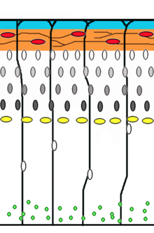
Corticogenesis: younger neurons migrate past older ones using radial glia as a scaffolding. Cajal-Retzius cells (red) release reelin (orange).
 
Neuronal migration is the method by which neurons travel from their origin or birthplace to their final position in the brain. There are several ways they can do this, e.g. by radial migration or tangential migration. This time lapse displays sequences of radial migration (also known as glial guidance) and somal translocation.

Tangential migration of interneurons from ganglionic eminence.

Radial migration

Neuronal precursor cells proliferate in the ventricular zone of the developing neocortex, where the principal neural stem cell is the radial glial cell. The first postmitotic cells must leave the stem cell niche and migrate outward to form the preplate, which is destined to become Cajal-Retzius cells and subplate neurons. These cells do so by somal translocation. Neurons migrating with this mode of locomotion are bipolar and attach the leading edge of the process to the pia. The soma is then transported to the pial surface by nucleokinesis, a process by which a microtubule "cage" around the nucleus elongates and contracts in association with the centrosome to guide the nucleus to its final destination. Radial glial cells, whose fibers serve as a scaffolding for migrating cells and a means of radial communication mediated by calcium dynamic activity, act as the main excitatory neuronal stem cell of the cerebral cortex or translocate to the cortical plate and differentiate either into astrocytes or neurons. Somal translocation can occur at any time during development.

Subsequent waves of neurons split the preplate by migrating along radial glial fibres to form the cortical plate. Each wave of migrating cells travel past their predecessors forming layers in an inside-out manner, meaning that the youngest neurons are the closest to the surface. It is estimated that glial guided migration represents 90% of migrating neurons in human and about 75% in rodents.

Tangential migration

Most interneurons migrate tangentially through multiple modes of migration to reach their appropriate location in the cortex. An example of tangential migration is the movement of interneurons from the ganglionic eminence to the cerebral cortex. One example of ongoing tangential migration in a mature organism, observed in some animals, is the rostral migratory stream connecting subventricular zone and olfactory bulb.

Axophilic migration

Many neurons migrating along the anterior-posterior axis of the body use existing axon tracts to migrate along; this is called axophilic migration. An example of this mode of migration is in GnRH-expressing neurons, which make a long journey from their birthplace in the nose, through the forebrain, and into the hypothalamus. Many of the mechanisms of this migration have been worked out, starting with the extracellular guidance cues that trigger intracellular signaling. These intracellular signals, such as calcium signaling, lead to actin  and microtubule cytoskeletal dynamics, which produce cellular forces that interact with the extracellular environment through cell adhesion proteins  to cause the movement of these cells.

Other modes of migration

There is also a method of neuronal migration called multipolar migration. This is seen in multipolar cells, which in the human, are abundantly present in the cortical intermediate zone. They do not resemble the cells migrating by locomotion or somal translocation. Instead these multipolar cells express neuronal markers and extend multiple thin processes in various directions independently of the radial glial fibers.

Neurotrophic factors

The survival of neurons is regulated by survival factors, called trophic factors. The neurotrophic hypothesis was formulated by Victor Hamburger and Rita Levi Montalcini based on studies of the developing nervous system. Victor Hamburger discovered that implanting an extra limb in the developing chick led to an increase in the number of spinal motor neurons. Initially he thought that the extra limb was inducing proliferation of motor neurons, but he and his colleagues later showed that there was a great deal of motor neuron death during normal development, and the extra limb prevented this cell death. According to the neurotrophic hypothesis, growing axons compete for limiting amounts of target-derived trophic factors and axons that fail to receive sufficient trophic support die by apoptosis. It is now clear that factors produced by a number of sources contribute to neuronal survival.
  • Nerve Growth Factor (NGF): Rita Levi Montalcini and Stanley Cohen purified the first trophic factor, Nerve Growth Factor (NGF), for which they received the Nobel Prize. There are three NGF-related trophic factors: BDNF, NT3, and NT4, which regulate survival of various neuronal populations. The Trk proteins act as receptors for NGF and related factors. Trk is a receptor tyrosine kinase. Trk dimerization and phosphorylation leads to activation of various intracellular signaling pathways including the MAP kinase, Akt, and PKC pathways.
  • CNTF: Ciliary neurotrophic factor is another protein that acts as a survival factor for motor neurons. CNTF acts via a receptor complex that includes CNTFRα, GP130, and LIFRβ. Activation of the receptor leads to phosphorylation and recruitment of the JAK kinase, which in turn phosphorylates LIFRβ. LIFRβ acts as a docking site for the STAT transcription factors. JAK kinase phosphorylates STAT proteins, which dissociate from the receptor and translocate to the nucleus to regulate gene expression.
  • GDNF: Glial derived neurotrophic factor is a member of the TGFb family of proteins, and is a potent trophic factor for striatal neurons. The functional receptor is a heterodimer, composed of type 1 and type 2 receptors. Activation of the type 1 receptor leads to phosphorylation of Smad proteins, which translocate to the nucleus to activate gene expression.

Synapse formation

Neuromuscular junction

Much of our understanding of synapse formation comes from studies at the neuromuscular junction. The transmitter at this synapse is acetylcholine. The acetylcholine receptor (AchR) is present at the surface of muscle cells before synapse formation. The arrival of the nerve induces clustering of the receptors at the synapse. McMahan and Sanes showed that the synaptogenic signal is concentrated at the basal lamina. They also showed that the synaptogenic signal is produced by the nerve, and they identified the factor as Agrin. Agrin induces clustering of AchRs on the muscle surface and synapse formation is disrupted in agrin knockout mice. Agrin transduces the signal via MuSK receptor to rapsyn. Fischbach and colleagues showed that receptor subunits are selectively transcribed from nuclei next to the synaptic site. This is mediated by neuregulins. 

In the mature synapse each muscle fiber is innervated by one motor neuron. However, during development many of the fibers are innervated by multiple axons. Lichtman and colleagues have studied the process of synapses elimination. This is an activity-dependent event. Partial blockage of the receptor leads to retraction of corresponding presynaptic terminals.

CNS synapses

Agrin appears not to be a central mediator of CNS synapse formation and there is active interest in identifying signals that mediate CNS synaptogenesis. Neurons in culture develop synapses that are similar to those that form in vivo, suggesting that synaptogenic signals can function properly in vitro. CNS synaptogenesis studies have focused mainly on glutamatergic synapses. Imaging experiments show that dendrites are highly dynamic during development and often initiate contact with axons. This is followed by recruitment of postsynaptic proteins to the site of contact. Stephen Smith and colleagues have shown that contact initiated by dendritic filopodia can develop into synapses. 

Induction of synapse formation by glial factors: Barres and colleagues made the observation that factors in glial conditioned media induce synapse formation in retinal ganglion cell cultures. Synapse formation in the CNS is correlated with astrocyte differentiation suggesting that astrocytes might provide a synaptogenic factor. The identity of the astrocytic factors is not yet known. 

Neuroligins and SynCAM as synaptogenic signals: Sudhof, Serafini, Scheiffele and colleagues have shown that neuroligins and SynCAM can act as factors that induce presynaptic differentiation. Neuroligins are concentrated at the postsynaptic site and act via neurexins concentrated in the presynaptic axons. SynCAM is a cell adhesion molecule that is present in both pre- and post-synaptic membranes.

Activity dependent mechanisms in the assembly of neural circuits

The processes of neuronal migration, differentiation and axon guidance are generally believed to be activity-independent mechanisms and rely on hard-wired genetic programs in the neurons themselves. New research findings however have implicated a role for activity-dependent mechanisms in mediating some aspects of the aforementioned processes such as the rate of neuronal migration, aspects of neuronal differentiation and axon pathfinding. Activity-dependent mechanisms influence neural circuit development and are crucial for laying out early connectivity maps and the continued refinement of synapses which occurs during development. There are two distinct types of neural activity we observe in developing circuits -early spontaneous activity and sensory-evoked activity. Spontaneous activity occurs early during neural circuit development even when sensory input is absent and is observed in many systems such as the developing visual system, auditory system, motor system, hippocampus, cerebellum, and neocortex.

Experimental techniques such as direct electrophysiological recording, fluorescence imaging using calcium indicators and optogenetic techniques have shed light on the nature and function of these early bursts of activity. They have distinct spatial and temporal patterns during development and their ablation during development has been known to result in deficits in network refinement in the visual system. In the immature retina, waves of spontaneous action potentials arise from the retinal ganglion cells and sweep across the retinal surface in the first few postnatal weeks. These waves are mediated by neurotransmitter acetylcholine in the initial phase and later on by glutamate. They are thought to instruct the formation of two sensory maps- the retinotopic map and eye-specific segregation. Retinotopic map refinement occurs in downstream visual targets in the brain-the superior colliculus (SC) and dorsal lateral geniculate nucleus (LGN). Pharmacological disruption and mouse models lacking the β2 subunit of the nicotinic acetylcholine receptor has shown that the lack of spontaneous activity leads to marked defects in retinotopy and eye-specific segregation.

In the developing auditory system, developing cochlea generate bursts of activity which spreads across the inner hair cells and spiral ganglion neurons which relay auditory information to the brain. ATP release from supporting cells triggers action potentials in inner hair cells. In the auditory system, spontaneous activity is thought to be involved in tonotopic map formation by segregating cochlear neuron axons tuned to high and low frequencies. In the motor system, periodic bursts of spontaneous activity are driven by excitatory GABA and glutamate during the early stages and by acetylcholine and glutamate at later stages. In the developing zebrafish spinal cord, early spontaneous activity is required for the formation of increasingly synchronous alternating bursts between ipsilateral and contralateral regions of the spinal cord and for the integration of new cells into the circuit. In the cortex, early waves of activity have been observed in the cerebellum and cortical slices. Once sensory stimulus becomes available, final fine-tuning of sensory-coding maps and circuit refinement begins to rely more and more on sensory-evoked activity as demonstrated by classic experiments about the effects of sensory deprivation during critical periods.

Contemporary diffusion-weigthted MRI techniques may also uncover the macroscopic process of axonal development. The connectome can be constructed from diffusion MRI data: the vertices of the graph correspond to anatomically labelled gray matter areas, and two such vertices, say u and v, are connected by an edge if the tractography phase of the data processing finds an axonal fiber that connects the two areas, corresponding to u and v

Numerous braingraphs, computed from the Human Connectome Project can be downloaded from the http://braingraph.org site. The Consensus Connectome Dynamics (CCD) is a remarkable phenomenon that was discovered by continuously decreasing the minimum confidence-parameter at the graphical interface of the Budapest Reference Connectome Server. The Budapest Reference Connectome Server (http://connectome.pitgroup.org) depicts the cerebral connections of n=418 subjects with a frequency-parameter k: For any k=1,2,...,n one can view the graph of the edges that are present in at least k connectomes. If parameter k is decreased one-by-one from k=n through k=1 then more and more edges appear in the graph, since the inclusion condition is relaxed. The surprising observation is that the appearance of the edges is far from random: it resembles a growing, complex structure, like a tree or a shrub (visualized on the animation on the left). 

It is hypothesized in  that the growing structure copies the axonal development of the human brain: the earliest developing connections (axonal fibers) are common at most of the subjects, and the subsequently developing connections have larger and larger variance, because their variances are accumulated in the process of axonal development.

Synapse elimination

Several motorneurons compete for each neuromuscular junction, but only one survives until adulthood. Competition in vitro has been shown to involve a limited neurotrophic substance that is released, or that neural activity infers advantage to strong post-synaptic connections by giving resistance to a toxin also released upon nerve stimulation. In vivo, it is suggested that muscle fibres select the strongest neuron through a retrograde signal.

Adult neurogenesis

Contrary to popular belief, neurogenesis also occurs in specific parts of the adult brain.
Major depressive episode

From Wikipedia, the free encyclopedia

Major depressive episode
SpecialtyPsychiatry

A major depressive episode (MDE) is a period characterized by the symptoms of major depressive disorder. Sufferers primarily have a depressed mood for two weeks or more, and a loss of interest or pleasure in everyday activities, accompanied by other symptoms such as feelings of emptiness, hopelessness, anxiety, worthlessness, guilt and irritability, changes in appetite, problems concentrating, remembering details or making decisions, and thoughts of suicide. Insomnia or hypersomnia, aches, pains, or digestive problems that are resistant to treatment may also be present. The description has been formalized in psychiatric diagnostic criteria such as the DSM-5 and ICD-10.

Biological, psychological, and social factors are believed to be involved in the cause of depression, although it is still not well understood. Factors like socioeconomic status, life experience, and personality tendencies play a role in the development of depression and may represent increases in risk for developing a major depressive episode. There are many theories as to how depression occurs. One interpretation is that neurotransmitters in the brain are out of balance, and this results in feelings of worthlessness and despair. Magnetic resonance imaging shows that brains of people who have depression look different than the brains of people not exhibiting signs of depression. A family history of depression increases the chance of being diagnosed.

Emotional pain and economic costs are associated with depression. In the United States and Canada, the costs associated with major depression are comparable to those related to heart disease, diabetes, and back problems and are greater than the costs of hypertension. According to the Nordic Journal of Psychiatry, there is a direct correlation between major depressive episode and unemployment.

Treatments for a major depressive episode include psychotherapy and antidepressants, although in more serious cases, hospitalization or intensive outpatient treatment may be required.

Signs and symptoms

The criteria below are based on the formal DSM-V criteria for a major depressive episode. A diagnosis of major depressive episode requires that the patient has experienced five or more of the symptoms below, and one of the symptoms must be either depressed mood or loss of interest or pleasure (although both are frequently present). These symptoms must be present for at least 2 weeks and represent a change from the patient's normal behavior.

Depressed mood and loss of interest (anhedonia)

Either depressed mood or a loss of interest or pleasure must be present for the diagnosis of a major depressive episode. Depressed mood is the most common symptom seen in major depressive episodes. Interest or pleasure in everyday activities can be decreased; this is referred to as anhedonia. These feelings must be present on an everyday basis for two weeks or longer to meet DSM-V criteria for a major depressive episode. In addition, the person may experience one or more of the following emotions: sadness, emptiness, hopelessness, indifference, anxiety, tearfulness, pessimism, emotional numbness, or irritability. In children and adolescents, a depressed mood often appears more irritable in nature. There may be a loss of interest in or desire for sex, or other activities once found to be pleasant. Friends and family of the depressed person may notice that they have withdrawn from friends, or neglected or quit doing activities that were once a source of enjoyment.

Sleep

Nearly every day, the person may sleep excessively, known as hypersomnia, or not enough, known as insomnia. Insomnia is the most common type of sleep disturbance for people who are clinically depressed. Symptoms of insomnia include trouble falling asleep, trouble staying asleep, or waking up too early in the morning. Hypersomnia is a less common type of sleep disturbance. It may include sleeping for prolonged periods at night or increased sleeping during the daytime. The sleep may not be restful, and the person may feel sluggish despite many hours of sleep, which may amplify their depressive symptoms and interfere with other aspects of their lives. Hypersomnia is often associated with an atypical depression, as well as seasonal affective disorder.

Feelings of guilt or worthlessness

Depressed people may have feelings of guilt that go beyond a normal level or are delusional. These feelings of guilt and/or worthlessness are excessive and inappropriate. Major depressive episodes are notable for a significant, often unrealistic, drop in self-esteem. The guilt and worthlessness experienced in a major depressive episode can range from subtle feelings of guilt to frank delusions or to shame and humiliation. Additionally, self-loathing is common in clinical depression, and can lead to a downward spiral when combined with other symptoms.

Loss of energy

Persons going through a major depressive episode often have a general lack of energy, as well as fatigue and tiredness, nearly every day for at least 2 weeks. A person may feel tired without having engaged in any physical activity, and day-to-day tasks become increasingly difficult. Job tasks or housework become very tiring, and the patient finds that their work begins to suffer.

Decreased concentration

Nearly every day, the person may be indecisive or have trouble thinking or concentrating. These issues cause significant difficulty in functioning for those involved in intellectually demanding activities, such as school and work, especially in difficult fields. Depressed people often describe a slowing of thought, inability to concentrate and make decisions, and being easily distracted. In the elderly, the decreased concentration caused by a major depressive episode may present as deficits in memory. This is referred to as pseudodementia and often goes away with treatment. Decreased concentration may be reported by the patient or observed by others.

Change in eating, appetite, or weight

In a major depressive episode, appetite is most often decreased, although a small percentage of people experience an increase in appetite. A person experiencing a depressive episode may have a marked loss or gain of weight (5% of their body weight in one month). A decrease in appetite may result in weight loss that is unintentional or when a person is not dieting. Some people experience an increase in appetite and may gain significant amounts of weight. They may crave certain types of food, such as sweets or carbohydrates. In children, failure to make expected weight gains may be counted towards this criteria. Overeating is often associated with atypical depression.

Motor activity

Nearly every day, others may see that the person's activity level is not normal. People suffering from depression may be overly active (psychomotor agitation) or be very lethargic (psychomotor retardation). Psychomotor agitation is marked by an increase in body activity which may result in restlessness, an inability to sit still, pacing, hand wringing, or fidgeting with clothes or objects. Psychomotor retardation results in a decrease in body activity or thinking. In this case, a depressed person may demonstrate a slowing of thinking, speaking, or body movement. They may speak more softly or say less than usual. To meet diagnostic criteria, changes in motor activity must be so abnormal that it can be observed by others. Personal reports of feeling restless or feeling slow do not count towards the diagnostic criteria.

Thoughts of death and suicide

A person going through a major depressive episode may have repeated thoughts about death (other than the fear of dying) or suicide (with or without a plan), or may have made a suicide attempt. The frequency and intensity of thoughts about suicide can range from believing that friends and family would be better off if one were dead, to frequent thoughts about committing suicide (generally related to wishing to stop the emotional pain), to detailed plans about how the suicide would be carried out. Those who are more severely suicidal may have made specific plans and decided upon a day and location for the suicide attempt.

Causes

The cause of a major depressive episode is not well understood. However, the mechanism is believed to be a combination of biological, psychological, and social factors. A major depressive episode can often follow an acute stress in someone's life. Evidence suggests that psychosocial stressors play a larger role in the first 1-2 depressive episodes, while having less influence in later episodes. People who experience a major depressive episode often have other mental health issues.
Other risk factors for a depressive episode include:
  • Family history of a mood disorder
  • Recent negative life events
  • Personality (insecure, worried, stress-sensitive, obsessive, unassertive, dependent)
  • Early childhood trauma
  • Postpartum
  • Lack of interpersonal relationships
One gene by itself has not been linked to depression. Studies show that depression can be passed down in families, but this is believed to be due to a combined effect of genetic and environmental factors. Other medical conditions, like hypothyroidism for example, may cause someone to experience similar symptoms as a major depressive episode, however this would be considered a mood disorder due to a general medical condition, according to the DSM-V.

Diagnosis

Screening

Healthcare providers may screen patients in the general population for depression using a screening tool, such as the Patient Healthcare Questionnaire-2 (PHQ-2). If the PHQ-2 screening is positive for depression, a provider may then administer the PHQ-9. The Geriatric Depression Scale is a screening tool that can be used in the elderly population.

Criteria

The two main symptoms in a major depressive episode are a depressed mood or a loss of interest or pleasure. From the list below, one bold symptom and four other symptoms must be present for a diagnosis of major depressive episode. These symptoms must be present for at least 2 weeks and must be causing significant distress or impairment in functioning.
  • Depressed mood
  • Loss of interest or pleasure
  • Change in appetite
  • Change in sleep
  • Change in body activity (psychomotor changes)
  • Loss of energy
  • Feelings of worthlessness and excessive or inappropriate guilt
  • Indecisiveness or a decrease in concentration
  • Suicidal ideation
To diagnose a major depressive episode, a trained healthcare provider must make sure that:
  • The symptoms do not meet the criteria for a mixed episode.
  • The symptoms must cause considerable distress or impair functioning at work, in social settings or in other important areas in order to qualify as an episode.
  • The symptoms are not due to the direct physiological effects of a substance (e.g., abuse of a drug or medication) or a general medical condition (e.g., hypothyroidism).

Workup

No labs are diagnostic of a depressive episode. But some labs can help rule out general medical conditions that may mimic the symptoms of a depressive episode. Healthcare providers may order some routine blood work, including routine blood chemistry, CBC with differential, thyroid function studies, and Vitamin B12 levels, before making a diagnosis.

Differential Diagnosis

There are other mental health disorders or medical conditions to consider before diagnosing a major depressive episode:

Treatment

Depression is a treatable illness. Treatments for a major depressive episode may be provided by mental health specialists (i.e. psychologist, psychiatrists, social workers, counselors, etc.), mental health centers or organizations, hospitals, outpatient clinics, social service agencies, private clinics, peer support groups, clergy, and employee assistance programs. The treatment plan could include psychotherapy alone, antidepressant medications alone, or a combination of medication and psychotherapy.

For major depressive episodes of severe intensity (multiple symptoms, minimal mood reactivity, severe functional impairment), combined psychotherapy and antidepressant medications are more effective than psychotherapy alone. Meta-analyses suggest that the combination of psychotherapy and antidepressant medications is more effective in treating mild and moderate forms of depression as well, compared to either type of treatment alone. Patients with severe symptoms may require outpatient treatment or hospitalization.

The treatment of a major depressive episode can be split into 3 phases:
  1. Acute phase: the goal of this phase is to resolve the current major depressive episode
  2. Continuation: this phase continues the same treatment from the acute phase for 4–8 months after the depressive episode has resolved and the goal is to prevent relapse
  3. Maintenance: this phase is not necessary for every patient but is often used for patients who have experienced 2-3 or more major depressive episodes. Treatment may be maintained indefinitely to prevent the occurrence and severity of future episodes.

Therapy

Psychotherapy, also known as talk therapy, counseling, or psychosocial therapy, is characterized by a patient talking about their condition and mental health issues with a trained therapist. Different types of psychotherapy are used as a treatment for depression. These include cognitive behavioral therapy, interpersonal therapy, dialectical behavior therapy, acceptance and commitment therapy, and mindfulness techniques. Evidence shows that cognitive behavioral therapy can be as effective as medication in the treatment of a major depressive episode.

Psychotherapy may be the first treatment used for mild to moderate depression, especially when psychosocial stressors are playing a large role. Psychotherapy alone may not be as effective for more severe forms of depression.

Some of the main forms of psychotherapies used for treatment of a major depressive episode along with what makes them unique are included below:

Medication

Prozac is one example of an SSRI, the class of antidepressant medications that is used as the first line in treatment of depression.
 
Medications used to treat depression include selective serotonin reuptake inhibitors (SSRIs), serotonin-norepinephrine reuptake inhibitors (SNRIs), norepinephrine-dopamine reuptake inhibitors (NDRIs), tricyclic antidepressants, monoamine oxidase inhibitors (MAOIs), and atypical antidepressants such as mirtazapine, which do not fit neatly into any of the other categories. Different antidepressants work better for different individuals. It is often necessary to try several before finding one that works best for a specific patient. Some people may find it necessary to combine medications, which could mean two antidepressants or an antipsychotic medication in addition to an antidepressant. If a person's close relative has responded well to a certain medication, that treatment will likely work well for him or her. Antidepressant medications are effective in the acute, continuation, and maintenance phases of treatment, as described above.

The treatment benefits of antidepressant medications are often not seen until 1–2 weeks into treatment, with maximum benefits being reached around 4–6 weeks. Most healthcare providers will monitor patients more closely during the acute phase of treatment and continue to monitor at longer intervals in the continuation and maintenance phases.

Sometimes, people stop taking antidepressant medications due to side effects, although side effects often become less severe over time. Suddenly stopping treatment or missing several doses may cause withdrawal-like symptoms. Some studies have shown that antidepressants may increase short-term suicidal thoughts or actions, especially in children, adolescents, and young adults. However, antidepressants are more likely to reduce a person's risk of suicide in the long run.

Below are listed the main classes of antidepressant medications, some of the most common drugs in each category, and their major side effects:

Alternative treatments

There are several treatment options that exist for people who have experienced several episodes of major depression or have not responded to several treatments. 

Electroconvulsive therapy is a treatment in which a generalized seizure is induced by means of electrical current. The mechanism of action of the treatment is not clearly understood but has been show to be most effective in the most severely depressed patients. For this reason, electroconvulsive therapy is preferred for the most severe forms of depression or depression that has not responded to other treatments, known as refractory depression.

Vagus nerve stimulation is another alternative treatment that has been proven to be effective in the treatment of depression, especially people that have been resistant to four or more treatments. Some of the unique benefits of vagus nerve stimulation include improved neurocognitive function and a sustained clinical response.

Transcranial magnetic stimulation is also an alternative treatment for a major depressive episode. It is a noninvasive treatment that is easily tolerated and shows an antidepressant effect, especially in more typical depression and younger adults.

Prognosis

If left untreated, a typical major depressive episode may last for about six months. About 20% of these episodes can last two years or more. About half of depressive episodes end spontaneously. However, even after the major depressive episode is over, 20% to 30% of patients have residual symptoms, which can be distressing and associated with disability. Fifty percent of people will have another major depressive episode after the first. However, the risk of relapse is decreased by taking antidepressant medications for more than 6 months.

Symptoms completely improve in six to eight weeks in sixty to seventy percent of patients. The combination of therapy and antidepressant medications has been shown to improve resolution of symptoms and outcomes of treatment.

Suicide is the 8th leading cause of death in the United States. The risk of suicide is increased during a major depressive episode. However, the risk is even more elevated during the first two phases of treatment. There are several factors associated with an increased risk of suicide, listed below:
  • Greater than 45 years of age
  • Caucasian
  • Male
  • History of suicide attempt or self-injurious behaviors
  • Family history of suicide or mental illness
  • Recent severe loss
  • Poor health
  • Detailed plan
  • Inability to accept help
  • Lack of social support
  • Psychotic features (auditory or visual hallucinations, disorganization of speech, behavior, or thought)
  • Alcohol or drug use or comorbid psychiatric disorder
  • Severe depression

Epidemiology

Estimates of the numbers of people suffering from major depressive episodes and major depressive disorder (MDD) vary significantly. Overall, 13-20% of people will experience significant depressive symptoms at some point in their life. The overall prevalence of MDD is slightly lower ranging from 3.7-6.7% of people. In their lifetime, 20% to 25% of women, and 7% to 12% of men will suffer a major depressive episode. The peak period of development is between the ages of 25 and 44 years. Onset of major depressive episodes or MDD often occurs to people in their mid-20s, and less often to those over 65. The prevalence of depressive symptoms in the elderly is around 1-2%. Elderly persons in nursing homes may have increased rates, up to 15-25%. African-Americans have higher rates of depressive symptoms compared to other races. Prepubescent girls are affected at a slightly higher rate than prepubescent boys.

In a National Institute of Mental Health study, researchers found that more than 40% of people with post-traumatic stress disorder suffered from depression four months after the traumatic event they experienced.

Women who have recently given birth may be at increased risk for having a major depressive episode. This is referred to as postpartum depression and is a different health condition than the baby blues, a low mood that resolves within 10 days after delivery.

Comorbid disorders

Major depressive episodes may show comorbidity (association) with other physical and mental health problems. About 20–25% of individuals with a chronic general medical condition will develop major depression. Common comorbid disorders include: eating disorders, substance-related disorders, panic disorder, and obsessive-compulsive disorder. Up to 25% of people who experience a major depressive episode have a pre-existing dysthymic disorder.

Some persons who have a fatal illness or are at the end of their life may experience depression, although this is not universal.

Mood disorder

From Wikipedia, the free encyclopedia

Mood disorder
SynonymsAffective disorder
A depressive man standing by a country pond in the pouring r Wellcome V0011388.jpg
A depressive man standing by a country pond in the pouring rain
SpecialtyPsychiatry
TypesBipolar disorder, cyclothymia, disruptive mood dysregulation disorder, dysthymia, major depressive disorder, premenstrual dysphoric disorder, seasonal affective disorder
MedicationAntidepressants, mood stabilizers

Mood disorder, also known as mood (affective) disorders, is a group of conditions where a disturbance in the person's mood is the main underlying feature. The classification is in the Diagnostic and Statistical Manual of Mental Disorders (DSM) and International Classification of Diseases (ICD).

Mood disorders fall into the basic groups of elevated mood, such as mania or hypomania; depressed mood, of which the best-known and most researched is major depressive disorder (MDD) (commonly called clinical depression, unipolar depression, or major depression); and moods which cycle between mania and depression, known as bipolar disorder (BD) (formerly known as manic depression). There are several sub-types of depressive disorders or psychiatric syndromes featuring less severe symptoms such as dysthymic disorder (similar to but milder than MDD) and cyclothymic disorder (similar to but milder than BD). Mood disorders may also be substance induced or occur in response to a medical condition.

English psychiatrist Henry Maudsley proposed an overarching category of affective disorder. The term was then replaced by mood-disorder, as the latter term refers to the underlying or longitudinal emotional state, whereas the former refers to the external expression observed by others.

Classification

Depressive disorders

  • Major depressive disorder (MDD), commonly called major depression, unipolar depression, or clinical depression, wherein a person has one or more major depressive episodes. After a single episode, Major Depressive Disorder (single episode) would be diagnosed. After more than one episode, the diagnosis becomes Major Depressive Disorder (Recurrent). Depression without periods of mania is sometimes referred to as unipolar depression because the mood remains at the bottom "pole" and does not climb to the higher, manic "pole" as in bipolar disorder.
Individuals with a major depressive episode or major depressive disorder are at increased risk for suicide. Seeking help and treatment from a health professional dramatically reduces the individual's risk for suicide. Studies have demonstrated that asking if a depressed friend or family member has thought of committing suicide is an effective way of identifying those at risk, and it does not "plant" the idea or increase an individual's risk for suicide in any way. Epidemiological studies carried out in Europe suggest that, at this moment, roughly 8.5 percent of the world's population have a depressive disorder. No age group seems to be exempt from depression, and studies have found that depression appears in infants as young as 6 months old who have been separated from their mothers.
  • Depressive disorder is frequent in primary care and general hospital practice but is often undetected. Unrecognized depressive disorder may slow recovery and worsen prognosis in physical illness, therefore it is important that all doctors be able to recognize the condition, treat the less severe cases, and identify those requiring specialist care.
Diagnosticians recognize several subtypes or course specifiers:
  • Atypical depression (AD) is characterized by mood reactivity (paradoxical anhedonia) and positivity, significant weight gain or increased appetite ("comfort eating"), excessive sleep or somnolence (hypersomnia), a sensation of heaviness in limbs known as leaden paralysis, and significant social impairment as a consequence of hypersensitivity to perceived interpersonal rejection. Difficulties in measuring this subtype have led to questions of its validity and prevalence.
  • Psychotic major depression (PMD), or simply psychotic depression, is the term for a major depressive episode, in particular of melancholic nature, wherein the patient experiences psychotic symptoms such as delusions or, less commonly, hallucinations. These are most commonly mood-congruent (content coincident with depressive themes).
  • Postpartum depression (PPD) is listed as a course specifier in DSM-IV-TR; it refers to the intense, sustained and sometimes disabling depression experienced by women after giving birth. Postpartum depression, which affects 10–15% of women, typically sets in within three months of labor, and lasts as long as three months. It is quite common for women to experience a short-term feeling of tiredness and sadness in the first few weeks after giving birth; however, postpartum depression is different because it can cause significant hardship and impaired functioning at home, work, or school as well as, possibly, difficulty in relationships with family members, spouses, or friends, or even problems bonding with the newborn. In the treatment of postpartum major depressive disorders and other unipolar depressions in women who are breastfeeding, nortriptyline, paroxetine (Paxil), and sertraline (Zoloft) are in general considered to be the preferred medications. Women with personal or family histories of mood disorders are at particularly high risk of developing postpartum depression.
  • Premenstrual dysphoric disorder (PMDD) is a severe and disabling form of premenstrual syndrome affecting 3–8% of menstruating women. The disorder consists of a "cluster of affective, behavioral and somatic symptoms" that recur monthly during the luteal phase of the menstrual cycle. PMDD was added to the list of depressive disorders in the Diagnostic and Statistical Manual of Mental Disorders in 2013. The exact pathogenesis of the disorder is still unclear and is an active research topic. Treatment of PMDD relies largely on antidepressants that modulate serotonin levels in the brain via serotonin reuptake inhibitors as well as ovulation suppression using contraception.
  • Seasonal affective disorder (SAD), also known as "winter depression" or "winter blues", is a specifier. Some people have a seasonal pattern, with depressive episodes coming on in the autumn or winter, and resolving in spring. The diagnosis is made if at least two episodes have occurred in colder months with none at other times over a two-year period or longer. It is commonly hypothesised that people who live at higher latitudes tend to have less sunlight exposure in the winter and therefore experience higher rates of SAD, but the epidemiological support for this proposition is not strong (and latitude is not the only determinant of the amount of sunlight reaching the eyes in winter). It is said that this disorder can be treated by light therapy. SAD is also more prevalent in people who are younger and typically affects more females than males.
  • Dysthymia is a condition related to unipolar depression, where the same physical and cognitive problems are evident, but they are not as severe and tend to last longer (usually at least 2 years). The treatment of dysthymia is largely the same as for major depression, including antidepressant medications and psychotherapy.
  • Double depression can be defined as a fairly depressed mood (dysthymia) that lasts for at least two years and is punctuated by periods of major depression.
  • Depressive Disorder Not Otherwise Specified (DD-NOS) is designated by the code 311 for depressive disorders that are impairing but do not fit any of the officially specified diagnoses. According to the DSM-IV, DD-NOS encompasses "any depressive disorder that does not meet the criteria for a specific disorder." It includes the research diagnoses of recurrent brief depression, and minor depressive disorder listed below.
  • Depressive personality disorder (DPD) is a controversial psychiatric diagnosis that denotes a personality disorder with depressive features. Originally included in the DSM-II, depressive personality disorder was removed from the DSM-III and DSM-III-R. Recently, it has been reconsidered for reinstatement as a diagnosis. Depressive personality disorder is currently described in Appendix B in the DSM-IV-TR as worthy of further study.
  • Recurrent brief depression (RBD), distinguished from major depressive disorder primarily by differences in duration. People with RBD have depressive episodes about once per month, with individual episodes lasting less than two weeks and typically less than 2–3 days. Diagnosis of RBD requires that the episodes occur over the span of at least one year and, in female patients, independently of the menstrual cycle. People with clinical depression can develop RBD, and vice versa, and both illnesses have similar risks.
  • Minor depressive disorder, or simply minor depression, which refers to a depression that does not meet full criteria for major depression but in which at least two symptoms are present for two weeks.

Bipolar disorders

  • Bipolar disorder (BD) (also called Manic Depression or Manic-Depressive Disorder), an unstable emotional condition characterized by cycles of abnormal, persistent high mood (mania) and low mood (depression), which was formerly known as "manic depression" (and in some cases rapid cycling, mixed states, and psychotic symptoms). Subtypes include:
  • Bipolar I is distinguished by the presence or history of one or more manic episodes or mixed episodes with or without major depressive episodes. A depressive episode is not required for the diagnosis of Bipolar I Disorder, but depressive episodes are usually part of the course of the illness.
  • Bipolar II consisting of recurrent intermittent hypomanic and depressive episodes or mixed episodes.
  • Cyclothymia is a form of bipolar disorder, consisting of recurrent hypomanic and dysthymic episodes, but no full manic episodes or full major depressive episodes.
  • Bipolar Disorder Not Otherwise Specified (BD-NOS), sometimes called "sub-threshold" bipolar, indicates that the patient has some symptoms in the bipolar spectrum (e.g., manic and depressive symptoms) but does not fully qualify for any of the three formal bipolar DSM-IV diagnoses mentioned above.
It is estimated that roughly 1% of the adult population has bipolar I, a further 1% has bipolar II or cyclothymia, and somewhere between 2% and 5% percent have "sub-threshold" forms of bipolar disorder. Furthermore the possibility of getting bipolar disorder when one parent is diagnosed with it is 15–30%. Risk when both parents have it is 50–75%. Also, while with bipolar siblings the risk is 15–25%, with identical twins it is about 70%.
A minority of people with bipolar disorder have high creativity, artistry or a particular gifted talent. Before the mania phase becomes too extreme, its energy, ambition, enthusiasm and grandiosity often bring people with this type of mood disorder life's masterpieces.

Substance-induced

A mood disorder can be classified as substance-induced if its etiology can be traced to the direct physiologic effects of a psychoactive drug or other chemical substance, or if the development of the mood disorder occurred contemporaneously with substance intoxication or withdrawal. Also, an individual may have a mood disorder coexisting with a substance abuse disorder. Substance-induced mood disorders can have features of a manic, hypomanic, mixed, or depressive episode. Most substances can induce a variety of mood disorders. For example, stimulants such as amphetamine, methamphetamine, and cocaine can cause manic, hypomanic, mixed, and depressive episodes.

Alcohol-induced

High rates of major depressive disorder occur in heavy drinkers and those with alcoholism. Controversy has previously surrounded whether those who abused alcohol and developed depression were self-medicating their pre-existing depression. But recent research has concluded that, while this may be true in some cases, alcohol misuse directly causes the development of depression in a significant number of heavy drinkers. Participants studied were also assessed during stressful events in their lives and measured on a Feeling Bad Scale. Likewise, they were also assessed on their affiliation with deviant peers, unemployment, and their partner’s substance use and criminal offending. High rates of suicide also occur in those who have alcohol-related problems. It is usually possible to differentiate between alcohol-related depression and depression that is not related to alcohol intake by taking a careful history of the patient. Depression and other mental health problems associated with alcohol misuse may be due to distortion of brain chemistry, as they tend to improve on their own after a period of abstinence.

Benzodiazepine-induced

Benzodiazepines, such as alprazolam, clonazepam, lorazepam and diazepam, can cause both depression and mania.

Benzodiazepines are a class of medication commonly used to treat anxiety, panic attacks and insomnia, and are also commonly misused and abused. Those with anxiety, panic and sleep problems commonly have negative emotions and thoughts, depression, suicidal ideations, and often have comorbid depressive disorders. While the anxiolytic and hypnotic effects of benzodiazepines disappear as tolerance develops, depression and impulsivity with high suicidal risk commonly persist. Unfortunately, these symptoms are “often interpreted as an exacerbation or as a natural evolution of previous disorders and the chronic use of sedatives is overlooked.” Benzodiazepines do not prevent the development of depression, can exacerbate preexisting depression, can cause depression in those with no history of it, and can lead to suicide attempts. Risk factors for attempted and completed suicide while using benzodiazepines include high dose prescriptions (even in those not misusing the medications), benzodiazepine intoxication, and underlying depression.

The long-term use of benzodiazepines may have a similar effect on the brain as alcohol, and are also implicated in depression. As with alcohol, the effects of benzodiazepine on neurochemistry, such as decreased levels of serotonin and norepinephrine, are believed to be responsible for the increased depression. Additionally, benzodiazepines can indirectly worsen mood by worsening sleep (i.e., benzodiazepine-induced sleep disorder). Like alcohol, benzodiazepines can put people to sleep but, while asleep, they disrupt sleep architecture: decreasing sleep time, delaying time to REM sleep, and decreasing deep sleep (the most restorative part of sleep for both energy and mood). Just as some antidepressants can cause or worsen anxiety in some patients due to being activating, benzodiazepines can cause or worsen depression due to being a central nervous system depressant—worsening thinking, concentration and problem solving (i.e., benzodiazepine-induced neurocognitive disorder). However, unlike antidepressants, in which the activating effects usually improve with continued treatment, benzodiazepine-induced depression is unlikely to improve until after stopping the medication. 

In a long-term follow-up study of patients dependent on benzodiazepines, it was found that 10 people (20%) had taken drug overdoses while on chronic benzodiazepine medication despite only two people ever having had any pre-existing depressive disorder. A year after a gradual withdrawal program, no patients had taken any further overdoses.

Just as with intoxication and chronic use, benzodiazepine withdrawal can also cause depression. While benzodiazepine-induced depressive disorder may be exacerbated immediately after discontinuation of benzodiazepines, evidence suggests that mood significantly improves after the acute withdrawal period to levels better than during use. Depression resulting from withdrawal from benzodiazepines usually subsides after a few months but in some cases may persist for 6–12 months.

Due to another medical condition

"Mood disorder due to a general medical condition" is used to describe manic or depressive episodes which occur secondary to a medical condition. There are many medical conditions that can trigger mood episodes, including neurological disorders (e.g. dementias), metabolic disorders (e.g. electrolyte disturbances), gastrointestinal diseases (e.g. cirrhosis), endocrine disease (e.g. thyroid abnormalities), cardiovascular disease (e.g. heart attack), pulmonary disease (e.g. chronic obstructive pulmonary disease), cancer, and autoimmune diseases .

Not otherwise specified

Mood disorder not otherwise specified (MD-NOS) is a mood disorder that is impairing but does not fit in with any of the other officially specified diagnoses. In the DSM-IV MD-NOS is described as "any mood disorder that does not meet the criteria for a specific disorder." MD-NOS is not used as a clinical description but as a statistical concept for filing purposes.

Most cases of MD-NOS represent hybrids between mood and anxiety disorders, such as mixed anxiety-depressive disorder or atypical depression. An example of an instance of MD-NOS is being in minor depression frequently during various intervals, such as once every month or once in three days. There is a risk for MD-NOS not to get noticed, and for that reason not to get treated.

Causes

Meta-analyses show that high scores on the personality domain neuroticism is a strong predictor for the development of mood disorders. A number of authors have also suggested that mood disorders are an evolutionary adaptation. A low or depressed mood can increase an individual's ability to cope with situations in which the effort to pursue a major goal could result in danger, loss, or wasted effort. In such situations, low motivation may give an advantage by inhibiting certain actions. This theory helps to explain why negative life incidents precede depression in around 80 percent of cases, and why they so often strike people during their peak reproductive years. These characteristics would be difficult to understand if depression were a dysfunction.

A depressed mood is a predictable response to certain types of life occurrences, such as loss of status, divorce, or death of a child or spouse. These are events that signal a loss of reproductive ability or potential, or that did so in humans' ancestral environment. A depressed mood can be seen as an adaptive response, in the sense that it causes an individual to turn away from the earlier (and reproductively unsuccessful) modes of behavior. 

A depressed mood is common during illnesses, such as influenza. It has been argued that this is an evolved mechanism that assists the individual in recovering by limiting his/her physical activity. The occurrence of low-level depression during the winter months, or seasonal affective disorder, may have been adaptive in the past, by limiting physical activity at times when food was scarce. It is argued that humans have retained the instinct to experience low mood during the winter months, even if the availability of food is no longer determined by the weather.

Much of what we know about the genetic influence of clinical depression is based upon research that has been done with identical twins. Identical twins both have exactly the same genetic code. It has been found that when one identical twin becomes depressed the other will also develop clinical depression approximately 76% of the time. When identical twins are raised apart from each other, they will both become depressed about 67% of the time. Because both twins become depressed at such a high rate, the implication is that there is a strong genetic influence. If it happened that when one twin becomes clinically depressed the other always develops depression, then clinical depression would likely be entirely genetic.

Bipolar disorder is also considered a mood disorder and it is hypothesized that it might be caused by mitochondrial dysfunction or mitochondrial disease.

Diagnosis

DSM-5

The DSM-5, released in May 2013, separates the mood disorder chapter from the DSM-TR-IV into two sections: Depressive and Related Disorders and Bipolar and Related Disorders. Bipolar Disorders falls in between Depressive Disorders and Schizophrenia Spectrum and Related Disorders “in recognition of their place as a bridge between the two diagnostic classes in terms of symptomatology, family history and genetics” (Ref. 1, p 123). Bipolar Disorders underwent a few changes in the DSM-5, most notably the addition of more specific symptomology related to hypomanic and mixed manic states. Depressive Disorders underwent the most changes, the addition of three new disorders: disruptive mood dysregulation disorder, persistent depressive disorder (previously dysthymia), and premenstrual dysphoric disorder (previously in Appendix B, the section for disorders needing further research). Disruptive mood dysregulation disorder is meant as a diagnosis for children and adolescents who would normally be diagnosed with bipolar disorder as a way to limit the bipolar diagnosis in this age cohort. Major depressive disorder (MDD) also underwent a notable change, in that the bereavement clause has been removed. Those previously exempt from a diagnosis of MDD due to bereavement are now candidates for the MDD diagnosis.

Treatment

There are different types of treatments available for mood disorders, such as therapy and medications. Behaviour therapy, cognitive behaviour therapy and interpersonal therapy have all shown to be potentially beneficial in depression. Major depressive disorder medications usually include antidepressants, while bipolar disorder medications can consist of antipsychotics, mood stabilizers, anticonvulsants and/or lithium. Lithium specifically has been proven to reduce suicide and all causes of mortality in people with mood disorders. If mitochondrial dysfunction or mitochondrial diseases are the cause of mood disorders like bipolar disorder, then it has been hypothesized that N-acetyl-cysteine (NAC), acetyl-L-carnitine (ALCAR), S-adenosylmethionine (SAMe), coenzyme Q10 (CoQ10), alpha-lipoic acid (ALA), creatine monohydrate (CM), and melatonin could be potential treatment options. In determining treatment, two different depression scales are used most frequently. One of the depression scales is a self report scale called Beck Depression Inventory (BDI) and the second is the Hamilton Depression Rating Scale (HAMD). HAMD is a clinical rating scale in which the patient is rated based on clinician observation. 

Epidemiology

According to a substantial amount of epidemiology studies conducted, women are twice as likely to develop certain mood disorders, such as major depression. Although there is an equal number of men and women diagnosed with bipolar II disorder, women have a slightly higher frequency of the disorder.

In 2011, mood disorders were the most common reason for hospitalization among children aged 1–17 years in the United States, with approximately 112,000 stays. Mood disorders were top principal diagnosis for Medicaid super-utilizers in the United States in 2012. Further, a study of 18 States found that mood disorders accounted for the highest number of hospital readmissions among Medicaid patients and the uninsured, with 41,600 Medicaid patients and 12,200 uninsured patients being readmitted within 30 days of their index stay—a readmission rate of 19.8 per 100 admissions and 12.7 per 100 admissions, respectively. In 2012, mood and other behavioral health disorders were the most common diagnoses for Medicaid-covered and uninsured hospital stays in the United States (6.1% of Medicaid stays and 5.2% of uninsured stays).

A study conducted in 1988 to 1994 amongst young American adults involved a selection of demographic and health characteristics. A population-based sample of 8,602 men and women ages 17–39 years participated. Lifetime prevalence were estimated based on six mood measures:
  1. major depressive episode (MDE) 8.6%,
  2. major depressive disorder with severity (MDE-s) 7.7%,
  3. dysthymia 6.2%,
  4. MDE-s with dysthymia 3.4%,
  5. any bipolar disorder 1.6%, and
  6. any mood disorder 11.5%.

Research

Kay Redfield Jamison and others have explored the possible links between mood disorders — especially bipolar disorder — and creativity. It has been proposed that a "ruminating personality type may contribute to both [mood disorders] and art."

Jane Collingwood notes an Oregon State University study that
...looked at the occupational status of a large group of typical patients and found that ‘those with bipolar illness appear to be disproportionately concentrated in the most creative occupational category.’ They also found that the likelihood of ‘engaging in creative activities on the job’ is significantly higher for bipolar than nonbipolar workers.
In Liz Paterek’s article "Bipolar Disorder and the Creative Mind" she wrote
Memory and creativity are related to mania. Clinical studies have shown that those in a manic state will rhyme, find synonyms, and use alliteration more than controls. This mental fluidity could contribute to an increase in creativity. Moreover, mania creates increases in productivity and energy. Those in a manic state are more emotionally sensitive and show less inhibition about attitudes, which could create greater expression. Studies performed at Harvard looked into the amount of original thinking in solving creative tasks. Bipolar individuals, whose disorder was not severe, tended to show greater degrees of creativity.
The relationship between depression and creativity appears to be especially strong among poets.

Inequality (mathematics)

From Wikipedia, the free encyclopedia https://en.wikipedia.org/wiki/Inequality...