Search This Blog

Saturday, May 30, 2020

Diphtheria

From Wikipedia, the free encyclopedia

Diphtheria
Diphtheria bull neck.5325 lores.jpg
Diphtheria can cause a swollen neck, sometimes referred to as a bull neck.
SpecialtyInfectious disease
SymptomsSore throat, fever, barky cough
Usual onset2–5 days post-exposure
CausesCorynebacterium diphtheriae (spread by direct contact and through the air)
Diagnostic methodThroat appearance, culture
PreventionDiphtheria vaccine
TreatmentAntibiotics, tracheostomy
Frequency4,500 (reported 2015)
Deaths2,100 (2015)

Diphtheria is an infection caused by the bacterium Corynebacterium diphtheriae. Signs and symptoms may vary from mild to severe. They usually start two to five days after exposure. Symptoms often come on fairly gradually, beginning with a sore throat and fever.  In severe cases, a grey or white patch develops in the throat. This can block the airway and create a barking cough as in croup. The neck may swell in part due to enlarged lymph nodes. A form of diphtheria which involves the skin, eyes or genitals also exists. Complications may include myocarditis, inflammation of nerves, kidney problems, and bleeding problems due to low levels of platelets. Myocarditis may result in an abnormal heart rate and inflammation of the nerves may result in paralysis.

Diphtheria is usually spread between people by direct contact or through the air. It may also be spread by contaminated objects. Some people carry the bacterium without having symptoms, but can still spread the disease to others. The three main types of C. diphtheriae cause different severities of disease. The symptoms are due to a toxin produced by the bacterium. Diagnosis can often be made based on the appearance of the throat with confirmation by microbiological culture. Previous infection may not protect against future infection.

A diphtheria vaccine is effective for prevention and available in a number of formulations. Three or four doses, given along with tetanus vaccine and pertussis vaccine, are recommended during childhood. Further doses of diphtheria-tetanus vaccine are recommended every ten years. Protection can be verified by measuring the antitoxin level in the blood. Diphtheria can be prevented in those exposed as well as treated with the antibiotics erythromycin or benzylpenicillin. A tracheotomy is sometimes needed to open the airway in severe cases.

In 2015, 4,500 cases were officially reported worldwide, down from nearly 100,000 in 1980. About a million cases a year are believed to have occurred before the 1980s. Diphtheria currently occurs most often in sub-Saharan Africa, India, and Indonesia. In 2015, it resulted in 2,100 deaths, down from 8,000 deaths in 1990. In areas where it is still common, children are most affected. It is rare in the developed world due to widespread vaccination but can re-emerge if vaccination rates decrease. In the United States, 57 cases were reported between 1980 and 2004. Death occurs in 5% to 10% of those diagnosed. The disease was first described in the 5th century BC by Hippocrates. The bacterium was identified in 1882 by Edwin Klebs.

Signs and symptoms

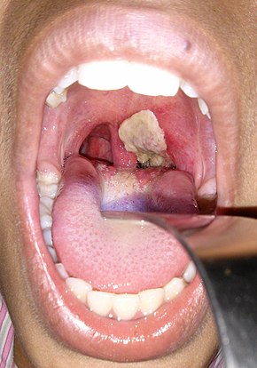
An adherent, dense, grey pseudomembrane covering the tonsils is classically seen in diphtheria.
 
A diphtheria skin lesion on the leg

The symptoms of diphtheria usually begin two to seven days after infection. Symptoms of diphtheria include fever of 38 °C (100.4 °F) or above; chills; fatigue; bluish skin coloration (cyanosis); sore throat; hoarseness; cough; headache; difficulty swallowing; painful swallowing; difficulty breathing; rapid breathing; foul-smelling and bloodstained nasal discharge; and lymphadenopathy. Within two to three days, diphtheria may destroy healthy tissues in the respiratory system. The dead tissue forms a thick, gray coating that can build up in the throat or nose. This thick gray coating is called a "pseudomembrane". It can cover tissues in the nose, tonsils, voice box, and throat, making it very hard to breathe and swallow. Symptoms can also include cardiac arrhythmias, myocarditis, and cranial and peripheral nerve palsies.

Diphtheritic croup

Laryngeal diphtheria can lead to a characteristic swollen neck and throat, or "bull neck". The swollen throat is often accompanied by a serious respiratory condition, characterized by a brassy or "barking" cough, stridor, hoarseness, and difficulty breathing; and historically referred to variously as "diphtheritic croup", "true croup", or sometimes simply as "croup". Diphtheritic croup is extremely rare in countries where diphtheria vaccination is customary. As a result, the term "" nowadays most often refers to an unrelated viral illness that produces similar but milder respiratory symptoms.

Transmission

Human-to-human transmission of diphtheria typically occurs through the air when an infected individual coughs or sneezes. Breathing in particles released from the infected individual leads to infection. Contact with any lesions on the skin can also lead to transmission of diphtheria, but this is uncommon. Indirect infections can occur, as well. If an infected individual touches a surface or object, the bacteria can be left behind and remain viable. Also, some evidence indicates diphtheria has the potential to be zoonotic, but this has yet to be confirmed. Corynebacterium ulcerans has been found in some animals, which would suggest zoonotic potential.

Mechanism

Diphtheria toxin is produced by C. diphtheriae only when infected with a bacteriophage that integrates the toxin-encoding genetic elements into the bacteria.

Diphtheria toxin is a single, 60-kDa-molecular weight protein composed of two peptide chains, fragment A and fragment B, held together by a disulfide bond. Fragment B is a recognition subunit that gains the toxin entry into the host cell by binding to the EGF-like domain of heparin-binding EGF-like growth factor on the cell surface. This signals the cell to internalize the toxin within an endosome via receptor-mediated endocytosis. Inside the endosome, the toxin is split by a trypsin-like protease into its individual A and B fragments. The acidity of the endosome causes fragment B to create pores in the endosome membrane, thereby catalysing the release of fragment A into the cell's cytoplasm.

Fragment A inhibits the synthesis of new proteins in the affected cell by catalyzing ADP-ribosylation of elongation factor EF-2—a protein that is essential to the translation step of protein synthesis. This ADP-ribosylation involves the transfer of an ADP-ribose from NAD+ to a diphthamide (a modified histidine) residue within the EF-2 protein. Since EF-2 is needed for the moving of tRNA from the A-site to the P-site of the ribosome during protein translation, ADP-ribosylation of EF-2 prevents protein synthesis.

ADP-ribosylation of EF-2 is reversed by giving high doses of nicotinamide (a form of vitamin B3), since this is one of the reaction's end products, and high amounts drive the reaction in the opposite direction.

Diagnosis

The current clinical case definition of diphtheria used by the United States' Centers for Disease Control and Prevention is based on both laboratory and clinical criteria.

Laboratory criteria

  • Isolation of C. diphtheriae from a Gram stain or throat culture from a clinical specimen
  • Histopathologic diagnosis of diphtheria by Albert's stain

Toxin demonstration

  • In vivo tests (guinea pig inoculation): Subcutaneous and intracutaneous tests
  • In vitro test: Elek's gel precipitation test, detection of tox gene by PCR, ELISA, ICA

Clinical criteria

  • Upper respiratory tract illness with sore throat
  • Low-grade fever (above 39 °C (102 °F) is rare)
  • An adherent, dense, grey pseudomembrane covering the posterior aspect of the pharynx: in severe cases, it can extend to cover the entire tracheobronchial tree.

Case classification

  • Probable: a clinically compatible case that is not laboratory-confirmed and is not epidemiologically linked to a laboratory-confirmed case
  • Confirmed: a clinically compatible case that is either laboratory-confirmed or epidemiologically linked to a laboratory-confirmed case
Empirical treatment should generally be started in a patient in whom suspicion of diphtheria is high.

Prevention

Quinvaxem is a widely administered pentavalent vaccine, which is a combination of five vaccines in one that protect babies from diphtheria, among other common childhood diseases. Diphtheria vaccine is usually combined at least with tetanus vaccine (Td) and often with pertussis (DTP, DTaP, TdaP, Tdap) vaccines, as well.

Treatment

The disease may remain manageable, but in more severe cases, lymph nodes in the neck may swell, and breathing and swallowing are more difficult. People in this stage should seek immediate medical attention, as obstruction in the throat may require intubation or a tracheotomy. Abnormal cardiac rhythms can occur early in the course of the illness or weeks later, and can lead to heart failure. Diphtheria can also cause paralysis in the eye, neck, throat, or respiratory muscles. Patients with severe cases are put in a hospital intensive care unit and given a diphtheria antitoxin (consisting of antibodies isolated from the serum of horses that have been challenged with diphtheria toxin). Since antitoxin does not neutralize toxin that is already bound to tissues, delaying its administration increases risk of death. Therefore, the decision to administer diphtheria antitoxin is based on clinical diagnosis, and should not await laboratory confirmation.

Antibiotics have not been demonstrated to affect healing of local infection in diphtheria patients treated with antitoxin. Antibiotics are used in patients or carriers to eradicate C. diphtheriae and prevent its transmission to others. The Centers for Disease Control and Prevention recommends either:
  • Metronidazole
  • Erythromycin is given (orally or by injection) for 14 days (40 mg/kg per day with a maximum of 2 g/d), or
  • Procaine penicillin G is given intramuscularly for 14 days (300,000 U/d for patients weighing <10 600="" and="" d="" for="" kg="" nbsp="" those="" u="" weighing="">10 kg); patients with allergies to penicillin G or erythromycin can use rifampin or clindamycin.
In cases that progress beyond a throat infection, diphtheria toxin spreads through the blood and can lead to potentially life-threatening complications that affect other organs, such as the heart and kidneys. Damage to the heart caused by the toxin affects the heart's ability to pump blood or the kidneys' ability to clear wastes. It can also cause nerve damage, eventually leading to paralysis. About 40% to 50% of those left untreated can die.

Epidemiology

Disability-adjusted life year for diphtheria per 100,000 inhabitants in 2004
  no data
  ≤ 1
  1–2
  2–3
  3–4
  4–5
  5–6
  6–7
  7–9
  9–10
  10–15
  15–50
  ≥ 50
Diphtheria cases reported to the World Health Organization between 1997 and 2006:
  no data
  1–49 reported cases
  Between 50 and 99 reported cases
  Over 100 reported cases

Diphtheria is fatal in between 5% and 10% of cases. In children under five years and adults over 40 years, the fatality rate may be as much as 20%. In 2013, it resulted in 3,300 deaths, down from 8,000 deaths in 1990.

The number of cases has changed over the course of the last 2 decades, specifically throughout developing countries. Better standards of living, mass immunization, improved diagnosis, prompt treatment, and more effective health care have led to the decrease in cases worldwide. However, although outbreaks are rare, they still occur worldwide, especially in developed nations such as Germany among unvaccinated children. In Nazi Germany contagious diseases such as diphtheria were among the leading causes of morbidity; they increased "after the mid-1920s, doubled again between 1932 and 1937, and reached extremely high levels during the war only to decline rapidly thereafter".

After the breakup of the former Soviet Union in the early 1990s, vaccination rates in its constituent countries fell so low that an explosion of diphtheria cases occurred. In 1991, 2,000 cases of diphtheria occurred in the USSR. Between 1991 and 1998 as many as 200,000 cases in the Commonwealth of Independent States were reported, with 5,000 deaths.

History

In 1613, Spain experienced an epidemic of diphtheria. The year is known as El Año de los Garrotillos (The Year of Strangulations) in the history of Spain.

In 1735, a diphtheria epidemic swept through New England.

Before 1826, diphtheria was known by different names across the world. In England, it was known as Boulogne sore throat, as it spread from France. In 1826, Pierre Bretonneau gave the disease the name diphthérite (from Greek diphthera "leather") describing the appearance of pseudomembrane in the throat.

In 1856, Victor Fourgeaud described an epidemic of diphtheria in California.

In 1878, Queen Victoria's daughter Princess Alice and her family became infected with diphtheria, causing two deaths, Princess Marie of Hesse and by Rhine and Princess Alice herself.

In 1883, Edwin Klebs identified the bacterium causing diphtheria and named it Klebs-Loeffler bacterium. The club shape of this bacterium helped Edwin to differentiate it from other bacteria. Over the period of time, it was called Microsporon diphtheriticum, Bacillus diphtheriae, and Mycobacterium diphtheriae. Current nomenclature is Corynebacterium diphtheriae

Friedrich Loeffler was the first person to cultivate C. diphtheriae in 1884. He used Koch's postulates to prove association between C. diphtheriae and diphtheria. He also showed that the bacillus produces an exotoxin.

A diphtheria immunisation scheme in London, 1941
 
Joseph P. O’Dwyer introduced the O'Dwyer tube for laryngeal intubation in patients with an obstructed larynx in 1885. It soon replaced tracheostomy as the emergency diphtheric intubation method.

In 1888, Emile Roux and Alexandre Yersin showed that a substance produced by C. diphtheriae caused symptoms of diphtheria in animals.

In 1890, Shibasaburo Kitasato and Emil von Behring immunized guinea pigs with heat-treated diphtheria toxin. They also immunized goats and horses in the same way and showed that an "antitoxin" made from serum of immunized animals could cure the disease in non-immunized animals. Behring used this antitoxin (now known to consist of antibodies that neutralize the toxin produced by C. diphtheriae) for human trials in 1891, but they were unsuccessful. Successful treatment of human patients with horse-derived antitoxin began in 1894, after production and quantification of antitoxin had been optimized. Von Behring won the first Nobel Prize in medicine in 1901 for his work on diphtheria.

In 1895, H. K. Mulford Company of Philadelphia started production and testing of diphtheria antitoxin in the United States. Park and Biggs described the method for producing serum from horses for use in diphtheria treatment.

In 1897, Paul Ehrlich developed a standardized unit of measure for diphtheria antitoxin. This was the first ever standardization of a biological product, and played an important role in future developmental work on sera and vaccines.

In 1901, 10 of 11 inoculated St. Louis children died from contaminated diphtheria antitoxin. The horse from which the antitoxin was derived died of tetanus. This incident, coupled with a tetanus outbreak in Camden, New Jersey, played an important part in initiating federal regulation of biologic products.

On 7 January 1904, Ruth Cleveland died of diphtheria at the age of 12 years in Princeton, New Jersey. Ruth was the eldest daughter of former President Grover Cleveland and the former first lady Frances Folsom.


In 1905, Franklin Royer, from Philadelphia's Municipal Hospital, published a paper urging timely treatment for diphtheria and adequate doses of antitoxin. In 1906, Clemens Pirquet and Béla Schick described serum sickness in children receiving large quantities of horse-derived antitoxin.


Between 1910 and 1911, Béla Schick developed the Schick test to detect pre-existing immunity to diphtheria in an exposed person. Only those who were not exposed to diphtheria were preferably vaccinated. A massive, five-year campaign was coordinated by Dr. Schick. As a part of the campaign, 85 million pieces of literature were distributed by the Metropolitan Life Insurance Company with an appeal to parents to "Save your child from diphtheria." A vaccine was developed in the next decade, and deaths began declining significantly in 1924.

A poster from the United Kingdom advertising diphtheria immunisation (published prior to 1962)

In 1919, in Dallas, Texas, 10 children were killed and 60 others made seriously ill by toxic antitoxin which had passed the tests of the New York State Health Department. Mulford Company of Philadelphia (manufacturers) paid damages in every case.

In the 1920s, each year an estimated 100,000 to 200,000 diphtheria cases and 13,000 to 15,000 deaths occurred in the United States. Children represented a large majority of these cases and fatalities. One of the most infamous outbreaks of diphtheria was in Nome, Alaska; the "Great Race of Mercy" to deliver diphtheria antitoxin is now celebrated by the Iditarod Trail Sled Dog Race.

In 1926, Alexander Thomas Glenny increased the effectiveness of diphtheria toxoid (a modified version of the toxin used for vaccination) by treating it with aluminum salts. Vaccination with toxoid was not widely used until the early 1930s.

In 1943, diphtheria outbreaks accompanied war and disruption in Europe. The 1 million cases in Europe resulted in 50,000 deaths.

In 1949, 68 of 606 children died after diphtheria immunization due to improper manufacture of aluminum phosphate toxoid.


In 1975, an outbreak of cutaneous diphtheria in Seattle, Washington, was reported.
In 1994, the Russian Federation had 39,703 diphtheria cases. By contrast, in 1990, only 1,211 cases were reported. Between 1990 and 1998, diphtheria caused 5000 deaths in the countries of the former Soviet Union.

In early May 2010, a case of diphtheria was diagnosed in Port-au-Prince, Haiti, after the devastating 2010 Haiti earthquake. The 15-year-old male patient died while workers searched for antitoxin.

In 2013, three children died of diphtheria in Hyderabad, India.

In early June 2015, a case of diphtheria was diagnosed at Vall d'Hebron University Hospital in Barcelona, Spain. The 6-year-old child who died of the illness had not been previously vaccinated due to parental opposition to vaccination. It was the first case of diphtheria in the country since 1986 as reported by "El Mundo" or from 1998, as reported by WHO.

In March 2016, a 3-year-old girl died of diphtheria in the University Hospital of Antwerp, Belgium.

In June 2016, a 3-year-old, 5-year-old, and 7-year-old girl died of diphtheria in Kedah, Malacca and Sabah, Malaysia.

In January 2017, more than 300 cases were recorded in Venezuela.

In November and December 2017, an outbreak of diphtheria occurred in Indonesia with more than 600 cases found and 38 fatalities.
In November 2019, two cases of diphtheria occurred in the Lothian area of Scotland. Additionally, in November 2019 an 8-year-old boy died of diphtheria in Athens, Greece.

Whooping cough

From Wikipedia, the free encyclopedia
 
Whooping cough
Other namesPertussis, 100-day cough
Pertussis.jpg
A young boy coughing due to pertussis.
SpecialtyInfectious disease
SymptomsRunny nose, fever, cough
ComplicationsVomiting, broken ribs, very tired
Duration~ 10 weeks
CausesBordetella pertussis (spread through the air)
Diagnostic methodNasopharyngeal swab
PreventionPertussis vaccine
TreatmentAntibiotics (if started early)
Frequency16.3 million (2015)
Deaths58,700 (2015)

Whooping cough, also known as pertussis or the 100-day cough, is a highly contagious bacterial disease. Initial symptoms are usually similar to those of the common cold with a runny nose, fever, and mild cough, but these are followed by weeks of severe coughing fits. Following a fit of coughing, a high-pitched whoop sound or gasp may occur as the person breathes in. The coughing may last for 10 or more weeks, hence the phrase "100-day cough". A person may cough so hard that they vomit, break ribs, or become very tired from the effort. Children less than one year old may have little or no cough and instead have periods where they do not breathe. The time between infection and the onset of symptoms is usually seven to ten days. Disease may occur in those who have been vaccinated, but symptoms are typically milder.

Pertussis is caused by the bacterium Bordetella pertussis. It is spread easily through the coughs and sneezes of an infected person. People are infectious from the start of symptoms until about three weeks into the coughing fits. Those treated with antibiotics are no longer infectious after five days. Diagnosis is by collecting a sample from the back of the nose and throat. This sample can then be tested by either culture or by polymerase chain reaction.

Prevention is mainly by vaccination with the pertussis vaccine. Initial immunization is recommended between six and eight weeks of age, with four doses to be given in the first two years of life. Protection from pertussis decreases over time, so additional doses of vaccine are often recommended for older children and adults. Antibiotics may be used to prevent the disease in those who have been exposed and are at risk of severe disease. In those with the disease, antibiotics are useful if started within three weeks of the initial symptoms, but otherwise have little effect in most people. In pregnant women and children less than one year old, antibiotics are recommended within six weeks of symptom onset. Antibiotics used include erythromycin, azithromycin, clarithromycin, or trimethoprim/sulfamethoxazole. Evidence to support interventions for the cough, other than antibiotics, is poor. About 50% of infected children less than a year old require hospitalization and nearly 0.5% (1 in 200) die.

An estimated 16.3 million people worldwide were infected in 2015. Most cases occur in the developing world, and people of all ages may be affected. In 2015, pertussis resulted in 58,700 deaths – down from 138,000 deaths in 1990. Outbreaks of the disease were first described in the 16th century. The bacterium that causes the infection was discovered in 1906. The pertussis vaccine became available in the 1940s.

Signs and symptoms

A boy with pertussis

The classic symptoms of pertussis are a paroxysmal cough, inspiratory whoop, and fainting, or vomiting after coughing. The cough from pertussis has been documented to cause subconjunctival hemorrhages, rib fractures, urinary incontinence, hernias, and vertebral artery dissection. Violent coughing can cause the pleura to rupture, leading to a pneumothorax. Vomiting after a coughing spell or an inspiratory whooping sound on coughing, almost doubles the likelihood that the illness is pertussis. The absence of a paroxysmal cough or posttussive emesis, though, makes it almost half as likely.

The illness usually starts with mild respiratory symptoms include mild coughing, sneezing, or a runny nose (known as the catarrhal stage). After one to two weeks, the coughing classically develops into uncontrollable fits, sometimes followed by a high-pitched "whoop" sound, as the person tries to inhale. About 50% of children and adults "whoop" at some point in diagnosed pertussis cases during the paroxysmal stage.

This stage usually lasts two to eight weeks, or sometimes longer. A gradual transition then occurs to the convalescent stage, which usually lasts one to four weeks. This stage is marked by a decrease in paroxysms of coughing, although paroxysms may occur with subsequent respiratory infection for many months after the onset of pertussis.

Symptoms of pertussis can be variable, especially between immunized and non-immunized people. Those that are immunized can present with a more mild infection; they may only have the paroxysmal cough for a couple of weeks, and it may lack the "whooping" characteristic. Although immunized people have a milder form of the infection, they can spread the disease to others who are not immune.

Incubation period

The time between exposure and the development of symptoms is on average 7–14 days (range 6–20 days), rarely as long as 42 days.

Cause

Pertussis is caused by the bacterium Bordetella pertussis. It is an airborne disease (through droplets) that spreads easily through the coughs and sneezes of an infected person.

Spread from other animals

Uncertainties have existed of B. pertussis and whooping cough as a zoonotic disease since around 1910 but in the 1930s, knowledge was gained that the bacteria lost their virulent power when repeatedly spread on agar media. This explained the difficulties to reproduce results from different studies as the pre-inoculating handlings of the bacteria were not standardized among scientists.

Today it is established that at least some primate species are highly susceptible to B. pertussis and develop clinical whooping cough in high incidence when exposed to low inoculation doses. The bacteria may be present in wild animal populations, but this is not confirmed by laboratory diagnosis, although whooping cough is known among wild gorillas. Several zoos also have a long-standing custom of vaccinating their primates against whooping cough.

Mechanism

After the bacteria are inhaled, they initially adhere to the ciliated epithelium in the nasopharynx. Surface proteins of B. pertussis, including filamentous hemaglutinin and pertactin, mediate attachment to the epithelium. The bacteria then multiply. In infants, who experience more severe disease, the bacteria spread down to the lungs.

The bacteria secretes a number of toxins. Tracheal cytotoxin, a fragment of peptidoglycan, kills ciliated epithelial cells and thereby inhibits the mucociliary elevator by which mucus and debris are removed. TCT may contribute to the cough characteristic of pertussis. The cough may also be caused by a yet-to-be identified "cough toxin". Pertussis toxin causes lymphocytosis by an unknown mechanism. The elevated number of white blood cells leads to pulmonary hypertension, a major cause of death by pertussis. In infants who develop encephalopathy, cerebral hemorrhage and cortical atrophy occur, likely due to hypoxia.

Diagnosis

Gram stain of Bordetella pertussis

Based on symptoms

A physician's overall impression is most effective in initially making the diagnosis. Single factors are much less useful. In adults with a cough of less than 8 weeks, vomiting after coughing or a "whoop" is supportive. If there are no bouts of coughing or there is a fever the diagnosis is unlikely. In children who have a cough of less than 4 weeks vomiting after coughing is somewhat supportive but not definitive.

Lab tests

Methods used in laboratory diagnosis include culturing of nasopharyngeal swabs on a nutrient medium (Bordet-Gengou medium), polymerase chain reaction (PCR), direct fluorescent antibody (DFA), and serological methods (e.g. complement fixation test). The bacteria can be recovered from the person only during the first three weeks of illness, rendering culturing and DFA useless after this period, although PCR may have some limited usefulness for an additional three weeks.

Serology may be used for adults and adolescents who have already been infected for several weeks to determine whether antibody against pertussis toxin or another virulence factor of B. pertussis is present at high levels in the blood of the person.

Differential diagnosis

A similar, milder disease is caused by B. parapertussis.

Prevention

The primary method of prevention for pertussis is vaccination. Evidence is insufficient to determine the effectiveness of antibiotics in those who have been exposed, but are without symptoms. Preventive antibiotics, however, are still frequently used in those who have been exposed and are at high risk of severe disease (such as infants).

Vaccine

Pertussis vaccines are effective at preventing illness and are recommended for routine use by the World Health Organization and the United States Centers for Disease Control and Prevention. The vaccine saved an estimated half a million lives in 2002.

The multicomponent acellular pertussis vaccine is 71–85% effective, with greater effectiveness against more severe strains. However, despite widespread vaccination, pertussis has persisted in vaccinated populations and is today "one of the most common vaccine-preventable diseases in Western countries". The 21st-century resurgences in pertussis infections is attributed to a combination of waning immunity and bacterial mutations that elude vaccines.

Immunization does not confer lifelong immunity; a 2011 CDC study indicated that protection may only last three to six years. This covers childhood, which is the time of greatest exposure and greatest risk of death from pertussis.

An effect of widespread immunization on society has been the shift of reported infections from children aged 1–9 years to infants, adolescents, and adults, with adolescents and adults acting as reservoirs for B. pertussis and infecting infants who have had fewer than three doses of vaccine.

Infection induces incomplete natural immunity that wanes over time. A 2005 study said estimates of the duration of infection-acquired immunity range from 7 to 20 years and the different results could be the result of differences in levels of circulating B. pertussis, surveillance systems, and case definitions used. The study said protective immunity after vaccination wanes after 4–12 years. One study suggested that the availability of vaccine exemptions increases the number of pertussis cases.

Some studies have suggested that while acellular pertussis vaccines are effective at preventing the disease, they have a limited impact on infection and transmission, meaning that vaccinated people could spread pertussis even though they may have only mild symptoms or none at all. Pertussis infection in these persons may be asymptomatic, or present as illness ranging from a mild cough to classic pertussis with persistent cough (i.e., lasting more than 7 days). Even though the disease may be milder in older persons, those who are infected may transmit the disease to other susceptible persons, including unimmunized or incompletely immunized infants. Older persons are often found to have the first case in a household with multiple pertussis cases, and are often the source of infection for children.

Treatment

The antibiotics erythromycin, clarithromycin, or azithromycin are typically the recommended treatment. Newer macrolides are frequently recommended due to lower rates of side effects. Trimethoprim-sulfamethoxazole (TMP/SMX) may be used in those with allergies to first-line agents or in infants who have a risk of pyloric stenosis from macrolides.

A reasonable guideline is to treat people age >1 year within 3 weeks of cough onset and infants age <1 10="" 3="" 6="" alter="" and="" antibiotics="" are="" as="" azithromycin="" be="" cough="" course="" days="" decrease="" diagnosed="" duration="" early="" effective="" eliminating="" erythromycin="" even="" for="" i="" if="" illness="" in="" infectiousness="" is="" late="" long-term="" longer="" no="" not="" of="" onset.="" person="" pertussis.="" pregnant="" prevent="" short-term="" should="" spread.="" spreading="" the="" they="" thus="" treatment="" used="" weeks="" when="" will="" within="" without="" women="" year="">B. pertussis
with fewer and less severe side effects.

People with pertussis are most infectious during the first two weeks following the onset of symptoms.

Effective treatments of the cough associated with this condition have not been developed. The use of over the counter cough medications is discouraged and has not been found helpful.

Prognosis

Disability-adjusted life year for pertussis per 100,000 inhabitants as of 2004.
  No data
  Less than 50
  50–100
  100–150
  150–200
  200–250
  250–300
  300–350
  350–400
  400–450
  450–500
  500–550
  More than 550

While most healthy older children and adults fully recover, infection in newborns is particularly severe. Pertussis is fatal in an estimated 0.5% of US infants under one year of age. First-year infants are also more likely to develop complications, such as: apneas (31%), pneumonia (12%), seizures (0.6%) and encephalopathy (0.15%). This may be due to the ability of the bacterium to suppress the immune system.

Epidemiology

Whooping cough deaths per million persons in 2012
  0–0.9
  1–1.9
  2–3
  4–4.9
  5–5.9
  6–32
  33–38
  39–44
  45–79

Worldwide, whooping cough affects around 16 million people yearly. One estimate for 2013 stated it resulted in about 61,000 deaths – down from 138,000 deaths in 1990. Another estimated 195,000 child deaths yearly from the disease worldwide. This is despite generally high coverage with the DTP and DTaP vaccines. Pertussis is one of the leading causes of vaccine-preventable deaths worldwide. About 90% of all cases occur in developing countries.

Before vaccines, an average of 178,171 cases was reported in the U.S., with peaks reported every two to five years; more than 93% of reported cases occurred in children under 10 years of age. The actual incidence was likely much higher. After vaccinations were introduced in the 1940s, pertussis incidence fell dramatically to approximately 1,000 by 1976. Incidence rates have increased since 1980. In 2015, rates in the United States were 20,762 people.

Pertussis is the only vaccine-preventable disease that is associated with increasing deaths in the U.S. The number of deaths increased from four in 1996 to 17 in 2001, almost all of which were infants under one year. In Canada, the number of pertussis infections has varied between 2,000 and 10,000 reported cases each year over the last ten years, and it is the most common vaccine-preventable illness in Toronto.

In 2009 Australia reported an average of 10,000 cases a year, and the number of cases had increased. In the U.S. pertussis in adults has increased significantly since about 2004.

In 2017, India had a reported 23,766 reported pertussis cases, making it one of the highest reported number of cases of the year. Other countries, such as Germany, had reported 16,183 cases, while Australia and China had a reported number of 12,114 and 10,390 pertussis cases.

US outbreaks

An epidemiologist tests blood samples for pertussis during a 2010 outbreak.
 
In 2010 ten infants in California died, and health authorities declared an epidemic encompassing 9,120 cases. They found that doctors had failed to correctly diagnose the infants' condition during several visits. Statistical analysis identified significant overlap in communities with a cluster of nonmedical child exemptions and cases. The number of exemptions varied widely among communities, but tended to be highly clustered. In some schools, more than three quarters of parents filed for vaccination exemptions. The data suggest vaccine refusal based on nonmedical reasons and personal belief exacerbated the outbreak. Other factors included reduced duration of immunity following the acellular vaccine and, the fact that most vaccinated adults and older children had not received a booster shot.

In April and May 2012 pertussis was declared to be at epidemic levels in Washington, with 3,308 cases. In December 2012 Vermont declared an epidemic of 522 cases. Wisconsin had the highest incidence rate, with 3,877 cases, although it did not make an official epidemic declaration.

History

Discovery

B. pertussis was discovered in 1906 by Jules Bordet and Octave Gengou, who also developed the first serology and vaccine. Efforts to develop an inactivated whole-cell vaccine began soon after B. pertussis was cultured that year. In the 1920s, Louis W. Sauer developed a weak vaccine for whooping cough at Evanston Hospital (Evanston, IL). In 1925 Danish physician Thorvald Madsen was the first to test a whole-cell vaccine on a wide scale. Madsen used the vaccine to control outbreaks in the Faroe Islands in the North Sea.

Vaccine

In 1932 an outbreak of whooping cough hit Atlanta, Georgia, prompting pediatrician Leila Denmark to begin her study of the disease. Over the next six years her work was published in the Journal of the American Medical Association, and in partnership with Emory University and Eli Lilly & Company, she developed the first pertussis vaccine. In 1942 American scientists Grace Eldering, Loney Gordon, and Pearl Kendrick combined the whole-cell pertussis vaccine with diphtheria and tetanus toxoids to generate the first DTP combination vaccine. To minimize the frequent side effects caused by the pertussis component, Japanese scientist Yuji Sato developed an acellular vaccine consisting of purified haemagglutinins (HAs: filamentous strep throat and leukocytosis-promoting-factor HA), which are secreted by B. pertussis. Sato's acellular pertussis vaccine was used in Japan starting in 1981. Later versions of the acellular vaccine in other countries consisted of additional defined components of B. pertussis and were often part of the DTaP combination vaccine.

Bronchitis

From Wikipedia, the free encyclopedia

Bronchitis
Bronchitis.jpg
Figure A shows the location of the lungs and bronchial tubes. Figure B is an enlarged view of a normal bronchial tube. Figure C is an enlarged view of a bronchial tube with bronchitis.
Pronunciation
  • bron-kye-tis
SpecialtyInfectious disease, pulmonology
SymptomsCoughing up mucus, wheezing, shortness of breath, chest discomfort[1]
TypesAcute, chronic
FrequencyAcute: ~5% of people a year
Chronic: ~5% of people

Bronchitis is inflammation of the bronchi (large and medium-sized airways) in the lungs that causes coughing. Symptoms include coughing up sputum, wheezing, shortness of breath, and chest pain. Bronchitis can be acute or chronic.

Acute bronchitis usually has a cough that lasts around three weeks, and is also known as a chest cold. In more than 90% of cases the cause is a viral infection. These viruses may be spread through the air when people cough or by direct contact. A small number of cases are caused by a bacterial infection such as Mycoplasma pneumoniae or Bordetella pertussis. Risk factors include exposure to tobacco smoke, dust, and other air pollution. Treatment of acute bronchitis typically involves rest, paracetamol (acetaminophen), and nonsteroidal anti-inflammatory drugs (NSAIDs) to help with the fever.

Chronic bronchitis is defined as a productive cough – one that produces sputum – that lasts for three months or more per year for at least two years.  Many people with chronic bronchitis have chronic obstructive pulmonary disease (COPD). Tobacco smoking is the most common cause, with a number of other factors such as air pollution and genetics playing a smaller role. Treatments include quitting smoking, vaccinations, rehabilitation, and often inhaled bronchodilators and steroids. Some people may benefit from long-term oxygen therapy.

Acute bronchitis is one of the most common diseases. About 5% of adults are affected and about 6% of children have at least one episode a year. Acute bronchitis is the most common type of bronchitis. In the United States, in 2018, 9.3 million people were diagnosed with chronic bronchitis.

Acute bronchitis

Bronchitis

Acute bronchitis, also known as a chest cold, is short term inflammation of the bronchi of the lungs. The most common symptom is a cough, that may or may not produce sputum. Other symptoms may include coughing up mucus, wheezing, shortness of breath, fever, and chest discomfort. Fever when present is mild. The infection may last from a few to ten days. The cough may persist for several weeks afterwards, with the total duration of symptoms usually around three weeks. Symptoms may last for up to six weeks.

Cause

In more than 90% of cases, the cause is a viral infection. These viruses may spread through the air when people cough or by direct contact. Risk factors include exposure to tobacco smoke, dust, and other air pollutants. A small number of cases are due to bacteria such as Mycoplasma pneumoniae or Bordetella pertussis.

Diagnosis

Diagnosis is typically based on a person's signs and symptoms. The color of the sputum does not indicate if the infection is viral or bacterial. Determining the underlying organism is usually not required. Other causes of similar symptoms include asthma, pneumonia, bronchiolitis, bronchiectasis, and COPD. A chest X-ray may be useful to detect pneumonia.

Another common sign of bronchitis is a cough which lasts ten days to three weeks. If the cough lasts for longer than a month, it may become chronic bronchitis. In addition, a fever may be present. Acute bronchitis is normally caused by a viral infection. Typically, these infections are rhinovirus, parainfluenza, or influenza. No specific testing is normally needed in order to diagnose acute bronchitis.

Treatment

One form of prevention is to avoid smoking and other lung irritants. Frequent hand washing may also be protective. Treatment for acute bronchitis usually involves rest, paracetamol (acetaminophen), and NSAIDs to help with the fever. Cough medicine has little support for its use, and is not recommended in children under the age of six. There is tentative evidence that salbutamol may be useful in treating wheezing; however, it may result in nervousness and tremors. Antibiotics should generally not be used. An exception is when acute bronchitis is due to pertussis. Tentative evidence supports honey and pelargonium to help with symptoms. Getting plenty of rest and drinking enough fluids are often recommended as well. Chinese medicinal herbs are of unclear effect.

Epidemiology

Acute bronchitis is one of the most common diseases. About 5% of adults are affected, and about 6% of children have at least one episode a year. It occurs more often in the winter. More than 10 million people in the US visit a doctor each year for this condition, with about 70% receiving antibiotics which are mostly not needed. There are efforts to decrease the use of antibiotics in acute bronchitis. Acute bronchitis is the most common type of bronchitis.

Chronic bronchitis

Chronic bronchitis is defined as a productive cough that lasts for three months or more per year for at least two years. When this occurs together with decreased airflow it is known as chronic obstructive pulmonary disease (COPD). Many people with chronic bronchitis have COPD however, most people with COPD do not have chronic bronchitis. Estimates of the number of people with COPD who have chronic bronchitis are 7 to 40%. Estimates of the number of people who smoke and have chronic bronchitis who also have COPD is 60%.

The term "chronic bronchitis" was used in previous definitions of COPD but is no longer included in the definition. The term is still used clinically. While both chronic bronchitis and emphysema are often associated with COPD, neither is needed to make the diagnosis. A Chinese consensus commented on symptomatic types of COPD that include chronic bronchitis and with frequent exacerbations.

Chronic bronchitis is a respiratory disease marked by overproduction of mucus and mucins. The excess mucus is produced by goblet cells and enlarged submucosal glands in response to long-term irritation. The mucous glands in the submucosa secrete more than the goblet cells. Mucins thicken mucus, and their concentration has been found to be high in cases of chronic bronchitis, and also to correlate with the severity of the disease. Excess mucus can narrow the airways, thereby limiting airflow and accelerating the decline in lung function, and result in COPD. Excess mucus shows itself as a chronic productive cough and its severity and volume of sputum can fluctuate in periods of acute exacerbations. In COPD, those with the chronic bronchitic phenotype with associated chronic excess mucus, experience a worse quality of life than those without.

The increased secretions are initially cleared by coughing. The cough is often worse soon after awakening, and the sputum produced may have a yellow or green color and may be streaked with specks of blood. In the early stages, a cough can maintain mucus clearance. However, with continued excessive secretion, mucus clearance is impaired and when the airways become obstructed a cough becomes ineffective. Effective mucociliary clearance depends on airway hydration, ciliary beating, and the rates of mucin secretion. Each of these factors is impaired in chronic bronchitis. Chronic bronchitis can lead to a higher number of exacerbations and a faster decline in lung function. The ICD-11 lists chronic bronchitis with emphysema (emphysematous bronchitis) as a "certain specified COPD".

Cause

Most cases of chronic bronchitis are caused by tobacco smoking. Chronic bronchitis in young adults who smoke is associated with a greater chance of developing COPD. There is an association between smoking cannabis and chronic bronchitis. In addition, chronic inhalation of air pollution, or irritating fumes or dust from hazardous exposures in occupations such as coal mining, grain handling, textile manufacturing, livestock farming, and metal moulding may also be a risk factor for the development of chronic bronchitis. Bronchitis caused in this way is often referred to as industrial bronchitis, or occupational bronchitis. Rarely genetic factors also play a role.

Air quality can also affect the respiratory system with higher levels of nitrogen dioxide and sulfur dioxide contributing to bronchial symptoms. Sulfur dioxide can cause inflammation which can aggravate chronic bronchitis and make infections more likely.

Air pollution in the workplace is the cause of several non-communicable diseases (NCDs) including chronic bronchitis.

Treatment

Decline in lung function in chronic bronchitis may be slowed by stopping smoking. Chronic bronchitis may be treated with a number of medications and occasionally oxygen therapy. Pulmonary rehabilitation may also be used.

A distinction has been made between exacerbations (sudden worsenings) of chronic bronchitis, and otherwise stable chronic bronchitis. Stable chronic bronchitis can be defined as the normal definition of chronic bronchitis, plus the absence of an acute exacerbation in the previous four weeks. A Cochrane review found that mucolytics in chronic bronchitis may slightly decrease the chance of developing an exacerbation. The mucolytic guaifenesin is a safe and effective treatment for stable chronic bronchitis. This has an advantage in that it is available as an extended use tablet which lasts for twelve hours. Another mucolytic fudosteine may also be used. In those with chronic bronchitis and severe COPD, the phosphodiesterase-4 inhibitor roflumilast may decrease significant exacerbations.

Epidemiology

Chronic bronchitis affects about 3.4% to 22% of the general population. Individuals over 45 years of age, smokers, those that live or work in areas with high air pollution, and anybody with asthma all have a higher risk of developing chronic bronchitis. This wide range is due to the different definitions of chronic bronchitis that can be diagnosed based on signs and symptoms or the clinical diagnosis of the disorder. Chronic bronchitis tends to affect men more often than women. While the primary risk factor for chronic bronchitis is smoking, there is still a 4%-22% chance that never smokers can get chronic bronchitis. This might suggest other risk factors such as the inhalation of fuels, dusts, fumes and genetic factor. In the United States, in 2016, 8.6 million people were diagnosed with chronic bronchitis, and there were 518 reported deaths. Per 100,000 of population the death rate of chronic bronchitis was 0.2.

History

The condition of bronchitis has been recognised for many centuries, in several different cultures including the Ancient Greek, Chinese, and Indian, with the presence of excess phlegm and cough noted in recognition of the same condition.

In Britain in 1808, a physician Charles Badham was the first person to describe the condition and name the acute form as acute bronchitis. This was written of in a book entitled Inflammatory conditions of the bronchia. In this book Badham distinguished three forms of bronchitis including acute and chronic. A second edition of this book was renamed An Essay on Bronchitis and published in 1814. Bradham used the term catarrh to refer to the cardinal symptoms of chronic cough and mucus hypersecretion of chronic bronchitis, and described chronic bronchitis as a disabling disorder.

In 1901 an article was published on the treatment of chronic bronchitis in the elderly. The symptoms described have remained unchanged. The cause was thought to be brought on by dampness, cold weather, and foggy conditions, and treatments were aimed towards various cough mixtures, respiratory stimulants, and tonics. It was noted that something other than the weather was thought to be at play. Exacerbations of the condition were also described at this time. Another physician Harry Campbell was referred to who had written in the British Medical Journal a week before. Campbell had suggested that the cause of chronic bronchitis was due to toxic substances, and recommended pure air, simple food, and exercise to remove them from the body.

A joint research programme was undertaken in Chicago and London from 1951 to 1953 in which the clinical features of one thousand cases of chronic bronchitis were detailed. The findings were published in the Lancet in 1953. It was stated that since its introduction by Badham, chronic bronchitis had become an increasingly popular diagnosis. The study had looked at various associations such as the weather, conditions at home, and at work, age of onset, childhood illnesses, smoking habits, and breathlessness. It was concluded that chronic bronchitis invariably led to emphysema, particularly when the bronchitis had persisted for a long time.

In 1957 it was noted that at the time there were many investigations being carried out into chronic bronchitis and emphysema in general, and among industrial workers exposed to dust. Excerpts were published dating from 1864 in which Charles Parsons had noted the occurring consequence of the development of emphysema from bronchitis. This was seen to be not always applicable. His findings were in association with his studies on chronic bronchitis among pottery workers.

A CIBA (now Novartis) meeting in 1959, and a meeting of the American Thoracic Society in 1962, defined chronic bronchitis as a component of COPD, in the terms that have not changed.

Eosinophilic bronchitis

Eosinophilic bronchitis is a chronic dry cough, defined by the presence of an increased number of a type of white blood cell known as eosinophils. It has a normal finding on X-ray and has no airflow limitation.

Protracted bacterial bronchitis

Protracted bacterial bronchitis in children, is defined as a chronic productive cough with a positive bronchoalveolar lavage that resolves with antibiotics. Protracted bacterial bronchitis is usually caused by Streptococcus pneumoniae, non-typable Haemophilus influenzae, or Moraxella catarrhalis. Protracted bacterial bronchitis (lasting more than 4 weeks) in children may be helped by antibiotics.

Plastic bronchitis

Plastic bronchitis is a rarely found condition in which thickened secretions plug the bronchi. The plugs are rubbery or plastic-feeling (thus the name). The light-colored plugs take the branching shape of the bronchi that they fill, and are known as bronchial casts. When these casts are coughed up, they are firmer in texture from typical phlegm or the short, softer mucus plugs seen in some people with asthma. However, some people with asthma have larger, firmer, more complex plugs. These differ from the casts seen in people whose plastic bronchitis is associated with congenital heart disease or lymphatic vessel abnormalities mainly because eosinophils and Charcot–Leyden crystals are present in the asthma-associated casts but not in the others.

Casts obstruct the airflow, and can result in the overinflation of the opposite lung. Plastic bronchitis usually occurs in children. Some cases may result from abnormalities in the lymphatic vessels. Advanced cases may show imaging similarities to bronchiectasis.

Aspergillus bronchitis

Aspergillus bronchitis is one of the Aspergillosis spectrum of diseases, in which the bronchi are specifically subject to a fungal infection. This differs from the other pulmonary aspergillosis conditions, in that it need not affect just the immunocompromised.

Operator (computer programming)

From Wikipedia, the free encyclopedia https://en.wikipedia.org/wiki/Operator_(computer_programmin...