Search This Blog

Friday, June 12, 2020

Head injury

From Wikipedia, the free encyclopedia

Head injury
Head wound received at Antietam 1862.jpg
Soldier wounded at the Battle of Antietam on September 17, 1862.

A head injury is any injury that results in trauma to the skull or brain. The terms traumatic brain injury and head injury are often used interchangeably in the medical literature. Because head injuries cover such a broad scope of injuries, there are many causes—including accidents, falls, physical assault, or traffic accidents—that can cause head injuries.

The number of new cases is 1.7 million in the United States each year, with about 3% of these incidents leading to death. Adults have head injuries more frequently than any age group resulting from falls, motor vehicle crashes, colliding or being struck by an object, or assaults. Children, however, may experience head injuries from accidental falls or intentional causes (such as being struck or shaken) leading to hospitalization. Acquired brain injury (ABI) is a term used to differentiate brain injuries occurring after birth from injury, from a genetic disorder, or from a congenital disorder.

Unlike a broken bone where trauma to the body is obvious, head trauma can sometimes be conspicuous or inconspicuous. In the case of an open head injury, the skull is cracked and broken by an object that makes contact with the brain. This leads to bleeding. Other obvious symptoms can be neurological in nature. The person may become sleepy, behave abnormally, lose consciousness, vomit, develop a severe headache, have mismatched pupil sizes, and/or be unable to move certain parts of the body. While these symptoms happen immediately after a head injury occurs, many problems can develop later in life. Alzheimer’s disease, for example, is much more likely to develop in a person who has experienced a head injury.

Brain damage, which is the destruction or degeneration of brain cells, is a common occurrence in those who experience a head injury. Neurotoxicity is another cause of brain damage that typically refers to selective, chemically induced neuron/brain damage.

Classification

Head injuries include both injuries to the brain and those to other parts of the head, such as the scalp and skull. Head injuries can be closed or open. A closed (non-missile) head injury is where the dura mater remains intact. The skull can be fractured, but not necessarily. A penetrating head injury occurs when an object pierces the skull and breaches the dura mater. Brain injuries may be diffuse, occurring over a wide area, or focal, located in a small, specific area. A head injury may cause skull fracture, which may or may not be associated with injury to the brain. Some patients may have linear or depressed skull fractures. If intracranial hemorrhage occurs, a hematoma within the skull can put pressure on the brain. Types of intracranial hemorrhage include subdural, subarachnoid, extradural, and intraparenchymal hematoma. Craniotomy surgeries are used in these cases to lessen the pressure by draining off the blood.

Brain injury can occur at the site of impact, but can also be at the opposite side of the skull due to a contrecoup effect (the impact to the head can cause the brain to move within the skull, causing the brain to impact the interior of the skull opposite the head-impact). While impact on the brain at the same site of injury to the skull is the coup effect. If the impact causes the head to move, the injury may be worsened, because the brain may ricochet inside the skull causing additional impacts, or the brain may stay relatively still (due to inertia) but be hit by the moving skull (both are contrecoup injuries).

Specific problems after head injury can include
  • Skull fracture
  • Lacerations to the scalp and resulting hemorrhage of the skin
  • Traumatic subdural hematoma, a bleeding below the dura mater which may develop slowly
  • Traumatic extradural, or epidural hematoma, bleeding between the dura mater and the skull
  • Traumatic subarachnoid hemorrhage
  • Cerebral contusion, a bruise of the brain
  • Concussion, a loss of function due to trauma
  • Dementia pugilistica, or "punch-drunk syndrome", caused by repetitive head injuries, for example in boxing or other contact sports
  • A severe injury may lead to a coma or death
  • Shaken baby syndrome – a form of child abuse

Concussion

coup bruise

A concussion is a form of a mild traumatic brain injury (TBI). This injury is a result due to a blow to the head that could make the person’s physical, cognitive, and emotional behaviors irregular. Symptoms may include clumsiness, fatigue, confusion, nausea, blurry vision, headaches, and others. Mild concussions are associated with sequelae. Severity is measured using various concussion grading systems.

A slightly greater injury is associated with both anterograde and retrograde amnesia (inability to remember events before or after the injury). The amount of time that the amnesia is present correlates with the severity of the injury. In all cases, the patients develop post concussion syndrome, which includes memory problems, dizziness, tiredness, sickness and depression. Cerebral concussion is the most common head injury seen in children.

Intracranial bleeding

Types of intracranial hemorrhage are roughly grouped into intra-axial and extra-axial. The hemorrhage is considered a focal brain injury; that is, it occurs in a localized spot rather than causing diffuse damage over a wider area.

Intra-axial bleeding

Intra-axial hemorrhage is bleeding within the brain itself, or cerebral hemorrhage. This category includes intraparenchymal hemorrhage, or bleeding within the brain tissue, and intraventricular hemorrhage, bleeding within the brain's ventricles (particularly of premature infants). Intra-axial hemorrhages are more dangerous and harder to treat than extra-axial bleeds.

Extra-axial bleeding

Extra-axial hemorrhage, bleeding that occurs within the skull but outside of the brain tissue, falls into three subtypes:
  • Epidural hemorrhage (extradural hemorrhage) which occur between the dura mater (the outermost meninx) and the skull, is caused by trauma. It may result from laceration of an artery, most commonly the middle meningeal artery. This is a very dangerous type of injury because the bleed is from a high-pressure system and deadly increases in intracranial pressure can result rapidly. However, it is the least common type of meningeal bleeding and is seen in 1% to 3% cases of head injury.
    • Patients have a loss of consciousness (LOC), then a lucid interval, then sudden deterioration (vomiting, restlessness, LOC)
    • Head CT shows lenticular (convex) deformity.
  • Subdural hemorrhage results from tearing of the bridging veins in the subdural space between the dura and arachnoid mater.
    • Head CT shows crescent-shaped deformity
  • Subarachnoid hemorrhage, which occur between the arachnoid and pia meningeal layers, like intraparenchymal hemorrhage, can result either from trauma or from ruptures of aneurysms or arteriovenous malformations. Blood is seen layering into the brain along sulci and fissures, or filling cisterns (most often the suprasellar cistern because of the presence of the vessels of the circle of Willis and their branch points within that space). The classic presentation of subarachnoid hemorrhage is the sudden onset of a severe headache (a thunderclap headache). This can be a very dangerous entity and requires emergent neurosurgical evaluation and sometimes urgent intervention.

Cerebral contusion

Cerebral contusion is bruising of the brain tissue. The piamater is not breached in contusion in contrary to lacerations. The majority of contusions occur in the frontal and temporal lobes. Complications may include cerebral edema and transtentorial herniation. The goal of treatment should be to treat the increased intracranial pressure. The prognosis is guarded.

Diffuse axonal injury

Diffuse axonal injury, or DAI, usually occurs as the result of an acceleration or deceleration motion, not necessarily an impact. Axons are stretched and damaged when parts of the brain of differing density slide over one another. Prognoses vary widely depending on the extent of the damage.

Signs and symptoms

Three categories used for classifying the severity of brain injuries are mild, moderate or severe.

Mild brain injuries

Symptoms of a mild brain injury include headaches, confusion, ringing ears, fatigue, changes in sleep patterns, mood or behavior. Other symptoms include trouble with memory, concentration, attention or thinking. Mental fatigue is a common debilitating experience and may not be linked by the patient to the original (minor) incident. Narcolepsy and sleep disorders are common misdiagnoses.

Moderate/severe brain injuries

Cognitive symptoms include confusion, aggressive, abnormal behavior, slurred speech, and coma or other disorders of consciousness. Physical symptoms include headaches that do not go away or worsen, vomiting or nausea, convulsions or seizures, abnormal dilation of the eyes, inability to awaken from sleep, weakness in the extremities and loss of coordination. In cases of severe brain injuries, the likelihood of areas with permanent disability is great, including neurocognitive deficits, delusions (often, to be specific, monothematic delusions), speech or movement problems, and intellectual disability. There may also be personality changes. The most severe cases result in coma or even persistent vegetative state.

Symptoms in children

Symptoms observed in children include changes in eating habits, persistent irritability or sadness, changes in attention, disrupted sleeping habits, or loss of interest in toys.

Presentation varies according to the injury. Some patients with head trauma stabilize and other patients deteriorate. A patient may present with or without neurological deficit. Patients with concussion may have a history of seconds to minutes unconsciousness, then normal arousal. Disturbance of vision and equilibrium may also occur. Common symptoms of head injury include coma, confusion, drowsiness, personality change, seizures, nausea and vomiting, headache and a lucid interval, during which a patient appears conscious only to deteriorate later.
Symptoms of skull fracture can include:
Because brain injuries can be life-threatening, even people with apparently slight injuries, with no noticeable signs or complaints, require close observation; They have a chance for severe symptoms later on. The caretakers of those patients with mild trauma who are released from the hospital are frequently advised to rouse the patient several times during the next 12 to 24 hours to assess for worsening symptoms.

The Glasgow Coma Scale (GCS) is a tool for measuring the degree of unconsciousness and is thus a useful tool for determining the severity of the injury. The Pediatric Glasgow Coma Scale is used in young children. The widely used PECARN Pediatric Head Injury/Trauma Algorithm helps physicians weigh risk-benefit of imaging in a clinical setting given multiple factors about the patient—including mechanism/location of the injury, age of the patient, and GCS score.

Location of brain damage predicts symptoms

Symptoms of brain injuries can also be influenced by the location of the injury and as a result, impairments are specific to the part of the brain affected. Lesion size is correlated with severity, recovery, and comprehension. Brain injuries often create impairment or disability that can vary greatly in severity.

Studies show there is a correlation between brain lesion and language, speech, and category-specific disorders. Wernicke's aphasia is associated with anomia, unknowingly making up words (neologisms), and problems with comprehension. The symptoms of Wernicke’s aphasia are caused by damage to the posterior section of the superior temporal gyrus.

Damage to the Broca’s area typically produces symptoms like omitting functional words (agrammatism), sound production changes, dyslexia, dysgraphia, and problems with comprehension and production. Broca’s aphasia is indicative of damage to the posterior inferior frontal gyrus of the brain.

An impairment following damage to a region of the brain does not necessarily imply that the damaged area is wholly responsible for the cognitive process which is impaired, however. For example, in pure alexia, the ability to read is destroyed by a lesion damaging both the left visual field and the connection between the right visual field and the language areas (Broca's area and Wernicke's area). However, this does not mean one suffering from pure alexia is incapable of comprehending speech—merely that there is no connection between their working visual cortex and language areas—as is demonstrated by the fact that pure alexics can still write, speak, and even transcribe letters without understanding their meaning.

Lesions to the fusiform gyrus often result in prosopagnosia, the inability to distinguish faces and other complex objects from each other. Lesions in the amygdala would eliminate the enhanced activation seen in occipital and fusiform visual areas in response to fear with the area intact. Amygdala lesions change the functional pattern of activation to emotional stimuli in regions that are distant from the amygdala.

Other lesions to the visual cortex have different effects depending on the location of the damage. Lesions to V1, for example, can cause blindsight in different areas of the brain depending on the size of the lesion and location relative to the calcarine fissure. Lesions to V4 can cause color-blindness, and bilateral lesions to MT/V5 can cause the loss of the ability to perceive motion. Lesions to the parietal lobes may result in agnosia, an inability to recognize complex objects, smells, or shapes, or amorphosynthesis, a loss of perception on the opposite side of the body.

Causes

Head injuries can be caused by a large variety of reasons. All of these causes can be put into two categories used to classify head injuries; those that occur from impact (blows) and those that occur from shaking. Common causes of head injury due to impact are motor vehicle traffic collisions, home and occupational accidents, falls, assault, and sports related accidents. Head injuries from shaking are most common amongst infants and children.

According to the United States CDC, 32% of traumatic brain injuries (another, more specific, term for head injuries) are caused by falls, 10% by assaults, 16.5% by being struck by or against something, 17% by motor vehicle accidents, and 21% by other/unknown ways. In addition, the highest rate of injury is among children ages 0–14 and adults age 65 and older. Brain injuries that include brain damage can also be brought on by exposure to toxic chemicals, lack of oxygen, tumors, infections, and stroke. Possible causes of widespread brain damage include birth hypoxia, prolonged hypoxia (shortage of oxygen), poisoning by teratogens (including alcohol), infection, and neurological illness. Brain tumors can increase intracranial pressure, causing brain damage.

Diagnosis

There are a few methods used to diagnose a head injury. A healthcare professional will ask the patient questions revolving around the injury as well as questions to help determine in what ways the injury is affecting function. In addition to this hearing, vision, balance, and reflexes may also be assessed as an indicator of the severity of the injury. A non-contrast CT of the head should be performed immediately in all those who have suffered a moderate or severe head injury. A CT is an imaging technique that allows physicians to see inside the head without surgery in order to determine if there is internal bleeding or swelling in the brain. Computed tomography (CT) has become the diagnostic modality of choice for head trauma due to its accuracy, reliability, safety, and wide availability. The changes in microcirculation, impaired auto-regulation, cerebral edema, and axonal injury start as soon as a head injury occurs and manifest as clinical, biochemical, and radiological changes. An MRI may also be conducted to determine if someone has abnormal growths or tumors in the brain or to determine if the patient has had a stroke.

Glasgow Coma Scale (GCS) is the most widely used scoring system used to assess the level of severity of a brain injury. This method is based on objective observations of specific traits to determine the severity of a brain injury. It is based on three traits eye-opening, verbal response, and motor response, gauged as described below. Based on the Glasgow Coma Scale severity is classified as follows, severe brain injuries score 3–8, moderate brain injuries score 9-12 and mild score 13–15.

There are several imaging techniques that can aid in diagnosing and assessing the extent of brain damage, such as computed tomography (CT) scan, magnetic resonance imaging (MRI), diffusion tensor imaging (DTI) and magnetic resonance spectroscopy (MRS), positron emission tomography (PET), single-photon emission tomography (SPECT). CT scans and MRI are the two techniques widely used and are the most effective. CT scans can show brain bleeds, fractures of the skull, fluid build up in the brain that will lead to increased cranial pressure. MRI is able to better detect smaller injuries, detect damage within the brain, diffuse axonal injury, injuries to the brainstem, posterior fossa, and subtemporal and sub frontal regions. However, patients with pacemakers, metallic implants, or other metal within their bodies are unable to have an MRI done. Typically the other imaging techniques are not used in a clinical setting because of the cost, lack of availability.

Management

Most head injuries are of a benign nature and require no treatment beyond analgesics such as acetaminophen. Non-steroidal painkillers such as ibuprofen are avoided since they could make any potential bleeding worse. Due to the high risk of even minor brain injuries, close monitoring for potential complications such as intracranial bleeding. If the brain has been severely damaged by trauma, a neurosurgical evaluation may be useful. Treatments may involve controlling elevated intracranial pressure. This can include sedation, paralytics, cerebrospinal fluid diversion. Second-line alternatives include decompressive craniectomy (Jagannathan et al. found a net 65% favorable outcomes rate in pediatric patients), barbiturate coma, hypertonic saline, and hypothermia. Although all of these methods have potential benefits, there has been no randomized study that has shown unequivocal benefit.

Clinicians will often consult clinical decision support rules such as the Canadian CT Head Rule or the New Orleans/Charity Head injury/Trauma Rule to decide if the patient needs further imaging studies or observation only. Rules like these are usually studied in depth by multiple research groups with large patient cohorts to ensure accuracy given the risk of adverse events in this area.

There is a subspecialty certification available for brain injury medicine that signifies expertise in the treatment of brain injury.

Prognosis

Prognosis, or the likely progress of a disorder, depends on the nature, location, and cause of the brain damage (see Traumatic brain injury, Focal and diffuse brain injury, Primary and secondary brain injury). 

In children with uncomplicated minor head injuries the risk of intracranial bleeding over the next year is rare at 2 cases per 1 million. In some cases transient neurological disturbances may occur, lasting minutes to hours. Malignant post traumatic cerebral swelling can develop unexpectedly in stable patients after an injury, as can post-traumatic seizures. Recovery in children with neurologic deficits will vary. Children with neurologic deficits who improve daily are more likely to recover, while those who are vegetative for months are less likely to improve. Most patients without deficits have full recovery. However, persons who sustain head trauma resulting in unconsciousness for an hour or more have twice the risk of developing Alzheimer's disease later in life.

Head injury may be associated with a neck injury. Bruises on the back or neck, neck pain, or pain radiating to the arms are signs of cervical spine injury and merit spinal immobilization via application of a cervical collar and possibly a longboard. If the neurological exam is normal this is reassuring. Reassessment is needed if there is a worsening headache, seizure, one-sided weakness, or has persistent vomiting.

To combat overuse of Head CT Scans yielding negative intracranial hemorrhage, which unnecessarily exposes patients to radiation and increase time in the hospital and cost of the visit, multiple clinical decision support rules have been developed to help clinicians weigh the option to scan a patient with a head injury. Among these are the Canadian Head CT rule, the PECARN Head Injury/Trauma Algorithm, and the New Orleans/Charity Head Injury/Trauma Rule all help clinicians make these decisions using easily obtained information and noninvasive practices.

Brain injuries are very hard to predict in the outcome. Many tests and specialists are needed to determine the likelihood of the prognosis. People with minor brain damage can have debilitating side effects; not just severe brain damage has debilitating effects. The side- effects of a brain injury depend on location and the body’s response to injury. Even a mild concussion can have long term effects that may not resolve.

History

The foundation for understanding human behavior and brain injury can be attributed to the case of Phineas Gage and the famous case studies by Paul Broca. The first case study on Phineas Gage’s head injury is one of the most astonishing brain injuries in history. In 1848, Phineas Gage was paving way for a new railroad line when he encountered an accidental explosion of a tamping iron straight through his frontal lobe. Gage observed to be intellectually unaffected but exemplified post-injury behavioral deficits. These deficits include: becoming sporadic, disrespectful, extremely profane, and gave no regard for other workers. Gage started having seizures in February 1860, dying only four months later on May 21, 1860.

Ten years later, Paul Broca examined two patients exhibiting impaired speech due to frontal lobe injuries. Broca’s first patient lacked productive speech. He saw this as an opportunity to address language localization. It wasn't until Leborgne, formally known as "tan", died when Broca confirmed the frontal lobe lesion from an autopsy. The second patient had similar speech impairments, supporting his findings on language localization. The results of both cases became a vital verification of the relationship between speech and the left cerebral hemisphere. The affected areas are known today as Broca’s area and Broca’s Aphasia.

A few years later, a German neuroscientist, Carl Wernicke, consulted on a stroke patient. The patient experienced neither speech nor hearing impairments but suffered from a few brain deficits. These deficits included: lacking the ability to comprehend what was spoken to him and the words written down. After his death, Wernicke examined his autopsy that found a lesion located in the left temporal region. This area became known as Wernicke's area. Wernicke later hypothesized the relationship between Wernicke's area and Broca's area, which was proven fact.

Epidemiology

Head injury is the leading cause of death in many countries.

Ötzi

From Wikipedia, the free encyclopedia

Ötzi
Ötzi the Iceman on a sheet-covered autopsy table
PronunciationGerman pronunciation: [ˈœtsi] (About this soundlisten)
Bornc. 3345 BCE
near the present village of Feldthurns (Velturno), north of Bolzano, Italy
Diedc. 3300 BCE (aged about 45)
Ötztal Alps, near Hauslabjoch on the border between Austria and Italy
Other namesÖtzi the Iceman
Similaun Man
Man from Hauslabjoch
Hauslabjoch mummy
Frozen Man
Frozen Fritz
Tyrolean Iceman
Similaun Man (Italian: Mummia del Similaun)
Known forOldest natural mummy of a Chalcolithic (Copper Age) European man
Height1.6 m (5 ft 3 in)
WebsiteSouth Tyrol Museum of Archaeology

Ötzi, also called the Iceman, is the natural mummy of a man who lived between 3400 and 3100 BCE. The mummy was found in September 1991 in the Ötztal Alps, hence the nickname "Ötzi", near Similaun mountain and Hauslabjoch on the border between Austria and Italy.

Ötzi is believed to have been murdered; an arrowhead has been found in his left shoulder, which would have caused a fatal wound. The circumstances of his death and those of his life are the subject of much investigation and speculation.

He is Europe's oldest known natural human mummy and has offered an unprecedented view of Chalcolithic (Copper Age) Europeans. His body and belongings are displayed in the South Tyrol Museum of Archaeology in Bolzano, South Tyrol, Italy.

Discovery

Ötzi is located in Alps
Ötzi
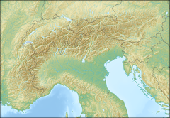
Discovery site marked on a map of the Alps
 
Ötzi was found on 19 September 1991 by two German tourists, at an elevation of 3,210 metres (10,530 ft) on the east ridge of the Fineilspitze in the Ötztal Alps on the Austrian–Italian border. The tourists, Helmut and Erika Simon, were walking off the path between the mountain passes Hauslabjoch and Tisenjoch. They believed that the body was of a recently deceased mountaineer. The next day, a mountain gendarme and the keeper of the nearby Similaunhütte first attempted to remove the body, which was frozen in ice below the torso, using a pneumatic drill and ice-axes, but had to give up due to bad weather. The next day, eight groups visited the site, among whom were mountaineers Hans Kammerlander and Reinhold Messner. The body was semi-officially extracted on 22 September and officially salvaged the following day. It was transported to the office of the medical examiner in Innsbruck, together with other objects found. On 24 September, the find was examined there by archaeologist Konrad Spindler of the University of Innsbruck. He dated the find to be "about four thousand years old", based on the typology of an axe among the retrieved objects.

Border dispute

At the Treaty of Saint-Germain-en-Laye of 1919, the border between North and South Tyrol was defined as the watershed of the rivers Inn and Etsch. Near Tisenjoch the glacier (which has since retreated) complicated establishing the watershed and the border was drawn too far north. Although Ötzi's find site drains to the Austrian side, surveys in October 1991 showed that the body had been located 92.56 m (101.22 yd) inside Italian territory as delineated in 1919 (Coordinates: 46°46′45.8″N 10°50′25.1″E.) The province of South Tyrol claimed property rights but agreed to let Innsbruck University finish its scientific examinations. Since 1998, it has been on display at the South Tyrol Museum of Archaeology in Bolzano, the capital of South Tyrol.

Scientific analyses

The corpse has been extensively examined, measured, X-rayed, and dated. Tissues and intestinal contents have been examined microscopically, as have the items found with the body. In August 2004, frozen bodies of three Austro-Hungarian soldiers killed during the Battle of San Matteo (1918) were found on the mountain Punta San Matteo in Trentino. One body was sent to a museum in the hope that research on how the environment affected its preservation would help unravel Ötzi's past.

Body

Ötzi the Iceman half uncovered, face down in a pool of water with iced banks
Ötzi the Iceman while still frozen in the glacier, photographed by Helmut Simon upon the discovery of the body in September 1991
 
By current estimates (2016), at the time of his death, Ötzi was 160 centimetres (5 ft 3 in) tall, weighed about 50 kilograms (110 lb), and was about 45 years of age. When his body was found, it weighed 13.750 kilograms (30 lb 5.0 oz). Because the body was covered in ice shortly after his death, it had only partially deteriorated. Initial reports claimed that his penis and most of his scrotum were missing, but this was later shown to be unfounded. Analysis of pollen, dust grains and the isotopic composition of his tooth enamel indicates that he spent his childhood near the present village of Feldthurns, north of Bolzano, but later went to live in valleys about 50 kilometres farther north.

In 2009, a CAT scan revealed that the stomach had shifted upward to where his lower lung area would normally be. Analysis of the contents revealed the partly digested remains of ibex meat, confirmed by DNA analysis, suggesting he had a meal less than two hours before his death. Wheat grains were also found. It is believed that Ötzi most likely had a few slices of a dried, fatty meat, probably bacon, which came from a wild goat in South Tyrol, Italy. Analysis of Ötzi's intestinal contents showed two meals (the last one consumed about eight hours before his death), one of chamois meat, the other of red deer and herb bread; both were eaten with roots and fruits. The grain also eaten with both meals was a highly processed einkorn wheat bran, quite possibly eaten in the form of bread. In the proximity of the body, and thus possibly originating from the Iceman's provisions, chaff and grains of einkorn and barley, and seeds of flax and poppy were discovered, as well as kernels of sloes (small plum-like fruits of the blackthorn tree) and various seeds of berries growing in the wild.

Hair analysis was used to examine his diet from several months before. Pollen in the first meal showed that it had been consumed in a mid-altitude conifer forest, and other pollens indicated the presence of wheat and legumes, which may have been domesticated crops. Pollen grains of hop-hornbeam were also discovered. The pollen was very well preserved, with the cells inside remaining intact, indicating that it had been fresh (estimated about two hours old) at the time of Ötzi's death, which places the event in the spring or early summer. Einkorn wheat is harvested in the late summer, and sloes in the autumn; these must have been stored from the previous year.

High levels of both copper particles and arsenic were found in Ötzi's hair. This, along with Ötzi's copper axe blade, which is 99.7% pure copper, has led scientists to speculate that Ötzi was involved in copper smelting.

By examining the proportions of Ötzi's tibia, femur and pelvis, Christopher Ruff has determined that Ötzi's lifestyle included long walks over hilly terrain. This degree of mobility is not characteristic of other Copper Age Europeans. Ruff proposes that this may indicate that Ötzi was a high-altitude shepherd.

Using modern 3D scanning technology, a facial reconstruction has been created for the South Tyrol Museum of Archaeology in Bolzano, Italy. It shows Ötzi looking old for his 45 years, with deep-set brown eyes, a beard, a furrowed face, and sunken cheeks. He is depicted looking tired and ungroomed.

Health

Ötzi apparently had whipworm (Trichuris trichiura), an intestinal parasite. During CT scans, it was observed that three or four of his right ribs had been cracked when he had been lying face down after death, or where the ice had crushed his body. One of his fingernails (of the two found) shows three Beau's lines indicating he was sick three times in the six months before he died. The last incident, two months before he died, lasted about two weeks. It was also found that his epidermis, the outer skin layer, was missing, a natural process from his mummification in ice. Ötzi's teeth showed considerable internal deterioration from cavities. These oral pathologies may have been brought about by his grain-heavy, high carbohydrate diet. DNA analysis in February 2012 revealed that Ötzi was lactose intolerant, supporting the theory that lactose intolerance was still common at that time, despite the increasing spread of agriculture and dairying.

Skeletal details and tattooing

Ötzi had a total of 61 tattoos, consisting of 19 groups of black lines ranging from 1 to 3 mm in thickness and 7 to 40 mm long. These include groups of parallel lines running along the longitudinal axis of his body and to both sides of the lumbar spine, as well as a cruciform mark behind the right knee and on the right ankle, and parallel lines around the left wrist. The greatest concentration of markings is found on his legs, which together exhibit 12 groups of lines. A microscopic examination of samples collected from these tattoos revealed that they were created from pigment manufactured out of fireplace ash or soot.

Radiological examination of Ötzi's bones showed "age-conditioned or strain-induced degeneration" corresponding to many tattooed areas, including osteochondrosis and slight spondylosis in the lumbar spine and wear-and-tear degeneration in the knee and especially in the ankle joints. It has been speculated that these tattoos may have been related to pain relief treatments similar to acupressure or acupuncture. If so, this is at least 2,000 years before their previously known earliest use in China (c. 1000 BCE). Recent research into archaeological evidence for ancient tattooing has confirmed that Ötzi is the oldest tattooed human mummy yet discovered.

Clothes and shoes

Archeoparc (Schnals valley / South Tyrol). Museum: Reconstruction of the neolithic clothes worn by Ötzi
 
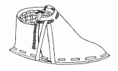
Ötzi wore a cloak made of woven grass and a coat, a belt, a pair of leggings, a loincloth and shoes, all made of leather of different skins. He also wore a bearskin cap with a leather chin strap. The shoes were waterproof and wide, seemingly designed for walking across the snow; they were constructed using bearskin for the soles, deer hide for the top panels, and a netting made of tree bark. Soft grass went around the foot and in the shoe and functioned like modern socks. The coat, belt, leggings and loincloth were constructed of vertical strips of leather sewn together with sinew. His belt had a pouch sewn to it that contained a cache of useful items: a scraper, drill, flint flake, bone awl and a dried fungus.

Line drawing of a right shoe
An artist's impression of Ötzi's right shoe

The shoes have since been reproduced by a Czech academic, who said that "because the shoes are actually quite complex, I'm convinced that even 5,300 years ago, people had the equivalent of a cobbler who made shoes for other people". The reproductions were found to constitute such excellent footwear that it was reported that a Czech company offered to purchase the rights to sell them. However, a more recent hypothesis by British archaeologist Jacqui Wood says that Ötzi's shoes were actually the upper part of snowshoes. According to this theory, the item currently interpreted as part of a backpack is actually the wood frame and netting of one snowshoe and animal hide to cover the face.

The leather loincloth and hide coat were made from sheepskin. Genetic analysis showed that the sheep species was nearer to modern domestic European sheep than to wild sheep; the items were made from the skins of at least four animals. Part of the coat was made from domesticated goat belonging to a mitochondrial haplogroup (a common female ancestor) that inhabits central Europe today. The coat was made from several animals from two different species and was stitched together with hides available at the time. The leggings were made from domesticated goat leather. A similar set of 6,500-year-old leggings discovered in Switzerland were made from goat leather which may indicate the goat leather was specifically chosen.

Shoelaces were made from the European genetic population of cattle. The quiver was made from wild roe deer, the fur hat was made from a genetic lineage of brown bear which lives in the region today. Writing in the journal Scientific Reports, researchers from Ireland and Italy reported their analysis of his clothing's mitochondrial DNA, which was extracted from nine fragments from six of his garments, including his loin cloth and fur cap.

Tools and equipment

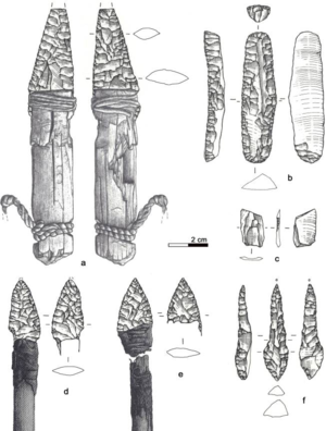
Ötzi lithic assemblage
a) Dagger, b) Endscraper, c) Borer, d) Arrowhead 14, e) Arrowhead 12, f) Small flake 
 
Other items found with the Iceman were a copper axe with a yew handle, a chert-bladed knife with an ash handle and a quiver of 14 arrows with viburnum and dogwood shafts. Two of the arrows, which were broken, were tipped with flint and had fletching (stabilizing fins), while the other 12 were unfinished and untipped. The arrows were found in a quiver with what is presumed to be a bow string, an unidentified tool, and an antler tool which might have been used for sharpening arrow points. There was also an unfinished yew longbow that was 1.82 metres (72 in) long.

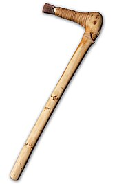
A replica of Ötzi's copper axe

In addition, among Ötzi's possessions were berries, two birch bark baskets, and two species of polypore mushrooms with leather strings through them. One of these, the birch fungus, is known to have anthelmintic properties, and was probably used for medicinal purposes. The other was a type of tinder fungus, included with part of what appeared to be a complex firelighting kit. The kit featured pieces of over a dozen different plants, in addition to flint and pyrite for creating sparks.

Ötzi's copper axe was of particular interest. His axe's haft is 60 centimetres (24 in) long and made from carefully worked yew with a right-angled crook at the shoulder, leading to the blade. The 9.5 centimetres (3.7 in) long axe head is made of almost pure copper, produced by a combination of casting, cold forging, polishing, and sharpening. Despite the fact that copper ore sources in the Alpines are known to have been exploited at the time, a study indicated that the copper in the axe came from southern Tuscany. It was let into the forked end of the crook and fixed there using birch-tar and tight leather lashing. The blade part of the head extends out of the lashing and shows clear signs of having been used to chop and cut. At the time, such an axe would have been a valuable possession, important both as a tool and as a status symbol for the bearer.

Genetic analysis

Ötzi's full genome has been sequenced; the report on this was published on 28 February 2012. The Y chromosome DNA of Ötzi belongs to a subclade of G defined by the SNPs M201, P287, P15, L223 and L91 (G-L91, ISOGG G2a2b, former "G2a4"). He was not typed for any of the subclades downstreaming from G-L91; however, an analysis of his BAM file revealed that he belongs to the L166 and FGC5672 subclades below L91. G-L91 is now mostly found in South Corsica.

Analysis of his mitochondrial DNA showed that Ötzi belongs to the K1 subclade, but cannot be categorized into any of the three modern branches of that subclade (K1a, K1b or K1c). The new subclade has provisionally been named K1ö for Ötzi. A multiplex assay study was able to confirm that the Iceman's mtDNA belongs to a previously unknown European mtDNA clade with a very limited distribution among modern data sets.

By autosomal DNA, Ötzi is most closely related to southern Europeans, especially to geographically isolated populations like Corsicans and Sardinians.

DNA analysis also showed him at high risk of atherosclerosis and lactose intolerance, with the presence of the DNA sequence of Borrelia burgdorferi, possibly making him the earliest known human with Lyme disease. A later analysis suggested the sequence may have been a different Borrelia species.

A 2012 paper by paleoanthropologist John Hawks suggests that Ötzi had a higher degree of Neanderthal ancestry than modern Europeans.

In October 2013, it was reported that 19 modern Tyrolean men were descendants of Ötzi or of a close relative of Ötzi. Scientists from the Institute of Legal Medicine at Innsbruck Medical University had analysed the DNA of over 3,700 Tyrolean male blood donors and found 19 who shared a particular genetic mutation with the 5,300-year-old man.

Blood

In May 2012, scientists announced the discovery that Ötzi still had intact blood cells. These are the oldest complete human blood cells ever identified. In most bodies this old, the blood cells are either shrunken or mere remnants, but Ötzi's have the same dimensions as living red blood cells and resembled a modern-day sample.

H. pylori analysis

In 2016, researchers reported on a study from the extraction of twelve samples from the gastrointestinal tract of Ötzi to analyze the origins of the Helicobacter pylori in his gut. The H. pylori strain found in his gastrointestinal tract was, surprisingly, the hpAsia2 strain, a strain today found primarily in South Asian and Central Asian populations, with extremely rare occurrences in modern European populations. The strain found in Ötzi's gut is most similar to three modern individuals from Northern India; the strain itself is, of course, older than the modern Northern Indian strain.

Cause of death

The Ötzi memorial near Tisenjoch. Ötzi was found ca. 70 m NE of here, a place indicated with a red mark (not in this photo). The mountain in the background is the Fineilspitze.
 
Naturalistic reconstruction of Ötzi – South Tyrol Museum of Archaeology

The cause of death remained uncertain until 10 years after the discovery of the body. It was initially believed that Ötzi died from exposure during a winter storm. Later it was speculated that Ötzi might have been a victim of a ritual sacrifice, perhaps for being a chieftain. This explanation was inspired by theories previously advanced for the first millennium BCE bodies recovered from peat bogs such as the Tollund Man and the Lindow Man.

Arrowhead and blood analyses

In 2001, X-rays and a CT scan revealed that Ötzi had an arrowhead lodged in his left shoulder when he died and a matching small tear on his coat. The discovery of the arrowhead prompted researchers to theorize Ötzi died of blood loss from the wound, which would probably have been fatal even if modern medical techniques had been available. Further research found that the arrow's shaft had been removed before death, and close examination of the body found bruises and cuts to the hands, wrists and chest and cerebral trauma indicative of a blow to the head. One of the cuts was to the base of his thumb that reached down to the bone but had no time to heal before his death. Currently, it is believed that Ötzi bled to death after the arrow shattered the scapula and damaged nerves and blood vessels before lodging near the lung.

Recent DNA analyses claim they revealed traces of blood from at least four other people on his gear: one from his knife, two from a single arrowhead, and a fourth from his coat. Interpretations of these findings were that Ötzi killed two people with the same arrow and was able to retrieve it on both occasions, and the blood on his coat was from a wounded comrade he may have carried over his back. Ötzi's posture in death (frozen body, face down, left arm bent across the chest) could support a theory that before death occurred and rigor mortis set in, the Iceman was turned onto his stomach in the effort to remove the arrow shaft.

Alternate theory of death

In 2010, it was proposed that Ötzi died at a much lower altitude and was buried higher in the mountains, as posited by archaeologist Alessandro Vanzetti of the Sapienza University of Rome and his colleagues. According to their study of the items found near Ötzi and their locations, it is possible that the iceman may have been placed above what has been interpreted as a stone burial mound but was subsequently moved with each thaw cycle that created a flowing watery mix driven by gravity before being re-frozen. While archaeobotanist Klaus Oeggl of the University of Innsbruck agrees that the natural process described probably caused the body to move from the ridge that includes the stone formation, he pointed out that the paper provided no compelling evidence to demonstrate that the scattered stones constituted a burial platform. Moreover, biological anthropologist Albert Zink argues that the iceman's bones display no dislocations that would have resulted from a downhill slide and that the intact blood clots in his arrow wound would show damage if the body had been moved up the mountain. In either case, the burial theory does not contradict the possibility of a violent cause of death.

Legal dispute

Italian law entitled the Simons to a finders' fee from the South Tyrolean provincial government of 25% of the value of Ötzi. In 1994 the authorities offered a "symbolic" reward of 10 million lire (€5,200), which the Simons declined. In 2003, the Simons filed a lawsuit which asked a court in Bolzano to recognize their role in Ötzi's discovery and declare them his "official discoverers". The court decided in the Simons' favor in November 2003, and at the end of December that year the Simons announced that they were seeking US$300,000 as their fee. The provincial government decided to appeal.

In addition, two people came forward to claim that they were part of the same mountaineering party that came across Ötzi and discovered the body first:
  • Magdalena Mohar Jarc, a retired Slovenian climber, who alleged that she discovered the corpse first after falling into a crevice, and shortly after returning to a mountain hut, asked Helmut Simon to take photographs of Ötzi. She cited Reinhold Messner, who was also present in the mountain hut, as the witness to this.
  • Sandra Nemeth, from Switzerland, who contended that she found the corpse before Helmut and Erika Simon, and that she spat on Ötzi to make sure that her DNA would be found on the body later. She asked for a DNA test on the remains, but experts believed that there was little chance of finding any trace.
In 2005 the rival claims were heard by a Bolzano court. The legal case angered Mrs. Simon, who alleged that neither woman was present on the mountain that day. In 2005, Mrs. Simon's lawyer said: "Mrs. Simon is very upset by all this and by the fact that these two new claimants have decided to appear 14 years after Ötzi was found." In 2008, however, Jarc stated for a Slovene newspaper that she wrote twice to the Bolzano court in regard to her claim but received no reply whatsoever.

In 2004, Helmut Simon died. Two years later, in June 2006, an appeals court affirmed that the Simons had indeed discovered the Iceman and were therefore entitled to a finder's fee. It also ruled that the provincial government had to pay the Simons' legal costs. After this ruling, Mrs. Erika Simon reduced her claim to €150,000. The provincial government's response was that the expenses it had incurred to establish a museum and the costs of preserving the Iceman should be considered in determining the finder's fee. It insisted it would pay no more than €50,000. In September 2006, the authorities appealed the case to Italy's highest court, the Court of Cassation.

On 29 September 2008 it was announced that the provincial government and Mrs. Simon had reached a settlement of the dispute, under which she would receive €150,000 in recognition of Ötzi's discovery by her and her late husband and the tourist income that it attracts.

"Ötzi's curse"

Influenced by the "Curse of the pharaohs" and the media theme of cursed mummies, claims have been made that Ötzi is cursed. The allegation revolves around the deaths of several people connected to the discovery, recovery and subsequent examination of Ötzi. It is alleged that they have died under mysterious circumstances. These people include co-discoverer Helmut Simon and Konrad Spindler, the first examiner of the mummy in Austria in 1991. To date, the deaths of seven people, of which four were accidental, have been attributed to the alleged curse. In reality hundreds of people were involved in the recovery of Ötzi and are still involved in studying the body and the artifacts found with it. The fact that a small percentage of them have died over the years has not been shown to be statistically significant.

Decomposition

From Wikipedia, the free encyclopedia

A rotten apple after it fell from the tree
 
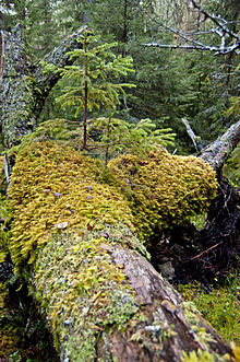
Decomposing fallen nurse log in a forest
 
Decomposition is the process by which dead organic substances are broken down into simpler organic or inorganic matter such as carbon dioxide , water, simple sugars and mineral salts. The process is a part of the nutrient cycle and is essential for recycling the finite matter that occupies physical space in the biosphere. Bodies of living organisms begin to decompose shortly after death. Animals, such as worms, also help decompose the organic materials. Organisms that do this are known as decomposers. Although no two organisms decompose in the same way, they all undergo the same sequential stages of decomposition. The science which studies decomposition is generally referred to as taphonomy from the Greek word taphos, meaning tomb. Decomposition can also be a gradual process for organisms that have extended periods of dormancy.

One can differentiate abiotic from biotic substance (biodegradation). The former means "degradation of a substance by chemical or physical processes, e.g., hydrolysis. The latter means "the metabolic breakdown of materials into simpler components by living organisms", typically by microorganisms.

Animal decomposition


Ants eating a dead snake

Decomposition begins at the moment of death, caused by two factors: 1.) autolysis, the breaking down of tissues by the body's own internal chemicals and enzymes, and 2.) putrefaction, the breakdown of tissues by bacteria. These processes release compounds such as cadaverine and putrescine, that are the chief source of the unmistakably putrid odor of decaying animal tissue. 

Prime decomposers are bacteria or fungi, though larger scavengers also play an important role in decomposition if the body is accessible to insects, mites and other animals. The most important arthropods that are involved in the process include carrion beetles, mites, the flesh-flies (Sarcophagidae) and blow-flies (Calliphoridae), such as the green-bottle fly seen in the summer. In North America, the most important non-insect animals that are typically involved in the process include mammal and bird scavengers, such as coyotes, dogs, wolves, foxes, rats, crows and vultures. Some of these scavengers also remove and scatter bones, which they ingest at a later time. Aquatic and marine environments have break-down agents that include bacteria, fish, crustaceans, fly larvae  and other carrion scavengers.

Stages of decomposition

Five general stages are used to describe the process of decomposition in vertebrate animals: fresh, bloat, active decay, advanced decay, and dry/remains. The general stages of decomposition are coupled with two stages of chemical decomposition: autolysis and putrefaction. These two stages contribute to the chemical process of decomposition, which breaks down the main components of the body. With death the microbiome of the living organism collapses and is followed by the necrobiome that undergoes predictable changes over time.

Fresh

Among those animals that have the heart, the "fresh" stage begins immediately after the heart stops beating. From the moment of death, the body begins cooling or warming to match the temperature of the ambient environment, during a stage called algor mortis. Shortly after death, within three to six hours, the muscular tissues become rigid and incapable of relaxing, during a stage called rigor mortis. Since blood is no longer being pumped through the body, gravity causes it to drain to the dependent portions of the body, creating an overall bluish-purple discolouration termed livor mortis or, more commonly, lividity.


Once the heart stops, the blood can no longer supply oxygen or remove carbon dioxide from the tissues. The resulting decrease in pH and other chemical changes causes cells to lose their structural integrity, bringing about the release of cellular enzymes capable of initiating the breakdown of surrounding cells and tissues. This process is known as autolysis.


Visible changes caused by decomposition are limited during the fresh stage, although autolysis may cause blisters to appear at the surface of the skin.

The small amount of oxygen remaining in the body is quickly depleted by cellular metabolism and aerobic microbes naturally present in respiratory and gastrointestinal tracts, creating an ideal environment for the proliferation of anaerobic organisms. These multiply, consuming the body's carbohydrates, lipids, and proteins, to produce a variety of substances including propionic acid, lactic acid, methane, hydrogen sulfide, and ammonia. The process of microbial proliferation within a body is referred to as putrefaction and leads to the second stage of decomposition, known as bloat.

Blowflies and flesh flies are the first carrion insects to arrive, and they seek a suitable oviposition site.

Bloat

The bloat stage provides the first clear visual sign that microbial proliferation is underway. In this stage, anaerobic metabolism takes place, leading to the accumulation of gases, such as hydrogen sulfide, carbon dioxide, methane, and nitrogen. The accumulation of gases within the bodily cavity causes the distention of the abdomen and gives a cadaver its overall bloated appearance. The gases produced also cause natural liquids and liquefying tissues to become frothy. As the pressure of the gases within the body increases, fluids are forced to escape from natural orifices, such as the nose, mouth, and anus, and enter the surrounding environment. The buildup of pressure combined with the loss of integrity of the skin may also cause the body to rupture.

Intestinal anaerobic bacteria transform haemoglobin into sulfhemoglobin and other colored pigments. The associated gases which accumulate within the body at this time aid in the transport of sulfhemoglobin throughout the body via the circulatory and lymphatic systems, giving the body an overall marbled appearance.

If insects have access, maggots hatch and begin to feed on the body's tissues. Maggot activity, typically confined to natural orifices, and masses under the skin, causes the skin to slip, and hair to detach from the skin. Maggot feeding, and the accumulation of gases within the body, eventually leads to post-mortem skin ruptures which will then further allow purging of gases and fluids into the surrounding environment. Ruptures in the skin allow oxygen to re-enter the body and provide more surface area for the development of fly larvae and the activity of aerobic microorganisms. The purging of gases and fluids results in the strong distinctive odors associated with decay.

Active decay

Active decay is characterized by the period of greatest mass loss. This loss occurs as a result of both the voracious feeding of maggots and the purging of decomposition fluids into the surrounding environment. The purged fluids accumulate around the body and create a cadaver decomposition island (CDI). Liquefaction of tissues and disintegration become apparent during this time and strong odors persist. The end of active decay is signaled by the migration of maggots away from the body to pupate.

Advanced decay

Decomposition is largely inhibited during advanced decay due to the loss of readily available cadaveric material. Insect activity is also reduced during this stage. When the carcass is located on soil, the area surrounding it will show evidence of vegetation death. The CDI surrounding the carcass will display an increase in soil carbon and nutrients, such as phosphorus, potassium, calcium, and magnesium; changes in pH; and a significant increase in soil nitrogen.

Dry/remains

During the dry/remains stage, the resurgence of plant growth around the CDI may occur and is a sign that the nutrients present in the surrounding soil have not yet returned to their normal levels. All that remains of the cadaver at this stage is dry skin, cartilage, and bones, which will become dry and bleached if exposed to the elements. If all soft tissue is removed from the cadaver, it is referred to as completely skeletonized, but if only portions of the bones are exposed, it is referred to as partially skeletonised.

Pig carcass in the different stages of decomposition:
Fresh > Bloat > Active decay > Advanced decay > Dry remains

Factors affecting decomposition of bodies

Exposure to the elements

A dead body that has been exposed to the open elements, such as water and air, will decompose more quickly and attract much more insect activity than a body that is buried or confined in special protective gear or artifacts. This is due, in part, to the limited number of insects that can penetrate a coffin and the lower temperatures under soil.

The rate and manner of decomposition in an animal body is strongly affected by several factors. In roughly descending degrees of importance, they are:
The speed at which decomposition occurs varies greatly. Factors such as temperature, humidity, and the season of death all determine how fast a fresh body will skeletonize or mummify. A basic guide for the effect of environment on decomposition is given as Casper's Law (or Ratio): if all other factors are equal, then, when there is free access of air a body decomposes twice as fast than if immersed in water and eight times faster than if buried in earth. Ultimately, the rate of bacterial decomposition acting on the tissue will depend upon the temperature of the surroundings. Colder temperatures decrease the rate of decomposition while warmer temperatures increase it. A dry body will not decompose efficiently. Moisture helps the growth of microorganisms that decompose the organic matter, but too much moisture could lead to anaerobic conditions slowing down the decomposition process.

The most important variable is a body's accessibility to insects, particularly flies. On the surface in tropical areas, invertebrates alone can easily reduce a fully fleshed corpse to clean bones in under two weeks. The skeleton itself is not permanent; acids in soils can reduce it to unrecognizable components. This is one reason given for the lack of human remains found in the wreckage of the Titanic, even in parts of the ship considered inaccessible to scavengers. Freshly skeletonized bone is often called "green" bone and has a characteristic greasy feel. Under certain conditions (normally cool, damp soil), bodies may undergo saponification and develop a waxy substance called adipocere, caused by the action of soil chemicals on the body's proteins and fats. The formation of adipocere slows decomposition by inhibiting the bacteria that cause putrefaction.

In extremely dry or cold conditions, the normal process of decomposition is halted – by either lack of moisture or temperature controls on bacterial and enzymatic action – causing the body to be preserved as a mummy. Frozen mummies commonly restart the decomposition process when thawed (see Ötzi the Iceman), whilst heat-desiccated mummies remain so unless exposed to moisture.

The bodies of newborns who never ingested food are an important exception to the normal process of decomposition. They lack the internal microbial flora that produce much of decomposition and quite commonly mummify if kept in even moderately dry conditions.

Anaerobic vs aerobic

Aerobic decomposition takes place in the presence of oxygen. This is most common to occur in nature. Living organisms that use oxygen to survive feed on the body. Anaerobic decomposition takes place in the absence of oxygen. This could be place where the body is buried in organic material and oxygen can not reach it. This process of putrefaction has a bad odor accompanied by it due to the hydrogen sulfide and organic matter containing sulfur.

Artificial preservation

Embalming is the practice of delaying decomposition of human and animal remains. Embalming slows decomposition somewhat, but does not forestall it indefinitely. Embalmers typically pay great attention to parts of the body seen by mourners, such as the face and hands. The chemicals used in embalming repel most insects, and slow down bacterial putrefaction by either killing existing bacteria in or on the body themselves or by "fixing" cellular proteins, which means that they cannot act as a nutrient source for subsequent bacterial infections. In sufficiently dry environments, an embalmed body may end up mummified and it is not uncommon for bodies to remain preserved to a viewable extent after decades. Notable viewable embalmed bodies include those of:

Environmental preservation

A body buried in a sufficiently dry environment may be well preserved for decades. This was observed in the case for murdered civil rights activist Medgar Evers, who was found to be almost perfectly preserved over 30 years after his death, permitting an accurate autopsy when the case of his murder was re-opened in the 1990s.

Bodies submerged in a peat bog may become naturally "embalmed", arresting decomposition and resulting in a preserved specimen known as a bog body. The generally cool and anoxic conditions in these environments limits the rate of microbial activity, thus limiting the potential for decomposition. The time for an embalmed body to be reduced to a skeleton varies greatly. Even when a body is decomposed, embalming treatment can still be achieved (the arterial system decays more slowly) but would not restore a natural appearance without extensive reconstruction and cosmetic work, and is largely used to control the foul odors due to decomposition.

An animal can be preserved almost perfectly, for millions of years in a resin such as amber. 

There are some examples where bodies have been inexplicably preserved (with no human intervention) for decades or centuries and appear almost the same as when they died. In some religious groups, this is known as incorruptibility. It is not known whether or for how long a body can stay free of decay without artificial preservation.

Importance to forensic sciences

Various sciences study the decomposition of bodies under the general rubric of forensic science because the usual motive for such studies is to determine the time and cause of death for legal purposes:
  • Forensic taphonomy specifically studies the processes of decomposition in order to apply the biological and chemical principles to forensic cases in order to determine post-mortem interval (PMI), post-burial interval as well as to locate clandestine graves.
  • Forensic pathology studies the clues to the cause of death found in the corpse as a medical phenomenon.
  • Forensic entomology studies the insects and other vermin found in corpses; the sequence in which they appear, the kinds of insects, and where they are found in their life cycle are clues that can shed light on the time of death, the length of a corpse's exposure, and whether the corpse was moved.
  • Forensic anthropology is the medico-legal branch of physical anthropology that studies skeletons and human remains, usually to seek clues as to the identity, age, sex, height and ethnicity of their former owner.
The University of Tennessee Anthropological Research Facility (better known as the Body Farm) in Knoxville, Tennessee has a number of bodies laid out in various situations in a fenced-in plot near the medical center. Scientists at the Body Farm study how the human body decays in various circumstances to gain a better understanding of decomposition.

Plant decomposition

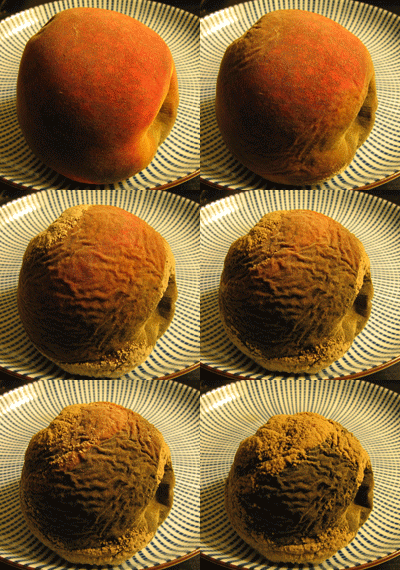
A decaying peach over a period of six days. Each frame is approximately 12 hours apart, as the fruit shrivels and becomes covered with mold.

Decomposition of plant matter occurs in many stages. It begins with leaching by water; the most easily lost and soluble carbon compounds are liberated in this process. Another early process is physical breakup or fragmentation of the plant material into smaller bits which have greater surface area for microbial colonization and attack. In smaller dead plants, this process is largely carried out by the soil invertebrate fauna, whereas in the larger plants, primarily parasitic life-forms such as insects and fungi play a major breakdown role and are not assisted by numerous detritivore species.

Following this, the plant detritus (consisting of cellulose, hemicellulose, microbial products, and lignin) undergoes chemical alteration by microbes. Different types of compounds decompose at different rates. This is dependent on their chemical structure.

For instance, lignin is a component of wood, which is relatively resistant to decomposition and can in fact only be decomposed by certain fungi, such as the black-rot fungi. Wood decomposition is a complex process involving fungi which transport nutrients to the nutritionally scarce wood from outside environment. Because of this nutritional enrichment the fauna of saproxylic insects may develop and in turn affect dead wood, contributing to wood decomposition and nutrient cycling in the forest floor. Lignin is one such remaining product of decomposing plants with a very complex chemical structure causing the rate of microbial breakdown to slow. Warmth increases the speed of plant decay, by the same amount regardless of the composition of the plant.

In most grassland ecosystems, natural damage from fire, insects that feed on decaying matter, termites, grazing mammals, and the physical movement of animals through the grass are the primary agents of breakdown and nutrient cycling, while bacteria and fungi play the main roles in further decomposition.

The chemical aspects of plant decomposition always involve the release of carbon dioxide. In fact, decomposition contributes over 90 percent of carbon dioxide released each year.

Food decomposition

The decomposition of food, either plant or animal, called spoilage in this context, is an important field of study within food science. Food decomposition can be slowed down by conservation. The spoilage of meat occurs, if the meat is untreated, in a matter of hours or days and results in the meat becoming unappetizing, poisonous or infectious. Spoilage is caused by the practically unavoidable infection and subsequent decomposition of meat by bacteria and fungi, which are borne by the animal itself, by the people handling the meat, and by their implements. Meat can be kept edible for a much longer time – though not indefinitely – if proper hygiene is observed during production and processing, and if appropriate food safety, food preservation and food storage procedures are applied.
Spoilage of food is attributed to contamination from microorganisms such as bacteria, molds, and yeasts, along with natural decay of the food. These decomposition bacteria reproduce at rapid rates under conditions of moisture and preferred temperatures. When the proper conditions are lacking the bacteria may form spores which lurk until suitable conditions arise to continue reproduction.

Rate of decomposition

The rate of decomposition is governed by three sets of factors—the physical environment (temperature, moisture and soil properties), the quantity and quality of the dead material available to decomposers, and the nature of the microbial community itself.

Decomposition rates are low under very wet or very dry conditions. Decomposition rates are highest in damp, moist conditions with adequate levels of oxygen. Wet soils tend to become deficient in oxygen (this is especially true in wetlands), which slows microbial growth. In dry soils, decomposition slows as well, but bacteria continue to grow (albeit at a slower rate) even after soils become too dry to support plant growth. When the rains return and soils become wet, the osmotic gradient between the bacterial cells and the soil water causes the cells to gain water quickly. Under these conditions, many bacterial cells burst, releasing a pulse of nutrients. Decomposition rates also tend to be slower in acidic soils. Soils which are rich in clay minerals tend to have lower decomposition rates, and thus, higher levels of organic matter. The smaller particles of clay result in a larger surface area that can hold water. The higher the water content of a soil, the lower the oxygen content and consequently, the lower the rate of decomposition. Clay minerals also bind particles of organic material to their surface, making them less accessible to microbes. Soil disturbance like tilling increases decomposition by increasing the amount of oxygen in the soil and by exposing new organic matter to soil microbes.

The quality and quantity of the material available to decomposers is another major factor that influences the rate of decomposition. Substances like sugars and amino acids decompose readily and are considered labile. Cellulose and hemicellulose, which are broken down more slowly, are "moderately labile". Compounds which are more resistant to decay, like lignin or cutin, are considered recalcitrant. Litter with a higher proportion of labile compounds decomposes much more rapidly than does litter with a higher proportion of recalcitrant material. Consequently, dead animals decompose more rapidly than dead leaves, which themselves decompose more rapidly than fallen branches. As organic material in the soil ages, its quality decreases. The more labile compounds decompose quickly, leaving an increasing proportion of recalcitrant material. Microbial cell walls also contain recalcitrant materials like chitin, and these also accumulate as the microbes die, further reducing the quality of older soil organic matter.

Censorship in the United States

From Wikipedia, the free encyclopedia https://en.wikipedia.org/wiki/Censorshi...