Search This Blog

Wednesday, December 11, 2024

Genome instability

From Wikipedia, the free encyclopedia
https://en.wikipedia.org/wiki/Genome_instability

Genome instability (also genetic instability or genomic instability) refers to a high frequency of mutations within the genome of a cellular lineage. These mutations can include changes in nucleic acid sequences, chromosomal rearrangements or aneuploidy. Genome instability does occur in bacteria. In multicellular organisms genome instability is central to carcinogenesis, and in humans it is also a factor in some neurodegenerative diseases such as amyotrophic lateral sclerosis or the neuromuscular disease myotonic dystrophy.

The sources of genome instability have only recently begun to be elucidated. A high frequency of externally caused DNA damage can be one source of genome instability since DNA damage can cause inaccurate translesion DNA synthesis past the damage or errors in repair, leading to mutation. Another source of genome instability may be epigenetic or mutational reductions in expression of DNA repair genes. Because endogenous (metabolically-caused) DNA damage is very frequent, occurring on average more than 60,000 times a day in the genomes of human cells, any reduced DNA repair is likely an important source of genome instability.

Usual genome situation

Usually, all cells in an individual in a given species (plant or animal) show a constant number of chromosomes, which constitute what is known as the karyotype defining this species (see also List of number of chromosomes of various organisms), although some species present a very high karyotypic variability. In humans, mutations that would change an amino acid within the protein coding region of the genome occur at an average of only 0.35 per generation (less than one mutated protein per generation).

Sometimes, in a species with a stable karyotype, random variations that modify the normal number of chromosomes may be observed. In other cases, there are structural alterations (e.g., chromosomal translocations, deletions) that modify the standard chromosomal complement. In these cases, it is indicated that the affected organism presents genome instability (also genetic instability, or even chromosomic instability). The process of genome instability often leads to a situation of aneuploidy, in which the cells present a chromosomic number that is either higher or lower than the normal complement for the species.

Causes of genome instability

DNA Replication Defects

In the cell cycle, DNA is usually most vulnerable during replication. The replisome must be able to navigate obstacles such as tightly wound chromatin with bound proteins, single and double stranded breaks which can lead to the stalling of the replication fork. Each protein or enzyme in the replisome must perform its function well to result in a perfect copy of DNA. Mutations of proteins such as DNA polymerase or DNA ligase can lead to impairment of replication and lead to spontaneous chromosomal exchanges. Proteins such as Tel1 and Mec1 (ATR, ATM in humans) can detect single and double-stranded breaks and recruit factors such as Rmr3 helicase to stabilize the replication fork in order to prevent its collapse. Mutations in Tel1, Mec1, and Rmr3 helicase result in a significant increase of chromosomal recombination. ATR responds specifically to stalled replication forks and single-stranded breaks resulting from UV damage while ATM responds directly to double-stranded breaks. These proteins also prevent progression into mitosis by inhibiting the firing of late replication origins until the DNA breaks are fixed by phosphorylating CHK1 and CHK2, which results in a signaling cascade arresting the cell in S-phase. For single stranded breaks, replication occurs until the location of the break, then the other strand is nicked to form a double stranded break, which can then be repaired by Break Induced Replication or homologous recombination using the sister chromatid as an error-free template. In addition to S-phase checkpoints, G1 and G2 checkpoints exist to check for transient DNA damage which could be caused by mutagens such as UV damage. An example is the Saccharomyces pombe gene rad9 which arrests the cells in late S/G2 phase in the presence of DNA damage caused by radiation. The yeast cells with defective rad9 failed to arrest following irradiation, continued cell division, and died rapidly; the cells with wild-type rad9 successfully arrested in late S/G2 phase and remained viable. The cells that arrested were able to survive due to the increased time in S/G2 phase allowing for DNA repair enzymes to function fully.

Fragile Sites

There are hotspots in the genome where DNA sequences are prone to gaps and breaks after inhibition of DNA synthesis such as in the aforementioned checkpoint arrest. These sites are called fragile sites, and can occur commonly as naturally present in most mammalian genomes or occur rarely as a result of mutations, such as DNA-repeat expansion. Rare fragile sites can lead to genetic disease such as fragile X mental retardation syndrome, myotonic dystrophy, Friedrich's ataxia, and Huntington's disease, most of which are caused by expansion of repeats at the DNA, RNA, or protein level. Although, seemingly harmful, these common fragile sites are conserved all the way to yeast and bacteria. These ubiquitous sites are characterized by trinucleotide repeats, most commonly CGG, CAG, GAA, and GCN. These trinucleotide repeats can form into hairpins, leading to difficulty of replication. Under replication stress, such as defective machinery or further DNA damage, DNA breaks and gaps can form at these fragile sites. Using a sister chromatid as repair is not a fool-proof backup as the surrounding DNA information of the n and n+1 repeat is virtually the same, leading to copy number variation. For example, the 16th copy of CGG might be mapped to the 13th copy of CGG in the sister chromatid since the surrounding DNA is both CGGCGGCGG..., leading to 3 extra copies of CGG in the final DNA sequence.

Transcription-associated instability

In both E. coli and Saccharomyces pombe, transcription sites tend to have higher recombination and mutation rates. The coding or non-transcribed strand accumulates more mutations than the template strand. This is due to the fact that the coding strand is single-stranded during transcription, which is chemically more unstable than double-stranded DNA. During elongation of transcription, supercoiling can occur behind an elongating RNA polymerase, leading to single-stranded breaks. When the coding strand is single-stranded, it can also hybridize with itself, creating DNA secondary structures that can compromise replication. In E. coli, when attempting to transcribe GAA triplets such as those found in Friedrich's ataxia, the resulting RNA and template strand can form mismatched loops between different repeats, leaving the complementary segment in the coding strand available to form its own loops which impede replication. Furthermore, replication of DNA and transcription of DNA are not temporally independent; they can occur at the same time and lead to collisions between the replication fork and RNA polymerase complex. In S. cerevisiae, Rrm3 helicase is found at highly transcribed genes in the yeast genome, which is recruited to stabilize a stalling replication fork as described above. This suggests that transcription is an obstacle to replication, which can lead to increased stress in the chromatin spanning the short distance between the unwound replication fork and transcription start site, potentially causing single-stranded DNA breaks. In yeast, proteins act as barriers at the 3' of the transcription unit to prevent further travel of the DNA replication fork.

Increase Genetic Variability

In some portions of the genome, variability is essential to survival. One such locale is the Ig genes. In a pre-B cell, the region consists of all V, D, and J segments. During development of the B cell, a specific V, D, and J segment is chosen to be spliced together to form the final gene, which is catalyzed by RAG1 and RAG2 recombinases. Activation-Induced Cytidine Deaminase (AID) then converts cytidine into uracil. Uracil normally does not exist in DNA, and thus the base is excised and the nick is converted into a double-stranded break which is repaired by non-homologous end joining (NHEJ). This procedure is very error-prone and leads to somatic hypermutation. This genomic instability is crucial in ensuring mammalian survival against infection. V, D, J recombination can ensure millions of unique B-cell receptors; however, random repair by NHEJ introduces variation which can create a receptor that can bind with higher affinity to antigens.

In neuronal and neuromuscular disease

Of about 200 neurological and neuromuscular disorders, 15 have a clear link to an inherited or acquired defect in one of the DNA repair pathways or excessive genotoxic oxidative stress. Five of them (xeroderma pigmentosum, Cockayne's syndrome, trichothiodystrophy, Down's syndrome, and triple-A syndrome) have a defect in the DNA nucleotide excision repair pathway. Six (spinocerebellar ataxia with axonal neuropathy-1, Huntington's disease, Alzheimer's disease, Parkinson's disease, Down's syndrome and amyotrophic lateral sclerosis) seem to result from increased oxidative stress, and the inability of the base excision repair pathway to handle the damage to DNA that this causes. Four of them (Huntington's disease, various spinocerebellar ataxias, Friedreich's ataxia and myotonic dystrophy types 1 and 2) often have an unusual expansion of repeat sequences in DNA, likely attributable to genome instability. Four (ataxia-telangiectasia, ataxia-telangiectasia-like disorder, Nijmegen breakage syndrome and Alzheimer's disease) are defective in genes involved in repairing DNA double-strand breaks. Overall, it seems that oxidative stress is a major cause of genomic instability in the brain. A particular neurological disease arises when a pathway that normally prevents oxidative stress is deficient, or a DNA repair pathway that normally repairs damage caused by oxidative stress is deficient.

In cancer

In cancer, genome instability can occur prior to or as a consequence of transformation. Genome instability can refer to the accumulation of extra copies of DNA or chromosomes, chromosomal translocations, chromosomal inversions, chromosome deletions, single-strand breaks in DNA, double-strand breaks in DNA, the intercalation of foreign substances into the DNA double helix, or any abnormal changes in DNA tertiary structure that can cause either the loss of DNA, or the misexpression of genes. Situations of genome instability (as well as aneuploidy) are common in cancer cells, and they are considered a "hallmark" for these cells. The unpredictable nature of these events are also a main contributor to the heterogeneity observed among tumour cells.

It is currently accepted that sporadic tumors (non-familial ones) are originated due to the accumulation of several genetic errors. An average cancer of the breast or colon can have about 60 to 70 protein altering mutations, of which about 3 or 4 may be "driver" mutations, and the remaining ones may be "passenger" mutations Any genetic or epigenetic lesion increasing the mutation rate will have as a consequence an increase in the acquisition of new mutations, increasing then the probability to develop a tumor. During the process of tumorogenesis, it is known that diploid cells acquire mutations in genes responsible for maintaining genome integrity (caretaker genes), as well as in genes that are directly controlling cellular proliferation (gatekeeper genes). Genetic instability can originate due to deficiencies in DNA repair, or due to loss or gain of chromosomes, or due to large scale chromosomal reorganizations. Losing genetic stability will favour tumor development, because it favours the generation of mutants that can be selected by the environment.

The tumor microenvironment has an inhibitory effect on DNA repair pathways contributing to genomic instability, which promotes tumor survival, proliferation, and malignant transformation.

Low frequency of mutations without cancer

The protein coding regions of the human genome, collectively called the exome, constitutes only 1.5% of the total genome. As pointed out above, ordinarily there are only an average of 0.35 mutations in the exome per generation (parent to child) in humans. In the entire genome (including non-protein coding regions) there are only about 70 new mutations per generation in humans.

Cause of mutations in cancer

The likely major underlying cause of mutations in cancer is DNA damage. For example, in the case of lung cancer, DNA damage is caused by agents in exogenous genotoxic tobacco smoke (e.g. acrolein, formaldehyde, acrylonitrile, 1,3-butadiene, acetaldehyde, ethylene oxide and isoprene). Endogenous (metabolically-caused) DNA damage is also very frequent, occurring on average more than 60,000 times a day in the genomes of human cells (see DNA damage (naturally occurring)). Externally and endogenously caused damages may be converted into mutations by inaccurate translesion synthesis or inaccurate DNA repair (e.g. by non-homologous end joining). In addition, DNA damages can also give rise to epigenetic alterations during DNA repair. Both mutations and epigenetic alterations (epimutations) can contribute to progression to cancer.

Very frequent mutations in cancer

As noted above, about 3 or 4 driver mutations and 60 passenger mutations occur in the exome (protein coding region) of a cancer. However, a much larger number of mutations occur in the non-protein-coding regions of DNA. The average number of DNA sequence mutations in the entire genome of a breast cancer tissue sample is about 20,000. In an average melanoma tissue sample (where melanomas have a higher exome mutation frequency) the total number of DNA sequence mutations is about 80,000.

Cause of high frequency of mutations in cancer

The high frequency of mutations in the total genome within cancers suggests that, often, an early carcinogenic alteration may be a deficiency in DNA repair. Mutation rates substantially increase (sometimes by 100-fold) in cells defective in DNA mismatch repair or in homologous recombinational DNA repair. Also, chromosomal rearrangements and aneuploidy increase in humans defective in DNA repair gene BLM.

A deficiency in DNA repair itself can allow DNA damages to accumulate, and error-prone translesion synthesis past some of those damages may give rise to mutations. In addition, faulty repair of these accumulated DNA damages may give rise to epigenetic alterations or epimutations. While a mutation or epimutation in a DNA repair gene itself would not confer a selective advantage, such a repair defect may be carried along as a passenger in a cell when the cell acquires an additional mutation/epimutation that does provide a proliferative advantage. Such cells, with both proliferative advantages and one or more DNA repair defects (causing a very high mutation rate), likely give rise to the 20,000 to 80,000 total genome mutations frequently seen in cancers.

DNA repair deficiency in cancer

In somatic cells, deficiencies in DNA repair sometimes arise by mutations in DNA repair genes, but much more often are due to epigenetic reductions in expression of DNA repair genes. Thus, in a sequence of 113 colorectal cancers, only four had somatic missense mutations in the DNA repair gene MGMT, while the majority of these cancers had reduced MGMT expression due to methylation of the MGMT promoter region. Five reports, listed in the article Epigenetics (see section "DNA repair epigenetics in cancer") presented evidence that between 40% and 90% of colorectal cancers have reduced MGMT expression due to methylation of the MGMT promoter region.

Similarly, for 119 cases of colorectal cancers classified as mismatch repair deficient and lacking DNA repair gene PMS2 expression, Pms2 was deficient in 6 due to mutations in the PMS2 gene, while in 103 cases PMS2 expression was deficient because its pairing partner MLH1 was repressed due to promoter methylation (PMS2 protein is unstable in the absence of MLH1). The other 10 cases of loss of PMS2 expression were likely due to epigenetic overexpression of the microRNA, miR-155, which down-regulates MLH1.

In cancer epigenetics (see section Frequencies of epimutations in DNA repair genes), there is a partial listing of epigenetic deficiencies found in DNA repair genes in sporadic cancers. These include frequencies of between 13–100% of epigenetic defects in genes BRCA1, WRN, FANCB, FANCF, MGMT, MLH1, MSH2, MSH4, ERCC1, XPF, NEIL1 and ATM located in cancers including breast, ovarian, colorectal and head and neck. Two or three epigenetic deficiencies in expression of ERCC1, XPF and/or PMS2 were found to occur simultaneously in the majority of the 49 colon cancers evaluated. Some of these DNA repair deficiencies can be caused by epimutations in microRNAs as summarized in the MicroRNA article section titled miRNA, DNA repair and cancer.

Lymphomas as a consequence of genome instability

Cancers usually result from disruption of a tumor repressor or dysregulation of an oncogene. Knowing that B-cells experience DNA breaks during development can give insight to the genome of lymphomas. Many types of lymphoma are caused by chromosomal translocation, which can arise from breaks in DNA, leading to incorrect joining. In Burkitt's lymphoma, c-myc, an oncogene encoding a transcription factor, is translocated to a position after the promoter of the immunoglobulin gene, leading to dysregulation of c-myc transcription. Since immunoglobulins are essential to a lymphocyte and highly expressed to increase detection of antigens, c-myc is then also highly expressed, leading to transcription of its targets, which are involved in cell proliferation. Mantle cell lymphoma is characterized by fusion of cyclin D1 to the immunoglobulin locus. Cyclin D1 inhibits Rb, a tumor suppressor, leading to tumorigenesis. Follicular lymphoma results from the translocation of the immunoglobulin promoter to the Bcl-2 gene, giving rise to high levels of Bcl-2 protein, which inhibits apoptosis. DNA-damaged B-cells no longer undergo apoptosis, leading to further mutations which could affect driver genes, leading to tumorigenesis. The location of translocation in the oncogene shares structural properties of the target regions of AID, suggesting that the oncogene was a potential target of AID, leading to a double-stranded break that was translocated to the immunoglobulin gene locus through NHEJ repair.

Neoplasm

From Wikipedia, the free encyclopedia
 
Neoplasm
Other namesTumor, tumour, carcinocytes
Colectomy specimen containing a malignant neoplasm, namely an invasive example of colorectal cancer (the crater-like, reddish, irregularly shaped tumor at top-center)

A neoplasm (/ˈnplæzəm, ˈnə-/) is a type of abnormal and excessive growth of tissue. The process that occurs to form or produce a neoplasm is called neoplasia. The growth of a neoplasm is uncoordinated with that of the normal surrounding tissue, and persists in growing abnormally, even if the original trigger is removed. This abnormal growth usually forms a mass, which may be called a tumour or tumor.

ICD-10 classifies neoplasms into four main groups: benign neoplasms, in situ neoplasms, malignant neoplasms, and neoplasms of uncertain or unknown behavior. Malignant neoplasms are also simply known as cancers and are the focus of oncology.

Prior to the abnormal growth of tissue, such as neoplasia, cells often undergo an abnormal pattern of growth, such as metaplasia or dysplasia. However, metaplasia or dysplasia does not always progress to neoplasia and can occur in other conditions as well. The word neoplasm is from Ancient Greek νέος- neo 'new' and πλάσμα plasma 'formation, creation'.

Types

A neoplasm can be benign, potentially malignant, or malignant (cancer).

  • Benign tumors include uterine fibroids, osteophytes, and melanocytic nevi (skin moles). They are circumscribed and localized and do not transform into cancer.
  • Potentially-malignant neoplasms include carcinoma in situ. They are localised, and do not invade and destroy but in time, may transform into cancer.
  • Malignant neoplasms are commonly called cancer. They invade and destroy the surrounding tissue, may form metastases and, if untreated or unresponsive to treatment, will generally prove fatal.
  • Secondary neoplasm refers to any of a class of cancerous tumor that is either a metastatic offshoot of a primary tumor, or an apparently unrelated tumor that increases in frequency following certain cancer treatments such as chemotherapy or radiotherapy.
  • Rarely there can be a metastatic neoplasm with no known site of the primary cancer and this is classed as a cancer of unknown primary origin.

Clonality

Neoplastic tumors are often heterogeneous and contain more than one type of cell, but their initiation and continued growth are usually dependent on a single population of neoplastic cells. These cells are presumed to be monoclonal – that is, they are derived from the same cell, and all carry the same genetic or epigenetic anomaly – evident of clonality. For lymphoid neoplasms, e.g. lymphoma and leukemia, clonality is proven by the amplification of a single rearrangement of their immunoglobulin gene (for B cell lesions) or T cell receptor gene (for T cell lesions). The demonstration of clonality is now considered to be necessary to identify a lymphoid cell proliferation as neoplastic.

Neoplasm vs. tumor

The word tumor or tumour comes from the Latin word for swelling, which is one of the cardinal signs of inflammation. The word originally referred to any form of swelling, neoplastic or not. In modern English, tumor (non-US spelling: tumour) is used as a synonym for a neoplasm (a solid or fluid-filled cystic lesion that may or may not be formed by an abnormal growth of neoplastic cells) that appears enlarged in size. Some neoplasms do not form a tumor; these include leukemia and most forms of carcinoma in situ. Tumor is also not synonymous with cancer. While cancer is by definition malignant, a tumor can be benign, precancerous, or malignant.

The terms mass and nodule are often used synonymously with tumor. Generally speaking, however, the term tumor is used generically, without reference to the physical size of the lesion. More specifically, the term mass is often used when the lesion has a maximal diameter of at least 20 millimeters (mm) in the greatest direction, while the term nodule is usually used when the size of the lesion is less than 20 mm in its greatest dimension (25.4 mm = 1 inch).

Causes

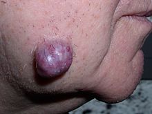
Neoplastic tumor of the cheek skin, here a benign neoplasm of the sweat glands called hidradenoma, which is not solid but is fluid-filled
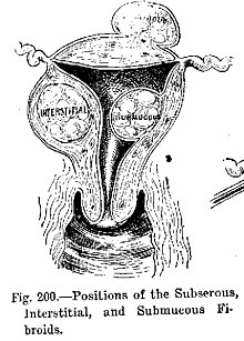
Diagram illustrating benign neoplasms, namely fibroids of the uterus

Tumors in humans occur as a result of accumulated genetic and epigenetic alterations within single cells, which cause the cell to divide and expand uncontrollably. A neoplasm can be caused by an abnormal proliferation of tissues, which can be caused by genetic mutations. Not all types of neoplasms cause a tumorous overgrowth of tissue (such as leukemia or carcinoma in situ), however similarities between neoplasmic growths and regenerative processes, e.g., dedifferentiation and rapid cell proliferation, have been pointed out.

Tumor growth has been studied using mathematics and continuum mechanics. Vascular tumors such as hemangiomas and lymphangiomas (formed from blood or lymph vessels) are thus looked at as being amalgams of a solid skeleton formed by sticky cells and an organic liquid filling the spaces in which cells can grow. Under this type of model, mechanical stresses and strains can be dealt with and their influence on the growth of the tumor and the surrounding tissue and vasculature elucidated. Recent findings from experiments that use this model show that active growth of the tumor is restricted to the outer edges of the tumor and that stiffening of the underlying normal tissue inhibits tumor growth as well.

Benign conditions that are not associated with an abnormal proliferation of tissue (such as sebaceous cysts) can also present as tumors, however, but have no malignant potential. Breast cysts (as occur commonly during pregnancy and at other times) are another example, as are other encapsulated glandular swellings (thyroid, adrenal gland, pancreas).

Encapsulated hematomas, encapsulated necrotic tissue (from an insect bite, foreign body, or other noxious mechanism), keloids (discrete overgrowths of scar tissue) and granulomas may also present as tumors.

Discrete localized enlargements of normal structures (ureters, blood vessels, intrahepatic or extrahepatic biliary ducts, pulmonary inclusions, or gastrointestinal duplications) due to outflow obstructions or narrowings, or abnormal connections, may also present as a tumor. Examples are arteriovenous fistulae or aneurysms (with or without thrombosis), biliary fistulae or aneurysms, sclerosing cholangitis, cysticercosis or hydatid cysts, intestinal duplications, and pulmonary inclusions as seen with cystic fibrosis. It can be dangerous to biopsy a number of types of tumor in which the leakage of their contents would potentially be catastrophic. When such types of tumors are encountered, diagnostic modalities such as ultrasound, CT scans, MRI, angiograms, and nuclear medicine scans are employed prior to (or during) biopsy or surgical exploration/excision in an attempt to avoid such severe complications.

Malignant neoplasms

DNA damage

The central role of DNA damage and epigenetic defects in DNA repair genes in malignant neoplasms

DNA damage is considered to be the primary underlying cause of malignant neoplasms known as cancers. Its central role in progression to cancer is illustrated in the figure in this section, in the box near the top. (The central features of DNA damage, epigenetic alterations and deficient DNA repair in progression to cancer are shown in red.) DNA damage is very common. Naturally occurring DNA damages (mostly due to cellular metabolism and the properties of DNA in water at body temperatures) occur at a rate of more than 10,000 new damages, on average, per human cell, per day. Additional DNA damages can arise from exposure to exogenous agents. Tobacco smoke causes increased exogenous DNA damage, and these DNA damages are the likely cause of lung cancer due to smoking. UV light from solar radiation causes DNA damage that is important in melanoma. Helicobacter pylori infection produces high levels of reactive oxygen species that damage DNA and contributes to gastric cancer. Bile acids, at high levels in the colons of humans eating a high fat diet, also cause DNA damage and contribute to colon cancer. Katsurano et al. indicated that macrophages and neutrophils in an inflamed colonic epithelium are the source of reactive oxygen species causing the DNA damages that initiate colonic tumorigenesis (creation of tumors in the colon). Some sources of DNA damage are indicated in the boxes at the top of the figure in this section.

Individuals with a germline mutation causing deficiency in any of 34 DNA repair genes (see article DNA repair-deficiency disorder) are at increased risk of cancer. Some germline mutations in DNA repair genes cause up to 100% lifetime chance of cancer (e.g., p53 mutations). These germline mutations are indicated in a box at the left of the figure with an arrow indicating their contribution to DNA repair deficiency.

About 70% of malignant (cancerous) neoplasms have no hereditary component and are called "sporadic cancers". Only a minority of sporadic cancers have a deficiency in DNA repair due to mutation in a DNA repair gene. However, a majority of sporadic cancers have deficiency in DNA repair due to epigenetic alterations that reduce or silence DNA repair gene expression. For example, of 113 sequential colorectal cancers, only four had a missense mutation in the DNA repair gene MGMT, while the majority had reduced MGMT expression due to methylation of the MGMT promoter region (an epigenetic alteration). Five reports present evidence that between 40% and 90% of colorectal cancers have reduced MGMT expression due to methylation of the MGMT promoter region.

Similarly, out of 119 cases of mismatch repair-deficient colorectal cancers that lacked DNA repair gene PMS2 expression, PMS2 was deficient in 6 due to mutations in the PMS2 gene, while in 103 cases PMS2 expression was deficient because its pairing partner MLH1 was repressed due to promoter methylation (PMS2 protein is unstable in the absence of MLH1). In the other 10 cases, loss of PMS2 expression was likely due to epigenetic overexpression of the microRNA, miR-155, which down-regulates MLH1.

In further examples, epigenetic defects were found at frequencies of between 13%-100% for the DNA repair genes BRCA1, WRN, FANCB, FANCF, MGMT, MLH1, MSH2, MSH4, ERCC1, XPF, NEIL1 and ATM. These epigenetic defects occurred in various cancers, including breast, ovarian, colorectal, and head and neck cancers. Two or three deficiencies in expression of ERCC1, XPF or PMS2 occur simultaneously in the majority of the 49 colon cancers evaluated by Facista et al. Epigenetic alterations causing reduced expression of DNA repair genes is shown in a central box at the third level from the top of the figure in this section, and the consequent DNA repair deficiency is shown at the fourth level.

When expression of DNA repair genes is reduced, DNA damages accumulate in cells at a higher than normal level, and these excess damages cause increased frequencies of mutation or epimutation. Mutation rates strongly increase in cells defective in DNA mismatch repair or in homologous recombinational repair (HRR).

During repair of DNA double strand breaks, or repair of other DNA damages, incompletely cleared sites of repair can cause epigenetic gene silencing. DNA repair deficiencies (level 4 in the figure) cause increased DNA damages (level 5 in the figure) which result in increased somatic mutations and epigenetic alterations (level 6 in the figure).

Field defects, normal-appearing tissue with multiple alterations (and discussed in the section below), are common precursors to development of the disordered and improperly proliferating clone of tissue in a malignant neoplasm. Such field defects (second level from bottom of figure) may have multiple mutations and epigenetic alterations.

Once a cancer is formed, it usually has genome instability. This instability is likely due to reduced DNA repair or excessive DNA damage. Because of such instability, the cancer continues to evolve and to produce sub clones. For example, a renal cancer, sampled in 9 areas, had 40 ubiquitous mutations, demonstrating tumor heterogeneity (i.e. present in all areas of the cancer), 59 mutations shared by some (but not all areas), and 29 "private" mutations only present in one of the areas of the cancer.

Field defects

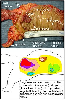
Longitudinally opened freshly resected colon segment showing a cancer and four polyps, plus a schematic diagram indicating a likely field defect (a region of tissue that precedes and predisposes to the development of cancer) in this colon segment. The diagram indicates sub-clones and sub-sub-clones that were precursors to the tumors.

Various other terms have been used to describe this phenomenon, including "field effect", "field cancerization", and "field carcinogenesis". The term "field cancerization" was first used in 1953 to describe an area or "field" of epithelium that has been preconditioned by (at that time) largely unknown processes so as to predispose it towards development of cancer. Since then, the terms "field cancerization" and "field defect" have been used to describe pre-malignant tissue in which new cancers are likely to arise.

Field defects are important in progression to cancer. However, in most cancer research, as pointed out by Rubin "The vast majority of studies in cancer research has been done on well-defined tumors in vivo, or on discrete neoplastic foci in vitro. Yet there is evidence that more than 80% of the somatic mutations found in mutator phenotype human colorectal tumors occur before the onset of terminal clonal expansion. Similarly, Vogelstein et al. point out that more than half of somatic mutations identified in tumors occurred in a pre-neoplastic phase (in a field defect), during growth of apparently normal cells. Likewise, epigenetic alterations present in tumors may have occurred in pre-neoplastic field defects.

An expanded view of field effect has been termed "etiologic field effect", which encompasses not only molecular and pathologic changes in pre-neoplastic cells but also influences of exogenous environmental factors and molecular changes in the local microenvironment on neoplastic evolution from tumor initiation to patient death.

In the colon, a field defect probably arises by natural selection of a mutant or epigenetically altered cell among the stem cells at the base of one of the intestinal crypts on the inside surface of the colon. A mutant or epigenetically altered stem cell may replace the other nearby stem cells by natural selection. Thus, a patch of abnormal tissue may arise. The figure in this section includes a photo of a freshly resected and lengthwise-opened segment of the colon showing a colon cancer and four polyps. Below the photo, there is a schematic diagram of how a large patch of mutant or epigenetically altered cells may have formed, shown by the large area in yellow in the diagram. Within this first large patch in the diagram (a large clone of cells), a second such mutation or epigenetic alteration may occur so that a given stem cell acquires an advantage compared to other stem cells within the patch, and this altered stem cell may expand clonally forming a secondary patch, or sub-clone, within the original patch. This is indicated in the diagram by four smaller patches of different colors within the large yellow original area. Within these new patches (sub-clones), the process may be repeated multiple times, indicated by the still smaller patches within the four secondary patches (with still different colors in the diagram) which clonally expand, until stem cells arise that generate either small polyps or else a malignant neoplasm (cancer).

In the photo, an apparent field defect in this segment of a colon has generated four polyps (labeled with the size of the polyps, 6mm, 5mm, and two of 3mm, and a cancer about 3 cm across in its longest dimension). These neoplasms are also indicated, in the diagram below the photo, by 4 small tan circles (polyps) and a larger red area (cancer). The cancer in the photo occurred in the cecal area of the colon, where the colon joins the small intestine (labeled) and where the appendix occurs (labeled). The fat in the photo is external to the outer wall of the colon. In the segment of colon shown here, the colon was cut open lengthwise to expose the inner surface of the colon and to display the cancer and polyps occurring within the inner epithelial lining of the colon.

If the general process by which sporadic colon cancers arise is the formation of a pre-neoplastic clone that spreads by natural selection, followed by formation of internal sub-clones within the initial clone, and sub-sub-clones inside those, then colon cancers generally should be associated with, and be preceded by, fields of increasing abnormality reflecting the succession of premalignant events. The most extensive region of abnormality (the outermost yellow irregular area in the diagram) would reflect the earliest event in formation of a malignant neoplasm.

In experimental evaluation of specific DNA repair deficiencies in cancers, many specific DNA repair deficiencies were also shown to occur in the field defects surrounding those cancers. The Table, below, gives examples for which the DNA repair deficiency in a cancer was shown to be caused by an epigenetic alteration, and the somewhat lower frequencies with which the same epigenetically caused DNA repair deficiency was found in the surrounding field defect.

Some of the small polyps in the field defect shown in the photo of the opened colon segment may be relatively benign neoplasms. Of polyps less than 10mm in size, found during colonoscopy and followed with repeat colonoscopies for 3 years, 25% were unchanged in size, 35% regressed or shrank in size while 40% grew in size.

Genome instability

Cancers are known to exhibit genome instability or a mutator phenotype. The protein-coding DNA within the nucleus is about 1.5% of the total genomic DNA. Within this protein-coding DNA (called the exome), an average cancer of the breast or colon can have about 60 to 70 protein altering mutations, of which about 3 or 4 may be "driver" mutations, and the remaining ones may be "passenger" mutations. However, the average number of DNA sequence mutations in the entire genome (including non-protein-coding regions) within a breast cancer tissue sample is about 20,000. In an average melanoma tissue sample (where melanomas have a higher exome mutation frequency) the total number of DNA sequence mutations is about 80,000. This compares to the very low mutation frequency of about 70 new mutations in the entire genome between generations (parent to child) in humans.

The high frequencies of mutations in the total nucleotide sequences within cancers suggest that often an early alteration in the field defects giving rise to a cancer (e.g. yellow area in the diagram in this section) is a deficiency in DNA repair. The large field defects surrounding colon cancers (extending to at about 10 cm on each side of a cancer) were shown by Facista et al. to frequently have epigenetic defects in 2 or 3 DNA repair proteins (ERCC1, XPF or PMS2) in the entire area of the field defect. Deficiencies in DNA repair cause increased mutation rates. A deficiency in DNA repair, itself, can allow DNA damages to accumulate, and error-prone translesion synthesis past some of those damages may give rise to mutations. In addition, faulty repair of these accumulated DNA damages may give rise to epimutations. These new mutations or epimutations may provide a proliferative advantage, generating a field defect. Although the mutations/epimutations in DNA repair genes do not, themselves, confer a selective advantage, they may be carried along as passengers in cells when the cells acquire additional mutations/epimutations that do provide a proliferative advantage.

Etymology

The term neoplasm is a synonym of tumor. Neoplasia denotes the process of the formation of neoplasms/tumors, and the process is referred to as a neoplastic process. The word neoplastic itself comes from Greek neo 'new' and plastic 'formed, molded'.

The term tumor derives from the Latin noun tumor 'a swelling', ultimately from the verb tumēre 'to swell'. In the British Commonwealth, the spelling tumour is commonly used, whereas in the U.S. the word is usually spelled tumor.

In its medical sense, tumor has traditionally meant an abnormal swelling of the flesh. The Roman medical encyclopedist Celsus (c. 30 BC–38 AD) described the four cardinal signs of acute inflammation as tumor, dolor, calor, and rubor (swelling, pain, increased heat, and redness). (His treatise, De Medicina, was the first medical book printed in 1478 following the invention of the movable-type printing press.)

In contemporary English, the word tumor is often used as a synonym for a cystic (liquid-filled) growth or solid neoplasm (cancerous or non-cancerous), with other forms of swelling often referred to as "swellings".

Related terms occur commonly in the medical literature, where the nouns tumefaction and tumescence (derived from the adjective tumescent) are current medical terms for non-neoplastic swelling. This type of swelling is most often caused by inflammation caused by trauma, infection, and other factors.

Tumors may be caused by conditions other than an overgrowth of neoplastic cells, however. Cysts (such as sebaceous cysts) are also referred to as tumors, even though they have no neoplastic cells. This is standard in medical-billing terminology (especially when billing for a growth whose pathology has yet to be determined).

Multicellular organism

From Wikipedia, the free encyclopedia
The nematode Caenorhabditis elegans stained to highlight the nuclei of its cells

A multicellular organism is an organism that consists of more than one cell, unlike unicellular organisms. All species of animals, land plants and most fungi are multicellular, as are many algae, whereas a few organisms are partially uni- and partially multicellular, like slime molds and social amoebae such as the genus Dictyostelium.

Multicellular organisms arise in various ways, for example by cell division or by aggregation of many single cells. Colonial organisms are the result of many identical individuals joining together to form a colony. However, it can often be hard to separate colonial protists from true multicellular organisms, because the two concepts are not distinct; colonial protists have been dubbed "pluricellular" rather than "multicellular". There are also macroscopic organisms that are multinucleate though technically unicellular, such as the Xenophyophorea that can reach 20 cm.

Evolutionary history

Occurrence

Multicellularity has evolved independently at least 25 times in eukaryotes, and also in some prokaryotes, like cyanobacteria, myxobacteria, actinomycetes, Magnetoglobus multicellularis or Methanosarcina. However, complex multicellular organisms evolved only in six eukaryotic groups: animals, symbiomycotan fungi, brown algae, red algae, green algae, and land plants. It evolved repeatedly for Chloroplastida (green algae and land plants), once for animals, once for brown algae, three times in the fungi (chytrids, ascomycetes, and basidiomycetes) and perhaps several times for slime molds and red algae. The first evidence of multicellular organization, which is when unicellular organisms coordinate behaviors and may be an evolutionary precursor to true multicellularity, is from cyanobacteria-like organisms that lived 3.0–3.5 billion years ago. To reproduce, true multicellular organisms must solve the problem of regenerating a whole organism from germ cells (i.e., sperm and egg cells), an issue that is studied in evolutionary developmental biology. Animals have evolved a considerable diversity of cell types in a multicellular body (100–150 different cell types), compared with 10–20 in plants and fungi.

Loss of multicellularity

Loss of multicellularity occurred in some groups. Fungi are predominantly multicellular, though early diverging lineages are largely unicellular (e.g., Microsporidia) and there have been numerous reversions to unicellularity across fungi (e.g., Saccharomycotina, Cryptococcus, and other yeasts). It may also have occurred in some red algae (e.g., Porphyridium), but they may be primitively unicellular. Loss of multicellularity is also considered probable in some green algae (e.g., Chlorella vulgaris and some Ulvophyceae). In other groups, generally parasites, a reduction of multicellularity occurred, in the number or types of cells (e.g., the myxozoans, multicellular organisms, earlier thought to be unicellular, are probably extremely reduced cnidarians).

Cancer

Multicellular organisms, especially long-living animals, face the challenge of cancer, which occurs when cells fail to regulate their growth within the normal program of development. Changes in tissue morphology can be observed during this process. Cancer in animals (metazoans) has often been described as a loss of multicellularity and an atavistic reversion towards a unicellular-like state. Many genes responsible for the establishment of multicellularity that originated around the appearance of metazoans are deregulated in cancer cells, including genes that control cell differentiation, adhesion and cell-to-cell communication. There is a discussion about the possibility of existence of cancer in other multicellular organisms or even in protozoa. For example, plant galls have been characterized as tumors, but some authors argue that plants do not develop cancer.

Separation of somatic and germ cells

In some multicellular groups, which are called Weismannists, a separation between a sterile somatic cell line and a germ cell line evolved. However, Weismannist development is relatively rare (e.g., vertebrates, arthropods, Volvox), as a great part of species have the capacity for somatic embryogenesis (e.g., land plants, most algae, many invertebrates).

Origin hypotheses

One hypothesis for the origin of multicellularity is that a group of function-specific cells aggregated into a slug-like mass called a grex, which moved as a multicellular unit. This is essentially what slime molds do. Another hypothesis is that a primitive cell underwent nucleus division, thereby becoming a coenocyte. A membrane would then form around each nucleus (and the cellular space and organelles occupied in the space), thereby resulting in a group of connected cells in one organism (this mechanism is observable in Drosophila). A third hypothesis is that as a unicellular organism divided, the daughter cells failed to separate, resulting in a conglomeration of identical cells in one organism, which could later develop specialized tissues. This is what plant and animal embryos do as well as colonial choanoflagellates.

Because the first multicellular organisms were simple, soft organisms lacking bone, shell, or other hard body parts, they are not well preserved in the fossil record. One exception may be the demosponge, which may have left a chemical signature in ancient rocks. The earliest fossils of multicellular organisms include the contested Grypania spiralis and the fossils of the black shales of the Palaeoproterozoic Francevillian Group Fossil B Formation in Gabon (Gabonionta). The Doushantuo Formation has yielded 600 million year old microfossils with evidence of multicellular traits.

Until recently, phylogenetic reconstruction has been through anatomical (particularly embryological) similarities. This is inexact, as living multicellular organisms such as animals and plants are more than 500 million years removed from their single-cell ancestors. Such a passage of time allows both divergent and convergent evolution time to mimic similarities and accumulate differences between groups of modern and extinct ancestral species. Modern phylogenetics uses sophisticated techniques such as alloenzymes, satellite DNA and other molecular markers to describe traits that are shared between distantly related lineages.

The evolution of multicellularity could have occurred in several different ways, some of which are described below:

The symbiotic theory

This theory suggests that the first multicellular organisms occurred from symbiosis (cooperation) of different species of single-cell organisms, each with different roles. Over time these organisms would become so dependent on each other that they would not be able to survive independently, eventually leading to the incorporation of their genomes into one multicellular organism. Each respective organism would become a separate lineage of differentiated cells within the newly created species.

This kind of severely co-dependent symbiosis can be seen frequently, such as in the relationship between clown fish and Riterri sea anemones. In these cases, it is extremely doubtful whether either species would survive very long if the other became extinct. However, the problem with this theory is that it is still not known how each organism's DNA could be incorporated into one single genome to constitute them as a single species. Although such symbiosis is theorized to have occurred (e.g., mitochondria and chloroplasts in animal and plant cells—endosymbiosis), it has happened only extremely rarely and, even then, the genomes of the endosymbionts have retained an element of distinction, separately replicating their DNA during mitosis of the host species. For instance, the two or three symbiotic organisms forming the composite lichen, although dependent on each other for survival, have to separately reproduce and then re-form to create one individual organism once more.

The cellularization (syncytial) theory

This theory states that a single unicellular organism, with multiple nuclei, could have developed internal membrane partitions around each of its nuclei. Many protists such as the ciliates or slime molds can have several nuclei, lending support to this hypothesis. However, the simple presence of multiple nuclei is not enough to support the theory. Multiple nuclei of ciliates are dissimilar and have clear differentiated functions. The macronucleus serves the organism's needs, whereas the micronucleus is used for sexual reproduction with exchange of genetic material. Slime molds syncitia form from individual amoeboid cells, like syncitial tissues of some multicellular organisms, not the other way round. To be deemed valid, this theory needs a demonstrable example and mechanism of generation of a multicellular organism from a pre-existing syncytium.

The colonial theory

The colonial theory of Haeckel, 1874, proposes that the symbiosis of many organisms of the same species (unlike the symbiotic theory, which suggests the symbiosis of different species) led to a multicellular organism. At least some - it is presumed land-evolved - multicellularity occurs by cells separating and then rejoining (e.g., cellular slime molds) whereas for the majority of multicellular types (those that evolved within aquatic environments), multicellularity occurs as a consequence of cells failing to separate following division. The mechanism of this latter colony formation can be as simple as incomplete cytokinesis, though multicellularity is also typically considered to involve cellular differentiation.

The advantage of the Colonial Theory hypothesis is that it has been seen to occur independently in 16 different protoctistan phyla. For instance, during food shortages the amoeba Dictyostelium groups together in a colony that moves as one to a new location. Some of these amoeba then slightly differentiate from each other. Other examples of colonial organisation in protista are Volvocaceae, such as Eudorina and Volvox, the latter of which consists of up to 500–50,000 cells (depending on the species), only a fraction of which reproduce. For example, in one species 25–35 cells reproduce, 8 asexually and around 15–25 sexually. However, it can often be hard to separate colonial protists from true multicellular organisms, as the two concepts are not distinct; colonial protists have been dubbed "pluricellular" rather than "multicellular".

The synzoospore theory

Some authors suggest that the origin of multicellularity, at least in Metazoa, occurred due to a transition from temporal to spatial cell differentiation, rather than through a gradual evolution of cell differentiation, as affirmed in Haeckel's gastraea theory.

GK-PID

About 800 million years ago, a minor genetic change in a single molecule called guanylate kinase protein-interaction domain (GK-PID) may have allowed organisms to go from a single cell organism to one of many cells.

The role of viruses

Genes borrowed from viruses and mobile genetic elements (MGEs) have recently been identified as playing a crucial role in the differentiation of multicellular tissues and organs and even in sexual reproduction, in the fusion of egg cells and sperm. Such fused cells are also involved in metazoan membranes such as those that prevent chemicals from crossing the placenta and the brain body separation. Two viral components have been identified. The first is syncytin, which came from a virus. The second identified in 2002 is called EFF-1, which helps form the skin of Caenorhabditis elegans, part of a whole family of FF proteins. Felix Rey, of the Pasteur Institute in Paris, has constructed the 3D structure of the EFF-1 protein and shown it does the work of linking one cell to another, in viral infections. The fact that all known cell fusion molecules are viral in origin suggests that they have been vitally important to the inter-cellular communication systems that enabled multicellularity. Without the ability of cellular fusion, colonies could have formed, but anything even as complex as a sponge would not have been possible.

Oxygen availability hypothesis

This theory suggests that the oxygen available in the atmosphere of early Earth could have been the limiting factor for the emergence of multicellular life. This hypothesis is based on the correlation between the emergence of multicellular life and the increase of oxygen levels during this time. This would have taken place after the Great Oxidation Event but before the most recent rise in oxygen. Mills concludes that the amount of oxygen present during the Ediacaran is not necessary for complex life and therefore is unlikely to have been the driving factor for the origin of multicellularity.

Snowball Earth hypothesis

A snowball Earth is a geological event where the entire surface of the Earth is covered in snow and ice. The term can either refer to individual events (of which there were at least two) or to the larger geologic period during which all the known total glaciations occurred.

The most recent snowball Earth took place during the Cryogenian period and consisted of two global glaciation events known as the Sturtian and Marinoan glaciations. Xiao et al. suggest that between the period of time known as the "Boring Billion" and the snowball Earth, simple life could have had time to innovate and evolve, which could later lead to the evolution of multicellularity.

The snowball Earth hypothesis in regards to multicellularity proposes that the Cryogenian period in Earth's history could have been the catalyst for the evolution of complex multicellular life. Brocks suggests that the time between the Sturtian Glacian and the more recent Marinoan Glacian allowed for planktonic algae to dominate the seas making way for rapid diversity of life for both plant and animal lineages. Complex life quickly emerged and diversified in what is known as the Cambrian explosion shortly after the Marinoan.

Predation hypothesis

The predation hypothesis suggests that to avoid being eaten by predators, simple single-celled organisms evolved multicellularity to make it harder to be consumed as prey. Herron et al. performed laboratory evolution experiments on the single-celled green alga, Chlamydomonas reinhardtii, using paramecium as a predator. They found that in the presence of this predator, C. reinhardtii does indeed evolve simple multicellular features.

Experimental evolution

It is impossible to know what happened when single cells evolved into multicellular organisms hundreds of millions of years ago. However, we can identify mutations that can turn single-celled organisms into multicellular ones. This would demonstrate the possibility of such an event. Unicellular species can relatively easily acquire mutations that make them attach to each other—the first step towards multicellularity. Multiple normally unicellular species have been evolved to exhibit such early steps:

  • yeast are long known to exhibit flocculation. One of the first yeast genes found to cause this phenotype is FLO1. A more strikingly clumped phenotype is called "snowflake", caused by the loss of a single transcription factor Ace2. "Snowflake" yeast grow into multicellular clusters that sediment quickly; they were identified by directed evolution. More recently (2024), snowflake yeast were subject to over 3,000 generations of further directed evolution, forming macroscopic assemblies on the scale of millimeters. Changes in multiple genes were identified. In addition, the authors reported that only anaerobic cultures of snowflake yeast evolved this trait, while the aerobic ones did not.
  • A range of green algae species have been experimentally evolved to form larger clumps. When Chlorella vulgaris is grown with a predator Ochromonas vallescia, it starts forming small colonies, which are harder to ingest due to the larger size. The same is true for Chlamydomonas reinhardtii under predation by Brachionus calyciflorus and Paramecium tetraurelia.

C. reinhartii normally starts as a motile single-celled propagule; this single cell asexually reproduces by undergoing 2–5 rounds of mitosis as a small clump of non-motile cells, then all cells become single-celled propagules and the clump dissolves. With a few generations under Paramecium predation, the "clump" becomes a persistent structure: only some cells become propagules. Some populations go further and evolved multi-celled propagules: instead of peeling off single cells from the clump, the clump now reproduces by peeling off smaller clumps.

Advantages

Multicellularity allows an organism to exceed the size limits normally imposed by diffusion: single cells with increased size have a decreased surface-to-volume ratio and have difficulty absorbing sufficient nutrients and transporting them throughout the cell. Multicellular organisms thus have the competitive advantages of an increase in size without its limitations. They can have longer lifespans as they can continue living when individual cells die. Multicellularity also permits increasing complexity by allowing differentiation of cell types within one organism.

Whether all of these can be seen as advantages however is debatable: The vast majority of living organisms are single celled, and even in terms of biomass, single celled organisms are far more successful than animals, although not plants. Rather than seeing traits such as longer lifespans and greater size as an advantage, many biologists see these only as examples of diversity, with associated tradeoffs.

Gene expression changes in the transition from uni- to multicellularity

During the evolutionary transition from unicellular organisms to multicellular organisms, the expression of genes associated with reproduction and survival likely changed. In the unicellular state, genes associated with reproduction and survival are expressed in a way that enhances the fitness of individual cells, but after the transition to multicellularity, the pattern of expression of these genes must have substantially changed so that individual cells become more specialized in their function relative to reproduction and survival. As the multicellular organism emerged, gene expression patterns became compartmentalized between cells that specialized in reproduction (germline cells) and those that specialized in survival (somatic cells). As the transition progressed, cells that specialized tended to lose their own individuality and would no longer be able to both survive and reproduce outside the context of the group.

Dyson sphere

From Wikipedia, the free encyclopedia
https://en.wikipedia.org/wiki/Dyson_sphere
A hypothetical depiction of a Dyson swarm surrounding a star
Freeman Dyson, the first scientist to explore the concept

A Dyson sphere is a hypothetical megastructure that encompasses a star and captures a large percentage of its power output. The concept is a thought experiment that attempts to imagine how a spacefaring civilization would meet its energy requirements once those requirements exceed what can be generated from the home planet's resources alone. Because only a tiny fraction of a star's energy emissions reaches the surface of any orbiting planet, building structures encircling a star would enable a civilization to harvest far more energy.

The first modern imagining of such a structure was by Olaf Stapledon in his science fiction novel Star Maker (1937). The concept was later explored by the physicist Freeman Dyson in his 1960 paper "Search for Artificial Stellar Sources of Infrared Radiation". Dyson speculated that such structures would be the logical consequence of the escalating energy needs of a technological civilization and would be a necessity for its long-term survival. A signature of such spheres detected in astronomical searches would be an indicator of extraterrestrial intelligence.

Since Dyson's paper, many variant designs involving an artificial structure or series of structures to encompass a star have been proposed in exploratory engineering or described in science fiction, often under the name "Dyson sphere". Fictional depictions often describe a solid shell of matter enclosing a star – an arrangement considered by Dyson himself to be impossible.

Origins

Inspired by the 1937 science fiction novel Star Maker by Olaf Stapledon, the physicist and mathematician Freeman Dyson was the first to formalize the concept of what became known as the "Dyson sphere" in his 1960 Science paper "Search for Artificial Stellar Sources of Infra-Red Radiation". Dyson theorized that as the energy requirements of an advanced technological civilization increased, there would come a time when it would need to systematically harvest the energy from its local star on a large scale. He speculated that this could be done via a system of structures orbiting the star, designed to intercept and collect its energy. He argued that as the structure would result in the large-scale conversion of starlight into far-infrared radiation, an earth-based search for sources of infrared radiation could identify stars supporting intelligent life.

Dyson did not detail how such a system could be constructed, simply referring to it in the paper as a "shell" or "biosphere". He later clarified that he did not have in mind a solid structure, saying: "A solid shell or ring surrounding a star is mechanically impossible. The form of 'biosphere' which I envisaged consists of a loose collection or swarm of objects traveling on independent orbits around the star." Such a concept has often been referred to as a Dyson swarm; however, in 2013, Dyson said he had come to regret that the concept had been named after him.

Search for megastructures

Dyson-style energy collectors around a distant star would absorb and re-radiate energy from the star. The wavelengths of such re-radiated energy may be atypical for the star's spectral type, due to the presence of heavy elements not naturally occurring within the star. If the percentage of such atypical wavelengths were to be significant, an alien megastructure could be detected at interstellar distances. This could indicate the presence of what has been called a Type II Kardashev civilization.

SETI has looked for such infrared-heavy spectra from solar analogs, as has Fermilab. Fermilab discovered 17 potential "ambiguous" candidates, of which four were in 2006 called "amusing but still questionable". Later searches also resulted in several candidates, all of which remain unconfirmed.

On 14 October 2015, Planet Hunters' citizen scientists discovered unusual light fluctuations of the star KIC 8462852 raising press speculation that a Dyson sphere may have been discovered. However, subsequent analysis showed that the results were consistent with the presence of dust. A further campaign in 2024 identified seven possible candidates for Dyson-spheres, but further investigation was said to be required.

Feasibility and science-based speculation

Although Dyson sphere systems are theoretically possible, building a stable megastructure around the Sun is currently far beyond humanity's engineering capacity. The number of craft required to obtain, transmit, and maintain a complete Dyson sphere exceeds present-day industrial capabilities. George Dvorsky has advocated the use of self-replicating robots to overcome this limitation in the relatively near term. Some have suggested that Dyson sphere habitats could be built around white dwarfs and even pulsars.

Stellar engines are hypothetical megastructures whose purpose is to extract useful energy from a star, sometimes for specific purposes. For example, Matrioshka brains have been proposed to extract energy for computation, while Shkadov thrusters would extract energy for propulsion. Some proposed stellar engine designs are based on the Dyson sphere.

From May until June 2024, speculation grew that potential signs of interstellar Dyson spheres had been discovered. The seven objects of interest – all located within a thousand light-years of Earth – are M-dwarfs, a class of stars that are smaller and less luminous than the Sun. However, the authors of the findings were careful not to make any overblown claims. Despite this, many media outlets picked up on the story. Less fantastical alternative explanations have been made, including a proposal that the infrared from the discoveries was caused by distant dust-obscured galaxies.

Fictional examples

A precursor to the concept of Dyson spheres was featured in the 1937 novel Star Maker by Olaf Stapledon, in which he described "every solar system... surrounded by a gauze of light-traps, which focused the escaping solar energy for intelligent use"; Dyson got his inspiration from this book and suggested that "Stapledon sphere" would be a more apt name for the concept. Fictional Dyson spheres are typically solid structures forming a continuous shell around the star in question, although Dyson himself considered that prospect to be mechanically implausible. They are sometimes used as the type of plot device known as a Big Dumb Object.

Dyson spheres appear as a background element in many works of fiction, including the 1964 novel The Wanderer by Fritz Leiber where aliens enclose multiple stars in this way. Dyson spheres are depicted in the 1975–1983 book series Saga of Cuckoo by Frederik Pohl and Jack Williamson, and one functions as the setting of Bob Shaw's 1975 novel Orbitsville and its sequels. In the 1992 episode "Relics" of the TV show Star Trek: The Next Generation, the USS Enterprise finds itself trapped in an abandoned Dyson Sphere; in a 2011 interview, Dyson said that he enjoyed the episode, although he considered the sphere depicted to be "nonsense". Michael Jan Friedman who wrote the novelization observed that in the TV episode itself the Dyson sphere was effectively a MacGuffin, with "just nothing about it" in the story, and decided to flesh out the plot element in his novelization.

Other science-fiction story examples include Tony Rothman's The World Is Round, Somtow Sucharitkul's Inquisitor series, Timothy Zahn's Spinneret, James White's Federation World, Stephen Baxter's The Time Ships, and Peter F. Hamilton's Pandora's Star. Variations on the Dyson Sphere concept include a single circular band in Larry Niven's 1970 novel Ringworld, a half sphere in the 2012 novel Bowl of Heaven by Gregory Benford and Niven, and nested spheres – also known as a Matrioshka brain – in Colin Kapp's 1980s Cageworld series and Brian Stableford's 1979–1990 Asgard trilogy.

Stableford himself observed that Dyson spheres are usually MacGuffins or largely deep in the backgrounds of stories, giving as examples Fritz Leiber's The Wanderer and Linda Nagata's Deception Well, whereas stories involving space exploration tend to employ the variants like Niven's Ringworld.He gives two reasons for this: firstly that Dyson spheres are simply too big to address, which Friedman also alluded to when pointing out that the reason his novelization of "Relics" did not go further into the sphere was that it was only four hundred pages and he had just shy of four weeks to write it; and secondly that, especially for hard science-fiction, Dyson spheres have certain engineering problems that complicate stories. In particular, since gravitational attraction is in equilibrium inside such a sphere (per the shell theorem), other means such as rotating the sphere have to be employed in order to keep things attached to the interior surface, which then leads to the problem of a gravity gradient that goes to zero at the rotational poles. Authors address this with various modifications of the idea such as the aforementioned Cageworld nesting, Dan Alderson's double sphere idea, and Niven's reduced Ringworld (discussed in "Bigger Than Worlds").

Fossil fuels lobby

  From Wikipedia, the free encyclopedia https://en.wikipedia.org/wiki/Fossil_fuels_lobby ...