Search This Blog

Saturday, May 18, 2019

Fight-or-flight response

From Wikipedia, the free encyclopedia

Dog and cat showing acute stress responses
The fight-or-flight response (also called hyperarousal, or the acute stress response) is a physiological reaction that occurs in response to a perceived harmful event, attack, or threat to survival. It was first described by Walter Bradford Cannon. His theory states that animals react to threats with a general discharge of the sympathetic nervous system, preparing the animal for fighting or fleeing. More specifically, the adrenal medulla produces a hormonal cascade that results in the secretion of catecholamines, especially norepinephrine and epinephrine. The hormones estrogen, testosterone, and cortisol, as well as the neurotransmitters dopamine and serotonin, also affect how organisms react to stress.

This response is recognised as the first stage of the general adaptation syndrome that regulates stress responses among vertebrates and other organisms.

Physiology

Autonomic nervous system

The autonomic nervous system is a control system that acts largely unconsciously and regulates heart rate, digestion, respiratory rate, pupillary response, urination, and sexual arousal. This system is the primary mechanism in control of the fight-or-flight response and its role is mediated by two different components: the sympathetic nervous system and the parasympathetic nervous system.

Sympathetic nervous system

The sympathetic nervous system originates in the spinal cord and its main function is to activate the physiological changes that occur during the fight-or-flight response. This component of the autonomic nervous system utilises and activates the release of norepinephrine in the reaction.

Parasympathetic nervous system

The parasympathetic nervous system originates in the sacral spinal cord and medulla, physically surrounding the sympathetic origin, and works in concert with the sympathetic nervous system. Its main function is to activate the "rest and digest" response and return the body to homeostasis after the fight or flight response. This system utilises and activates the release of the neurotransmitter acetylcholine.

Reaction

The fight-or-flight response
The reaction begins in the amygdala, which triggers a neural response in the hypothalamus. The initial reaction is followed by activation of the pituitary gland and secretion of the hormone ACTH. The adrenal gland is activated almost simultaneously, via the sympathetic nervous system, and releases the hormone epinephrine. The release of chemical messengers results in the production of the hormone cortisol, which increases blood pressure, blood sugar, and suppresses the immune system. The initial response and subsequent reactions are triggered in an effort to create a boost of energy. This boost of energy is activated by epinephrine binding to liver cells and the subsequent production of glucose. Additionally, the circulation of cortisol functions to turn fatty acids into available energy, which prepares muscles throughout the body for response. Catecholamine hormones, such as adrenaline (epinephrine) or noradrenaline (norepinephrine), facilitate immediate physical reactions associated with a preparation for violent muscular action and:

Function of physiological changes

The physiological changes that occur during the fight or flight response are activated in order to give the body increased strength and speed in anticipation of fighting or running. Some of the specific physiological changes and their functions include:
  • Increased blood flow to the muscles activated by diverting blood flow from other parts of the body.
  • Increased blood pressure, heart rate, blood sugars, and fats in order to supply the body with extra energy.
  • The blood clotting function of the body speeds up in order to prevent excessive blood loss in the event of an injury sustained during the response.
  • Increased muscle tension in order to provide the body with extra speed and strength.

Emotional components

Emotion regulation

In the context of the fight or flight response, emotional regulation is used proactively to avoid threats of stress or to control the level of emotional arousal.

Emotional reactivity

During the reaction, the intensity of emotion that is brought on by the stimulus will also determine the nature and intensity of the behavioral response. Individuals with higher levels of emotional reactivity may be prone to anxiety and aggression, which illustrates the implications of appropriate emotional reaction in the fight or flight response.

Cognitive components

Content specificity

The specific components of cognitions in the fight or flight response seem to be largely negative. These negative cognitions may be characterised by: attention to negative stimuli, the perception of ambiguous situations as negative, and the recurrence of recalling negative words. There also may be specific negative thoughts associated with emotions commonly seen in the reaction.

Perception of control

Perceived control relates to an individual's thoughts about control over situations and events. Perceived control should be differentiated from actual control because an individual's beliefs about their abilities may not reflect their actual abilities. Therefore, overestimation or underestimation of perceived control can lead to anxiety and aggression.

Social information processing

The social information processing model proposes a variety of factors that determine behavior in the context of social situations and preexisting thoughts. The attribution of hostility, especially in ambiguous situations, seems to be one of the most important cognitive factors associated with the fight or flight response because of its implications towards aggression.

Other animals

Evolutionary perspective

An evolutionary psychology explanation is that early animals had to react to threatening stimuli quickly and did not have time to psychologically and physically prepare themselves. The fight or flight response provided them with the mechanisms to rapidly respond to threats against survival.

Examples

A typical example of the stress response is a grazing zebra. If the zebra sees a lion closing in for the kill, the stress response is activated as a means to escape its predator. The escape requires intense muscular effort, supported by all of the body’s systems. The sympathetic nervous system’s activation provides for these needs. A similar example involving fight is of a cat about to be attacked by a dog. The cat shows accelerated heartbeat, piloerection (hair standing on end), and pupil dilation, all signs of sympathetic arousal. Note that the zebra and cat still maintain homeostasis in all states.

Varieties of responses

Bison hunted by dogs
Animals respond to threats in many complex ways. Rats, for instance, try to escape when threatened, but will fight when cornered. Some animals stand perfectly still so that predators will not see them. Many animals freeze or play dead when touched in the hope that the predator will lose interest.
Other animals have alternative self-protection methods. Some species of cold-blooded animals change color swiftly, to camouflage themselves. These responses are triggered by the sympathetic nervous system, but, in order to fit the model of fight or flight, the idea of flight must be broadened to include escaping capture either in a physical or sensory way. Thus, flight can be disappearing to another location or just disappearing in place. And often both fight and flight are combined in a given situation.
The fight or flight actions also have polarity – the individual can either fight or flee against something that is threatening, such as a hungry lion, or fight for or fly towards something that is needed, such as the safety of the shore from a raging river.
A threat from another animal does not always result in immediate fight or flight. There may be a period of heightened awareness, during which each animal interprets behavioral signals from the other. Signs such as paling, piloerection, immobility, sounds, and body language communicate the status and intentions of each animal. There may be a sort of negotiation, after which fight or flight may ensue, but which might also result in playing, mating, or nothing at all. An example of this is kittens playing: each kitten shows the signs of sympathetic arousal, but they never inflict real damage.

Muscarinic acetylcholine receptor

From Wikipedia, the free encyclopedia

Acetylcholine - the natural agonist of muscarinic and nicotinic receptors.
 
Muscarine - an agonist used to distinguish between these two classes of receptors. Not normally found in the body.
 
Atropine - an antagonist.
 
Muscarinic acetylcholine receptors, or mAChRs, are acetylcholine receptors that form G protein-coupled receptor complexes in the cell membranes of certain neurons and other cells. They play several roles, including acting as the main end-receptor stimulated by acetylcholine released from postganglionic fibers in the parasympathetic nervous system

Muscarinic receptors are so named because they are more sensitive to muscarine than to nicotine. Their counterparts are nicotinic acetylcholine receptors (nAChRs), receptor ion channels that are also important in the autonomic nervous system. Many drugs and other substances (for example pilocarpine and scopolamine) manipulate these two distinct receptors by acting as selective agonists or antagonists.

Function

Acetylcholine (ACh) is a neurotransmitter found in the brain, neuromuscular junctions and the autonomic ganglia. Muscarinic receptors are used in the following roles:

Recovery receptors

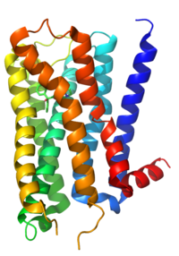
The structure of Muscarinic acetylcholine receptor M2.
 
ACh is always used as the transmitter within the autonomic ganglion. Nicotinic receptors on the postganglionic neuron are responsible for the initial fast depolarization (Fast EPSP) of that neuron. As a consequence of this, nicotinic receptors are often cited as the receptor on the postganglionic neurons at the ganglion. However, the subsequent hyperpolarization (IPSP) and slow depolarization (Slow EPSP) that represent the recovery of the postganglionic neuron from stimulation are actually mediated by muscarinic receptors, types M2 and M1 respectively (discussed below).

Peripheral autonomic fibers (sympathetic and parasympathetic fibers) are categorized anatomically as either preganglionic or postganglionic fibers, then further generalized as either adrenergic fibers, releasing noradrenaline, or cholinergic fibers, both releasing acetylcholine and expressing acetylcholine receptors. Both preganglionic sympathetic fibers and preganglionic parasympathetic fibers are cholinergic. Most postganglionic sympathetic fibers are adrenergic: their neurotransmitter is norepinephrine; postganglionic sympathetic fibers to the sweat glands, piloerectile muscles of the body hairs, and the skeletal muscle arterioles do not use adrenaline/noradrenaline.

The adrenal medulla is considered a sympathetic ganglion and, like other sympathetic ganglia, is supplied by cholinergic preganglionic sympathetic fibers: acetylcholine is the neurotransmitter utilized at this synapse. The chromaffin cells of the adrenal medulla act as "modified neurons", releasing adrenaline and noradrenaline into the bloodstream as hormones instead of as neurotransmitters. The other postganglionic fibers of the peripheral autonomic system belong to the parasympathetic division; all are cholinergic fibers, and use acetylcholine as the neurotransmitter.

Postganglionic neurons

Another role for these receptors is at the junction of the innervated tissues and the postganglionic neurons in the parasympathetic division of the autonomic nervous system. Here acetylcholine is again used as a neurotransmitter, and muscarinic receptors form the principal receptors on the innervated tissue.

Innervated tissue

Very few parts of the sympathetic system use cholinergic receptors. In sweat glands the receptors are of the muscarinic type. The sympathetic nervous system also has some preganglionic nerves terminating at the chromaffin cells in the adrenal medulla, which secrete epinephrine and norepinephrine into the bloodstream. Some believe that chromaffin cells are modified postganglionic CNS fibers. In the adrenal medulla, acetylcholine is used as a neurotransmitter, and the receptor is of the nicotinic type. 

The somatic nervous system uses a nicotinic receptor to acetylcholine at the neuromuscular junction.

Higher central nervous system

Muscarinic acetylcholine receptors are also present and distributed throughout the local nervous system, in post-synaptic and pre-synaptic positions. There is also some evidence for postsynaptic receptors on sympathetic neurons allowing the parasympathetic nervous system to inhibit sympathetic effects.

Presynaptic membrane of the neuromuscular junction

It is known that muscarinic acetylcholine receptors also appear on the pre-synaptic membrane of somatic neurons in the neuro-muscular junction, where they are involved in the regulation of acetylcholine release.

Form of muscarinic receptors

Muscarinic acetylcholine receptors belong to a class of metabotropic receptors that use G proteins as their signaling mechanism. In such receptors, the signaling molecule (the ligand) binds to a receptor that has seven transmembrane regions; in this case, the ligand is ACh. This receptor is bound to intracellular proteins, known as G proteins, which begin the information cascade within the cell.

By contrast, nicotinic receptors use a ligand-gated ion channel mechanism for signaling. In this case, binding of the ligands with the receptor causes an ion channel to open, permitting either one or more specific type(s) of ion (e.g., K+, Na+, Ca2+) to diffuse into or out of the cell.

Receptor isoforms

Classification

By the use of selective radioactively labeled agonist and antagonist substances, five subtypes of muscarinic receptors have been determined, named M1-M5 (using an upper case M and subscript number). M1,M3,M5 receptors are coupled with Gq proteins, while M2 and M4 receptors are coupled with Gi/o proteins. There are other classification systems. For example, the drug pirenzepine is a muscarinic antagonist (decreases the effect of ACh), which is much more potent at M1 receptors than it is at other subtypes. The acceptance of the various subtypes has proceeded in numerical order: therefore, sources that recognize only the M1/M2 distinction exist. More recent studies tend to recognize M3 and the most recent M4.

Genetic differences

Meanwhile, geneticists and molecular biologists have characterised five genes that appear to encode muscarinic receptors, named m1-m5 (lowercase m; no subscript number). The first four code for pharmacologic types M1-M4. The fifth, M5, corresponds to a subtype of receptor that had until recently not been detected pharmacologically. The receptors m1 and m2 were determined based upon partial sequencing of M1 and M2 receptor proteins. The others were found by searching for homology, using bioinformatic techniques.

Difference in G proteins

G proteins contain an alpha-subunit that is critical to the functioning of receptors. These subunits can take a number of forms. There are four broad classes of form of G-protein: Gs, Gi, Gq, and G12/13. Muscarinic receptors vary in the G protein to which they are bound, with some correlation according to receptor type. G proteins are also classified according to their susceptibility to cholera toxin (CTX) and pertussis toxin (PTX, whooping cough). Gs and some subtypes of Gi (Gαt and Gαg) are susceptible to CTX. Only Gi is susceptible to PTX, with the exception of one subtype of Gi (Gαz) which is immune. Also, only when bound with an agonist, those G proteins normally sensitive to PTX also become susceptible to CTX.

The various G-protein subunits act differently upon secondary messengers, upregulating Phospholipases, downregulating cAMP, etc. 

Because of the strong correlations to muscarinic receptor type, CTX and PTX are useful experimental tools in investigating these receptors.

M1 receptor

This receptor is found mediating slow EPSP at the ganglion in the postganglionic nerve[citation needed], is common in exocrine glands and in the CNS.

It is predominantly found bound to G proteins of class Gq, which use upregulation of phospholipase C and, therefore, inositol trisphosphate and intracellular calcium as a signaling pathway. A receptor so bound would not be susceptible to CTX or PTX. However, Gi (causing a downstream decrease in cAMP) and Gs (causing an increase in cAMP) have also been shown to be involved in interactions in certain tissues, and so would be susceptible to PTX and CTX, respectively.

M2 receptor

The M2 muscarinic receptors are located in the heart, where they act to slow the heart rate down to normal sinus rhythm, by slowing the speed of depolarization. In humans under resting conditions vagal activity dominates over sympathetic activity. Hence inhibition of m2 receptors (e.g. by atropine) will cause a raise in heart rate. They also moderately reduce contractile forces of the atrial cardiac muscle, and reduce conduction velocity of the atrioventricular node (AV node). It also serves to slightly decrease the contractile forces of the ventricular muscle. 

M2 muscarinic receptors act via a Gi type receptor, which causes a decrease in cAMP in the cell, inhibition of voltage-gated Ca2+ channels, and increasing efflux of K+, in general, leading to inhibitory-type effects.

M3 receptor

The M3 muscarinic receptors are located at many places in the body. They are located in the smooth muscles of the blood vessels, as well as in the lungs. Because the M3 receptor is Gq-coupled and mediates an increase in intracellular calcium, it typically causes contraction of smooth muscle, such as that observed during bronchoconstriction and bladder voiding. However, with respect to vasculature, activation of M3 on vascular endothelial cells causes increased synthesis of nitric oxide, which diffuses to adjacent vascular smooth muscle cells and causes their relaxation, thereby explaining the paradoxical effect of parasympathomimetics on vascular tone and bronchiolar tone. Indeed, direct stimulation of vascular smooth muscle, M3 mediates vasconstriction in pathologies wherein the vascular endothelium is disrupted. The M3 receptors are also located in many glands, which help to stimulate secretion in, for example, the salivary glands, as well as other glands of the body. 

Like the M1 muscarinic receptor, M3 receptors are G proteins of class Gq that upregulate phospholipase C and, therefore, inositol trisphosphate and intracellular calcium as a signaling pathway.

M4 receptor

M4 receptors are found in the CNS. 

Receptors work via Gi receptors to decrease cAMP in the cell and, thus, produce generally inhibitory effects. Possible bronchospasm may result if stimulated by muscarinic agonists

M5 receptor

Location of M5 receptors is not well known. 

Like the M1 and M3 muscarinic receptor, M5 receptors are coupled with G proteins of class Gq that upregulate phospholipase C and, therefore, inositol trisphosphate and intracellular calcium as a signaling pathway.

Pharmacological application

Ligands targeting the mAChR that are currently approved for clinical use include non-selective antagonists for the treatment of Parkinson's disease, atropine (to dilate the pupil), scopolamine (used to prevent motion sickness), and ipratropium (used in the treatment of COPD).

Nicotinic acetylcholine receptor

From Wikipedia, the free encyclopedia

Nicotinic acetylcholine receptors, or nAChRs, are receptor polypeptides that respond to the neurotransmitter acetylcholine. Nicotinic receptors also respond to drugs, including the nicotinic receptor agonist nicotine. They are found in the central and peripheral nervous system, muscle, and many other tissues of many organisms, including humans. At the neuromuscular junction they are the primary receptor in muscle for motor nerve-muscle communication that controls muscle contraction. In the peripheral nervous system: (1) they transmit outgoing signals from the presynaptic to the postsynaptic cells within the sympathetic and parasympathetic nervous system, and (2) they are the receptors found on skeletal muscle that receive acetylcholine released to signal for muscular contraction. In the immune system, nAChRs regulate inflammatory processes and signal through distinct intracellular pathways. In insects, the cholinergic system is limited to the central nervous system.

The nicotinic receptors are considered cholinergic receptors, since they respond to acetylcholine. Nicotinic receptors get their name from nicotine, which does not stimulate the muscarinic acetylcholine receptor, but instead selectively binds to the nicotinic receptor. The muscarinic acetylcholine receptor likewise gets its name from a chemical that selectively attaches to that receptor — muscarine. Acetylcholine itself binds to both muscarinic and nicotinic acetylcholine receptors. 

As ionotropic receptors, nAChRs are directly linked to ion channels. New evidence suggests that these receptors can also use second messengers (as metabotropic receptors do) in some cases. Nicotinic acetylcholine receptors are the best-studied of the ionotropic receptors.

Since nicotinic receptors help transmit outgoing signals for the sympathetic and parasympathetic systems, nicotinic receptor antagonists such as hexamethonium interfere with the transmission of these signals. Thus, for example, nicotinic receptor antagonists interfere with the baroreflex that normally corrects changes in blood pressure by sympathetic and parasympathetic stimulation of the heart.

Structure

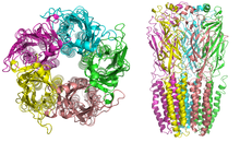
Nicotinic receptor structure
 
Nicotinic receptors, with a molecular mass of 290 kDa, are made up of five subunits, arranged symmetrically around a central pore. Each subunit comprises four transmembrane domains with both the N- and C-terminus located extracellularly. They possess similarities with GABAA receptors, glycine receptors, and the type 3 serotonin receptors (which are all ionotropic receptors), or the signature Cys-loop proteins.

In vertebrates, nicotinic receptors are broadly classified into two subtypes based on their primary sites of expression: muscle-type nicotinic receptors and neuronal-type nicotinic receptors. In the muscle-type receptors, found at the neuromuscular junction, receptors are either the embryonic form, composed of α1, β1, γ, and δ subunits in a 2:1:1:1 ratio, or the adult form composed of α1, β1, δ, and ε subunits in a 2:1:1:1 ratio. The neuronal subtypes are various homomeric (all one type of subunit) or heteromeric (at least one α and one β) combinations of twelve different nicotinic receptor subunits: α2−α10 and β2−β4. Examples of the neuronal subtypes include: (α4)3(β2)2, (α4)2(β2)3, (α3)2(β4)3, α4α6β3(β2)2, (α7)5, and many others. In both muscle-type and neuronal-type receptors, the subunits are very similar to one another, especially in the hydrophobic regions. 

A number of electron microscopy and x-ray crystallography studies have provided very high resolution structural information for muscle and neuronal nAChRs and their binding domains.

Binding to the receptor

As with all ligand-gated ion channels, opening of the nAChR channel pore requires the binding of a chemical messenger. Several different terms are used to refer to the molecules that bind receptors, such as ligand, agonist, or transmitter. As well as the endogenous agonist acetylcholine, agonists of the nAChR include nicotine, epibatidine, and choline. Nicotinic antagonists that block the receptor include mecamylamine, dihydro-β-erythroidine, and hexamethonium

In muscle-type nAChRs, the acetylcholine binding sites are located at the α and either ε or δ subunits interface. In neuronal nAChRs, the binding site is located at the interface of an α and a β subunit or between two α subunits in the case of α7 receptors. The binding site is located in the extracellular domain near the N terminus. When an agonist binds to the site, all present subunits undergo a conformational change and the channel is opened and a pore with a diameter of about 0.65 nm opens.

Opening the channel

Nicotinic AChRs may exist in different interconvertible conformational states. Binding of an agonist stabilises the open and desensitised states. In normal physiological conditions, the receptor needs exactly two molecules of ACh to open. Opening of the channel allows positively charged ions to move across it; in particular, sodium enters the cell and potassium exits. The net flow of positively charged ions is inward. 

The nAChR is a non-selective cation channel, meaning that several different positively charged ions can cross through. It is permeable to Na+ and K+, with some subunit combinations that are also permeable to Ca2+. The amount of sodium and potassium the channels allow through their pores (their conductance) varies from 50–110 pS, with the conductance depending on the specific subunit composition as well as the permeant ion.

Many neuronal nAChRs can affect the release of other neurotransmitters. The channel usually opens rapidly and tends to remain open until the agonist diffuses away, which usually takes about 1 millisecond. However, AChRs can spontaneously open with no ligands bound or can spontaneously close with ligands bound, and mutations in the channel can shift the likelihood of either event. Therefore, ACh binding changes the probability of pore opening, which increases as more ACh binds. 

The nAChR is unable to bind ACh when bound to any of the snake venom α-neurotoxins. These α-neurotoxins antagonistically bind tightly and noncovalently to nAChRs of skeletal muscles and in neurons, thereby blocking the action of ACh at the postsynaptic membrane, inhibiting ion flow and leading to paralysis and death. The nAChR contains two binding sites for snake venom neurotoxins. Progress towards discovering the dynamics of binding action of these sites has proved difficult, although recent studies using normal mode dynamics have aided in predicting the nature of both the binding mechanisms of snake toxins and of ACh to nAChRs. These studies have shown that a twist-like motion caused by ACh binding is likely responsible for pore opening, and that one or two molecules of α-bungarotoxin (or other long-chain α-neurotoxin) suffice to halt this motion. The toxins seem to lock together neighboring receptor subunits, inhibiting the twist and therefore, the opening motion.

Effects

The activation of receptors by nicotine modifies the state of neurons through two main mechanisms. On one hand, the movement of cations causes a depolarization of the plasma membrane (which results in an excitatory postsynaptic potential in neurons) leading to the activation of voltage-gated ion channels. On the other hand, the entry of calcium acts, either directly or indirectly, on different intracellular cascades. This leads, for example, to the regulation of the activity of some genes or the release of neurotransmitters.

Receptor regulation

Receptor desensitisation

Ligand-bound desensitisation of receptors was first characterised by Katz and Thesleff in the nicotinic acetylcholine receptor.

Prolonged or repeated exposure to a stimulus often results in decreased responsiveness of that receptor toward a stimulus, termed desensitisation. nAChR function can be modulated by phosphorylation by the activation of second messenger-dependent protein kinases. PKA and PKC, as well as tyrosine kinases, have been shown to phosphorylate the nAChR resulting in its desensitisation. It has been reported that, after prolonged receptor exposure to the agonist, the agonist itself causes an agonist-induced conformational change in the receptor, resulting in receptor desensitisation. Desensitised receptors can revert to a prolonged open state when an agonist is bound in the presence of a positive allosteric modulator, for example PNU-120596. Also, there is evidence that indicates specific chaperone molecules have regulatory effects on these receptors.

Roles

The subunits of the nicotinic receptors belong to a multigene family (16 members in humans) and the assembly of combinations of subunits results in a large number of different receptors. These receptors, with highly variable kinetic, electrophysiological and pharmacological properties, respond to nicotine differently, at very different effective concentrations. This functional diversity allows them to take part in two major types of neurotransmission. Classical synaptic transmission (wiring transmission) involves the release of high concentrations of neurotransmitter, acting on immediately neighboring receptors. In contrast, paracrine transmission (volume transmission) involves neurotransmitters released by synaptic boutons, which then diffuse through the extra-cellular medium until they reach their receptors, which may be distant. Nicotinic receptors can also be found in different synaptic locations; for example the muscle nicotinic receptor always functions post-synaptically. The neuronal forms of the receptor can be found both post-synaptically (involved in classical neurotransmission) and pre-synaptically where they can influence the release of multiple neurotransmitters.

Subunits

17 vertebrate nAChR subunits have been identified, which are divided into muscle-type and neuronal-type subunits. However, although an α8 subunit/gene is present in avian species such as the chicken, it is not present in human or mammalian species.

The nAChR subunits have been divided into 4 subfamilies (I-IV) based on similarities in protein sequence. In addition, subfamily III has been further divided into 3 types. 

Neuronal-type Muscle-type
I II III IV
α9, α10 α7, α8 1 2 3 α1, β1, δ, γ, ε
α2, α3, α4, α6 β2, β4 β3, α5

Neuromuscular junction

From Wikipedia, the free encyclopedia

At the neuromuscular junction, the nerve fiber is able to transmit a signal to the muscle fiber by releasing ACh (and other substances), causing muscle contraction.
 
Muscles will contract or relax when they receive signals from the nervous system. The neuromuscular junction is the site of the signal exchange. The steps of this process in vertebrates occur as follows:(1) The action potential reaches the axon terminal. (2) Voltage-dependent calcium gates open, allowing calcium to enter the axon terminal. (3) Neurotransmitter vesicles fuse with the presynaptic membrane and ACh is released into the synaptic cleft via exocytosis. (4) ACh binds to postsynaptic receptors on the sarcolemma. (5) This binding causes ion channels to open and allows sodium and other cations to flow across the membrane into the muscle cell. (6) The flow of sodium ions across the membrane into and potassium ions out of the muscle cell generates an action potential which travels to the myofibril and results in muscle contraction.Labels:A: Motor Neuron AxonB: Axon TerminalC. Synaptic CleftD. Muscle CellE. Part of a Myofibril
 
Neuromuscular junctions
Electron micrograph of neuromuscular junction (cross-section).jpg
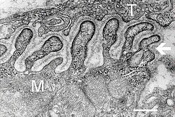
Electron micrograph showing a cross section through the neuromuscular junction. T is the axon terminal, M is the muscle fiber. The arrow shows junctional folds with basal lamina. Active zones are visible on the tips between the folds. Scale is 0.3 µm. Source: NIMH
Synapse diag4.png
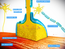
Detailed view of a neuromuscular junction:
Details
Identifiers
Latinsynapssis neuromuscularis; junctio neuromuscularis
MeSHD009469
THH2.00.06.1.02001
FMA61803

A neuromuscular junction (or myoneural junction) is a chemical synapse formed by the contact between a motor neuron and a muscle fiber. It is at the neuromuscular junction that a motor neuron is able to transmit a signal to the muscle fiber, causing muscle contraction

Muscles require innervation to function—and even just to maintain muscle tone, avoiding atrophy. Synaptic transmission at the neuromuscular junction begins when an action potential reaches the presynaptic terminal of a motor neuron, which activates voltage-dependent calcium channels to allow calcium ions to enter the neuron. Calcium ions bind to sensor proteins (synaptotagmin) on synaptic vesicles, triggering vesicle fusion with the cell membrane and subsequent neurotransmitter release from the motor neuron into the synaptic cleft. In vertebrates, motor neurons release acetylcholine (ACh), a small molecule neurotransmitter, which diffuses across the synaptic cleft and binds to nicotinic acetylcholine receptors (nAChRs) on the cell membrane of the muscle fiber, also known as the sarcolemma. nAChRs are ionotropic receptors, meaning they serve as ligand-gated ion channels. The binding of ACh to the receptor can depolarize the muscle fiber, causing a cascade that eventually results in muscle contraction. 

Neuromuscular junction diseases can be of genetic and autoimmune origin. Genetic disorders, such as Duchenne muscular dystrophy, can arise from mutated structural proteins that comprise the neuromuscular junction, whereas autoimmune diseases, such as myasthenia gravis, occur when antibodies are produced against nicotinic acetylcholine receptors on the sarcolemma.

Structure and function

motor endplate

Quantal transmission

At the neuromuscular junction presynaptic motor axons terminate 30 nanometers from the cell membrane or sarcolemma of a muscle fiber. The sarcolemma at the junction has invaginations called postjunctional folds, which increase its surface area facing the synaptic cleft. These postjunctional folds form the motor endplate, which is studded with nicotinic acetylcholine receptors (nAChRs) at a density of 10,000 receptors/micrometer2. The presynaptic axons terminate in bulges called terminal boutons (or presynaptic terminals) that project toward the postjunctional folds of the sarcolemma. In the frog each motor nerve terminal contains about 300,000 vesicles, with an average diameter of 0.05 micrometers. The vesicles contain acetylcholine. Some of these vesicles are gathered into groups of fifty, positioned at active zones close to the nerve membrane. Active zones are about 1 micrometer apart. The 30 nanometer cleft between nerve ending and endplate contains a meshwork of acetylcholinesterase (AChE) at a density of 2,600 enzyme molecules/micrometer2, held in place by the structural proteins dystrophin and rapsyn. Also present is the receptor tyrosine kinase protein MuSK, a signaling protein involved in the development of the neuromuscular junction, which is also held in place by rapsyn.

About once every second in a resting junction randomly one of the synaptic vesicles fuses with the presynaptic neuron's cell membrane in a process mediated by SNARE proteins. Fusion results in the emptying of the vesicle's contents of 7000-10,000 acetylcholine molecules into the synaptic cleft, a process known as exocytosis. Consequently exocytosis releases acetylcholine in packets that are called quanta. The acetylcholine quantum diffuses through the acetylcholinesterase meshwork, where the high local transmitter concentration occupies all of the binding sites on the enzyme in its path. The acetylcholine that reaches the endplate activates ~2,000 acetylcholine receptors, opening their ion channels which permits sodium ions to move into the endplate producing a depolarization of ~0.5 mV known as a miniature endplate potential (MEPP). By the time the acetylcholine is released from the receptors the acetylcholinesterase has destroyed its bound ACh, which takes about ~0.16 ms, and hence is available to destroy the ACh released from the receptors.

When the motor nerve is stimulated there is a delay of only 0.5 to 0.8 msec between the arrival of the nerve impulse in the motor nerve terminals and the first response of the endplate  The arrival of the motor nerve action potential at the presynaptic neuron terminal opens voltage-dependent calcium channels and Ca2+ ions flow from the extracellular fluid into the presynaptic neuron's cytosol. This influx of Ca2+ causes several hundred neurotransmitter-containing vesicles to fuse with the presynaptic neuron's cell membrane through SNARE proteins to release their acetylcholine quanta by exocytosis. The endplate depolarization by the released acetylcholine is called an endplate potential (EPP). The EPP is accomplished when ACh binds the nicotinic acetylcholine receptors (nAChR) at the motor end plate, and causes an influx of sodium ions. This influx of sodium ions generates the EPP (depolarization), and triggers an action potential which travels along the sarcolemma and into the muscle fiber via the transverse tubules (T-tubules) by means of voltage-gated sodium channels. The conduction of action potentials along the transverse tubules stimulates the opening of voltage-gated Ca2+ channels which are mechanically coupled to Ca2+ release channels in the sarcoplasmic reticulum. The Ca2+ then diffuses out of the sarcoplasmic reticulum to the myofibrils so it can stimulate contraction. The endplate potential is thus responsible for setting up an action potential in the muscle fiber which triggers muscle contraction. The transmission from nerve to muscle is so rapid because each quantum of acetylcholine reaches the endplate in millimolar concentrations, high enough to combine with a receptor with a low affinity, which then swiftly releases the bound transmitter.

Acetylcholine receptors

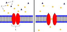
  1. Ion channel linked receptor
  2. Ions
  3. Ligand (such as acetylcholine)
When ligands bind to the receptor, the ion channel portion of the receptor opens, allowing ions to pass across the cell membrane.
 
Acetylcholine is a neurotransmitter synthesized from dietary choline and acetyl-CoA (ACoA), and is involved in the stimulation of muscle tissue in vertebrates as well as in some invertebrate animals. In vertebrate animals, the acetylcholine receptor subtype that is found at the neuromuscular junction of skeletal muscles is the nicotinic acetylcholine receptor (nAChR), which is a ligand-gated ion channel. Each subunit of this receptor has a characteristic "cys-loop", which is composed of a cysteine residue followed by 13 amino acid residues and another cysteine residue. The two cysteine residues form a disulfide linkage which results in the "cys-loop" receptor that is capable of binding acetylcholine and other ligands. These cys-loop receptors are found only in eukaryotes, but prokaryotes possess ACh receptors with similar properties. Not all species use a cholinergic neuromuscular junction; e.g. crayfish and fruit flies have a glutamatergic neuromuscular junction.

AChRs at the skeletal neuromuscular junction form heteropentamers composed of two α, one β, one ɛ, and one δ subunits. When a single ACh ligand binds to one of the α subunits of the ACh receptor it induces a conformational change at the interface with the second AChR α subunit. This conformational change results in the increased affinity of the second α subunit for a second ACh ligand. AChRs therefore exhibit a sigmoidal dissociation curve due to this cooperative binding. The presence of the inactive, intermediate receptor structure with a single-bound ligand keeps ACh in the synapse that might otherwise be lost by cholinesterase hydrolysis or diffusion. The persistence of these ACh ligands in the synapse can cause a prolonged post-synaptic response.

Development

The development of the neuromuscular junction requires signaling from both the motor neuron's terminal and the muscle cell's central region. During development, muscle cells produce acetylcholine receptors (AChRs) and express them in the central regions in a process called prepatterning. Agrin, a heparin proteoglycan, and MuSK kinase are thought to help stabilize the accumulation of AChR in the central regions of the myocyte. MuSK is a receptor tyrosine kinase—meaning that it induces cellular signaling by binding phosphate molecules to self regions like tyrosines, and to other targets in the cytoplasm. Upon activation by its ligand agrin, MuSK signals via two proteins called "Dok-7" and "rapsyn", to induce "clustering" of acetylcholine receptors. ACh release by developing motor neurons produces postsynaptic potentials in the muscle cell that positively reinforces the localization and stabilization of the developing neuromuscular junction.

These findings were demonstrated in part by mouse "knockout" studies. In mice which are deficient for either agrin or MuSK, the neuromuscular junction does not form. Further, mice deficient in Dok-7 did not form either acetylcholine receptor clusters or neuromuscular synapses.

The development of neuromuscular junctions is mostly studied in model organisms, such as rodents. In addition, in 2015 an all-human neuromuscular junction has been created in vitro using human embryonic stem cells and somatic muscle stem cells. In this model presynaptic motor neurons are activated by optogenetics and in response synaptically connected muscle fibers twitch upon light stimulation.

Research methods

José del Castillo and Bernard Katz used ionophoresis to determine the location and density of nicotinic acetylcholine receptors (nAChRs) at the neuromuscular junction. With this technique, a microelectrode was placed inside the motor endplate of the muscle fiber, and a micropipette filled with acetylcholine (ACh) is placed directly in front of the endplate in the synaptic cleft. A positive voltage was applied to the tip of the micropipette, which caused a burst of positively charged ACh molecules to be released from the pipette. These ligands flowed into the space representing the synaptic cleft and bound to AChRs. The intracellular microelectrode monitored the amplitude of the depolarization of the motor endplate in response to ACh binding to nicotinic (ionotropic) receptors. Katz and del Castillo showed that the amplitude of the depolarization (excitatory postsynaptic potential) depended on the proximity of the micropipette releasing the ACh ions to the endplate. The farther the micropipette was from the motor endplate, the smaller the depolarization was in the muscle fiber. This allowed the researchers to determine that the nicotinic receptors were localized to the motor endplate in high density.

Toxins are also used to determine the location of acetylcholine receptors at the neuromuscular junction. α-Bungarotoxin is a toxin found in the snake species Bungarus multicinctus that acts as an ACh antagonist and binds to AChRs irreversibly. By coupling assayable enzymes such as horseradish peroxidase (HRP) or fluorescent proteins such as green fluorescent protein (GFP) to the α-bungarotoxin, AChRs can be visualized and quantified.

Toxins that affect the neuromuscular junction

Nerve gases

Botulinum toxin injected in human face

Botulinum toxin

Botulinum toxin (aka botulinum neurotoxin, BoNT, and sold under the trade name Botox) inhibits the release of acetylcholine at the neuromuscular junction by interfering with SNARE proteins. This toxin crosses into the nerve terminal through the process of endocytosis and subsequently interferes with SNARE proteins, which are necessary for ACh release. By doing so, it induces a transient flaccid paralysis and chemical denervation localized to the striated muscle that it has affected. The inhibition of the ACh release does not set in until approximately two weeks after the injection is made. Three months after the inhibition occurs, neuronal activity begins to regain partial function, and six months, complete neuronal function is regained.

Tetanus toxin

Tetanus toxin, also known as tetanospasmin is a potent neurotoxin produced by Clostridium tetani and causes the disease state, tetanus. The LD50 of this toxin has been measured to be approximately 1 ng/kg, making it second only to Botulinum toxin D as the deadliest toxin in the world. It functions very similarly to botunlinum neurotoxin (BoNT) by attaching and endocytosing into the presynaptic nerve terminal and interfering with SNARE protein complexes. It differs from BoNT in a few ways, most apparently in its end state, wherein tetanospasmin demonstrates a rigid / spastic paralysis as opposed to the flaccid paralysis demonstrated with BoNT.

Latrotoxin

Latrotoxin (α-Latrotoxin) found in venom of widow spiders also affects the neuromuscular junction by causing the release of acetylcholine from the presynaptic cell. Mechanisms of action include binding to receptors on the presynaptic cell activating the IP3/DAG pathway and release of calcium from intracellular stores and pore formation resulting in influx of calcium ions directly. Either mechanism causes increased calcium in presynaptic cell, which then leads to release of synaptic vesicles of acetylcholine. Latrotoxin causes pain, muscle contraction and if untreated potentially paralysis and death.

Snake venom

Snake venoms act as toxins at the neuromuscular junction and can induce weakness and paralysis. Venoms can act as both presynaptic and postsynaptic neurotoxins.

Presynaptic neurotoxins, commonly known as β-neurotoxins, affect the presynaptic regions of the neuromuscular junction. The majority of these neurotoxins act by inhibiting the release of neurotransmitters, such as acetylcholine, into the synapse between neurons. However, some of these toxins have also been known to enhance neurotransmitter release. Those that inhibit neurotransmitter release create a neuromuscular blockade that prevents signaling molecules from reaching their postsynaptic target receptors. In doing so, the victim of these snake bite suffer from profound weakness. Such neurotoxins do not respond well to anti-venoms. After one hour of inoculation of these toxins, including notexin and taipoxin, many of the affected nerve terminals show signs of irreversible physical damage, leaving them devoid of any synaptic vesicles.

Postsynaptic neurotoxins, otherwise known as α-neurotoxins, act oppositely to the presynaptic neurotoxins by binding to the postsynaptic acetylcholine receptors. This prevents interaction between the acetylcholine released by the presynaptic terminal and the receptors on the postsynaptic cell. In effect, the opening of sodium channels associated with these acetylcholine receptors is prohibited, resulting in a neuromuscular blockade, similar to the effects seen due to presynaptic neurotoxins. This causes paralysis in the muscles involved in the affected junctions. Unlike presynaptic neurotoxins, postsynaptic toxins are more easily affected by anti-venoms, which accelerate the dissociation of the toxin from the receptors, ultimately causing a reversal of paralysis. These neurotoxins experimentally and qualitatively aid in the study of acetylcholine receptor density and turnover, as well as in studies observing the direction of antibodies toward the affected acetylcholine receptors in patients diagnosed with myasthenia gravis.

Diseases

Any disorder that compromises the synaptic transmission between a motor neuron and a muscle cell is categorized under the umbrella term of neuromuscular diseases. These disorders can be inherited or acquired and can vary in their severity and mortality. In general, most of these disorders tend to be caused by mutations or autoimmune disorders. Autoimmune disorders, in the case of neuromuscular diseases, tend to be humoral mediated, B cell mediated, and result in an antibody improperly created against a motor neuron or muscle fiber protein that interferes with synaptic transmission or signaling.

Autoimmune

Myasthenia gravis

Myasthenia gravis is an autoimmune disorder where the body makes antibodies against either the acetylcholine receptor (AchR) (in 80% of cases), or against postsynaptic muscle-specific kinase (MuSK) (0–10% of cases). In seronegative myasthenia gravis low density lipoprotein receptor-related protein 4 is targeted by IgG1, which acts as a competitive inhibitor of its ligand, preventing the ligand from binding its receptor. It is not known if seronegative myasthenia gravis will respond to standard therapies.
Neonatal MG
Neonatal MG is an autoimmune disorder that affects 1 in 8 children born to mothers who have been diagnosed with myasthenia gravis (MG). MG can be transferred from the mother to the fetus by the movement of AChR antibodies through the placenta. Signs of this disease at birth include weakness, which responds to anticholinesterase medications, as well as fetal akinesia, or the lack of fetal movement. This form of the disease is transient, lasting for about three months. However, in some cases, neonatal MG can lead to other health effects, such as arthrogryposis and even fetal death. These conditions are thought to be initiated when maternal AChR antibodies are directed to the fetal AChR and can last until the 33rd week of gestation, when the γ subunit of AChR is replaced by the ε subunit.

Lambert-Eaton myasthenic syndrome

Lambert-Eaton myasthenic syndrome (LEMS) is an autoimmune disorder that affects the presynaptic portion of the neuromuscular junction. This rare disease can be marked by a unique triad of symptoms: proximal muscle weakness, autonomic dysfunction, and areflexia. Proximal muscle weakness is a product of pathogenic autoantibodies directed against P/Q-type voltage-gated calcium channels, which in turn leads to a reduction of acetylcholine release from motor nerve terminals on the presynaptic cell. Examples of autonomic dysfunction caused by LEMS include erectile dysfunction in men, constipation, and, most commonly, dry mouth. Less common dysfunctions include dry eyes and altered perspiration. Areflexia is a condition in which tendon reflexes are reduced and it may subside temporarily after a period of exercise.

50–60% of the patients that are diagnosed with LEMS also have present an associated tumor, which is typically small-cell lung carcinoma (SCLC). This type of tumor also expresses voltage-gated calcium channels. Oftentimes, LEMS also occurs alongside myasthenia gravis.

Treatment for LEMS consists of using 3,4-diaminopyridine as a first measure, which serves to increase the compound muscle action potential as well as muscle strength by lengthening the time that voltage-gated calcium channels remain open after blocking voltage-gated potassium channels. In the US, treatment with 3,4-diaminopyridine for eligible LEMS patients is available at no cost under an expanded access program. Further treatment includes the use of prednisone and azathioprine in the event that 3,4-diaminopyridine does not aid in treatment.

Neuromyotonia

Neuromyotonia (NMT), otherwise known as Isaac’s syndrome, is unlike many other diseases present at the neuromuscular junction. Rather than causing muscle weakness, NMT leads to the hyperexcitation of motor nerves. NMT causes this hyperexcitation by producing longer depolarizations by down-regulating voltage-gated potassium channels, which causes greater neurotransmitter release and repetitive firing. This increase in rate of firing leads to more active transmission and as a result, greater muscular activity in the affected individual. NMT is also believed to be of autoimmune origin due to its associations with autoimmune symptoms in the individual affected.

Genetic

Congenital myasthenic syndromes

Congenital myasthenic syndromes (CMS) are very similar to both MG and LEMS in their functions, but the primary difference between CMS and those diseases is that CMS is of genetic origins. Specifically, these syndromes are diseases incurred due to mutations, typically recessive, in 1 of at least 10 genes that affect presynaptic, synaptic, and postsynaptic proteins in the neuromuscular junction. Such mutations usually arise in the ε-subunit of AChR, thereby affecting the kinetics and expression of the receptor itself. Single nucleotide substitutions or deletions may cause loss of function in the subunit. Other mutations, such as those affecting acetylcholinesterase and acetyltransferase, can also cause the expression of CMS, with the latter being associated specifically with episodic apnea. These syndromes can present themselves at different times within the life of an individual. They may arise during the fetal phase, causing fetal akinesia, or the perinatal period, during which certain conditions, such as arthrogryposis, ptosis, hypotonia, ophthalmoplegia, and feeding or breathing difficulties, may be observed. They could also activate during adolescence or adult years, causing the individual to develop slow-channel syndrome.

Treatment for particular subtypes of CMS (postsynaptic fast-channel CMS) is similar to treatment for other neuromuscular disorders. 3,4-Diaminopyridine, the first-line treatment for LEMS, is under development as an orphan drug for CMS in the US, and available to eligible patients under an expanded access program at no cost.

Bulbospinal muscular atrophy

Bulbospinal muscular atrophy, also known as Kennedy’s disease, is a rare recessive trinucleotide, polyglutamine disorder that is linked to the X chromosome. Because of its linkage to the X chromosome, it is typically transmitted through females. However, Kennedy’s disease is only present in adult males and the onset of the disease is typically later in life. This disease is specifically caused by the expansion of a CAG-tandem repeat in exon 1 found on the androgen-receptor (AR) gene on chromosome Xq11-12. Poly-Q-expanded AR accumulates in the nuclei of cells, where it begins to fragment. After fragmentation, degradation of the cell begins, leading to a loss of both motor neurons and dorsal root ganglia.

Symptoms of Kennedy’s disease include weakness and wasting of the facial bulbar and extremity muscles, as well as sensory and endocrinological disturbances, such as gynecomastia and reduced fertility. Other symptoms include elevated testosterone and other sexual hormone levels, development of hyper-CK-emia, abnormal conduction through motor and sensory nerves, and neuropathic or in rare cases myopathic alterations on biopsies of muscle cells.

Duchenne muscular dystrophy

Duchenne muscular dystrophy is an X-linked genetic disorder that results in the absence of the structural protein dystrophin at the neuromuscular junction. It affects 1 in 3,600–6,000 males and frequently causes death by the age of 30. The absence of dystrophin causes muscle degeneration, and patients present with the following symptoms: abnormal gait, hypertrophy in the calf muscles, and elevated creatine kinase. If left untreated, patients may suffer from respiratory distress, which can lead to death.

Operator (computer programming)

From Wikipedia, the free encyclopedia https://en.wikipedia.org/wiki/Operator_(computer_programmin...