Search This Blog

Monday, February 4, 2019

DNA damage theory of aging

From Wikipedia, the free encyclopedia

The DNA damage theory of aging proposes that aging is a consequence of unrepaired accumulation of naturally occurring DNA damages. Damage in this context is a DNA alteration that has an abnormal structure. Although both mitochondrial and nuclear DNA damage can contribute to aging, nuclear DNA is the main subject of this analysis. Nuclear DNA damage can contribute to aging either indirectly (by increasing apoptosis or cellular senescence) or directly (by increasing cell dysfunction).

Several review articles have shown that deficient DNA repair, allowing greater accumulation of DNA damages, causes premature aging; and that increased DNA repair facilitates greater longevity. Mouse models of nucleotide-excision–repair syndromes reveal a striking correlation between the degree to which specific DNA repair pathways are compromised and the severity of accelerated aging, strongly suggesting a causal relationship. Human populations studies show that single-nucleotide polymorphisms in DNA repair genes, causing up-regulation of their expression, correlate with increases in longevity. Lombard et al. compiled a lengthy list of mouse mutational models with pathologic features of premature aging, all caused by different DNA repair defects. Freitas and de Magalhães presented a comprehensive review and appraisal of the DNA damage theory of aging, including a detailed analysis of many forms of evidence linking DNA damage to aging. As an example, they described a study showing that centenarians of 100 to 107 years of age had higher levels of two DNA repair enzymes, PARP1 and Ku70, than general-population old individuals of 69 to 75 years of age. Their analysis supported the hypothesis that improved DNA repair leads to longer life span. Overall, they concluded that while the complexity of responses to DNA damage remains only partly understood, the idea that DNA damage accumulation with age is the primary cause of aging remains an intuitive and powerful one.

In humans and other mammals, DNA damage occurs frequently and DNA repair processes have evolved to compensate. In estimates made for mice, DNA lesions occur on average 25 to 115 times per minute in each cell, or about 36,000 to 160,000 per cell per day. Some DNA damage may remain in any cell despite the action of repair processes. The accumulation of unrepaired DNA damage is more prevalent in certain types of cells, particularly in non-replicating or slowly replicating cells, such as cells in the brain, skeletal and cardiac muscle.

DNA damage and mutation

8-Hydroxydeoxyguanosine

To understand the DNA damage theory of aging it is important to distinguish between DNA damage and mutation, the two major types of errors that occur in DNA. Damage and mutation are fundamentally different. DNA damage is any physical abnormality in the DNA, such as single and double strand breaks, 8-hydroxydeoxyguanosine residues and polycyclic aromatic hydrocarbon adducts. DNA damage can be recognized by enzymes, and thus can be correctly repaired using the complementary undamaged sequence in a homologous chromosome if it is available for copying. If a cell retains DNA damage, transcription of a gene can be prevented and thus translation into a protein will also be blocked. Replication may also be blocked and/or the cell may die. Descriptions of reduced function, characteristic of aging and associated with accumulation of DNA damage, are given later in this article. 

In contrast to DNA damage, a mutation is a change in the base sequence of the DNA. A mutation cannot be recognized by enzymes once the base change is present in both DNA strands, and thus a mutation cannot be repaired. At the cellular level, mutations can cause alterations in protein function and regulation. Mutations are replicated when the cell replicates. In a population of cells, mutant cells will increase or decrease in frequency according to the effects of the mutation on the ability of the cell to survive and reproduce. Although distinctly different from each other, DNA damages and mutations are related because DNA damages often cause errors of DNA synthesis during replication or repair and these errors are a major source of mutation. 

Given these properties of DNA damage and mutation, it can be seen that DNA damages are a special problem in non-dividing or slowly dividing cells, where unrepaired damages will tend to accumulate over time. On the other hand, in rapidly dividing cells, unrepaired DNA damages that do not kill the cell by blocking replication will tend to cause replication errors and thus mutation. The great majority of mutations that are not neutral in their effect are deleterious to a cell’s survival. Thus, in a population of cells comprising a tissue with replicating cells, mutant cells will tend to be lost. However, infrequent mutations that provide a survival advantage will tend to clonally expand at the expense of neighboring cells in the tissue. This advantage to the cell is disadvantageous to the whole organism, because such mutant cells can give rise to cancer. Thus DNA damages in frequently dividing cells, because they give rise to mutations, are a prominent cause of cancer. In contrast, DNA damages in infrequently dividing cells are likely a prominent cause of aging. 

The first person to suggest that DNA damage, as distinct from mutation, is the primary cause of aging was Alexander in 1967. By the early 1980s there was significant experimental support for this idea in the literature. By the early 1990s experimental support for this idea was substantial, and furthermore it had become increasingly evident that oxidative DNA damage, in particular, is a major cause of aging.

In a series of articles from 1970 to 1977, PV Narasimh Acharya, Phd. (1924–1993) theorized and presented evidence that cells undergo "irreparable DNA damage", whereby DNA crosslinks occur when both normal cellular repair processes fail and cellular apoptosis does not occur. Specifically, Acharya noted that double-strand breaks and a "cross-linkage joining both strands at the same point is irreparable because neither strand can then serve as a template for repair. The cell will die in the next mitosis or in some rare instances, mutate."

Age-associated accumulation of DNA damage and decline in gene expression

In tissues composed of non- or infrequently replicating cells, DNA damage can accumulate with age and lead either to loss of cells, or, in surviving cells, loss of gene expression. Accumulated DNA damage is usually measured directly. Numerous studies of this type have indicated that oxidative damage to DNA is particularly important. The loss of expression of specific genes can be detected at both the mRNA level and protein level.

Brain

The adult brain is composed in large part of terminally differentiated non-dividing neurons. Many of the conspicuous features of aging reflect a decline in neuronal function. Accumulation of DNA damage with age in the mammalian brain has been reported during the period 1971 to 2008 in at least 29 studies. This DNA damage includes the oxidized nucleoside 8-oxo-2'-deoxyguanosine (8-oxo-dG), single- and double-strand breaks, DNA-protein crosslinks and malondialdehyde adducts (reviewed in Bernstein et al.). Increasing DNA damage with age has been reported in the brains of the mouse, rat, gerbil, rabbit, dog, and human.

Rutten et al. showed that single-strand breaks accumulate in the mouse brain with age. Young 4-day-old rats have about 3,000 single-strand breaks and 156 double-strand breaks per neuron, whereas in rats older than 2 years the level of damage increases to about 7,400 single-strand breaks and 600 double-strand breaks per neuron. Sen et al. showed that DNA damages which block the polymerase chain reaction in rat brain accumulate with age. Swain and Rao observed marked increases in several types of DNA damages in aging rat brain, including single-strand breaks, double-strand breaks and modified bases (8-OHdG and uracil). Wolf et al. also showed that the oxidative DNA damage 8-OHdG accumulates in rat brain with age. Similarly, it was shown that as humans age from 48 to 97 years, 8-OHdG accumulates in the brain.

Lu et al. studied the transcriptional profiles of the human frontal cortex of individuals ranging from 26 to 106 years of age. This led to the identification of a set of genes whose expression was altered after age 40. These genes play central roles in synaptic plasticity, vesicular transport and mitochondrial function. In the brain, promoters of genes with reduced expression have markedly increased DNA damage. In cultured human neurons, these gene promoters are selectively damaged by oxidative stress. Thus Lu et al. concluded that DNA damage may reduce the expression of selectively vulnerable genes involved in learning, memory and neuronal survival, initiating a program of brain aging that starts early in adult life.

Muscle

Muscle strength, and stamina for sustained physical effort, decline in function with age in humans and other species. Skeletal muscle is a tissue composed largely of multinucleated myofibers, elements that arise from the fusion of mononucleated myoblasts. Accumulation of DNA damage with age in mammalian muscle has been reported in at least 18 studies since 1971. Hamilton et al. reported that the oxidative DNA damage 8-OHdG accumulates in heart and skeletal muscle (as well as in brain, kidney and liver) of both mouse and rat with age. In humans, increases in 8-OHdG with age were reported for skeletal muscle. Catalase is an enzyme that removes hydrogen peroxide, a reactive oxygen species, and thus limits oxidative DNA damage. In mice, when catalase expression is increased specifically in mitochondria, oxidative DNA damage (8-OHdG) in skeletal muscle is decreased and lifespan is increased by about 20%. These findings suggest that mitochondria are a significant source of the oxidative damages contributing to aging.

Protein synthesis and protein degradation decline with age in skeletal and heart muscle, as would be expected, since DNA damage blocks gene transcription. In 2005, Piec et al. found numerous changes in protein expression in rat skeletal muscle with age, including lower levels of several proteins related to myosin and actin. Force is generated in striated muscle by the interactions between myosin thick filaments and actin thin filaments.

Liver

Liver hepatocytes do not ordinarily divide and appear to be terminally differentiated, but they retain the ability to proliferate when injured. With age, the mass of the liver decreases, blood flow is reduced, metabolism is impaired, and alterations in microcirculation occur. At least 21 studies have reported an increase in DNA damage with age in liver. For instance, Helbock et al. estimated that the steady state level of oxidative DNA base alterations increased from 24,000 per cell in the liver of young rats to 66,000 per cell in the liver of old rats.

Kidney

In kidney, changes with age include reduction in both renal blood flow and glomerular filtration rate, and impairment in the ability to concentrate urine and to conserve sodium and water. DNA damages, particularly oxidative DNA damages, increase with age (at least 8 studies). For instance Hashimoto et al. showed that 8-OHdG accumulates in rat kidney DNA with age.

Long-lived stem cells

Tissue-specific stem cells produce differentiated cells through a series of increasingly more committed progenitor intermediates. In hematopoiesis (blood cell formation), the process begins with long-term hematopoietic stem cells that self-renew and also produce progeny cells that upon further replication go through a series of stages leading to differentiated cells without self-renewal capacity. In mice, deficiencies in DNA repair appear to limit the capacity of hematopoietic stem cells to proliferate and self-renew with age. Sharpless and Depinho reviewed evidence that hematopoietic stem cells, as well as stem cells in other tissues, undergo intrinsic aging. They speculated that stem cells grow old, in part, as a result of DNA damage. DNA damage may trigger signalling pathways, such as apoptosis, that contribute to depletion of stem cell stocks. This has been observed in several cases of accelerated aging and may occur in normal aging too.

A key aspect of hair loss with age is the aging of the hair follicle. Ordinarily, hair follicle renewal is maintained by the stem cells associated with each follicle. Aging of the hair follicle appears to be due to the DNA damage that accumulates in renewing stem cells during aging.

Mutation theories of aging

A popular idea, that has failed to gain significant experimental support, is the idea that mutation, as distinct from DNA damage, is the primary cause of aging. As discussed above, mutations tend to arise in frequently replicating cells as a result of errors of DNA synthesis when template DNA is damaged, and can give rise to cancer. However, in mice there is no increase in mutation in the brain with aging. Mice defective in a gene (Pms2) that ordinarily corrects base mispairs in DNA have about a 100-fold elevated mutation frequency in all tissues, but do not appear to age more rapidly. On the other hand, mice defective in one particular DNA repair pathway show clear premature aging, but do not have elevated mutation.

One variation of the idea that mutation is the basis of aging, that has received much attention, is that mutations specifically in mitochondrial DNA are the cause of aging. Several studies have shown that mutations accumulate in mitochondrial DNA in infrequently replicating cells with age. DNA polymerase gamma is the enzyme that replicates mitochondrial DNA. A mouse mutant with a defect in this DNA polymerase is only able to replicate its mitochondrial DNA inaccurately, so that it sustains a 500-fold higher mutation burden than normal mice. These mice showed no clear features of rapidly accelerated aging. Overall, the observations discussed in this section indicate that mutations are not the primary cause of aging.

Dietary restriction

In rodents, caloric restriction slows aging and extends lifespan. At least 4 studies have shown that caloric restriction reduces 8-OHdG damages in various organs of rodents. One of these studies showed that caloric restriction reduced accumulation of 8-OHdG with age in rat brain, heart and skeletal muscle, and in mouse brain, heart, kidney and liver. More recently, Wolf et al. showed that dietary restriction reduced accumulation of 8-OHdG with age in rat brain, heart, skeletal muscle, and liver. Thus reduction of oxidative DNA damage is associated with a slower rate of aging and increased lifespan.

Inherited defects that cause premature aging

If DNA damage is the underlying cause of aging, it would be expected that humans with inherited defects in the ability to repair DNA damages should age at a faster pace than persons without such a defect. Numerous examples of rare inherited conditions with DNA repair defects are known. Several of these show multiple striking features of premature aging, and others have fewer such features. Perhaps the most striking premature aging conditions are Werner syndrome (mean lifespan 47 years), Huchinson-Gilford Progeria (mean lifespan 13 years), and Cockayne syndrome (mean lifespan 13 years). 

Werner syndrome is due to an inherited defect in an enzyme (a helicase and exonuclease) that acts in base excision repair of DNA (e.g. see Harrigan et al.). 

Hutchinson-Guilford Progeria is due to a defect in Lamin A protein which forms a scaffolding within the cell nucleus to organize chromatin and is needed for repair of double-strand breaks in DNA. A-type lamins promote genetic stability by maintaining levels of proteins that have key roles in the DNA repair processes of non-homologous end joining and homologous recombination. Mouse cells deficient for maturation of prelamin A show increased DNA damage and chromosome aberrations and are more sensitive to DNA damaging agents.

Cockayne Syndrome is due to a defect in a protein necessary for the repair process, transcription coupled nucleotide excision repair, which can remove damages, particularly oxidative DNA damages, that block transcription.

In addition to these three conditions, several other human syndromes, that also have defective DNA repair, show several features of premature aging. These include ataxia telangiectasia, Nijmegen breakage syndrome, some subgroups of xeroderma pigmentosum, trichothiodystrophy, Fanconi anemia, Bloom syndrome and Rothmund-Thomson syndrome.

Ku bound to DNA
 
In addition to human inherited syndromes, experimental mouse models with genetic defects in DNA repair show features of premature aging and reduced lifespan. In particular, mutant mice defective in Ku70, or Ku80, or double mutant mice deficient in both Ku70 and Ku80 exhibit early aging. The mean lifespans of the three mutant mouse strains were similar to each other, at about 37 weeks, compared to 108 weeks for the wild-type control. Six specific signs of aging were examined, and the three mutant mice were found to display the same aging signs as the control mice, but at a much earlier age. Cancer incidence was not increased in the mutant mice. Ku70 and Ku80 form the heterodimer Ku protein essential for the non-homologous end joining (NHEJ) pathway of DNA repair, active in repairing DNA double-strand breaks. This suggests an important role of NHEJ in longevity assurance.

Defects in DNA repair cause features of premature aging

Many authors have noted an association between defects in the DNA damage response and premature aging. If a DNA repair protein is deficient, unrepaired DNA damages tend to accumulate. Such accumulated DNA damages appear to cause features of premature aging (segmental progeria). Table 1 lists 18 DNA repair proteins which, when deficient, cause numerous features of premature aging.

Table 1. DNA repair proteins that, when deficient, cause features of accelerated aging (segmental progeria).
Protein Pathway Description
ATR Nucleotide excision repair deletion of ATR in adult mice leads to a number of disorders including hair loss and graying, kyphosis, osteoporosis, premature involution of the thymus, fibrosis of the heart and kidney and decreased spermatogenesis
DNA-PKcs Non-homologous end joining shorter lifespan, earlier onset of aging related pathologies; higher level of DNA damage persistence
ERCC1 Nucleotide excision repair, Interstrand cross link repair deficient transcription coupled NER with time-dependent accumulation of transcription-blocking damages; mouse life span reduced from 2.5 years to 5 months; Ercc1−/− mice are leukopenic and thrombocytopenic, and there is extensive adipose transformation of the bone marrow, hallmark features of normal aging in mice
ERCC2 (XPD) Nucleotide excision repair (also transcription as part of TFIIH) some mutations in ERCC2 cause Cockayne syndrome in which patients have segmental progeria with reduced stature, mental retardation, cachexia (loss of subcutaneous fat tissue), sensorineural deafness, retinal degeneration, and calcification of the central nervous system; other mutations in ERCC2 cause trichothiodystrophy in which patients have segmental progeria with brittle hair, short stature, progressive cognitive impairment and abnormal face shape; still other mutations in ERCC2 cause xeroderma pigmentosum (without a progeroid syndrome) and with extreme sun-mediated skin cancer predisposition
ERCC4 (XPF) Nucleotide excision repair, Interstrand cross link repair, Single-strand annealing, Microhomology-mediated end joining mutations in ERCC4 cause symptoms of accelerated aging that affect the neurologic, hepatobiliary, musculoskeletal, and hematopoietic systems, and cause an old, wizened appearance, loss of subcutaneous fat, liver dysfunction, vision and hearing loss, renal insufficiency, muscle wasting, osteopenia, kyphosis and cerebral atrophy
ERCC5 (XPG) Nucleotide excision repair, Homologous recombinational repair, Base excision repair mice with deficient ERCC5 show loss of subcutaneous fat, kyphosis, osteoporosis, retinal photoreceptor loss, liver aging, extensive neurodegeneration, and a short lifespan of 4–5 months
ERCC6 (Cockayne syndrome B or CS-B) Nucleotide excision repair [especially transcription coupled repair (TC-NER) and interstrand crosslink repair] premature aging features with shorter life span and photosensitivity, deficient transcription coupled NER with accumulation of unrepaired DNA damages, also defective repair of oxidatively generated DNA damages including 8-oxoguanine, 5-hydroxycytosine and cyclopurines
ERCC8 (Cockayne syndrome A or CS-A) Nucleotide excision repair [especially transcription coupled repair (TC-NER) and interstrand crosslink repair] premature aging features with shorter life span and photosensitivity, deficient transcription coupled NER with accumulation of unrepaired DNA damages, also defective repair of oxidatively generated DNA damages including 8-oxoguanine, 5-hydroxycytosine and cyclopurines
GTF2H5 (TTDA) Nucleotide excision repair deficiency causes trichothiodystrophy (TTD) a premature-ageing and neuroectodermal disease; humans with GTF2H5 mutations have a partially inactivated protein with retarded repair of 6-4-photoproducts
Ku70 Non-homologous end joining shorter lifespan, earlier onset of aging related pathologies; persistent foci of DNA double-strand break repair proteins
Ku80 Non-homologous end joining shorter lifespan, earlier onset of aging related pathologies; defective repair of spontaneous DNA damage
Lamin A Non-homologous end joining, Homologous recombination increased DNA damage and chromosome aberrations; progeria; aspects of premature aging; altered expression of numerous DNA repair factors
NRMT1 Nucleotide excision repair mutation in NRMT1 causes decreased body size, female-specific infertility, kyphosis, decreased mitochondrial function, and early-onset liver degeneration
RECQL4 Base excision repair, Nucleotide excision repair, Homologous recombination, Non-homologous end joining mutations in RECQL4 cause Rothmund-Thomson syndrome, with alopecia, sparse eyebrows and lashes, cataracts and osteoporosis
SIRT6 Base excision repair, Nucleotide excision repair, Homologous recombination, Non-homologous end joining SIRT6-deficient mice develop profound lymphopenia, loss of subcutaneous fat and lordokyphosis, and these defects overlap with aging-associated degenerative processes
SIRT7 Non-homologous end joining mice defective in SIRT7 show phenotypic and molecular signs of accelerated aging such as premature pronounced curvature of the spine, reduced life span, and reduced non-homologous end joining
Werner syndrome helicase Homologous recombination, Non-homologous end joining,Base excision repair, Replication arrest recovery shorter lifespan, earlier onset of aging related pathologies, genome instability
ZMPSTE24 Homologous recombination lack of Zmpste24 prevents lamin A formation and causes progeroid phenotypes in mice and humans, increased DNA damage and chromosome aberrations, sensitivity to DNA-damaging agents and deficiency in homologous recombination

Increased DNA repair and extended longevity

Table 2 lists DNA repair proteins whose increased expression is connected to extended longevity.

Table 2. DNA repair proteins that, when highly- or over-expressed, cause (or are associated with) extended longevity.
Protein Pathway Description
NDRG1 Direct reversal long-lived Snell dwarf, GHRKO, and PAPPA-KO mice have increased expression of NDRG1; higher expression of NDRG1 can promote MGMT protein stability and enhanced DNA repair
NUDT1 (MTH1) Oxidized nucleotide removal degrades 8-oxodGTP; prevents the age-dependent accumulation of DNA 8-oxoguanine A transgenic mouse in which the human hMTH1 8-oxodGTPase is expressed, giving over-expression of hMTH1, increases the median lifespan of mice to 914 days vs. 790 days for wild-type mice. Mice with over-expressed hMTH1 have behavioral changes of reduced anxiety and enhanced investigation of environmental and social cues
PARP1 Base excision repair, Nucleotide excision repair, Microhomology-mediated end joining, Single-strand break repair PARP1 activity in blood cells of thirteen mammalian species (rat, guinea pig, rabbit, marmoset, sheep, pig, cattle, pigmy chimpanzee, horse, donkey, gorilla, elephant and man) correlates with maximum lifespan of the species.
SIRT1 Nucleotide excision repair, Homologous recombination, Non-homologous end joining Increased expression of SIRT1 in male mice extends the lifespan of mice fed a standard diet, accompanied by improvements in health, including enhanced motor coordination, performance, bone mineral density, and insulin sensitivity
SIRT6 Base excision repair, Nucleotide excision repair, Homologous recombination, Non-homologous end joining male, but not female, transgenic mice overexpressing Sirt6 have a significantly longer lifespan than wild-type mice

Lifespan in different mammalian species

Studies comparing DNA repair capacity in different mammalian species have shown that repair capacity correlates with lifespan. The initial study of this type, by Hart and Setlow, showed that the ability of skin fibroblasts of seven mammalian species to perform DNA repair after exposure to a DNA damaging agent correlated with lifespan of the species. The species studied were shrew, mouse, rat, hamster, cow, elephant and human. This initial study stimulated many additional studies involving a wide variety of mammalian species, and the correlation between repair capacity and lifespan generally held up. In one of the more recent studies, Burkle et al. studied the level of a particular enzyme, Poly ADP ribose polymerase, which is involved in repair of single-strand breaks in DNA. They found that the lifespan of 13 mammalian species correlated with the activity of this enzyme. 

The DNA repair transcriptomes of the liver of humans, naked mole-rats and mice were compared. The maximum lifespans of humans, naked mole-rat, and mouse are respectively ~120, 30 and 3 years. The longer-lived species, humans and naked mole rats expressed DNA repair genes, including core genes in several DNA repair pathways, at a higher level than did mice. In addition, several DNA repair pathways in humans and naked mole-rats were up-regulated compared with mouse. These findings suggest that increased DNA repair facilitates greater longevity.

Over the past decade, a series of papers have shown that the mitochondrial DNA (mtDNA) base composition correlates with animal species maximum life span. The mitochondrial DNA base composition is though to reflect its nucleotide-specific (guanine, cytosine, thymidine and adenine) different mutation rates (i.e., accumulation of guanine in the mitochondrial DNA of an animal species is due to low guanine mutation rate in the mitochondria of that species).

Centenarians

Lymphoblastoid cell lines established from blood samples of humans who lived past 100 years (centenarians) have significantly higher activity of the DNA repair protein Poly (ADP-ribose) polymerase (PARP) than cell lines from younger individuals (20 to 70 years old). The lymphocytic cells of centenarians have characteristics typical of cells from young people, both in their capability of priming the mechanism of repair after H2O2 sublethal oxidative DNA damage and in their PARP capacity.

Menopause

As women age, they experience a decline in reproductive performance leading to menopause. This decline is tied to a decline in the number of ovarian follicles. Although 6 to 7 million oocytes are present at mid-gestation in the human ovary, only about 500 (about 0.05%) of these ovulate, and the rest are lost. The decline in ovarian reserve appears to occur at an increasing rate with age, and leads to nearly complete exhaustion of the reserve by about age 51. As ovarian reserve and fertility decline with age, there is also a parallel increase in pregnancy failure and meiotic errors resulting in chromosomally abnormal conceptions. 

Titus et al. have proposed an explanation for the decline in ovarian reserve with age. They showed that as women age, double-strand breaks accumulate in the DNA of their primordial follicles. Primordial follicles are immature primary oocytes surrounded by a single layer of granulosa cells. An enzyme system is present in oocytes that normally accurately repairs DNA double-strand breaks. This repair system is referred to as homologous recombinational repair, and it is especially active during meiosis. Titus et al. also showed that expression of four key DNA repair genes that are necessary for homologous recombinational repair (BRCA1, MRE11, Rad51 and ATM) decline in oocytes with age. This age-related decline in ability to repair double-strand damages can account for the accumulation of these damages, which then likely contributes to the decline in ovarian reserve. 

Women with an inherited mutation in the DNA repair gene BRCA1 undergo menopause prematurely, suggesting that naturally occurring DNA damages in oocytes are repaired less efficiently in these women, and this inefficiency leads to early reproductive failure. Genomic data from about 70,000 women were analyzed to identify protein-coding variation associated with age at natural menopause. Pathway analyses identified a major association with DNA damage response genes, particularly those expressed during meiosis and including a common coding variant in the BRCA1 gene.

Atherosclerosis

The most important risk factor for cardiovascular problems is chronological aging. Several research groups have reviewed evidence for a key role of DNA damage in vascular aging.

Atherosclerotic plaque contains vascular smooth muscle cells, macrophages and endothelial cells and these have been found to accumulate 8-oxoG, a common type of oxidative DNA damage. DNA strand breaks also increased in atherosclerotic plaques, thus linking DNA damage to plaque formation.

Werner syndrome (WS), a premature aging condition in humans, is caused by a genetic defect in a RecQ helicase that is employed in several DNA repair processes. WS patients develop a substantial burden of atherosclerotic plaques in their coronary arteries and aorta. These findings link excessive unrepaired DNA damage to premature aging and early atherosclerotic plaque development.

DNA damage and the epigenetic clock

Endogenous, naturally occurring DNA damages are frequent, and in humans include an average of about 10,000 oxidative damages per day and 50 double-strand DNA breaks per cell cycle.

Several reviews summarize evidence that the methylation enzyme DNMT1 is recruited to sites of oxidative DNA damage. Recruitment of DNMT1 leads to DNA methylation at the promoters of genes to inhibit transcription during repair. In addition, the 2018 review describes recruitment of DNMT1 during repair of DNA double-strand breaks. DNMT1 localization results in increased DNA methylation near the site of recombinational repair, associated with altered expression of the repaired gene. In general, repair-associated hyper-methylated promoters are restored to their former methylation level after DNA repair is complete. However, these reviews also indicate that transient recruitment of epigenetic modifiers can occasionally result in subsequent stable epigenetic alterations and gene silencing after DNA repair has been completed. 

In human and mouse DNA, cytosine followed by guanine (CpG) is the least frequent dinucleotide, making up less than 1% of all dinucleotides (see CG suppression). At most CpG sites cytosine is methylated to form 5-methylcytosine. As indicated in the article CpG site, in mammals, 70% to 80% of CpG cytosines are methylated. However, in vertebrates there are CpG islands, about 300 to 3,000 base pairs long, with interspersed DNA sequences that deviate significantly from the average genomic pattern by being CpG-rich. These CpG islands are predominantly nonmethylated. In humans, about 70% of promoters located near the transcription start site of a gene (proximal promoters) contain a CpG island (see CpG islands in promoters). If the initially nonmethylated CpG sites in a CpG island become largely methylated, this causes stable silencing of the associated gene. 

For humans, after adulthood is reached and during subsequent aging, the majority of CpG sequences slowly lose methylation (called epigenetic drift). However, the CpG islands that control promoters tend to gain methylation with age. The gain of methylation at CpG islands in promoter regions is correlated with age, and has been used to create an epigenetic clock.

There may be some relationship between the epigenetic clock and epigenetic alterations accumulating after DNA repair. Both unrepaired DNA damage accumulated with age and accumulated methylation of CpG islands would silence genes in which they occur, interfere with protein expression, and contribute to the aging phenotype.

Free-radical theory of aging

From Wikipedia, the free encyclopedia

The free radical theory of aging (FRTA) states that organisms age because cells accumulate free radical damage over time. A free radical is any atom or molecule that has a single unpaired electron in an outer shell. While a few free radicals such as melanin are not chemically reactive, most biologically relevant free radicals are highly reactive. For most biological structures, free radical damage is closely associated with oxidative damage. Antioxidants are reducing agents, and limit oxidative damage to biological structures by passivating them from free radicals.

Strictly speaking, the free radical theory is only concerned with free radicals such as superoxide ( O2 ), but it has since been expanded to encompass oxidative damage from other reactive oxygen species such as hydrogen peroxide (H2O2), or peroxynitrite (OONO).

Denham Harman first proposed the free radical theory of aging in the 1950s, and in the 1970s extended the idea to implicate mitochondrial production of reactive oxygen species.

In some model organisms, such as yeast and Drosophila, there is evidence that reducing oxidative damage can extend lifespan. However, in mice, only 1 of the 18 genetic alterations (SOD-1 deletion) that block antioxidant defenses, shortened lifespan. Similarly, in roundworms (Caenorhabditis elegans), blocking the production of the naturally occurring antioxidant superoxide dismutase has recently been shown to increase lifespan. Whether reducing oxidative damage below normal levels is sufficient to extend lifespan remains an open and controversial question.

Background

The free radical theory of aging was conceived by Denham Harman in the 1950s, when prevailing scientific opinion held that free radicals were too unstable to exist in biological systems. This was also before anyone invoked free radicals as a cause of degenerative diseases. Two sources inspired Harman: 1) the rate of living theory, which holds that lifespan is an inverse function of metabolic rate which in turn is proportional to oxygen consumption, and 2) Rebbeca Gershman's observation that hyperbaric oxygen toxicity and radiation toxicity could be explained by the same underlying phenomenon: oxygen free radicals. Noting that radiation causes "mutation, cancer and aging", Harman argued that oxygen free radicals produced during normal respiration would cause cumulative damage which would eventually lead to organismal loss of functionality, and ultimately death.

In later years, the free radical theory was expanded to include not only aging per se, but also age-related diseases. Free radical damage within cells has been linked to a range of disorders including cancer, arthritis, atherosclerosis, Alzheimer's disease, and diabetes. There has been some evidence to suggest that free radicals and some reactive nitrogen species trigger and increase cell death mechanisms within the body such as apoptosis and in extreme cases necrosis.

In 1972, Harman modified his original theory. In its current form, this theory proposes that reactive oxygen species that are produced in the mitochondria, causes damage to certain macromolecules including lipids, proteins and most importantly mitochondrial DNA. This damage then causes mutations which lead to an increase of ROS production and greatly enhance the accumulation of free radicals within cells. This mitochondrial theory has been more widely accepted that it could play a major role in contributing to the aging process.

Since Harman first proposed the free radical theory of aging, there have been continual modifications and extensions to his original theory.

Processes

In chemistry, a free radical is any atom, molecule, or ion with an unpaired valence electron
 
Free radicals are atoms or molecules containing unpaired electrons. Electrons normally exist in pairs in specific orbitals in atoms or molecules. Free radicals, which contain only a single electron in any orbital, are usually unstable toward losing or picking up an extra electron, so that all electrons in the atom or molecule will be paired.

Note that the unpaired electron does not imply charge - free radicals can be positively charged, negatively charged, or neutral.

Damage occurs when the free radical encounters another molecule and seeks to find another electron to pair its unpaired electron. The free radical often pulls an electron off a neighboring molecule, causing the affected molecule to become a free radical itself. The new free radical can then pull an electron off the next molecule, and a chemical chain reaction of radical production occurs. The free radicals produced in such reactions often terminate by removing an electron from a molecule which becomes changed or cannot function without it, especially in biology. Such an event causes damage to the molecule, and thus to the cell that contains it (since the molecule often becomes dysfunctional).
The chain reaction caused by free radicals can lead to cross-linking of atomic structures. In cases where the free radical-induced chain reaction involves base pair molecules in a strand of DNA, the DNA can become cross-linked.

DNA cross-linking can in turn lead to various effects of aging, especially cancer. Other cross-linking can occur between fat and protein molecules, which leads to wrinkles. Free radicals can oxidize LDL, and this is a key event in the formation of plaque in arteries, leading to heart disease and stroke. These are examples of how the free-radical theory of aging has been used to neatly "explain" the origin of many chronic diseases.

Free radicals that are thought to be involved in the process of aging include superoxide and nitric oxide. Specifically, an increase in superoxide affects aging whereas a decrease in nitric oxide formation, or its bioavailability, does the same.

Antioxidants are helpful in reducing and preventing damage from free radical reactions because of their ability to donate electrons which neutralize the radical without forming another. Ascorbic acid, for example, can lose an electron to a free radical and remain stable itself by passing its unstable electron around the antioxidant molecule.

This has led to the hypothesis that large amounts of antioxidants, with their ability to decrease the numbers of free radicals, might lessen the radical damage causing chronic diseases, and even radical damage responsible for aging.

Evidence

Numerous studies have demonstrated a role for free radicals in the aging process and thus tentatively support the free radical theory of aging. Studies have shown a significant increase in superoxide radical (SOR) formation and lipid peroxidation in aging rats. Chung et al. suggest ROS production increases with age and indicated the conversion of XDH to XOD may be an important contributing factor. This was supported by a study that showed superoxide production by xanthine oxidase and NO synthase in mesenteric arteries was higher in older rats than young ones.

Hamilton et al. examined the similarities in impaired endothelial function in hypertension and aging in humans and found a significant overproduction of superoxide in both. This finding is supported by a 2007 study which found that endothelial oxidative stress develops with aging in healthy men and is related to reductions in endothelium-dependant dilation. Furthermore, a study using cultured smooth muscle cells displayed increased reactive oxygen species (ROS) in cells derived from older mice. These findings were supported by a second study using Leydig cells isolated from the testes of young and old rats.

The Choksi et al. experiment with Ames dwarf (DW) mice suggests the lower levels of endogenous ROS production in DW mice may be a factor in their resistance to oxidative stress and long life. Lener et al. suggest Nox4 activity increases oxidative damage in human umbilical vein endothelial cells via superoxide overproduction. Furthermore, Rodriguez-Manas et al. found endothelial dysfunction in human vessels is due to the collective effect of vascular inflammation and oxidative stress.

Sasaki et al. reported superoxide-dependent chemiluminescence was inversely proportionate to maximum lifespan in mice, Wistar rats, and pigeons. They suggest ROS signalling may be a determinant in the aging process. Mendoza-Nunez et al. propose an age of 60 years or older may be linked with increased oxidative stress. Miyazawa found mitochondrial superoxide anion production can lead to organ atrophy and dysfunction via mitochondrial- mediated apoptosis. In addition, they suggest mitochondrial superoxide anion plays an essential part in aging. Lund et al. demonstrated the role of endogenous extracellular superoxide dismutase in protecting against endothelial dysfunction during the aging process using mice.

Modifications of the free radical theory of aging

One of the main criticisms of the free radical theory of aging is directed at the suggestion that free radicals are responsible for the damage of biomolecules, thus being a major reason for cellular senescence and organismal aging. Several modifications have been proposed to integrate current research into the overall theory.

Mitochondrial theory of aging

Major sources of Reactive oxygen species in living systems
 
Mitochondrial theory of aging was first proposed in 1978, and shortly thereafter the Mitochondrial free radical theory of aging was introduced in 1980. The theory implicates the mitochondria as the chief target of radical damage, since there is a known chemical mechanism by which mitochondria can produce Reactive oxygen species (ROS), mitochondrial components such as mtDNA are not as well protected as nuclear DNA, and by studies comparing damage to nuclear and mtDNA that demonstrate higher levels of radical damage on the mitochondrial molecules. Electrons may escape from metabolic processes in the mitochondria like the Electron transport chain, and these electrons may in turn react with water to form ROS such as the superoxide radical, or via an indirect route the hydroxyl radical. These radicals then damage the mitochondria's DNA and proteins, and these damage components in turn are more liable to produce ROS byproducts. Thus a positive feedback loop of oxidative stress is established that, over time, can lead to the deterioration of cells and later organs and the entire body.

This theory has been widely debated and it is still unclear how ROS induced mtDNA mutations develop. Conte et al. suggest iron-substituted zinc fingers may generate free radicals due the zinc finger proximity to DNA and thus lead to DNA damage.

Afanas'ev suggests the superoxide dismutation activity of CuZnSOD demonstrates an important link between life span and free radicals. The link between CuZnSOD and life span was demonstrated by Perez et al. who indicated mice life span was affected by the deletion of the Sod1 gene which encodes CuZnSOD.

Contrary to the usually observed association between mitochondrial ROS (mtROS) and a decline in longevity, Yee et al. recently observed increased longevity mediated by mtROS signaling in an apoptosis pathway. This serves to support the possibility that observed correlations between ROS damage and aging are not necessarily indicative of the causal involvement of ROS in the aging process but are more likely due to their modulating signal transduction pathways that are part of cellular responses to the aging process.

Epigenetic oxidative redox shift (EORS) theory of aging

Brewer proposed a theory which integrates the free radical theory of aging with the insulin signalling effects in aging. Brewer’s theory suggests "sedentary behavior associated with age triggers an oxidized redox shift and impaired mitochondrial function". This mitochondrial impairment leads to more sedentary behaviour and accelerated aging.

Metabolic stability theory of aging

The metabolic stability theory of aging suggests it is the cells ability to maintain stable concentration of ROS which is the primary determinant of lifespan. This theory criticizes the free radical theory because it ignores that ROS are specific signalling molecules which are necessary for maintaining normal cell functions.

Mitohormesis

Oxidative stress may promote life expectancy of Caenorhabditis elegans by inducing a secondary response to initially increased levels of reactive oxygen species. This observation was initially named mitohormesis, or mitochondrial hormesis on a purely hypothetical basis. In mammals, the question of the net effect of reactive oxygen species on aging is even less clear. Recent epidemiological findings support the process of mitohormesis in humans, and even suggest that the intake of exogenous antioxidants may increase disease prevalence in humans (according to the theory, because they prevent the stimulation of the organism's natural response to the oxidant compounds which not only neutralizes them but provides other benefits as well).

Effects of calorie restriction

Studies have demonstrated that calorie restriction displays positive effects on the lifespan of organisms even though it is accompanied by increases in oxidative stress. Many studies suggest this may be due to anti-oxidative action, oxidative stress suppression, or oxidative stress resistance which occurs in calorie restriction. Fontana et al. suggest calorie restriction influenced numerous signal pathways through the reduction of insulin-like growth factor I (IGF-1). Additionally they suggest antioxidant SOD and catalase are involved in the inhibition of this nutrient signalling pathway.

The increase in life expectancy observed during some calorie restriction studies which can occur with lack of decreases or even increases in O2 consumption is often inferred as opposing the mitochondrial free radical theory of aging. However, Barja showed significant decreases in mitochondrial oxygen radical production (per unit of O2 consumed) occur during dietary restriction, aerobic exercise, chronic exercise, and hyperthyroidism. Additionally, mitochondrial oxygen radical generation is lower in long-lived birds than in short-lived mammals of comparable body size and metabolic rate. Thus, mitochondrial ROS production must be regulated independently of O2 consumption in a variety of species, tissues and physiologic states.

Challenges to the free radical theory of aging

Naked Mole-rat

The naked mole-rat is a long-lived (32 years) rodent. As reviewed by Lewis et al., (2013), levels of reactive oxygen species (ROS) production in the naked mole rat are similar to that of another rodent, the relatively short-lived mouse (4 years). They concluded that it is not oxidative stress that modulates health-span and longevity in these rodents, but rather other cytoprotective mechanisms that allow animals to deal with high levels of oxidative damage and stress. In the naked mole-rat, a likely important cytoprotective mechanism that could provide longevity assurance is elevated expression of DNA repair genes involved in several key DNA repair pathways. Compared with the mouse, the naked mole rat had significantly higher expression levels of genes essential for the DNA repair pathways of DNA mismatch repair, non-homologous end joining and base excision repair.

Birds

Among birds, parrots live about 5-times longer than quail. Reactive oxygen species (ROS) production in heart, skeletal muscle, liver and intact erythrocytes was found to be similar in parrots and quail and showed no correspondence with longevity difference. These findings were concluded to cast doubt on the robustness of the oxidative stress theory of aging.

Reactive oxygen species

From Wikipedia, the free encyclopedia
 
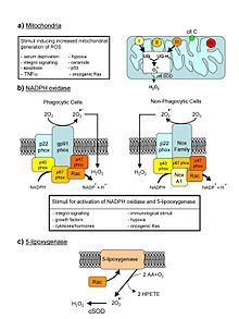
Major cellular sources of ROS in living non-photosynthetic cells. From a review by Novo and Parola, 2008.
 
Reactive oxygen species (ROS) are chemically reactive chemical species containing oxygen. Examples include peroxides, superoxide, hydroxyl radical, singlet oxygen, and alpha-oxygen

In a biological context, ROS are formed as a natural byproduct of the normal metabolism of oxygen and have important roles in cell signaling and homeostasis. However, during times of environmental stress (e.g., UV or heat exposure), ROS levels can increase dramatically. This may result in significant damage to cell structures. Cumulatively, this is known as oxidative stress. The production of ROS is strongly influenced by stress factor responses in plants, these factors that increase ROS production include drought, salinity, chilling, nutrient deficiency, metal toxicity and UV-B radiation. ROS are also generated by exogenous sources such as ionizing radiation.

Formation and decomposition

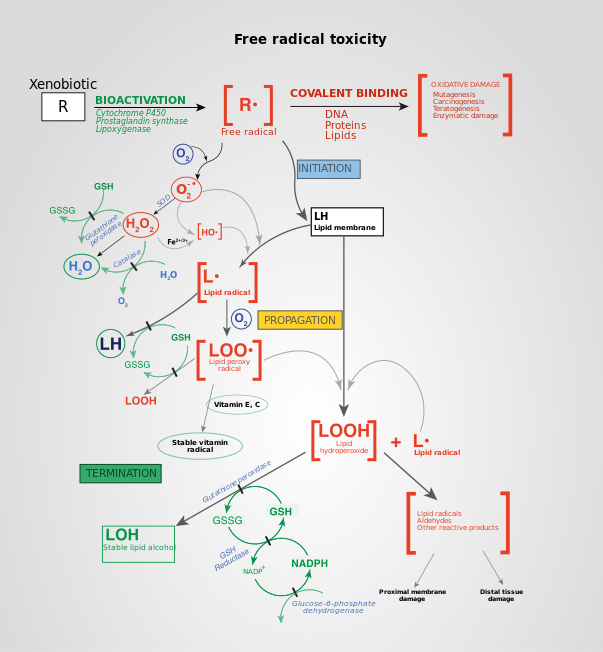
Free radical mechanisms in tissue injury. Free radical toxicity induced by xenobiotics and the subsequent detoxification by cellular enzymes (termination).
 
The reduction of molecular oxygen (O2) produces superoxide (O
2
) and is the precursor of most other reactive oxygen species:
O2 + eO
2
Dismutation of superoxide produces hydrogen peroxide (H2O2):
2 H+ + O
2
+ O
2
→ H2O2 + O2
Hydrogen peroxide in turn may be partially reduced to hydroxyl radical (OH) or fully reduced to water:
H2O2 + e → HO + OH
2 H+ + 2 e + H2O2 → 2 H2O

Exogenous ROS

Exogenous ROS can be produced from pollutants, tobacco, smoke, drugs, xenobiotics, or radiation. 

Ionizing radiation can generate damaging intermediates through the interaction with water, a process termed radiolysis. Since water comprises 55–60% of the human body, the probability of radiolysis is quite high under the presence of ionizing radiation. In the process, water loses an electron and becomes highly reactive. Then through a three-step chain reaction, water is sequentially converted to hydroxyl radical (OH), hydrogen peroxide (H2O2), superoxide radical (O
2
), and ultimately oxygen (O2). 

The hydroxyl radical is extremely reactive and immediately removes electrons from any molecule in its path, turning that molecule into a free radical and thus propagating a chain reaction. However, hydrogen peroxide is actually more damaging to DNA than the hydroxyl radical, since the lower reactivity of hydrogen peroxide provides enough time for the molecule to travel into the nucleus of the cell, subsequently reacting with macromolecules such as DNA.

Endogenous ROS

ROS are produced intracellularly through multiple mechanisms and depending on the cell and tissue types, the major sources being the "professional" producers of ROS: NADPH oxidase (NOX) complexes (7 distinct isoforms) in cell membranes, mitochondria, peroxisomes, and endoplasmic reticulum. Mitochondria convert energy for the cell into a usable form, adenosine triphosphate (ATP). The process in which ATP is produced, called oxidative phosphorylation, involves the transport of protons (hydrogen ions) across the inner mitochondrial membrane by means of the electron transport chain. In the electron transport chain, electrons are passed through a series of proteins via oxidation-reduction reactions, with each acceptor protein along the chain having a greater reduction potential than the previous. The last destination for an electron along this chain is an oxygen molecule. In normal conditions, the oxygen is reduced to produce water; however, in about 0.1–2% of electrons passing through the chain (this number derives from studies in isolated mitochondria, though the exact rate in live organisms is yet to be fully agreed upon), oxygen is instead prematurely and incompletely reduced to give the superoxide radical (O
2
), most well documented for Complex I and Complex III. Superoxide is not particularly reactive by itself, but can inactivate specific enzymes or initiate lipid peroxidation in its protonated form, hydroperoxyl HO
2
. The pKa of hydroperoxyl is 4.8. Thus, at physiological pH, the majority will exist as superoxide anion.

If too much damage is present in mitochondria, a cell undergoes apoptosis or programmed cell death. Bcl-2 proteins are layered on the surface of the mitochondria, detect damage, and activate a class of proteins called Bax, which punch holes in the mitochondrial membrane, causing cytochrome C to leak out. This cytochrome C binds to Apaf-1, or apoptotic protease activating factor-1, which is free-floating in the cell's cytoplasm. Using energy from the ATPs in the mitochondrion, the Apaf-1 and cytochrome C bind together to form apoptosomes. The apoptosomes bind to and activate caspase-9, another free-floating protein. The caspase-9 then cleaves the proteins of the mitochondrial membrane, causing it to break down and start a chain reaction of protein denaturation and eventually phagocytosis of the cell.

Superoxide dismutase

Superoxide dismutases (SOD) are a class of enzymes that catalyze the dismutation of superoxide into oxygen and hydrogen peroxide. As such, they are an important antioxidant defense in nearly all cells exposed to oxygen. In mammals and most chordates, three forms of superoxide dismutase are present. SOD1 is located primarily in the cytoplasm, SOD2 in the mitochondria and SOD3 is extracellular. The first is a dimer (consists of two units), while the others are tetramers (four subunits). SOD1 and SOD3 contain copper and zinc ions, while SOD2 has a manganese ion in its reactive centre. The genes are located on chromosomes 21, 6, and 4, respectively (21q22.1, 6q25.3 and 4p15.3-p15.1). 

The SOD-catalysed dismutation of superoxide may be written with the following half-reactions:
  • M(n+1)+ − SOD + O
    2
    → Mn+ − SOD + O2
  • Mn+ − SOD + O
    2
    + 2H+ → M(n+1)+ − SOD + H2O2.
where M = Cu (n = 1); Mn (n = 2); Fe (n = 2); Ni (n = 2). In this reaction the oxidation state of the metal cation oscillates between n and n + 1. 

Catalase, which is concentrated in peroxisomes located next to mitochondria, reacts with the hydrogen peroxide to catalyze the formation of water and oxygen. Glutathione peroxidase reduces hydrogen peroxide by transferring the energy of the reactive peroxides to a very small sulfur-containing protein called glutathione. The sulfur contained in these enzymes acts as the reactive center, carrying reactive electrons from the peroxide to the glutathione. Peroxiredoxins also degrade H2O2, within the mitochondria, cytosol, and nucleus.
  • 2 H2O2 → 2 H2O + O2      (catalase)
  • 2GSH + H2O2 → GS–SG + 2H2O      (glutathione peroxidase)

Singlet oxygen

Another type of reactive oxygen species is singlet oxygen (1O2) which is produced for example as byproduct of photosynthesis in plants. In the presence of light and oxygen, photosensitizers such as chlorophyll may convert triplet (3O2) to singlet oxygen:
Singlet oxygen is highly reactive, especially with organic compounds that contain double bonds. The resulting damage caused by singlet oxygen reduces the photosynthetic efficiency of chloroplasts. In plants exposed to excess light, the increased production of singlet oxygen can result in cell death. Various substances such as carotenoids, tocopherols and plastoquinones contained in chloroplasts quench singlet oxygen and protect against its toxic effects. In addition to direct toxicity, singlet oxygen acts a signaling molecule. Oxidized products of β-carotene arising from the presence of singlet oxygen act as second messengers that can either protect against singlet oxygen induced toxicity or initiate programmed cell death. Levels of jasmonate play a key role in the decision between cell acclimation or cell death in response to elevated levels of this reactive oxygen species.

Damaging effects

Effects of ROS on cell metabolism are well documented in a variety of species. These include not only roles in apoptosis (programmed cell death) but also positive effects such as the induction of host defense genes and mobilization of ion transport systems. This implicates them in control of cellular function. In particular, platelets involved in wound repair and blood homeostasis release ROS to recruit additional platelets to sites of injury. These also provide a link to the adaptive immune system via the recruitment of leukocytes.

Reactive oxygen species are implicated in cellular activity to a variety of inflammatory responses including cardiovascular disease. They may also be involved in hearing impairment via cochlear damage induced by elevated sound levels, in ototoxicity of drugs such as cisplatin, and in congenital deafness in both animals and humans. ROS are also implicated in mediation of apoptosis or programmed cell death and ischaemic injury. Specific examples include stroke and heart attack.

In general, harmful effects of reactive oxygen species on the cell are most often:
  1. damage of DNA or RNA
  2. oxidations of polyunsaturated fatty acids in lipids (lipid peroxidation)
  3. oxidations of amino acids in proteins
  4. oxidative deactivation of specific enzymes by oxidation of co-factors

Pathogen response

When a plant recognizes an attacking pathogen, one of the first induced reactions is to rapidly produce superoxide (O
2
) or hydrogen peroxide (H
2
O
2
) to strengthen the cell wall. This prevents the spread of the pathogen to other parts of the plant, essentially forming a net around the pathogen to restrict movement and reproduction.

In the mammalian host, ROS is induced as an antimicrobial defense. To highlight the importance of this defense, individuals with chronic granulomatous disease who have deficiencies in generating ROS, are highly susceptible to infection by a broad range of microbes including Salmonella enterica, Staphylococcus aureus, Serratia marcescens, and Aspergillus spp. 

The exact manner in which ROS defends the host from invading microbe is not fully understood. One of the more likely modes of defense is damage to microbial DNA. Studies using Salmonella demonstrated that DNA repair mechanisms were required to resist killing by ROS. More recently, a role for ROS in antiviral defense mechanisms has been demonstrated via Rig-like helicase-1 and mitochondrial antiviral signaling protein. Increased levels of ROS potentiate signaling through this mitochondria-associated antiviral receptor to activate interferon regulatory factor (IRF)-3, IRF-7, and nuclear factor kappa B (NF-κB), resulting in an antiviral state. Respiratory epithelial cells were recently demonstrated to induce mitrochondrial ROS in response to influenza infection. This induction of ROS led to the induction of type III interferon and the induction of an antiviral state, limiting viral replication. In host defense against mycobacteria, ROS play a role, although direct killing is likely not the key mechanism; rather, ROS likely affect ROS-dependent signalling controls, such as cytokine production, autophagy, and granuloma formation.

Reactive oxygen species are also implicated in activation, anergy and apoptosis of T cells.

Oxidative damage

In aerobic organisms the energy needed to fuel biological functions is produced in the mitochondria via the electron transport chain. In addition to energy, reactive oxygen species (ROS) with the potential to cause cellular damage are produced. ROS can damage lipid, DNA, RNA, and proteins, which, in theory, contributes to the physiology of aging

ROS are produced as a normal product of cellular metabolism. In particular, one major contributor to oxidative damage is hydrogen peroxide (H2O2), which is converted from superoxide that leaks from the mitochondria. Catalase and superoxide dismutase ameliorate the damaging effects of hydrogen peroxide and superoxide, respectively, by converting these compounds into oxygen and hydrogen peroxide (which is later converted to water), resulting in the production of benign molecules. However, this conversion is not 100% efficient, and residual peroxides persist in the cell. While ROS are produced as a product of normal cellular functioning, excessive amounts can cause deleterious effects. Memory capabilities decline with age, evident in human degenerative diseases such as Alzheimer's disease, which is accompanied by an accumulation of oxidative damage. Current studies demonstrate that the accumulation of ROS can decrease an organism's fitness because oxidative damage is a contributor to senescence. In particular, the accumulation of oxidative damage may lead to cognitive dysfunction, as demonstrated in a study in which old rats were given mitochondrial metabolites and then given cognitive tests. Results showed that the rats performed better after receiving the metabolites, suggesting that the metabolites reduced oxidative damage and improved mitochondrial function. Accumulating oxidative damage can then affect the efficiency of mitochondria and further increase the rate of ROS production. The accumulation of oxidative damage and its implications for aging depends on the particular tissue type where the damage is occurring. Additional experimental results suggest that oxidative damage is responsible for age-related decline in brain functioning. Older gerbils were found to have higher levels of oxidized protein in comparison to younger gerbils. Treatment of old and young mice with a spin trapping compound caused a decrease in the level of oxidized proteins in older gerbils but did not have an effect on younger gerbils. In addition, older gerbils performed cognitive tasks better during treatment but ceased functional capacity when treatment was discontinued, causing oxidized protein levels to increase. This led researchers to conclude that oxidation of cellular proteins is potentially important for brain function.

Cause of aging

According to the free radical theory of aging, oxidative damage initiated by reactive oxygen species is a major contributor to the functional decline that is characteristic of aging. While studies in invertebrate models indicate that animals genetically engineered to lack specific antioxidant enzymes (such as SOD), in general, show a shortened lifespan (as one would expect from the theory), the converse manipulation, increasing the levels of antioxidant enzymes, has yielded inconsistent effects on lifespan (though some studies in Drosophila do show that lifespan can be increased by the overexpression of MnSOD or glutathione biosynthesizing enzymes). Also contrary to this theory, deletion of mitochondrial SOD2 can extend lifespan in Caenorhabditis elegans.

In mice, the story is somewhat similar. Deleting antioxidant enzymes, in general, yields shorter lifespan, though overexpression studies have not (with some recent exceptions) consistently extended lifespan. Study of a rat model of premature aging found increased oxidative stress, reduced antioxidant enzyme activity and substantially greater DNA damage in the brain neocortex and hippocampus of the prematurely aged rats than in normally aging control rats. The DNA damage 8-OHdG is a product of ROS interaction with DNA. Numerous studies have shown that 8-OHdG increases in different mammalian organs with age.

Male infertility

Exposure of spermatozoa to oxidative stress is a major causative agent of male infertility. Sperm DNA fragmentation, caused by oxidative stress, appears to be an important factor in the etiology of male infertility. A high level of the oxidative DNA damage 8-OHdG is associated with abnormal spermatozoa and male infertility.

Cancer

ROS are constantly generated and eliminated in the biological system and are required to drive regulatory pathways. Under normal physiological conditions, cells control ROS levels by balancing the generation of ROS with their elimination by scavenging system. But under oxidative stress conditions, excessive ROS can damage cellular proteins, lipids and DNA, leading to fatal lesions in cell that contribute to carcinogenesis. 

Cancer cells exhibit greater ROS stress than normal cells do, partly due to oncogenic stimulation, increased metabolic activity and mitochondrial malfunction. ROS is a double-edged sword. On one hand, at low levels, ROS facilitates cancer cell survival since cell-cycle progression driven by growth factors and receptor tyrosine kinases (RTK) require ROS for activation and chronic inflammation, a major mediator of cancer, is regulated by ROS. On the other hand, a high level of ROS can suppress tumor growth through the sustained activation of cell-cycle inhibitor and induction of cell death as well as senescence by damaging macromolecules. In fact, most of the chemotherapeutic and radiotherapeutic agents kill cancer cells by augmenting ROS stress. The ability of cancer cells to distinguish between ROS as a survival or apoptotic signal is controlled by the dosage, duration, type, and site of ROS production. Modest levels of ROS are required for cancer cells to survive, whereas excessive levels kill them.

Metabolic adaptation in tumors balances the cells' need for energy with equally important need for macromolecular building blocks and tighter control of redox balance. As a result, production of NADPH is greatly enhanced, which functions as a cofactor to provide reducing power in many enzymatic reactions for macromolecular biosynthesis and at the same time rescuing the cells from excessive ROS produced during rapid proliferation. Cells counterbalance the detrimental effects of ROS by producing antioxidant molecules, such as reduced glutathione (GSH) and thioredoxin (TRX), which rely on the reducing power of NADPH to maintain their activities.

Most risk factors associated with cancer interact with cells through the generation of ROS. ROS then activate various transcription factors such as nuclear factor kappa-light-chain-enhancer of activated B cells (NF-κB), activator protein-1 (AP-1), hypoxia-inducible factor-1α and signal transducer and activator of transcription 3 (STAT3), leading to expression of proteins that control inflammation; cellular transformation; tumor cell survival; tumor cell proliferation; and invasion, agiogenesis as well as metastasis. And ROS also control the expression of various tumor suppressor genes such as p53, retinoblastoma gene (Rb), and phosphatase and tensin homolog (PTEN).

Carcinogenesis

ROS-related oxidation of DNA is one of the main causes of mutations, which can produce several types of DNA damage, including non-bulky (8-oxoguanine and formamidopyrimidine) and bulky (cyclopurine and etheno adducts) base modifications, abasic sites, non-conventional single-strand breaks, protein-DNA adducts, and intra/interstrand DNA crosslinks. It has been estimated that endogenous ROS produced via normal cell metabolism modify approximately 20,000 bases of DNA per day in a single cell. 8-oxoguanine is the most abundant among various oxidized nitrogeneous bases observed. During DNA replication, DNA polymerase mispairs 8-oxoguanine with adenine, leading to a G→T transversion mutation. The resulting genomic instability directly contributes to carcinogenesis. Cellular transformation leads to cancer and interaction of atypical PKC-ζ isoform with p47phox controls ROS production and transformation from apoptotic cancer stem cells through blebbishield emergency program.

Cell proliferation

Uncontrolled proliferation is a hallmark of cancer cells. Both exogenous and endogenous ROS have been shown to enhance proliferation of cancer cells. The role of ROS in promoting tumor proliferation is further supported by the observation that agents with potential to inhibit ROS generation can also inhibit cancer cell proliferation. Although ROS can promote tumor cell proliferation, a great increase in ROS has been associated with reduced cancer cell proliferation by induction of G2/M cell cycle arrest; increased phosphorylation of ataxia telangiectasia mutated (ATM), checkpoint kinase 1 (Chk 1), Chk 2; and reduced cell division cycle 25 homolog c (CDC25).

Cell death

A cancer cell can die in three ways: apoptosis, necrosis, and autophagy. Excessive ROS can induce apoptosis through both the extrinsic and intrinsic pathways. In the extrinsic pathway of apoptosis, ROS are generated by Fas ligand as an upstream event for Fas activation via phosphorylation, which is necessary for subsequent recruitment of Fas-associated protein with death domain and caspase 8 as well as apoptosis induction. In the intrinsic pathway, ROS function to facilitate cytochrome c release by activating pore-stabilizing proteins (Bcl-2 and Bcl-xL) as well as inhibiting pore-destabilizing proteins (Bcl-2-associated X protein, Bcl-2 homologous antagonist/killer). The intrinsic pathway is also known as the caspase cascade and is induced through mitochondrial damage which triggers the release of cytochrome c. DNA damage, oxidative stress, and loss of mitochondrial membrane potential lead to the release of the pro-apoptotic proteins mentioned above stimulating apoptosis. Mitochondrial damage is closely linked to apoptosis and since mitochondria are easily targeted there is potential for cancer therapy.

The cytotoxic nature of ROS is a driving force behind apoptosis, but in even higher amounts, ROS can result in both apoptosis and necrosis, a form of uncontrolled cell death, in cancer cells.

Numerous studies have shown the pathways and associations between ROS levels and apoptosis, but a newer line of study has connected ROS levels and autophagy. ROS can also induce cell death through autophagy, which is a self-catabolic process involving sequestration of cytoplasmic contents (exhausted or damaged organelles and protein aggregates) for degradation in lysosomes. Therefore, autophagy can also regulate the cell’s health in times of oxidative stress. Autophagy can be induced by ROS levels through many different pathways in the cell in an attempt to dispose of harmful organelles and prevent damage, such as carcinogens, without inducing apoptosis. Autophagic cell death can be prompted by the over expression of autophagy where the cell digests too much of itself in an attempt to minimize the damage and can no longer survive. When this type of cell death occurs, an increase or loss of control of autophagy regulating genes is commonly co-observed. Thus, once a more in-depth understanding of autophagic cell death is attained and its relation to ROS, this form of programmed cell death may serve as a future cancer therapy. Autophagy and apoptosis are two different cell death mechanisms brought on by high levels of ROS in the cells, however; autophagy and apoptosis rarely act through strictly independent pathways. There is a clear connection between ROS and autophagy and a correlation seen between excessive amounts of ROS leading to apoptosis. The depolarization of the mitochondrial membrane is also characteristic of the initiation of autophagy. When mitochondria are damaged and begin to release ROS, autophagy is initiated to dispose of the damaging organelle. If a drug targets mitochondria and creates ROS, autophagy may dispose of so many mitochondria and other damaged organelles that the cell is no longer viable. The extensive amount of ROS and mitochondrial damage may also signal for apoptosis. The balance of autophagy within the cell and the crosstalk between autophagy and apoptosis mediated by ROS is crucial for a cell’s survival. This crosstalk and connection between autophagy and apoptosis could be a mechanism targeted by cancer therapies or used in combination therapies for highly resistant cancers.

Tumor cell invasion, angiogenesis and metastasis

After growth factor stimulation of RTKs, ROS can trigger activation of signaling pathways involved in cell migration and invasion such as members of the mitogen activated protein kinase (MAPK) family – extracellular regulated kinase (ERK), c-jun NH-2 terminal kinase (JNK) and p38 MAPK. ROS can also promote migration by augmenting phosphorylation of the focal adhesion kinase (FAK) p130Cas and paxilin.

Both in vitro and in vivo, ROS have been shown to induce transcription factors and modulate signaling molecules involved in angiogenesis (MMP, VEGF) and metastasis (upregulation of AP-1, CXCR4, AKT and downregulation of PTEN).

Chronic inflammation and cancer

Experimental and epidemiologic research over the past several years has indicated close associations among ROS, chronic inflammation, and cancer. ROS induces chronic inflammation by the induction of COX-2, inflammatory cytokines (TNFα, interleukin 1 (IL-1), IL-6), chemokines (IL-8, CXCR4) and pro-inflammatory transcription factors (NF-κB). These chemokines and chemokine receptors, in turn, promote invasion and metastasis of various tumor types.

Cancer therapy

Both ROS-elevating and ROS-eliminating strategies have been developed with the former being predominantly used. Cancer cells with elevated ROS levels depend heavily on the antioxidant defense system. ROS-elevating drugs further increase cellular ROS stress level, either by direct ROS-generation (e.g. motexafin gadolinium, elesclomol) or by agents that abrogate the inherent antioxidant system such as SOD inhibitor (e.g. ATN-224, 2-methoxyestradiol) and GSH inhibitor (e.g. PEITC, buthionine sulfoximine (BSO)). The result is an overall increase in endogenous ROS, which when above a cellular tolerability threshold, may induce cell death. On the other hand, normal cells appear to have, under lower basal stress and reserve, a higher capacity to cope with additional ROS-generating insults than cancer cells do. Therefore, the elevation of ROS in all cells can be used to achieve the selective killing of cancer cells. 

Radiotherapy also relies on ROS toxicity to eradicate tumor cells. Radiotherapy uses X-rays, γ-rays as well as heavy particle radiation such as protons and neutrons to induce ROS-mediated cell death and mitotic failure.

Due to the dual role of ROS, both prooxidant and antioxidant-based anticancer agents have been developed. However, modulation of ROS signaling alone seems not to be an ideal approach due to adaptation of cancer cells to ROS stress, redundant pathways for supporting cancer growth and toxicity from ROS-generating anticancer drugs. Combinations of ROS-generating drugs with pharmaceuticals that can break the redox adaptation could be a better strategy for enhancing cancer cell cytotoxicity.

James Watson and others have proposed that lack of intracellular ROS due to a lack of physical exercise may contribute to the malignant progression of cancer, because spikes of ROS are needed to correctly fold proteins in the endoplasmatic reticulum and low ROS levels may thus aspecifically hamper the formation of tumor suppressor proteins. Since physical exercise induces temporary spikes of ROS, this may explain why physical exercise is beneficial for cancer patient prognosis. Moreover, high inducers of ROS such as 2-deoxy-D-glucose and carbohydrate-based inducers of cellular stress induce cancer cell death more potently because they exploit cancer cell high avidity for sugars.

Positive role of ROS in memory

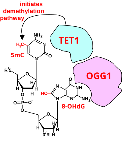
Initiation of DNA demethylation at a CpG site. In adult somatic cells DNA methylation typically occurs in the context of CpG dinucleotides (CpG sites), forming 5-methylcytosine-pG, or 5mCpG. Reactive oxygen species (ROS) may attack guanine at the dinucleotide site, forming 8-hydroxy-2'-deoxyguanosine (8-OHdG), and resulting in a 5mCp-8-OHdG dinucleotide site. The base excision repair enzyme OGG1 targets 8-OHdG and binds to the lesion without immediate excision. OGG1, present at a 5mCp-8-OHdG site recruits TET1 and TET1 oxidizes the 5mC adjacent to the 8-OHdG. This initiates demethylation of 5mC.
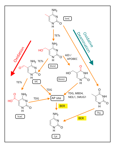
Demethylation of 5-Methylcytosine (5mC) in neuron DNA. As reviewed in 2018, in brain neurons, 5mC is oxidized by the ten-eleven translocation (TET) family of dioxygenases (TET1, TET2, TET3) to generate 5-hydroxymethylcytosine (5hmC). In successive steps TET enzymes further hydroxylate 5hmC to generate 5-formylcytosine (5fC) and 5-carboxylcytosine (5caC). Thymine-DNA glycosylase (TDG) recognizes the intermediate bases 5fC and 5caC and excises the glycosidic bond resulting in an apyrimidinic site (AP site). In an alternative oxidative deamination pathway, 5hmC can be oxidatively deaminated by activity-induced cytidine deaminase/apolipoprotein B mRNA editing complex (AID/APOBEC) deaminases to form 5-hydroxymethyluracil (5hmU) or 5mC can be converted to thymine (Thy). 5hmU can be cleaved by TDG, single-strand-selective monofunctional uracil-DNA glycosylase 1 (SMUG1), Nei-Like DNA Glycosylase 1 (NEIL1), or methyl-CpG binding protein 4 (MBD4). AP sites and T:G mismatches are then repaired by base excision repair (BER) enzymes to yield cytosine (Cyt).
 
Two reviews summarize the large body of evidence, reported largely between 1996 and 2011, for the critical and essential role of ROS in memory formation. A recent additional body of evidence indicates that both the formation and storage of memory depend on epigenetic modifications in neurons, including alterations in neuronal DNA methylation. The two bodies of information on memory formation appear to be connected in 2016 by the work of Zhou et al, who showed that ROS have a central role in epigenetic DNA demethylation

In mammalian nuclear DNA, a methyl group can be added, by a DNA methyltransferase, to the 5th carbon of cytosine to form 5mC (see red methyl group added to form 5mC near the top of the first figure). The DNA methyltransferases most often form 5mC within the dinucleotide sequence "cytosine-phosphate-guanine" to form 5mCpG. This addition is a major type of epigenetic alteration and it can silence gene expression. Methylated cytosine can also be demethylated, an epigenetic alteration that can increase the expression of a gene. A major enzyme involved in demethylating 5mCpG is TET1. However, TET1 is only able to act on 5mCpG if an ROS has first acted on the guanine to form 8-hydroxy-2'-deoxyguanosine (8-OHdG), resulting in a 5mCp-8-OHdG dinucleotide (see first figure). However, TET1 is only able to act on the 5mC part of the dinucleotide when the base excision repair enzyme OGG1 binds to the 8-OHdG lesion without immediate excision. Adherence of OGG1 to the 5mCp-8-OHdG site recruits TET1 and TET1 then oxidizes the 5mC adjacent to 8-OHdG, as shown in the first figure, initiating a demethylation pathway shown in the second figure. 

In 2016 Halder et al. using mice, and in 2017 Duke et al. using rats, subjected the rodents to contextual fear conditioning, causing an especially strong long-term memory to form. At 24 hours after the conditioning, in the hippocampus of rats, the expression of 1,048 genes was down-regulated (usually associated with hypermethylated gene promoters) and the expression of 564 genes was up-regulated (often associated with hypomethylated gene promoters). At 24 hours after training, 9.2% of the genes in the rat genome of hippocampus neurons were differentially methylated. However while the hippocampus is essential for learning new information it does not store information itself. In the mouse experiments of Halder, 1,206 differentially methylated genes were seen in the hippocampus one hour after contextual fear conditioning but these were reversed and not seen after four weeks. In contrast with the absence of long-term methylation changes in the hippocampus, substantial differential methylation could be detected in cortical neurons during memory maintenance. There were 1,223 differentially methylated genes in the anterior cingulate cortex of mice four weeks after contextual fear conditioning. 

The thousands of CpG sites being demethylated during memory formation depend on ROS in an initial step. The altered protein expression in neurons, controlled in part by ROS-dependent demethylation of CpG sites in gene promoters within neuron DNA, are central to memory formation.

Entropy (information theory)

From Wikipedia, the free encyclopedia https://en.wikipedia.org/wiki/Entropy_(information_theory) In info...