Search This Blog

Tuesday, April 2, 2019

Rubella

From Wikipedia, the free encyclopedia

Rubella
SynonymsGerman measles, three-day-measles
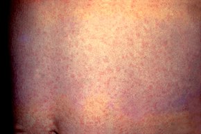
Rash of rubella on back (crop).JPG
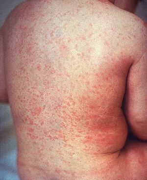
A rash due to rubella on a child's back. The area affected is similar to that of measles but the rash is less intensely red.
SpecialtyInfectious disease
SymptomsRash, swollen lymph nodes, fever, sore throat, feeling tired
ComplicationsTesticular swelling, inflammation of nerves, congenital rubella syndrome, miscarriage
Usual onset2 weeks after exposure
Duration3 days
CausesRubella virus (spread through the air)
Diagnostic methodFinding the virus in the blood, throat, or urine, antibody tests
PreventionRubella vaccine
TreatmentSupportive care
FrequencyCommon in many areas

Rubella, also known as German measles or three-day measles, is an infection caused by the rubella virus. This disease is often mild with half of people not realizing that they are infected. A rash may start around two weeks after exposure and last for three days. It usually starts on the face and spreads to the rest of the body. The rash is sometimes itchy and is not as bright as that of measles. Swollen lymph nodes are common and may last a few weeks. A fever, sore throat, and fatigue may also occur. In adults joint pain is common. Complications may include bleeding problems, testicular swelling, and inflammation of nerves. Infection during early pregnancy may result in a child born with congenital rubella syndrome (CRS) or miscarriage. Symptoms of CRS include problems with the eyes such as cataracts, ears such as deafness, heart, and brain. Problems are rare after the 20th week of pregnancy.

Rubella is usually spread through the air via coughs of people who are infected. People are infectious during the week before and after the appearance of the rash. Babies with CRS may spread the virus for more than a year. Only humans are infected. Insects do not spread the disease. Once recovered, people are immune to future infections. Testing is available that can verify immunity. Diagnosis is confirmed by finding the virus in the blood, throat, or urine. Testing the blood for antibodies may also be useful.

Rubella is preventable with the rubella vaccine with a single dose being more than 95% effective. Often it is given in combination with the measles vaccine and mumps vaccine, known as the MMR vaccine. When some, but less than 80% of the people are vaccinated, more women might make it to childbearing age without developing immunity by infection or vaccination and CRS rates could increase. Once infected there is no specific treatment.

Rubella is a common infection in many areas of the world. Each year about 100,000 cases of congenital rubella syndrome occur. Rates of disease have decreased in many areas as a result of vaccination. There are ongoing efforts to eliminate the disease globally. In April 2015 the World Health Organization declared the Americas free of rubella transmission. The name "rubella" is from Latin and means little red. It was first described as a separate disease by German physicians in 1814 resulting in the name "German measles".

Signs and symptoms

Young boy displaying the characteristic maculopapular rash of rubella
 
Generalized rash on the abdomen due to rubella
 
Rubella has symptoms that are similar to those of flu. However, the primary symptom of rubella virus infection is the appearance of a rash (exanthem) on the face which spreads to the trunk and limbs and usually fades after three days (that is why it is often referred to as three-day measles). The facial rash usually clears as it spreads to other parts of the body. Other symptoms include low grade fever, swollen glands (sub-occipital and posterior cervical lymphadenopathy), joint pains, headache, and conjunctivitis.

The swollen glands or lymph nodes can persist for up to a week and the fever rarely rises above 38 °C (100.4 °F). The rash of German measles is typically pink or light red. The rash causes itching and often lasts for about three days. The rash disappears after a few days with no staining or peeling of the skin. When the rash clears up, the skin might shed in very small flakes where the rash covered it. Forchheimer's sign occurs in 20% of cases, and is characterized by small, red papules on the area of the soft palate.

Rubella can affect anyone of any age. Adult women are particularly prone to arthritis and joint pains.

In children rubella normally causes symptoms which last two days and include:
  • Rash beginning on the face which spreads to the rest of the body.
  • Low fever of less than 38.3 °C (101 °F).
  • Posterior cervical lymphadenopathy.
In older children and adults additional symptoms may be present including:
  • Swollen glands
  • Coryza (cold-like symptoms)
  • Aching joints (especially in young women)
Severe complications of rubella include:
Coryza in rubella may convert to pneumonia, either direct viral pneumonia or secondary bacterial pneumonia, and bronchitis (either viral bronchitis or secondary bacterial bronchitis).

Congenital rubella syndrome

Cataracts due to congenital rubella syndrome
 
Rubella can cause congenital rubella syndrome in the newborn, the most severe sequela of rubella. The syndrome (CRS) follows intrauterine infection by the rubella virus and comprises cardiac, cerebral, ophthalmic and auditory defects. It may also cause prematurity, low birth weight, and neonatal thrombocytopenia, anemia and hepatitis. The risk of major defects or organogenesis is highest for infection in the first trimester. CRS is the main reason a vaccine for rubella was developed.

Many mothers who contract rubella within the first critical trimester either have a miscarriage or a stillborn baby. If the fetus survives the infection, it can be born with severe heart disorders (patent ductus arteriosus being the most common), blindness, deafness, or other life-threatening organ disorders. The skin manifestations are called "blueberry muffin lesions". For these reasons, rubella is included on the TORCH complex of perinatal infections. 

About 100,000 cases of this condition occur each year.

Cause

Transmission electron micrograph of rubella viruses
 
The disease is caused by rubella virus, a togavirus that is enveloped and has a single-stranded RNA genome. The virus is transmitted by the respiratory route and replicates in the nasopharynx and lymph nodes. The virus is found in the blood 5 to 7 days after infection and spreads throughout the body. The virus has teratogenic properties and is capable of crossing the placenta and infecting the fetus where it stops cells from developing or destroys them. During this incubation period, the patient is contagious typically for about one week before he/she develops a rash and for about one week thereafter. 

Increased susceptibility to infection might be inherited as there is some indication that HLA-A1 or factors surrounding A1 on extended haplotypes are involved in virus infection or non-resolution of the disease.

Diagnosis

Rubella virus specific IgM antibodies are present in people recently infected by rubella virus, but these antibodies can persist for over a year, and a positive test result needs to be interpreted with caution. The presence of these antibodies along with, or a short time after, the characteristic rash confirms the diagnosis.

Prevention

Rubella infections are prevented by active immunisation programs using live attenuated virus vaccines. Two live attenuated virus vaccines, RA 27/3 and Cendehill strains, were effective in the prevention of adult disease. However their use in prepubertal females did not produce a significant fall in the overall incidence rate of CRS in the UK. Reductions were only achieved by immunisation of all children.

The vaccine is now usually given as part of the MMR vaccine. The WHO recommends the first dose be given at 12 to 18 months of age with a second dose at 36 months. Pregnant women are usually tested for immunity to rubella early on. Women found to be susceptible are not vaccinated until after the baby is born because the vaccine contains live virus.

The immunisation program has been quite successful. Cuba declared the disease eliminated in the 1990s, and in 2004 the Centers for Disease Control and Prevention announced that both the congenital and acquired forms of rubella had been eliminated from the United States. The World Health Organisation declared Australia rubella free in October 2018.

Screening for rubella susceptibility by history of vaccination or by serology is recommended in the United States for all women of childbearing age at their first preconception counseling visit to reduce incidence of congenital rubella syndrome (CRS). It is recommended that all susceptible non-pregnant women of childbearing age should be offered rubella vaccination. Due to concerns about possible teratogenicity, use of MMR vaccine is not recommended during pregnancy. Instead, susceptible pregnant women should be vaccinated as soon as possible in the postpartum period.

Treatment

There is no specific treatment for rubella; however, management is a matter of responding to symptoms to diminish discomfort. Treatment of newborn babies is focused on management of the complications. Congenital heart defects and cataracts can be corrected by direct surgery.

Management for ocular congenital rubella syndrome (CRS) is similar to that for age-related macular degeneration, including counseling, regular monitoring, and the provision of low vision devices, if required.

Prognosis

Rubella infection of children and adults is usually mild, self-limiting and often asymptomatic. The prognosis in children born with CRS is poor.

Epidemiology

Rubella occurs worldwide. The virus tends to peak during the spring in countries with temperate climates. Before the vaccine against rubella was introduced in 1969, widespread outbreaks usually occurred every 6–9 years in the United States and 3–5 years in Europe, mostly affecting children in the 5-9 year old age group. Since the introduction of vaccine, occurrences have become rare in those countries with high uptake rates. 

Vaccination has interrupted the transmission of rubella in the Americas: no endemic case has been observed since February 2009. Vaccination is still strongly recommended as the virus could be reintroduced from other continents should vaccination rates in the Americas drop. During the epidemic in the U.S. between 1962–1965, rubella virus infections during pregnancy were estimated to have caused 30,000 stillbirths and 20,000 children to be born impaired or disabled as a result of CRS. Universal immunisation producing a high level of herd immunity is important in the control of epidemics of rubella.

In the UK, there remains a large population of men susceptible to rubella who have not been vaccinated. Outbreaks of rubella occurred amongst many young men in the UK in 1993 and in 1996 the infection was transmitted to pregnant women, many of whom were immigrants and were susceptible. Outbreaks still arise, usually in developing countries where the vaccine is not as accessible.

In Japan, 15,000 cases of rubella and 43 cases of congenital rubella syndrome were reported to the National Epidemiological Surveillance of Infectious Diseases between October 15, 2012, and March 2, 2014 during the 2012–13 rubella outbreak in Japan. They mainly occurred in men of ages 31 to 51 and young adults aged 24–34.

History

Rubella was first described in the mid-eighteenth century. Friedrich Hoffmann made the first clinical description of rubella in 1740, which was confirmed by de Bergen in 1752 and Orlow in 1758.

In 1814, George de Maton first suggested that it be considered a disease distinct from both measles and scarlet fever. All these physicians were German, and the disease was known as Rötheln (contemporary German Röteln), hence the common name of "German measles". Henry Veale, an English Royal Artillery surgeon, described an outbreak in India. He coined the name "rubella" (from the Latin word, meaning "little red") in 1866.

It was formally recognised as an individual entity in 1881, at the International Congress of Medicine in London. In 1914, Alfred Fabian Hess theorised that rubella was caused by a virus, based on work with monkeys. In 1938, Hiro and Tosaka confirmed this by passing the disease to children using filtered nasal washings from acute cases.

In 1940, there was a widespread epidemic of rubella in Australia. Subsequently, ophthalmologist Norman McAllister Gregg found 78 cases of congenital cataracts in infants and 68 of them were born to mothers who had caught rubella in early pregnancy. Gregg published an account, Congenital Cataract Following German Measles in the Mother, in 1941. He described a variety of problems now known as congenital rubella syndrome (CRS) and noticed that the earlier the mother was infected, the worse the damage was. Since no vaccine was yet available, some popular magazines promoted the idea of "German measles parties" for infected children to spread the disease to other children (especially girls) to immunize them for life and protect them from later catching the disease when pregnant. The virus was isolated in tissue culture in 1962 by two separate groups led by physicians Paul Douglas Parkman and Thomas Huckle Weller.

There was a pandemic of rubella between 1962 and 1965, starting in Europe and spreading to the United States. In the years 1964–65, the United States had an estimated 12.5 million rubella cases. This led to 11,000 miscarriages or therapeutic abortions and 20,000 cases of congenital rubella syndrome. Of these, 2,100 died as neonates, 12,000 were deaf, 3,580 were blind, and 1,800 were mentally retarded. In New York alone, CRS affected 1% of all births.

In 1969 a live attenuated virus vaccine was licensed. In the early 1970s, a triple vaccine containing attenuated measles, mumps and rubella (MMR) viruses was introduced. By 2006, confirmed cases in the Americas had dropped below 3000 a year. However, a 2007 outbreak in Argentina, Brazil, and Chile pushed the cases to 13,000 that year.

On January 22, 2014, the World Health Organization (WHO) and the Pan American Health Organization declared and certified Colombia free of rubella and became the first Latin American country to eliminate the disease within its borders. On April 29, 2015, the Americas became the first WHO region to officially eradicate the disease. The last non-imported cases occurred in 2009 in Argentina and Brazil. Pan-American Health Organization director remarked "The fight against rubella has taken more than 15 years, but it has paid off with what I believe will be one of the most important pan-American public health achievements of the 21st Century." The declaration was made after 165 million health records and genetically confirming that all recent cases were caused by known imported strains of the virus. Rubella is still common in some regions of the world and Susan E. Reef, team lead for rubella at the C.D.C.’s global immunization division, who joined in the announcement, said there was no chance it would be eradicated worldwide before 2020. Rubella is the third disease to be eliminated from the western hemisphere with vaccination after smallpox and polio.

Etymology

The name rubella is sometimes confused with rubeola, an alternative name for measles in English-speaking countries; the diseases are unrelated. In some other European languages, like Spanish, rubella and rubeola are synonyms, and rubeola is not an alternative name for measles. Thus, in Spanish, "rubeola" refers to rubella and "sarampión" refers to measles.

Cardiology

From Wikipedia, the free encyclopedia

Cardiology
Heart diagram blood flow en.svg
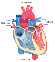
Blood flow diagram of the human heart. Blue components indicate de-oxygenated blood pathways and red components indicate oxygenated blood pathways.
SystemCardiovascular
SubdivisionsInterventional, Nuclear
Significant diseasesHeart disease, Cardiovascular disease, Atherosclerosis, Cardiomyopathy, Hypertension (High Blood Pressure)
Significant testsBlood tests, electrophysiology study, cardiac imaging, ECG, echocardiograms, stress test
SpecialistCardiologist
GlossaryGlossary of medicine

Cardiology (from Greek καρδίᾱ kardiā, "heart" and -λογία -logia, "study") is a branch of medicine that deals with the disorders of the heart as well as some parts of the circulatory system. The field includes medical diagnosis and treatment of congenital heart defects, coronary artery disease, heart failure, valvular heart disease and electrophysiology. Physicians who specialize in this field of medicine are called cardiologists, a specialty of internal medicine. Pediatric cardiologists are pediatricians who specialize in cardiology. Physicians who specialize in cardiac surgery are called cardiothoracic surgeons or cardiac surgeons, a specialty of general surgery.

Although the cardiovascular system is inextricably linked to blood, cardiology is relatively unconcerned with hematology and its diseases. Some obvious exceptions that affect the function of the heart would be blood tests (electrolyte disturbances, troponins), decreased oxygen carrying capacity (anemia, hypovolemic shock), and coagulopathies.

Specializations

All cardiologists study the disorders of the heart, but the study of adult and child heart disorders are through different training pathways. Therefore, an adult cardiologist (often simply called "cardiologist") is inadequately trained to take care of children, and pediatric cardiologists are not trained to take care of adult heart disease. The surgical aspects are not included in cardiology and are in the domain of cardiothoracic surgery. For example, coronary artery bypass surgery (CABG), cardiopulmonary bypass and valve replacement are surgical procedures performed by surgeons, not cardiologists. However the insertion of stents and pacemakers are performed by cardiologists

Adult cardiology

Cardiology is a specialty of internal medicine. To be a cardiologist in the United States, a three-year residency in internal medicine is followed by a three-year fellowship in cardiology. It is possible to specialize further in a sub-specialty. Recognized sub-specialties in the United States by the ACGME are cardiac electrophysiology, echocardiography, interventional cardiology, and nuclear cardiology. Recognized subspecialties in the United States by the American Osteopathic Association Bureau of Osteopathic Specialists (AOABOS) include clinical cardiac electrophysiology and interventional cardiology.

Per doximity, adult cardiologists make an average of $436,849 in the United States.

Cardiac electrophysiology

Cardiac electrophysiology is the science of elucidating, diagnosing, and treating the electrical activities of the heart. The term is usually used to describe studies of such phenomena by invasive (intracardiac) catheter recording of spontaneous activity as well as of cardiac responses to programmed electrical stimulation (PES). These studies are performed to assess complex arrhythmias, elucidate symptoms, evaluate abnormal electrocardiograms, assess risk of developing arrhythmias in the future, and design treatment. These procedures increasingly include therapeutic methods (typically radiofrequency ablation, or cryoablation) in addition to diagnostic and prognostic procedures. Other therapeutic modalities employed in this field include antiarrhythmic drug therapy and implantation of pacemakers and automatic implantable cardioverter-defibrillators (AICD).

The cardiac electrophysiology study (EPS) typically measures the response of the injured or cardiomyopathic myocardium to PES on specific pharmacological regimens in order to assess the likelihood that the regimen will successfully prevent potentially fatal sustained ventricular tachycardia (VT) or ventricular fibrillation VF (VF) in the future. Sometimes a series of EPS drug trials must be conducted to enable the cardiologist to select the one regimen for long-term treatment that best prevents or slows the development of VT or VF following PES. Such studies may also be conducted in the presence of a newly implanted or newly replaced cardiac pacemaker or AICD.
Clinical cardiac electrophysiology
Clinical cardiac electrophysiology is a branch of the medical specialty of cardiology and is concerned with the study and treatment of rhythm disorders of the heart. Cardiologists with expertise in this area are usually referred to as electrophysiologists. Electrophysiologists are trained in the mechanism, function, and performance of the electrical activities of the heart. Electrophysiologists work closely with other cardiologists and cardiac surgeons to assist or guide therapy for heart rhythm disturbances (arrhythmias). They are trained to perform interventional and surgical procedures to treat cardiac arrhythmia. 

The training required to become an electrophysiologist is long and requires 7 to 8 years after medical school (in the U.S.). Three years of internal medicine residency, three years of Clinical Cardiology fellowship, and one to two (in most instances) years of clinical cardiac electrophysiology.

Cardiogeriatrics

Cardiogeriatrics, or geriatric cardiology, is the branch of cardiology and geriatric medicine that deals with the cardiovascular disorders in elderly people.

Cardiac disorders such as coronary heart disease, including myocardial infarction, heart failure, cardiomyopathy, and arrhythmias such as atrial fibrillation, are common and are a major cause of mortality in elderly people. Vascular disorders such as atherosclerosis and peripheral arterial disease cause significant morbidity and mortality in aged people.

Echocardiography

Echocardiography uses standard two-dimensional, three-dimensional, and Doppler ultrasound to create images of the heart. 

Echocardiography has become routinely used in the diagnosis, management, and follow-up of patients with any suspected or known heart diseases. It is one of the most widely used diagnostic tests in cardiology. It can provide a wealth of helpful information, including the size and shape of the heart (internal chamber size quantification), pumping capacity, and the location and extent of any tissue damage. An echocardiogram can also give physicians other estimates of heart function, such as a calculation of the cardiac output, ejection fraction, and diastolic function (how well the heart relaxes).
Echocardiography can help detect cardiomyopathies, such as hypertrophic cardiomyopathy, dilated cardiomyopathy, and many others. The use of stress echocardiography may also help determine whether any chest pain or associated symptoms are related to heart disease. The biggest advantage to echocardiography is that it is not invasive (does not involve breaking the skin or entering body cavities) and has no known risks or side effects.

Interventional cardiology

Interventional cardiology is a branch of cardiology that deals specifically with the catheter based treatment of structural heart diseases. A large number of procedures can be performed on the heart by catheterization. This most commonly involves the insertion of a sheath into the femoral artery (but, in practice, any large peripheral artery or vein) and cannulating the heart under X-ray visualization (most commonly Fluoroscopy). 

The main advantages of using the interventional cardiology or radiology approach are the avoidance of the scars and pain, and long post-operative recovery. Additionally, interventional cardiology procedure of primary angioplasty is now the gold standard of care for an acute Myocardial infarction. This procedure can also be done proactively, when areas of the vascular system become occluded from Atherosclerosis. The Cardiologist will thread this sheath through the vascular system to access the heart. This sheath has a balloon and a tiny wire mesh tube wrapped around it, and if the cardiologist finds a blockage or Stenosis, they can inflate the balloon at the occlusion site in the vascular system to flatten or compress the plaque against the vascular wall. Once that is complete a Stent is placed as a type of scaffold to hold the vasculature open permanently.

Pediatric cardiology

Helen B. Taussig is known as the founder of pediatric cardiology. She became famous through her work with Tetralogy of Fallot, a congenital heart defect in which oxygenated and deoxygenated blood enters the circulatory system resulting from a ventricular septal defect (VSD) right beneath the aorta. This condition causes newborns to have a bluish-tint, cyanosis, and have a deficiency of oxygen to their tissues, hypoxemia. She worked with Alfred Blalock and Vivien Thomas at the Johns Hopkins Hospital where they experimented with dogs to look at how they would attempt to surgically cure these "blue babies." They eventually figured out how to do just that by the anastomosis of the systemic artery to the pulmonary artery and called this the Blalock-Taussig Shunt.

Tetralogy of Fallot, pulmonary atresia, double outlet right ventricle, transposition of the great arteries, persistent truncus arteriosus, and Ebsteins anomaly are various congenital cyanotic heart diseases. Congenital cyanotic heart diseases is where something is wrong with the heart of a newborn and it is not oxygenating the blood efficiently. 

Tetralogy of Fallot

Tetralogy of Fallot

Tetralogy of Fallot is the most common congenital heart disease arising in 1–3 cases per 1,000 births. The cause of this defect is a ventricular septal defect (VSD) and an overriding aorta. These two defects combined causes deoxygenated blood to bypass the lungs and going right back into the circulatory system. The modified Blalock-Taussig shunt is usually used to fix the circulation. This procedure is done by placing a graft between the subclavian artery and the ipsilateral pulmonary artery to restore the correct blood flow.

Pulmonary Atresia

Pulmonary Atresia happens in 7–8 per 100,000 births and is characterized by the aorta branching out of the right ventricle. This causes the deoxygenated blood to bypass the lungs and enter the circulatory system. Surgeries can fix this by redirecting the aorta and fixing the right ventricle and pulmonary artery connection. 

There are two types of pulmonary atresia, classified by whether or not the baby also has a ventricular septal defect.
  • Pulmonary atresia with an intact ventricular septum: This type of pulmonary atresia is associated with complete and intact septum between the ventricles.
  • Pulmonary atresia with a ventricular septal defect: This type of pulmonary atresia happens when a ventricular septal defect allows blood to flow into and out of the right ventricle.

Double Outlet Right Ventricle (DORV)

Double outlet right ventricle is when both great arteries, the pulmonary artery and the aorta, are connected to the right ventricle. There is usually a VSD in different particular places depending on the variations of DORV, typically 50% are subaortic and 30%. The surgeries that can be done to fix this defect can vary due to the different physiology and blood flow in the defected heart. One way it can be cured is by a VSD closure and placing conduits to restart the blood flow between the left ventricle and the aorta and between the right ventricle and the pulmonary artery. Another way is systemic-to-pulmonary artery shunt in cases associated with pulmonary stenosis. Also, a balloon atrial septostomy can be done to fix DORV with the Taussig-Bing anomaly.

Transposition of Great Arteries

Dextro-transposition of the Great Arteries
 
There are two different types of transposition of the great arteries, Dextro-transposition of the great arteries and Levo-transposition of the great arteries, depending on where the chambers and vessels connect. Dextro-transposition happens in about 1 in 4,000 newborns and is when the right ventricle pumps blood into the aorta and deoxygenated blood enters the blood stream. The temporary procedure is to create an atrial septal defect (ASD). A permanent fix is more complicated and involves redirecting the pulmonary return to the right atrium and the systemic return to the left atrium, which is known as the Senning procedure. The Rastelli procedure can also be done by rerouting the left ventricular outflow, dividing the pulmonary trunk, and placing a conduit in between the right ventricle and pulmonary trunk. Levo-transposition happens in about 1 in 13,000 newborns and is characterized by the left ventricle pumping blood into the lungs and the right ventricle pumping the blood into the aorta. This may not produce problems at the beginning, but will eventually due to the different pressures each ventricle uses to pump blood. Switching the left ventricle to be the systemic ventricle and the right ventricle to pump blood into the pulmonary artery can repair levo-transposition.

Persistent Truncus Arteriosis

Persistent truncus arteriosus is when the truncus arteriosus fails to split into the aorta and pulmonary trunk. This occurs in about 1 in 11,000 live births and allows both oxygenated and deoxygenated blood into the body. The repair consists of a VSD closure and the Rastelli procedure.

Ebstein Anomaly

Ebstein's anomaly is characterized by a right atrium that is significantly enlarged and a heart that is shaped like a box. This is very rare and happens in less than 1% of congenital heart disease cases. The surgical repair varies depending on the severity of the disease.

Pediatric cardiology is a sub-specialty of pediatrics. To become a pediatric cardiologist in the United States, one must complete a three-year residency in pediatrics, followed by a three-year fellowship in pediatric cardiology. Per doximity, pediatric cardiologists make an average of $303,917 in the United States.

The heart

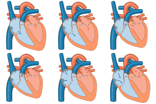
Blood flow through the valves

As the center focus of cardiology, the heart has numerous anatomical features (e.g., atria, ventricles, heart valves) and numerous physiological features (e.g., systole, heart sounds, afterload) that have been encyclopedically documented for many centuries. 

Disorders of the heart lead to heart disease and cardiovascular disease and can lead to a significant number of deaths: cardiovascular disease is the leading cause of death in the United States and caused 24.95% of total deaths in 2008.

The primary responsibility of the heart is to pump blood throughout the body. It pumps blood from the body — called the systemic circulation — through the lungs — called the pulmonary circulation — and then back out to the body. This means that the heart is connected to and affects the entirety of the body. Simplified, the heart is a circuit of the Circulation. While plenty is known about the healthy heart, the bulk of study in cardiology is in disorders of the heart and restoration, and where possible, of function. 

The heart is a muscle that squeezes blood and functions like a pump. Each part of the heart is susceptible to failure or dysfunction and the heart can be divided into the mechanical and the electrical parts. 

The electrical part of the heart is centered on the periodic contraction (squeezing) of the muscle cells that is caused by the cardiac pacemaker located in the sinoatrial node. The study of the electrical aspects is a sub-field of electrophysiology called cardiac electrophysiology and is epitomized with the electrocardiogram (ECG/EKG). The action potentials generated in the pacemaker propagate throughout the heart in a specific pattern. The system that carries this potential is called the electrical conduction system. Dysfunction of the electrical system manifests in many ways and may include Wolff–Parkinson–White syndrome, ventricular fibrillation, and heart block

The mechanical part of the heart is centered on the fluidic movement of blood and the functionality of the heart as a pump. The mechanical part is ultimately the purpose of the heart and many of the disorders of the heart disrupt the ability to move blood. Failure to move sufficient blood can result in failure in other organs and may result in death if severe. Heart failure is one condition in which the mechanical properties of the heart have failed or are failing, which means insufficient blood is being circulated.

Coronary circulation

Coronary circulation is the circulation of blood in the blood vessels of the heart muscle (myocardium). The vessels that deliver oxygen-rich blood to the myocardium are known as coronary arteries. The vessels that remove the deoxygenated blood from the heart muscle are known as cardiac veins. These include the great cardiac vein, the middle cardiac vein, the small cardiac vein and the anterior cardiac veins

As the left and right coronary arteries run on the surface of the heart, they can be called epicardial coronary arteries. These arteries, when healthy, are capable of autoregulation to maintain coronary blood flow at levels appropriate to the needs of the heart muscle. These relatively narrow vessels are commonly affected by atherosclerosis and can become blocked, causing angina or a heart attack. The coronary arteries that run deep within the myocardium are referred to as subendocardial. 

The coronary arteries are classified as "end circulation", since they represent the only source of blood supply to the myocardium; there is very little redundant blood supply, which is why blockage of these vessels can be so critical.

Cardiac examination

The cardiac examination (also called the "precordial exam"), is performed as part of a physical examination, or when a patient presents with chest pain suggestive of a cardiovascular pathology. It would typically be modified depending on the indication and integrated with other examinations especially the respiratory examination

Like all medical examinations, the cardiac examination follows the standard structure of inspection, palpation and auscultation.

Heart disorders

Cardiology is concerned with the normal functionality of the heart and the deviation from a healthy heart. Many disorders involve the heart itself but some are outside of the heart and in the vascular system. Collectively, the two together are termed the cardiovascular system and diseases of one part tend to affect the other.

Hypertension

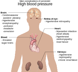
Diagram illustrating the main complications of persistent high blood pressure

Hypertension, also known as "high blood pressure"", is a long term medical condition in which the blood pressure in the arteries is persistently elevated. High blood pressure usually does not cause symptoms. Long term high blood pressure, however, is a major risk factor for coronary artery disease, stroke, heart failure, peripheral vascular disease, vision loss, and chronic kidney disease.

Lifestyle factors can increase the risk of hypertension. These include excess salt in the diet, excess body weight, smoking, and alcohol. Hypertension can also be caused by other diseases, or as a side-effect of drugs. 

Blood pressure is expressed by two measurements, the systolic and diastolic pressures, which are the maximum and minimum pressures, respectively. Normal blood pressure at rest is within the range of 100–140 millimeters mercury (mmHg) systolic and 60–90 mmHg diastolic. High blood pressure is present if the resting blood pressure is persistently at or above 140/90 mmHg for most adults. Different numbers apply to children. Ambulatory blood pressure monitoring over a 24-hour period appears more accurate than office best blood pressure measurement.

Lifestyle changes and medications can lower blood pressure and decrease the risk of health complications. Lifestyle changes include weight loss, decreased salt intake, physical exercise, and a healthy diet. If lifestyle changes are not sufficient then blood pressure medications are used. Up to three medications can control blood pressure in 90% of people. The treatment of moderately high arterial blood pressure (defined as >160/100 mmHg) with medications is associated with an improved life expectancy. The effect of treatment of blood pressure between 140/90 mmHg and 160/100 mmHg is less clear, with some reviews finding benefit and others finding a lack of evidence for benefit. High blood pressure affects between 16 and 37% of the population globally. In 2010 hypertension was believed to have been a factor in 18% (9.4 million) deaths.

Essential vs Secondary hypertension

Essential hypertension is the form of hypertension that by definition has no identifiable cause. It is the most common type of hypertension, affecting 95% of hypertensive patients, it tends to be familial and is likely to be the consequence of an interaction between environmental and genetic factors. Prevalence of essential hypertension increases with age, and individuals with relatively high blood pressure at younger ages are at increased risk for the subsequent development of hypertension. Hypertension can increase the risk of cerebral, cardiac, and renal events.

Secondary hypertension is a type of hypertension which is caused by an identifiable underlying secondary cause. It is much less common than essential hypertension, affecting only 5% of hypertensive patients. It has many different causes including endocrine diseases, kidney diseases, and tumors. It also can be a side effect of many medications.

Complications of hypertension

Main complications of persistent high blood pressure

Complications of hypertension are clinical outcomes that result from persistent elevation of blood pressure. Hypertension is a risk factor for all clinical manifestations of atherosclerosis since it is a risk factor for atherosclerosis itself. It is an independent predisposing factor for heart failure, coronary artery disease, stroke, renal disease, and peripheral arterial disease. It is the most important risk factor for cardiovascular morbidity and mortality, in industrialized countries.

Cardiac arrhythmia

Cardiac arrhythmia, also known as "cardiac dysrhythmia" or "irregular heartbeat", is a group of conditions in which the heartbeat is irregular, too fast, or too slow. A heart rate that is too fast – above 100 beats per minute in adults – is called tachycardia and a heart rate that is too slow – below 60 beats per minute – is called bradycardia. Many types of arrhythmia have no symptoms. When symptoms are present these may include palpitations or feeling a pause between heartbeats. More seriously there may be lightheadedness, passing out, shortness of breath, or chest pain. While most types of arrhythmia are not serious, some predispose a person to complications such as stroke or heart failure. Others may result in cardiac arrest.

There are four main types of arrhythmia: extra beats, supraventricular tachycardias, ventricular arrhythmias, and bradyarrhythmias. Extra beats include premature atrial contractions, premature ventricular contractions, and premature junctional contractions. Supraventricular tachycardias include atrial fibrillation, atrial flutter, and paroxysmal supraventricular tachycardia. Ventricular arrhythmias include ventricular fibrillation and ventricular tachycardia. Arrhythmias are due to problems with the electrical conduction system of the heart. Arrhythmias may occur in children; however, the normal range for the heart rate is different and depends on age. A number of tests can help with diagnosis including an electrocardiogram (ECG) and Holter monitor.

Most arrhythmias can be effectively treated. Treatments may include medications, medical procedures such as a pacemaker, and surgery. Medications for a fast heart rate may include beta blockers or agents that attempt to restore a normal heart rhythm such as procainamide. This later group may have more significant side effects especially if taken for a long period of time. Pacemakers are often used for slow heart rates. Those with an irregular heartbeat are often treated with blood thinners to reduce the risk of complications. Those who have severe symptoms from an arrhythmia may receive urgent treatment with a jolt of electricity in the form of cardioversion or defibrillation.

Arrhythmia affects millions of people. In Europe and North America, as of 2014, atrial fibrillation affects about 2% to 3% of the population. Atrial fibrillation and atrial flutter resulted in 112,000 deaths in 2013, up from 29,000 in 1990. Sudden cardiac death is the cause of about half of deaths due to cardiovascular disease or about 15% of all deaths globally. About 80% of sudden cardiac death is the result of ventricular arrhythmias. Arrhythmias may occur at any age but are more common among older people.

Coronary artery disease

Coronary artery disease, also known as "ischemic heart disease", is a group of diseases that includes: stable angina, unstable angina, myocardial infarction, and sudden cardiac death. It is within the group of cardiovascular diseases of which it is the most common type. A common symptom is chest pain or discomfort which may travel into the shoulder, arm, back, neck, or jaw. Occasionally it may feel like heartburn. Usually symptoms occur with exercise or emotional stress, last less than a few minutes, and get better with rest. Shortness of breath may also occur and sometimes no symptoms are present. The first sign is occasionally a heart attack. Other complications include heart failure or an irregular heartbeat.

Risk factors include: high blood pressure, smoking, diabetes, lack of exercise, obesity, high blood cholesterol, poor diet, and excessive alcohol, among others. Other risks include depression. The underlying mechanism involves atherosclerosis of the arteries of the heart. A number of tests may help with diagnoses including: electrocardiogram, cardiac stress testing, coronary computed tomographic angiography, and coronary angiogram, among others.

Prevention is by eating a healthy diet, regular exercise, maintaining a healthy weight and not smoking. Sometimes medication for diabetes, high cholesterol, or high blood pressure are also used. There is limited evidence for screening people who are at low risk and do not have symptoms. Treatment involves the same measures as prevention. Additional medications such as antiplatelets including aspirin, beta blockers, or nitroglycerin may be recommended. Procedures such as percutaneous coronary intervention (PCI) or coronary artery bypass surgery (CABG) may be used in severe disease. In those with stable CAD it is unclear if PCI or CABG in addition to the other treatments improve life expectancy or decreases heart attack risk.

In 2013 CAD was the most common cause of death globally, resulting in 8.14 million deaths (16.8%) up from 5.74 million deaths (12%) in 1990. The risk of death from CAD for a given age has decreased between 1980 and 2010 especially in developed countries. The number of cases of CAD for a given age has also decreased between 1990 and 2010. In the United States in 2010 about 20% of those over 65 had CAD, while it was present in 7% of those 45 to 64, and 1.3% of those 18 to 45. Rates are higher among men than women of a given age.

Cardiac arrest

Cardiac arrest is a sudden stop in effective blood flow due to the failure of the heart to contract effectively. Symptoms include loss of consciousness and abnormal or absent breathing. Some people may have chest pain, shortness of breath, or nausea before this occurs. If not treated within minutes, death usually occurs.

The most common cause of cardiac arrest is coronary artery disease. Less common causes include major blood loss, lack of oxygen, very low potassium, heart failure, and intense physical exercise. A number of inherited disorders may also increase the risk including long QT syndrome. The initial heart rhythm is most often ventricular fibrillation. The diagnosis is confirmed by finding no pulse. While a cardiac arrest may be caused by heart attack or heart failure these are not the same.

Prevention includes not smoking, physical activity, and maintaining a healthy weight. Treatment for cardiac arrest is immediate cardiopulmonary resuscitation (CPR) and, if a shockable rhythm is present, defibrillation. Among those who survive targeted temperature management may improve outcomes. An implantable cardiac defibrillator may be placed to reduce the chance of death from recurrence.

In the United States, cardiac arrest outside of hospital occurs in about 13 per 10,000 people per year (326,000 cases). In hospital cardiac arrest occurs in an additional 209,000 Cardiac arrest becomes more common with age. It affects males more often than females. The percentage of people who survive with treatment is about 8%. Many who survive have significant disability. Many U.S. television shows, however, have portrayed unrealistically high survival rates of 67%.

Congenital heart defects

A congenital heart defect, also known as a "congenital heart anomaly" or "congenital heart disease", is a problem in the structure of the heart that is present at birth. Signs and symptoms depend on the specific type of problem. Symptoms can vary from none to life-threatening. When present they may include rapid breathing, bluish skin, poor weight gain, and feeling tired. It does not cause chest pain. Most congenital heart problems do not occur with other diseases. Complications that can result from heart defects include heart failure.

The cause of a congenital heart defect is often unknown. Certain cases may be due to infections during pregnancy such as rubella, use of certain medications or drugs such as alcohol or tobacco, parents being closely related, or poor nutritional status or obesity in the mother. Having a parent with a congenital heart defect is also a risk factor. A number of genetic conditions are associated with heart defects including Down syndrome, Turner syndrome, and Marfan syndrome. Congenital heart defects are divided into two main groups: cyanotic heart defects and non-cyanotic heart defects, depending on whether the child has the potential to turn bluish in color. The problems may involve the interior walls of the heart, the heart valves, or the large blood vessels that lead to and from the heart.

Congenital heart defects are partly preventable through rubella vaccination, the adding of iodine to salt, and the adding of folic acid to certain food products. Some defects do not need treatment. Other may be effectively treated with catheter based procedures or heart surgery. Occasionally a number of operations may be needed. Occasionally heart transplantation is required. With appropriate treatment outcomes, even with complex problems, are generally good.

Heart defects are the most common birth defect. In 2013 they were present in 34.3 million people globally. They affect between 4 and 75 per 1,000 live births depending upon how they are diagnosed. About 6 to 19 per 1,000 cause a moderate to severe degree of problems. Congenital heart defects are the leading cause of birth defect-related deaths. In 2013 they resulted in 323,000 deaths down from 366,000 deaths in 1990.

Diagnostic tests in cardiology

Diagnostic tests in cardiology are the methods of identifying heart conditions associated with healthy vs. unhealthy, pathologic heart function. The starting point is obtaining a medical history, followed by Auscultation. Then blood tests, electrophysiological procedures, and cardiac imaging can be ordered for further analysis. Electrophysiological procedures include electrocardiogram, cardiac monitoring, cardiac stress testing, and the electrophysiology study.

Cardiology community

Associations

Journals

Open government

From Wikipedia, the free encyclopedia https://en.wikipedia.org/wiki/Open_gover...