Search This Blog

Tuesday, January 30, 2024

Blood–brain barrier

 
Blood–brain barrier
Solute permeability at the BBB vs. choroid plexus

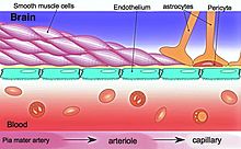
The blood–brain barrier (BBB) is a highly selective semipermeable border of endothelial cells that regulates the transfer of solutes and chemicals between the circulatory system and the central nervous system, thus protecting the brain from harmful or unwanted substances in the blood. The blood–brain barrier is formed by endothelial cells of the capillary wall, astrocyte end-feet ensheathing the capillary, and pericytes embedded in the capillary basement membrane. This system allows the passage of some small molecules by passive diffusion, as well as the selective and active transport of various nutrients, ions, organic anions, and macromolecules such as glucose and amino acids that are crucial to neural function.

The blood–brain barrier restricts the passage of pathogens, the diffusion of solutes in the blood, and large or hydrophilic molecules into the cerebrospinal fluid, while allowing the diffusion of hydrophobic molecules (O2, CO2, hormones) and small non-polar molecules. Cells of the barrier actively transport metabolic products such as glucose across the barrier using specific transport proteins. The barrier also restricts the passage of peripheral immune factors, like signaling molecules, antibodies, and immune cells, into the CNS, thus insulating the brain from damage due to peripheral immune events.

Specialized brain structures participating in sensory and secretory integration within brain neural circuits—the circumventricular organs and choroid plexus—have in contrast highly permeable capillaries.

Structure
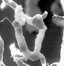
Part of a network of capillaries supplying brain cells
The astrocytes type 1 surrounding capillaries in the brain
Sketch showing constitution of blood vessels inside the brain

The BBB results from the selectivity of the tight junctions between the endothelial cells of brain capillaries, restricting the passage of solutes. At the interface between blood and the brain, endothelial cells are adjoined continuously by these tight junctions, which are composed of smaller subunits of transmembrane proteins, such as occludin, claudins (such as Claudin-5), junctional adhesion molecule (such as JAM-A). Each of these tight junction proteins is stabilized to the endothelial cell membrane by another protein complex that includes scaffolding proteins such as tight junction protein 1 (ZO1) and associated proteins.

The BBB is composed of endothelial cells restricting passage of substances from the blood more selectively than endothelial cells of capillaries elsewhere in the body. Astrocyte cell projections called astrocytic feet (also known as "glia limitans") surround the endothelial cells of the BBB, providing biochemical support to those cells. The BBB is distinct from the quite similar blood-cerebrospinal fluid barrier, which is a function of the choroidal cells of the choroid plexus, and from the blood-retinal barrier, which can be considered a part of the whole realm of such barriers.

Not all vessels in the human brain exhibit BBB properties. Some examples of this include the circumventricular organs, the roof of the third and fourth ventricles, capillaries in the pineal gland on the roof of the diencephalon and the pineal gland. The pineal gland secretes the hormone melatonin "directly into the systemic circulation", thus melatonin is not affected by the blood–brain barrier.

Development

The BBB appears to be functional by the time of birth. P-glycoprotein, a transporter, exists already in the embryonal endothelium.

Measurement of brain uptake of various blood-borne solutes showed that newborn endothelial cells were functionally similar to those in adults, indicating that a selective BBB is operative at birth.

In mice, Claudin-5 loss during development is lethal and results in size-selective loosening of the BBB.

Function

The blood–brain barrier acts effectively to protect brain tissue from circulating pathogens and other potentially toxic substances. Accordingly, blood-borne infections of the brain are rare. Infections of the brain that do occur are often difficult to treat. Antibodies are too large to cross the blood–brain barrier, and only certain antibiotics are able to pass. In some cases, a drug has to be administered directly into the cerebrospinal fluid where it can enter the brain by crossing the blood-cerebrospinal fluid barrier.

Circumventricular organs

Circumventricular organs (CVOs) are individual structures located adjacent to the fourth ventricle or third ventricle in the brain, and are characterized by dense capillary beds with permeable endothelial cells unlike those of the blood–brain barrier. Included among CVOs having highly permeable capillaries are the area postrema, subfornical organ, vascular organ of the lamina terminalis, median eminence, pineal gland, and three lobes of the pituitary gland.

Permeable capillaries of the sensory CVOs (area postrema, subfornical organ, vascular organ of the lamina terminalis) enable rapid detection of circulating signals in systemic blood, while those of the secretory CVOs (median eminence, pineal gland, pituitary lobes) facilitate transport of brain-derived signals into the circulating blood. Consequently, the CVO permeable capillaries are the point of bidirectional blood–brain communication for neuroendocrine function.

Specialized permeable zones

The border zones between brain tissue "behind" the blood–brain barrier and zones "open" to blood signals in certain CVOs contain specialized hybrid capillaries that are leakier than typical brain capillaries, but not as permeable as CVO capillaries. Such zones exist at the border of the area postrema—nucleus tractus solitarii (NTS), and median eminence—hypothalamic arcuate nucleus. These zones appear to function as rapid transit regions for brain structures involved in diverse neural circuits—like the NTS and arcuate nucleus—to receive blood signals which are then transmitted into neural output. The permeable capillary zone shared between the median eminence and hypothalamic arcuate nucleus is augmented by wide pericapillary spaces, facilitating bidirectional flow of solutes between the two structures, and indicating that the median eminence is not only a secretory organ, but may also be a sensory organ.

Therapeutic research

As a drug target

The blood–brain barrier is formed by the brain capillary endothelium and excludes from the brain 100% of large-molecule neurotherapeutics and more than 98% of all small-molecule drugs. Overcoming the difficulty of delivering therapeutic agents to specific regions of the brain presents a major challenge to treatment of most brain disorders. In its neuroprotective role, the blood–brain barrier functions to hinder the delivery of many potentially important diagnostic and therapeutic agents to the brain. Therapeutic molecules and antibodies that might otherwise be effective in diagnosis and therapy do not cross the BBB in adequate amounts to be clinically effective. The BBB represents an obstacle to some drugs reaching the brain, thus to overcome this barrier some peptides able to naturally cross the BBB have been widely investigated as a drug delivery system.

Mechanisms for drug targeting in the brain involve going either "through" or "behind" the BBB. Modalities for drug delivery to the brain in unit doses through the BBB entail its disruption by osmotic means, or biochemically by the use of vasoactive substances, such as bradykinin, or even by localized exposure to high-intensity focused ultrasound (HIFU).

Other methods used to get through the BBB may entail the use of endogenous transport systems, including carrier-mediated transporters, such as glucose and amino acid carriers, receptor-mediated transcytosis for insulin or transferrin, and the blocking of active efflux transporters such as p-glycoprotein. Some studies have shown that vectors targeting BBB transporters, such as the transferrin receptor, have been found to remain entrapped in brain endothelial cells of capillaries, instead of being ferried across the BBB into the targeted area.

Nanoparticles

Nanotechnology is under preliminary research for its potential to facilitate the transfer of drugs across the BBB. Capillary endothelial cells and associated pericytes may be abnormal in tumors and the blood–brain barrier may not always be intact in brain tumors. Other factors, such as astrocytes, may contribute to the resistance of brain tumors to therapy using nanoparticles. Fat soluble molecules less than 400 daltons in mass can freely diffuse past the BBB through lipid mediated passive diffusion.

Damage in injury and disease

The blood–brain barrier may become damaged in select neurological diseases, as indicated by neuroimaging studies of Alzheimer's disease, amyotrophic lateral sclerosis, epilepsy, ischemic stroke, and brain trauma, and in systemic diseases, such as liver failure. Effects such as impaired glucose transport and endothelial degeneration may lead to metabolic dysfunction within the brain, and an increased permeability of the BBB to proinflammatory factors, potentially allowing antibiotics and phagocytes to move across the BBB.

Prediction

There have been many attempts to correlate the experimental blood–brain barrier permeability with physicochemical properties. In 1988, the first QSAR study of brain–blood distribution conducted reported the in vivo values in rats for a large number of H2 receptor histamine agonists.

The first papers modelling blood-brain barrier permeability identified three properties, i.e., molecular volume, lipophilicity, and hydrogen bonding potential, as contributing to solute transport through the blood-brain barrier. A 2022 dataset selected different classification models based on molecular fingerprints, MACCS166 keys and molecular descriptors.

History

A 1898 study observed that low-concentration "bile salts" failed to affect behavior when injected into the blood of animals. Thus, in theory, the salts failed to enter the brain.

Two years later, Max Lewandowsky may have been the first to coin the term "blood–brain barrier" in 1900, referring to the hypothesized semipermeable membrane. There is some debate over the creation of the term blood–brain barrier as it is often attributed to Lewandowsky, but it does not appear in his papers. The creator of the term may have been Lina Stern. Stern was a Russian scientist who published her work in Russian and French. Due to the language barrier between her publications and English-speaking scientists, this could have made her work a lesser-known origin of the term.

All the while, bacteriologist Paul Ehrlich was studying staining, a procedure that is used in many microscopy studies to make fine biological structures visible using chemical dyes. As Ehrlich injected some of these dyes (notably the aniline dyes that were then widely used), the dye stained all of the organs of some kinds of animals except for their brains. At that time, Ehrlich attributed this lack of staining to the brain simply not picking up as much of the dye.

However, in a later experiment in 1913, Edwin Goldmann (one of Ehrlich's students) injected the dye directly into the cerebrospinal fluid of animal brains. He found then the brains did become dyed, but the rest of the body did not, demonstrating the existence of a compartmentalization between the two. At that time, it was thought that the blood vessels themselves were responsible for the barrier, since no obvious membrane could be found.
Drug delivery to the brain

From Wikipedia, the free encyclopedia

Drug delivery to the brain is the process of passing therapeutically active molecules across the blood–brain barrier into the brain. This is a complex process that must take into account the complex anatomy of the brain as well as the restrictions imposed by the special junctions of the blood–brain barrier.

Anatomy

The blood–brain barrier is formed by special tight junctions between endothelial cells lining brain blood vessels. Blood vessels of all tissues contain this monolayer of endothelial cells, however only brain endothelial cells have tight junctions preventing passive diffusion of most substances into the brain tissue. The structure of these tight junctions was first determined in the 1960s by Tom Reese, Morris Kranovsky, and Milton Brightman. Furthermore, astrocytic "end feet", the terminal regions of the astrocytic processes, surround the outside of brain capillary endothelial cells". The astrocytes are glial cells restricted to the brain and spinal cord and help maintain blood-brain barrier properties in brain endothelial cells.

Physiology

The main function of the blood–brain barrier is to protect the brain and keep it isolated from harmful toxins that are potentially in the blood stream. It accomplishes this because of its structure, as is usual in the body that structure defines its function. The tight junctions between the endothelial cells prevent large molecules as well as many ions from passing between the junction spaces. This forces molecules to go through the endothelial cells in order to enter the brain tissue, meaning that they must pass through the cell membranes of the endothelial cells. Because of this, the only molecules that are easily able to transverse the blood–brain barrier are ones that are very lipid-soluble. These are not the only molecules that can transverse the blood–brain barrier; glucose, oxygen and carbon dioxide are not lipid-soluble but are actively transported across the barrier, to support normal cellular function of the brain. The fact that molecules have to fully transverse the endothelial cells makes them a perfect barricade to unspecified particles from entering the brain, working to protect the brain at all costs. Also, because most molecules are transported across the barrier, it does a very effective job of maintaining homeostasis for the most vital organ of the human body.

Drug delivery to the blood–brain barrier

Because of the difficulty for drugs to pass through the blood–brain barrier, a study was conducted to determine the factors that influence a compound’s ability to transverse the blood–brain barrier. In this study, they examined several different factors to investigate diffusion across the blood–brain barrier. They used lipophilicity, Gibbs Adsorption Isotherm, a Co CMC Plot, and the surface area of the drug to water and air. They began by looking at compounds whose blood–brain permeability was known and labeled them either CNS+ or CNS- for compounds that easily transverse the barrier and those that did not. They then set out to analyze the above factors to determine what is necessary to transverse the blood–brain barrier. What they found was a little surprising; lipophilicity is not the leading characteristic for a drug to pass through the barrier. This is surprising because one would think that the most effective way to make a drug move through a lipophilic barrier is to increase its lipophilicity, it turns out that it is a complex function of all of these characteristics that makes a drug able to pass through the blood–brain barrier. The study found that barrier permittivity is "based on the measurement of the surface activity and as such takes into account the molecular properties of both hydrophobic and charged residues of the molecule of interest." They found that there is not a simple answer to what compounds transverse the blood–brain barrier and what does not. Rather, it is based on the complex analysis of the surface activity of the molecule as well as relative size.

Problems faced in drug delivery

Other problems persist besides just simply getting through the blood–brain barrier. The first of these is that a lot of times, even if a compound transverses the barrier, it does not do it in a way that the drug is in a therapeutically relevant concentration. This can have many causes, the most simple being that the way the drug was produced only allows a small amount to pass through the barrier. Another cause of this would be the binding to other proteins in the body rendering the drug ineffective to either be therapeutically active or able to pass through the barrier with the adhered protein. Another problem that must be accounted for is the presence of enzymes in the brain tissue that could render the drug inactive. The drug may be able to pass through the membrane fine, but will be deconstructed once it is inside the brain tissue rendering it useless. All of these are problems that must be addressed and accounted for in trying to deliver effective drug solutions to the brain tissue.

Possible solutions

Exosomes to deliver treatments across the blood–brain barrier

A group from the University of Oxford led by Prof. Matthew Wood claims that exosomes can cross the blood–brain barrier and deliver siRNAs, antisense oligonucleotides, chemotherapeutic agents and proteins specifically to neurons after inject them systemically (in blood). Because these exosomes are able to cross the blood–brain barrier, this protocol could solve the issue of poor delivery of medications to the central nervous system and cure Alzheimer's, Parkinson's Disease and brain cancer, among other diseases. The laboratory has been recently awarded a major new €30 million project leading experts from 14 academic institutions, two biotechnology companies and seven pharmaceutical companies to translate the concept to the clinic.

Pro-drugs

This is the process of disguising medically active molecules with lipophilic molecules that allow it to better sneak through the blood–brain barrier. Drugs can be disguised using more lipophilic elements or structures. This form of the drug will be inactive because of the lipophilic molecules but then would be activated, by either enzyme degradation or some other mechanism for removal of the lipophilic disguise to release the drug into its active form. There are still some major drawbacks to these pro-drugs. The first of which is that the pro-drug may be able to pass through the barrier and then also re-pass through the barrier without ever releasing the drug in its active form. The second is the sheer size of these types of molecules makes it still difficult to pass through the blood–brain barrier.

Peptide masking

Similar to the idea of pro-drugs, another way of masking the drugs chemical composition is by masking a peptide’s characteristics by combining with other molecular groups that are more likely to pass through the blood–brain barrier. An example of this is using a cholesteryl molecule instead of cholesterol that serves to conceal the water soluble characteristics of the drug. This type of masking as well as aiding in traversing the blood–brain barrier. It also can work to mask the drug peptide from peptide-degrading enzymes in the brain Also a "targetor" molecule could be attached to the drug that helps it pass through the barrier and then once inside the brain, is degraded in such a way that the drug cannot pass back through the brain. Once the drug cannot pass back through the barrier the drug can be concentrated and made effective for therapeutic use. However drawbacks to this exist as well. Once the drug is in the brain there is a point where it needs to be degraded to prevent overdose to the brain tissue. Also if the drug cannot pass back through the blood–brain barrier, it compounds the issues of dosage and intense monitoring would be required. For this to be effective there must be a mechanism for the removal of the active form of the drug from the brain tissue.

Receptor-mediated permabilitizers

These are drug compounds that increase the permeability of the blood–brain barrier. By decreasing the restrictiveness of the barrier, it is much easier to get a molecule to pass through it. These drugs increase the permeability of the blood–brain barrier temporarily by increasing the osmotic pressure in the blood which loosens the tight junctions between the endothelial cells. By loosening the tight junctions normal injection of drugs through an [IV] can take place and be effective to enter the brain. This must be done in a very controlled environment because of the risk associated with these drugs. Firstly, the brain can be flooded with molecules that are floating through the blood stream that are usually blocked by the barrier. Secondly, when the tight junctions loosen, the homeostasis of the brain can also be thrown off which can result in seizures and the compromised function of the brain.

Nanoparticles

The most promising drug delivery system is using nanoparticle delivery systems, these are systems where the drug is bound to a nanoparticle capable of traversing the blood–brain barrier. The most promising compound for the nanoparticles is Human Serum Albumin (HSA). The main benefits of this is that particles made of HSA are well tolerated without serious side effects as well as the albumin functional groups can be utilized for surface modification that allows for specific cell uptake. These nanoparticles have been shown to transverse the blood–brain barrier carrying host drugs. To enhance the effectiveness of nanoparticles, scientists are attempting to coat the nanoparticles to make them more effective to cross the blood–brain barrier. Studies have shown that "the overcoating of the [nanoparticles] with polysorbate 80 yielded doxorubicin concentrations in the brain of up to 6 μg/g after i.v. injection of 5 mg/kg" as compared to no detectable increase in an injection of the drug alone or the uncoated nanoparticle. This is very new science and technology so the real effectiveness of this process has not been fully understood. However young the research is, the results are promising pointing to nanotechnology as the way forward in treating a variety of brain diseases.

Loaded microbubble-enhanced focused ultrasound

Microbubbles are small "bubbles" of mono-lipids that are able to pass through the blood–brain barrier. They form a lipophilic bubble that can easily move through the barrier. One barrier to this however is that these microbubbles are rather large, which prevents their diffusion into the brain. This is counteracted by a focused ultrasound. The ultrasound increases the permeability of the blood–brain barrier by causing interference in the tight junctions in localized areas. This combined with the microbubbles allows for a very specific area of diffusion for the microbubbles, because they can only diffuse where the ultrasound is disrupting the barrier. The hypothesis and usefulness of these is the possibility of loading a microbubble with an active drug to diffuse through the barrier and target a specific area. There are several important factors in making this a viable solution for drug delivery. The first is that the loaded microbubble must not be substantially greater than the unloaded bubble. This ensures that the diffusion will be similar and the ultrasound disruption will be enough to induce diffusion. A second factor that must be determined is the stability of the loaded micro-bubble. This means is the drug fully retained in the bubble or is there leakage. Lastly, it must be determined how the drug is to be released from the microbubble once it passes through the blood–brain barrier. Studies have shown the effectiveness of this method for getting drugs to specific sites in the brain in animal models.

Hippocampal prosthesis

From Wikipedia, the free encyclopedia
https://en.wikipedia.org/wiki/Hippocampal_prosthesis

A hippocampus prosthesis is a type of cognitive prosthesis (a prosthesis implanted into the nervous system in order to improve or replace the function of damaged brain tissue). Prosthetic devices replace normal function of a damaged body part; this can be simply a structural replacement (e.g. reconstructive surgery or glass eye) or a rudimentary, functional replacement (e.g. a pegleg or hook).

However, prosthetics involving the brain have some special categories and requirements. "Input" prosthetics, such as retinal or cochlear implant, supply signals to the brain that the patient eventually learns to interpret as sight or sound. "Output" prosthetics use brain signals to drive a bionic arm, hand or computer device, and require considerable training during which the patient learns to generate the desired action via their thoughts. Both of these types of prosthetics rely on the plasticity of the brain to adapt to the requirement of the prosthesis, thus allowing the user to "learn" the use of his new body part.

A cognitive or "brain-to-brain" prosthesis involves neither learned input nor output signals, but the native signals used normally by the area of the brain to be replaced (or supported). Thus, such a device must be able to fully replace the function of a small section of the nervous system—using that section's normal mode of operation. In order to achieve this, developers require a deep understanding of the functioning of the nervous system. The scope of design must include a reliable mathematical model as well as the technology in order to properly manufacture and install a cognitive prosthesis. The primary goal of an artificial hippocampus is to provide a cure for Alzheimer's disease and other hippocampus—related problems. To do so, the prosthesis has to be able to receive information directly from the brain, analyze the information and give an appropriate output to the cerebral cortex; in other words, it must behave just like a natural hippocampus. At the same time, the artificial organ must be completely autonomous, since any exterior power source will greatly increase the risk of infection.

Hippocampus

Role

The hippocampus is part of the human limbic system, which interacts with the neocortex and other parts of the brain to produce emotions. As a part of the limbic system, the hippocampus plays its part in the formation of emotion in addition to its other roles, such as consolidation of new memories, navigation, and spatial orientation. The hippocampus is responsible for the formation of long term recognition memories. In other words, this is the part of the brain that allows us to associate a face with a name. Because of its close relationship with memory formation, damage to the hippocampus is closely related to Alzheimer's disease.

Anatomy

The hippocampus is a bilateral structure, situated under the neocortex. Each hippocampus is "composed of several different subsystem[s] that form a closed feedback loop, with input from the neocortex entering via the entorhinal cortex, propagating through the intrinsic subregions of the hippocampus and returning to the neocortex." In an electronic sense, the hippocampus is composed of a slice of parallel circuits.

Essential requirements

Biocompatibility

Since the prosthesis will be permanently implanted inside the brain, long term biocompatibility is required. We must also take into account the tendency for supporting braincells like astrocytes to encapsulate the implant. (This is a natural response for braincells, in order to protect neurons), thus impairing its function.

Bio-mimetic

Being biomimetic means that the implant must be able to fulfill the properties a real biological neuron. To do so we must have an in–depth understanding of brain behavior to build a solid mathematical model to be based upon. The field of computational neuroscience has made headway in this endeavor.

First, we must take into account that, like most of biological processes, the behaviors of neurons are highly nonlinear and depend on many factors: input frequency patterns, etc. Also, a good model must take into account the fact that the expression of a single nerve cell is negligible, since the processes are carried by groups of neurons interacting in network. Once installed, the device must assume all (or at least most) of the function of the damaged hippocampus for a prolonged period of time. First, the artificial neurons must be able to work together in network just like real neurons. Then, they must be able, working and effective synaptics connections with the existing neurons of the brain; therefore a model for silicon/neurons interface will be required.

Size

The implant must be small enough to be implantable while minimizing collateral damage during and after the implantation.

Bidirectional communication

In order to fully assume the function of the damaged hippocampus, the prosthesis must be able to communicate with the existing tissue in a bidirectional manner. in other words, the implant must be able to receive information from the brain and give an appropriate and compressible feedback to the surrounding nerve cell.

Personalized

The structural and functional characteristic of the brain varies greatly between individuals; therefore any neural implant has to be specific to each individual, which requires a precise model of the hippocampus and the use of advanced brain imagery to determine individual variance.

Surgical requirement

Since the prosthesis will be installed inside the brain, the operation itself will be much like a tumor removal operation. Although collateral damage will be inevitable, the effect on the patient will be minimal.

Model

"In order to incorporate the nonlinear dynamics of biological neurons into neuron models to develop a prosthesis, it is first necessary to measure them accurately. We have developed and applied methods for quantifying the nonlinear dynamics of hippocampal neurons (Berger et al., 1988a,b, 1991, 1992, 1994; Dalal et al., 1997) using principles of nonlinear systems theory (Lee and Schetzen, 1965; Krausz, 1975; P. Z. Marmarelis and Marmarelis, 1978; Rugh, 1981; Sclabassi et al., 1988). In this approach, properties of neurons are assessed experimentally by applying a random interval train of electrical impulses as an input and electrophysiologically recording the evoked output of the target neuron during stimulation (figure 12.2A). The input train consists of a series of impulses (as many as 4064), with interimpulse intervals varying according to a Poisson process having a mean of 500 ms and a range of 0.2–5000 ms. Thus, the input is "broadband" and stimulates the neuron over most of its operating range; that is, the statistical properties of the random train are highly consistent with the known physiological properties of hippocampal neurons. Nonlinear response properties are expressed in terms of the relation between progressively higher-order temporal properties of a sequence of input events and the probability of neuronal output, and are modeled as the kernels of a functional power series."

Technology involved

Imaging

Technology such as EEG, MEG, fMRI and other type of imaging technology are essential to the installation of the implant, which requires a high precision in order to minimize collateral damage (since the hippocampus is situated inside the cortex), as well as the proper function of the device.

Silicon/neuron interface

A silicon/neuron interface will be needed for the proper interaction of the silicon neurons of the prosthesis and the biological neurons of the brain.

Neuron network processor

In the brain, tasks are carried out by groups of interconnected neuronal network rather than a single cell, which means that any prosthesis must be able to simulate this network behavior. To do so, we will need a high number and density of silicon neurons to produce an effective prosthesis; therefore, a High-density Hippocampal Neuron Network Processor will be required in order for the prosthesis to carry out the task of a biological hippocampus. In addition, a neuron/silicon interface will be essential to the bidirectional communication of the implanted prosthesis. The choice of material and the design must ensure long term viability and bio compatibility while ensuring the density and the specificity of the interconnections.

Power supply

Appropriate power supply is still a major issue for any neural implant. Because the prostheses are implanted inside the brain, long term biocompatibility aside, the power supply will require several specification. First, the power supply must be self recharging. Unlike other prostheses, infection is a much greater issue for neural implant, due to the sensitivity of the brain; therefore an external power source is not envisagable. Because the brain is also highly heat sensitive, the power and the device itself must not generate too much heat to avoid disrupting brain function.

Prosthetic neuronal memory silicon chips

A prosthetic neuronal memory silicon chip is a device that imitates the brain's process of creating long-term memories. A prototype for this device was designed by Theodore Berger, a biomedical engineer and neurologist at University of Southern California. Berger started to work on the design in the early 1990s. He partnered with research colleagues that have been able to implant electrodes into rats and monkeys to test restoration of memory function. Recent work shows that the system can form long-term memories in many different behavioral situations. Berger and colleagues hope to eventually use these chips as electronic implants for humans whose brains that suffer from diseases such as Alzheimer's that disrupt neuronal networks.

Technology and medical application

To begin making a brain prosthesis, Berger and his collaborator Vasilis Marmarelis, a biomedical engineer at USC, worked with the hippocampus slices of rats. Since they knew that neuronal signals travel from one side of the hippocampus to the other, the researchers sent random pulses into the hippocampus, recorded the signals at specific locales to see how they were changed, and then derived equations representing the changes. They then programmed those equations into the computer chips.

Next, they had to determine whether a chip could be used as a prosthesis, or implant, for a damaged region in the hippocampus. To do this, they had to figure out whether they could avoid a central component of the pathway in the brain slices. They put electrodes in the region, which carried electrical pulses to an external chip. The chip then executed the transformations that are normally carried out in the hippocampus, and other electrodes sent the signals back to the slice of brain.

Memory codes

In 1996, Dr. Sam A. Deadwyler of Wake Forest Baptist Medical Center in Winston-Salem, NC, studied the activity patterns of collections of hippocampal neurons while rats performed a task requiring short-term memory.  These 'ensembles' or collections of neurons fired in different patterns in both time and 'space' (in this case, space referred to different neurons distributed throughout the hippocampus) depending on the type of behavior required in the task.  More importantly, Deadwyler and his colleagues could identify patterns that clearly distinguished between the various stimuli in the task including position (similar to place cells), behavioral responses, and what part of the task was occurring.  Analyses based on the neural ensemble activity alone without looking at those variables could identify and even 'predict' some of those variables even before they occurred.  In fact, the patterns would even identify when the rat was about to make an error in the task. Over the following ten years, Deadwyler's laboratory refined the analysis to identify the 'codes' and improved the ability to predict correct and error responses, even to the point of being able to have untrained rats perform the memory task using hippocampal stimulation with codes obtained from fully trained rats. The discovery of the memory codes in hippocampus led Deadwyler to join efforts with Berger for future studies in which Berger's team would develop models of memory function in hippocampus, and Deadwyler's team would test the models in rats and monkeys, and eventually move into human studies.

Trials on rats and monkeys

To transition to awake, behaving animals, Berger partnered with Deadwyler and Dr. Robert E. Hampson of Wake Forest to test a prototype of the memory prosthetic connected to rat and monkey brains via electrodes to analyze information just like the actual hippocampus. The prosthetic model allowed even a damaged hippocampus to generate new memories. In one demonstration, Deadwyler and Hampson impaired the rats' ability to form long-term memories by using pharmacological agents. These disrupted the neural circuitry that transfers messages between two subregions of the hippocampus. These subregions, CA1 and CA3, interact to create long-term memories. The rats were unable to remember which lever they needed to pull to obtain the reward. The researchers then developed an artificial hippocampus that could duplicate the pattern of interaction between CA3-CA1 interactions by analyzing the neural spikes in the cells with an electrode array, and then playing back the same pattern on the same array. After stimulating the rat hippocampi using the mathematical model of the prosthesis, their ability to identify the correct lever to pull improved dramatically. This artificial hippocampus played a significant role in the developmental stage of a memory prosthetic, as it went on to show that if a prosthetic device and its associated electrodes were implanted in the animals with a malfunctioning hippocampus, the device could potentially restore the memory capability to that of normal rats.

Goals for the future

The research teams at USC and Wake Forest are working to possibly make this system applicable to humans whose brains suffer damage from Alzheimer's, stroke, or injury, the disruption of neural networks often stops long-term memories from forming. The system designed by Berger and implemented by Deadwyler and Hampson allows the signal processing to take place that would occur naturally in undamaged neurons. Ultimately, they hope to restore the ability to create long-term memories by implanting chips such as these into the brain.

Recent development

Theodore Berger and his colleagues at the University of Southern California in Los Angeles have developed a working hippocampal prosthesis that passed the live tissue test in slices of brain tissue in 2004,. In 2011, in collaboration with Drs. Sam A. Deadwyler and Robert E. Hampson at Wake Forest Baptist Medical Center successfully tested a proof-of-concept hippocampal prosthesis in awake, behaving rats. The prosthesis was in the form of multisite electrodes positioned to record from both the input and output "sides" of the damaged hippocampus, the input is gathered and analyzed by external computation chips, an appropriate feedback is computed, then used to stimulate the appropriate output pattern in the brain so that the prosthesis functioned like a real hippocampus. In 2012, the team tested a further implementation in macaques prefrontal cortex, further developing the neural prosthesis technology. In 2013, Hampson et al. successfully tested a hippocampal prosthesis on non-human primates. While the device does not yet consist of a fully implantable "chip," these tests, from rat to monkey, demonstrate the effectiveness of the device as a neural prosthetic, and supports application to human trials.

Proof of concept for a human hippocampal prosthetic

In 2018, a team led by Robert E. Hampson at Wake Forest Baptist Medical, and including Berger and Deadwyler, became the first to demonstrate effectiveness of the prosthetic model in human patients. The subjects underwent implantation of electrodes in the brain at Wake Forest as part of a medical diagnostic procedure for epilepsy. While in the hospital, patients with electrodes in hippocampus volunteered to perform a memory task on computer while hippocampal neural activity was recorded in order for Berger and his team at USC team to customize the hippocampal prosthetic model for that patient. With model in hand, the Wake Forest team was able to demonstrate up to 37% improvement in memory function in patients with memory impaired by disease. The improvement was demonstrated for memories up to 75 minutes after stimulation by the hippocampal prosthetic model. As of 2018, studies are planned to test memory codes for additional attributes and features of items to be remembered as well as duration of memory facilitation in excess of 24 hours.

Monday, January 29, 2024

Synapse

From Wikipedia, the free encyclopedia
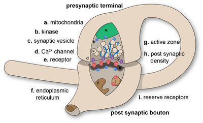
Diagram of a chemical synaptic connection.

In the nervous system, a synapse is a structure that permits a neuron (or nerve cell) to pass an electrical or chemical signal to another neuron or to the target effector cell.

Synapses are essential to the transmission of nervous impulses from one neuron to another, playing a key role in enabling rapid and direct communication by creating circuits. In addition, a synapse serves as a junction where both the transmission and processing of information occur, making it a vital means of communication between neurons. Neurons are specialized to pass signals to individual target cells, and synapses are the means by which they do so. At a synapse, the plasma membrane of the signal-passing neuron (the presynaptic neuron) comes into close apposition with the membrane of the target (postsynaptic) cell. Both the presynaptic and postsynaptic sites contain extensive arrays of molecular machinery that link the two membranes together and carry out the signaling process. In many synapses, the presynaptic part is located on an axon and the postsynaptic part is located on a dendrite or soma. Astrocytes also exchange information with the synaptic neurons, responding to synaptic activity and, in turn, regulating neurotransmission. Synapses (at least chemical synapses) are stabilized in position by synaptic adhesion molecules (SAMs) projecting from both the pre- and post-synaptic neuron and sticking together where they overlap; SAMs may also assist in the generation and functioning of synapses. Moreover, SAMs coordinate the formation of synapses, with various types working together to achieve the remarkable specificity of synapses. In essence, SAMs function in both excitatory and inhibitory synapses, likely serving as devices for signal transmission.

History

Santiago Ramón y Cajal proposed that neurons are not continuous throughout the body, yet still communicate with each other, an idea known as the neuron doctrine. The word "synapse" was introduced in 1897 by the English neurophysiologist Charles Sherrington in Michael Foster's Textbook of Physiology. Sherrington struggled to find a good term that emphasized a union between two separate elements, and the actual term "synapse" was suggested by the English classical scholar Arthur Woollgar Verrall, a friend of Foster. The word was derived from the Greek synapsis (σύναψις), meaning "conjunction", which in turn derives from synaptein (συνάπτειν), from syn (σύν) "together" and haptein (ἅπτειν) "to fasten".

However, while the synaptic gap remained a theoretical construct, and was sometimes reported as a discontinuity between contiguous axonal terminations and dendrites or cell bodies, histological methods using the best light microscopes of the day could not visually resolve their separation which is now known to be about 20 nm. It needed the electron microscope in the 1950s to show the finer structure of the synapse with its separate, parallel pre- and postsynaptic membranes and processes, and the cleft between the two.

Types

An example of chemical synapse by the release of neurotransmitters like acetylcholine or glutamic acid.

Chemical and electrical synapses are two ways of synaptic transmission.

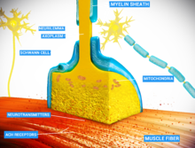
  • In a chemical synapse, electrical activity in the presynaptic neuron is converted (via the activation of voltage-gated calcium channels) into the release of a chemical called a neurotransmitter that binds to receptors located in the plasma membrane of the postsynaptic cell. The neurotransmitter may initiate an electrical response or a secondary messenger pathway that may either excite or inhibit the postsynaptic neuron. Chemical synapses can be classified according to the neurotransmitter released: glutamatergic (often excitatory), GABAergic (often inhibitory), cholinergic (e.g. vertebrate neuromuscular junction), and adrenergic (releasing norepinephrine). Because of the complexity of receptor signal transduction, chemical synapses can have complex effects on the postsynaptic cell.
  • In an electrical synapse, the presynaptic and postsynaptic cell membranes are connected by special channels called gap junctions that are capable of passing an electric current, causing voltage changes in the presynaptic cell to induce voltage changes in the postsynaptic cell.In fact, gap junctions facilitate the direct flow of electrical current without the need for neurotransmitters, as well as small molecules like calcium. Thus, the main advantage of an electrical synapse is the rapid transfer of signals from one cell to the next.
  • Mixed chemical electrical synapses are synaptic sites that feature both a gap junction and neurotransmitter release. This combination allows a signal to have both a fast component (electrical) and a slow component (chemical).

The formation of neural circuits in nervous systems, appears to heavily depend on the crucial interactions between chemical and electrical synapses. Thus, these interactions govern the generation of synaptic transmission. Synaptic communication is distinct from an ephaptic coupling, in which communication between neurons occurs via indirect electric fields. An autapse is a chemical or electrical synapse that forms when the axon of one neuron synapses onto dendrites of the same neuron.

Excitatory and inhibitory

  1. Excitatory synapse: Enhances the probability of depolarization in postsynaptic neurons and the initiation of an action potential.
  2. Inhibitory Synapse: Diminishes the probability of depolarization in postsynaptic neurons and the initiation of an action potential.

An influx of Na+ driven by excitatory neurotransmitters opens cation channels, depolarizing the postsynaptic membrane toward the action potential threshold. In contrast, inhibitory neurotransmitters cause the postsynaptic membrane to become less depolarized by opening either Cl- or K+ channels, reducing firing. Depending on their release location, the receptors they bind to, and the ionic circumstances they encounter, various transmitters can be either excitatory or inhibitory. For instance, acetylcholine can either excite or inhibit depending on the type of receptors it binds to. For example, glutamate serves as an excitatory neurotransmitter, in contrast to GABA, which acts as an inhibitory neurotransmitter. Additionally, dopamine is a neurotransmitter that exerts dual effects, displaying both excitatory and inhibitory impacts through binding to distinct receptors.

The membrane potential prevents Cl- from entering the cell, even when its concentration is much higher outside than inside. The resting potential for Cl- in many neurons is quite negative, nearly equal to the resting potential. Opening Cl- channels tends to buffer the membrane potential, but this effect is countered when the membrane starts to depolarize, allowing more negatively charged Cl- ions to enter the cell. Consequently, it becomes more difficult to depolarize the membrane and excite the cell when Cl- channels are open. Similar effects result from the opening of K+ channels. The significance of inhibitory neurotransmitters is evident from the effects of toxins that impede their activity. For instance, strychnine binds to glycine receptors, blocking the action of glycine and leading to muscle spasms, convulsions, and death.

Interfaces

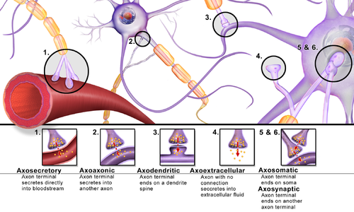
Synapses can be classified by the type of cellular structures serving as the pre- and post-synaptic components. The vast majority of synapses in the mammalian nervous system are classical axo-dendritic synapses (axon synapsing upon a dendrite), however, a variety of other arrangements exist. These include but are not limited to axo-axonic, dendro-dendritic, axo-secretory, axo-ciliary, somato-dendritic, dendro-somatic, and somato-somatic synapses.

In fact, the axon can synapse onto a dendrite, onto a cell body, or onto another axon or axon terminal, as well as into the bloodstream or diffusely into the adjacent nervous tissue.

Different types of synapses

Conversion of chemical into electrical signals

Neurotransmitters are tiny signal molecules stored in membrane-enclosed synaptic vesicles and released via exocytosis. Indeed, a change in electrical potential in the presynaptic cell triggers the release of these molecules. By attaching to transmitter-gated ion channels, the neurotransmitter causes an electrical alteration in the postsynaptic cell and rapidly diffuses across the synaptic cleft. Once released, the neurotransmitter is swiftly eliminated, either by being absorbed by the nerve terminal that produced it, taken up by nearby glial cells, or broken down by specific enzymes in the synaptic cleft. Numerous Na+-dependent neurotransmitter carrier proteins recycle the neurotransmitters and enable the cells to maintain rapid rates of release.

At chemical synapses, transmitter-gated ion channels play a vital role in rapidly converting extracellular chemical impulses into electrical signals. These channels are located in the postsynaptic cell's plasma membrane at the synapse region, and they temporarily open in response to neurotransmitter molecule binding, causing a momentary alteration in the membrane's permeability. Additionally, transmitter-gated channels are comparatively less sensitive to the membrane potential than voltage-gated channels, which is why they are unable to generate self-amplifying excitement on their own. However, they result in graded variations in membrane potential due to local permeability, influenced by the amount and duration of neurotransmitter released at the synapse.

Release of neurotransmitters

Neurotransmitters bind to ionotropic receptors on postsynaptic neurons, either causing their opening or closing. The variations in the quantities of neurotransmitters released from the presynaptic neuron may play a role in regulating the effectiveness of synaptic transmission. In fact, the concentration of cytoplasmic calcium is involved in regulating the release of neurotransmitters from presynaptic neurons.

The chemical transmission involves several sequential processes:

  1. Synthesizing neurotransmitters within the presynaptic neuron.
  2. Loading the neurotransmitters into secretory vesicles.
  3. Controlling the release of neurotransmitters into the synaptic cleft.
  4. Binding of neurotransmitters to postsynaptic receptors.
  5. Ceasing the activity of the released neurotransmitters.

Synaptic polarization

The function of neurons depends upon cell polarity. The distinctive structure of nerve cells allows action potentials to travel directionally (from dendrites to cell body down the axon), and for these signals to then be received and carried on by post-synaptic neurons or received by effector cells. Nerve cells have long been used as models for cellular polarization, and of particular interest are the mechanisms underlying the polarized localization of synaptic molecules. PIP2 signaling regulated by IMPase plays an integral role in synaptic polarity.

Phosphoinositides (PIP, PIP2, and PIP3) are molecules that have been shown to affect neuronal polarity. A gene (ttx-7) was identified in Caenorhabditis elegans that encodes myo-inositol monophosphatase (IMPase), an enzyme that produces inositol by dephosphorylating inositol phosphate. Organisms with mutant ttx-7 genes demonstrated behavioral and localization defects, which were rescued by expression of IMPase. This led to the conclusion that IMPase is required for the correct localization of synaptic protein components. The egl-8 gene encodes a homolog of phospholipase Cβ (PLCβ), an enzyme that cleaves PIP2. When ttx-7 mutants also had a mutant egl-8 gene, the defects caused by the faulty ttx-7 gene were largely reversed. These results suggest that PIP2 signaling establishes polarized localization of synaptic components in living neurons.

Presynaptic modulation

Modulation of neurotransmitter release by G-protein-coupled receptors (GPCRs) is a prominent presynaptic mechanism for regulation of synaptic transmission. The activation of GPCRs located at the presynaptic terminal, can decrease the probability of neurotransmitter release. This presynaptic depression involves activation of Gi/o-type G-proteins that mediate different inhibitory mechanisms, including inhibition of voltage-gated calcium channels, activation of potassium channels, and direct inhibition of the vesicle fusion process. Endocannabinoids, synthesized in and released from postsynaptic neuronal elements, and their cognate receptors, including the (GPCR) CB1 receptor, located at the presynaptic terminal, are involved in this modulation by an retrograde signaling process, in which these compounds are synthesized in and released from postsynaptic neuronal elements, and travel back to the presynaptic terminal to act on the CB1 receptor for short-term or long-term synaptic depression, that cause a short or long lasting decrease in neurotransmitter release.

Effects of drugs on ligand-gated ion channels

Drugs have long been considered crucial targets for transmitter-gated ion channels. The majority of medications utilized to treat schizophrenia, anxiety, depression, and sleeplessness work at chemical synapses, and many of these pharmaceuticals function by binding to transmitter-gated channels. For instance, some drugs like barbiturates and tranquilizers bind to GABA receptors and enhance the inhibitory effect of GABA neurotransmitter. Thus, reduced concentration of GABA enables the opening of Cl- channels.

Furthermore, psychoactive drugs could potentially target many other synaptic signalling machinery components. In fact, numerous neurotransmitters are released by Na+-driven carriers and are subsequently removed from the synaptic cleft. By inhibiting such carriers, synaptic transmission is strengthened as the action of the transmitter is prolonged. For example, Prozac is one of antidepressant medications that works by preventing the absorption of serotonin neurotransmitter. Also, other antidepressants operate by inhibiting the reabsorption of both serotonin and norepinephrine.

Biogenesis

In nerve terminals, synaptic vesicles are produced quickly to compensate for their rapid depletion during neurotransmitter release. Their biogenesis involves segregating synaptic vesicle membrane proteins from other cellular proteins and packaging those distinct proteins into vesicles of appropriate size. Besides, it entails the endocytosis of synaptic vesicle membrane proteins from the plasma membrane.

Synaptoblastic and synaptoclastic refer to synapse-producing and synapse-removing activities within the biochemical signalling chain. This terminology is associated with the Bredesen Protocol for treating Alzheimer's disease, which conceptualizes Alzheimer's as an imbalance between these processes. As of October 2023, studies concerning this protocol remain small and few results have been obtained within a standardized control framework.

Role in memory

Potentiation and depression

It is widely accepted that the synapse plays a key role in the formation of memory. The stability of long-term memory can persist for many years; nevertheless, synapses, the neurological basis of memory, are very dynamic. The formation of synaptic connections significantly depends on activity-dependent synaptic plasticity observed in various synaptic pathways. Indeed, the connection between memory formation and alterations in synaptic efficacy enables the reinforcement of neuronal interactions between neurons. As neurotransmitters activate receptors across the synaptic cleft, the connection between the two neurons is strengthened when both neurons are active at the same time, as a result of the receptor's signaling mechanisms. The strength of two connected neural pathways is thought to result in the storage of information, resulting in memory. This process of synaptic strengthening is known as long-term potentiation (LTP).

By altering the release of neurotransmitters, the plasticity of synapses can be controlled in the presynaptic cell. The postsynaptic cell can be regulated by altering the function and number of its receptors. Changes in postsynaptic signaling are most commonly associated with a N-methyl-d-aspartic acid receptor (NMDAR)-dependent LTP and long-term depression (LTD) due to the influx of calcium into the post-synaptic cell, which are the most analyzed forms of plasticity at excitatory synapses.

Mechanism of protein kinase

Moreover, Ca2+/calmodulin (CaM)-dependent protein kinase II (CaMKII) is best recognized for its roles in the brain, particularly in the neocortex and hippocampal regions because it serves as a ubiquitous mediator of cellular Ca2+ signals. CaMKII is abundant in the nervous system, mainly concentrated in the synapses in the nerve cells. Indeed, CaMKII has been definitively identified as a key regulator of cognitive processes, such as learning, and neural plasticity. The first concrete experimental evidence for the long-assumed function of CaMKII in memory storage was demonstrated

While Ca2+/CaM binding stimulates CaMKII activity, Ca2+-independent autonomous CaMKII activity can also be produced by a number of other processes. CaMKII becomes active by autophosphorylating itself upon Ca2+/calmodulin binding. CaMKII is still active and phosphorylates itself even after Ca2+ is cleaved; as a result, the brain stores long-term memories using this mechanism. Nevertheless, when the CaMKII enzyme is dephosphorylated by a phosphatase enzyme, it becomes inactive, and memories are lost. Hence, CaMKII plays a vital role in both the induction and maintenance of LTP. 

Experimental models

For technical reasons, synaptic structure and function have been historically studied at unusually large model synapses, for example:

Synapses and Diseases

Synapses function as ensembles within particular brain networks to control the amount of neuronal activity, which is essential for memory, learning, and behavior. Consequently, synaptic disruptions might have negative effects. In fact, alterations in cell-intrinsic molecular systems or modifications to environmental biochemical processes can lead to synaptic dysfunction. The synapse is the primary unit of information transfer in the nervous system, and correct synaptic contact creation during development is essential for normal brain function. In addition, several mutations have been connected to neurodevelopmental disorders, and that compromised function at different synapse locations is a hallmark of neurodegenerative diseases.

Synaptic defects are causally associated with early appearing neurological diseases, including autism spectrum disorders (ASD), schizophrenia (SCZ), and bipolar disorder (BP). On the other hand, in late-onset degenerative pathologies, such as Alzheimer's (AD), Parkinson's (PD), and Huntington's (HD) diseases, synaptopathy is thought to be the inevitable end-result of an ongoing pathophysiological cascade. These diseases are identified by a gradual loss in cognitive and behavioral function and a steady loss of brain tissue. Moreover, these deteriorations have been mostly linked to the gradual build-up of protein aggregates in neurons, the composition of which may vary based on the pathology; all have the same deleterious effects on neuronal integrity. Furthermore, the high number of mutations linked to synaptic structure and function, as well as dendritic spine alterations in post-mortem tissue, has led to the association between synaptic defects and neurodevelopmental disorders, such as ASD and SCZ, characterized by abnormal behavioral or cognitive phenotypes.

Nevertheless, due to limited access to human tissue at late stages and a lack of thorough assessment of the essential components of human diseases in the available experimental animal models, it has been difficult to fully grasp the origin and role of synaptic dysfunction in neurological disorders. 

Additional images

Rydberg atom

From Wikipedia, the free encyclopedia https://en.wikipedia.org/wiki/Rydberg_atom Figure 1: Electron orbi...