Search This Blog

Saturday, December 25, 2021

Glia

From Wikipedia, the free encyclopedia

Glia
Glial Cell Types.png
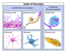
Illustration of the four different types of glial cells found in the central nervous system: ependymal cells (light pink), astrocytes (green), microglial cells (dark red), and oligodendrocytes (light blue).
Details
PrecursorNeuroectoderm for macroglia, and hematopoietic stem cells for microglia
SystemNervous system
Identifiers
MeSHD009457
TA98A14.0.00.005
THH2.00.06.2.00001
FMA54536 54541, 54536

Glia, also called glial cells (singular gliocyte) or neuroglia, are non-neuronal cells in the central nervous system (brain and spinal cord) and the peripheral nervous system that do not produce electrical impulses. They maintain homeostasis, form myelin in the peripheral nervous system, and provide support and protection for neurons. In the central nervous system, glial cells include oligodendrocytes, astrocytes, ependymal cells, and microglia, and in the peripheral nervous system glial cells include Schwann cells and satellite cells. They have four main functions: (1) to surround neurons and hold them in place; (2) to supply nutrients and oxygen to neurons; (3) to insulate one neuron from another; (4) to destroy pathogens and remove dead neurons. They also play a role in neurotransmission and synaptic connections, and in physiological processes like breathing. While glia were thought to outnumber neurons by a ratio of 10:1, recent studies using newer methods and reappraisal of historical quantitative evidence suggests an overall ratio of less than 1:1, with substantial variation between different brain tissues.

Glial cells have far more cellular diversity and functions than neurons, and glial cells can respond to and manipulate neurotransmission in many ways. Additionally, they can affect both the preservation and consolidation of memories.

Glia were discovered in 1856, by the pathologist Rudolf Virchow in his search for a "connective tissue" in the brain. The term derives from Greek γλία and γλοία "glue" (English: /ˈɡlə/ or /ˈɡlə/), and suggests the original impression that they were the glue of the nervous system.

Types

Neuroglia of the brain shown by Golgi's method
 
Astrocytes can be identified in culture because, unlike other mature glia, they express glial fibrillary acidic protein (GFAP)
 
Glial cells in a rat brain stained with an antibody against GFAP
 
Different types of neuroglia

Macroglia

Derived from ectodermal tissue.

Location Name Description
CNS Astrocytes

The most abundant type of macroglial cell in the CNS, astrocytes (also called astroglia) have numerous projections that link neurons to their blood supply while forming the blood-brain barrier. They regulate the external chemical environment of neurons by removing excess potassium ions, and recycling neurotransmitters released during synaptic transmission. Astrocytes may regulate vasoconstriction and vasodilation by producing substances such as arachidonic acid, whose metabolites are vasoactive.

Astrocytes signal each other using ATP. The gap junctions (also known as electrical synapses) between astrocytes allow the messenger molecule IP3 to diffuse from one astrocyte to another. IP3 activates calcium channels on cellular organelles, releasing calcium into the cytoplasm. This calcium may stimulate the production of more IP3 and cause release of ATP through channels in the membrane made of pannexins. The net effect is a calcium wave that propagates from cell to cell. Extracellular release of ATP, and consequent activation of purinergic receptors on other astrocytes, may also mediate calcium waves in some cases.

In general, there are two types of astrocytes, protoplasmic and fibrous, similar in function but distinct in morphology and distribution. Protoplasmic astrocytes have short, thick, highly branched processes and are typically found in gray matter. Fibrous astrocytes have long, thin, less branched processes and are more commonly found in white matter.

It has recently been shown that astrocyte activity is linked to blood flow in the brain, and that this is what is actually being measured in fMRI. They also have been involved in neuronal circuits playing an inhibitory role after sensing changes in extracellular calcium.

CNS Oligodendrocytes

Oligodendrocytes are cells that coat axons in the central nervous system (CNS) with their cell membrane, forming a specialized membrane differentiation called myelin, producing the myelin sheath. The myelin sheath provides insulation to the axon that allows electrical signals to propagate more efficiently.

CNS Ependymal cells

Ependymal cells, also named ependymocytes, line the spinal cord and the ventricular system of the brain. These cells are involved in the creation and secretion of cerebrospinal fluid (CSF) and beat their cilia to help circulate the CSF and make up the blood-CSF barrier. They are also thought to act as neural stem cells.

CNS Radial glia

Radial glia cells arise from neuroepithelial cells after the onset of neurogenesis. Their differentiation abilities are more restricted than those of neuroepithelial cells. In the developing nervous system, radial glia function both as neuronal progenitors and as a scaffold upon which newborn neurons migrate. In the mature brain, the cerebellum and retina retain characteristic radial glial cells. In the cerebellum, these are Bergmann glia, which regulate synaptic plasticity. In the retina, the radial Müller cell is the glial cell that spans the thickness of the retina and, in addition to astroglial cells, participates in a bidirectional communication with neurons.

PNS Schwann cells

Similar in function to oligodendrocytes, Schwann cells provide myelination to axons in the peripheral nervous system (PNS). They also have phagocytotic activity and clear cellular debris that allows for regrowth of PNS neurons.

PNS Satellite cells

Satellite glial cells are small cells that surround neurons in sensory, sympathetic, and parasympathetic ganglia. These cells help regulate the external chemical environment. Like astrocytes, they are interconnected by gap junctions and respond to ATP by elevating the intracellular concentration of calcium ions. They are highly sensitive to injury and inflammation and appear to contribute to pathological states, such as chronic pain.

PNS Enteric glial cells

Are found in the intrinsic ganglia of the digestive system. Glia cells are thought to have many roles in the enteric system, some related to homeostasis and muscular digestive processes.

Microglia

Microglia are specialized macrophages capable of phagocytosis that protect neurons of the central nervous system. They are derived from the earliest wave of mononuclear cells that originate in yolk sac blood islands early in development, and colonize the brain shortly after the neural precursors begin to differentiate.

These cells are found in all regions of the brain and spinal cord. Microglial cells are small relative to macroglial cells, with changing shapes and oblong nuclei. They are mobile within the brain and multiply when the brain is damaged. In the healthy central nervous system, microglia processes constantly sample all aspects of their environment (neurons, macroglia and blood vessels). In a healthy brain, microglia direct the immune response to brain damage and play an important role in the inflammation that accompanies the damage. Many diseases and disorders are associated with deficient microglia, such as Alzheimer's disease, Parkinson's disease, and ALS.

Other

Pituicytes from the posterior pituitary are glial cells with characteristics in common to astrocytes. Tanycytes in the median eminence of the hypothalamus are a type of ependymal cell that descend from radial glia and line the base of the third ventricle. Drosophila melanogaster, the fruit fly, contains numerous glial types that are functionally similar to mammalian glia but are nonetheless classified differently.

Total number

In general, neuroglial cells are smaller than neurons. There are approximately 85 billion glia cells in the human brain, about the same number as neurons. Glial cells make up about half the total volume of the brain and spinal cord. The glia to neuron-ratio varies from one part of the brain to another. The glia to neuron-ratio in the cerebral cortex is 3.72 (60.84 billion glia (72%); 16.34 billion neurons), while that of the cerebellum is only 0.23 (16.04 billion glia; 69.03 billion neurons). The ratio in the cerebral cortex gray matter is 1.48, with 3.76 for the gray and white matter combined. The ratio of the basal ganglia, diencephalon and brainstem combined is 11.35.

The total number of glia cells in the human brain is distributed into the different types with oligodendrocytes being the most frequent (45–75%), followed by astrocytes (19–40%) and microglia (about 10% or less).

Development

23-week fetal brain culture astrocyte
 

Most glia are derived from ectodermal tissue of the developing embryo, in particular the neural tube and crest. The exception is microglia, which are derived from hemopoietic stem cells. In the adult, microglia are largely a self-renewing population and are distinct from macrophages and monocytes, which infiltrate an injured and diseased CNS.

In the central nervous system, glia develop from the ventricular zone of the neural tube. These glia include the oligodendrocytes, ependymal cells, and astrocytes. In the peripheral nervous system, glia derive from the neural crest. These PNS glia include Schwann cells in nerves and satellite glial cells in ganglia.

Capacity to divide

Glia retains the ability to undergo cell divisions in adulthood, whereas most neurons cannot. The view is based on the general inability of the mature nervous system to replace neurons after an injury, such as a stroke or trauma, where very often there is a substantial proliferation of glia, or gliosis, near or at the site of damage. However, detailed studies have found no evidence that 'mature' glia, such as astrocytes or oligodendrocytes, retain mitotic capacity. Only the resident oligodendrocyte precursor cells seem to keep this ability once the nervous system matures.

Glial cells are known to be capable of mitosis. By contrast, scientific understanding of whether neurons are permanently post-mitotic, or capable of mitosis, is still developing. In the past, glia had been considered to lack certain features of neurons. For example, glial cells were not believed to have chemical synapses or to release transmitters. They were considered to be the passive bystanders of neural transmission. However, recent studies have shown this to not be entirely true.

Functions

Some glial cells function primarily as the physical support for neurons. Others provide nutrients to neurons and regulate the extracellular fluid of the brain, especially surrounding neurons and their synapses. During early embryogenesis, glial cells direct the migration of neurons and produce molecules that modify the growth of axons and dendrites. Some glial cells display regional diversity in the CNS and their functions may vary between the CNS regions.

Neuron repair and development

Glia are crucial in the development of the nervous system and in processes such as synaptic plasticity and synaptogenesis. Glia have a role in the regulation of repair of neurons after injury. In the central nervous system (CNS), glia suppress repair. Glial cells known as astrocytes enlarge and proliferate to form a scar and produce inhibitory molecules that inhibit regrowth of a damaged or severed axon. In the peripheral nervous system (PNS), glial cells known as Schwann cells (or also as neuri-lemmocytes) promote repair. After axonal injury, Schwann cells regress to an earlier developmental state to encourage regrowth of the axon. This difference between the CNS and the PNS, raises hopes for the regeneration of nervous tissue in the CNS. For example, a spinal cord may be able to be repaired following injury or severance.

Myelin sheath creation

Oligodendrocytes are found in the CNS and resemble an octopus: they have bulbous cell bodies with up to fifteen arm-like processes. Each process reaches out to an axon and spirals around it, creating a myelin sheath. The myelin sheath insulates the nerve fiber from the extracellular fluid and speeds up signal conduction along the nerve fiber. In the peripheral nervous system, Schwann cells are responsible for myelin production. These cells envelop nerve fibers of the PNS by winding repeatedly around them. This process creates a myelin sheath, which not only aids in conductivity but also assists in the regeneration of damaged fibers.

Neurotransmission

Astrocytes are crucial participants in the tripartite synapse. They have several crucial functions, including clearance of neurotransmitters from within the synaptic cleft, which aids in distinguishing between separate action potentials and prevents toxic build-up of certain neurotransmitters such as glutamate, which would otherwise lead to excitotoxicity. Furthermore, astrocytes release gliotransmitters such as glutamate, ATP, and D-serine in response to stimulation.

Clinical significance

Neoplastic glial cells stained with an antibody against GFAP (brown), from a brain biopsy

While glial cells in the PNS frequently assist in regeneration of lost neural functioning, loss of neurons in the CNS does not result in a similar reaction from neuroglia. In the CNS, regrowth will only happen if the trauma was mild, and not severe. When severe trauma presents itself, the survival of the remaining neurons becomes the optimal solution. However, some studies investigating the role of glial cells in Alzheimer's disease are beginning to contradict the usefulness of this feature, and even claim it can "exacerbate" the disease. In addition to impacting the potential repair of neurons in Alzheimer's disease, scarring and inflammation from glial cells have been further implicated in the degeneration of neurons caused by amyotrophic lateral sclerosis.

In addition to neurodegenerative diseases, a wide range of harmful exposure, such as hypoxia, or physical trauma, can lead to the end result of physical damage to the CNS. Generally, when damage occurs to the CNS, glial cells cause apoptosis among the surrounding cellular bodies. Then, there is a large amount of microglial activity, which results in inflammation, and finally, there is a heavy release of growth inhibiting molecules.

History

Although glial cells and neurons were probably first observed at the same time in the early 19th century, unlike neurons whose morphological and physiological properties were directly observable for the first investigators of the nervous system, glial cells had been considered to be merely “glue” that held neurons together until the mid-20th century.

Glia were first described in 1856 by the pathologist Rudolf Virchow in a comment to his 1846 publication on connective tissue. A more detailed description of glial cells was provided in the 1858 book 'Cellular Pathology' by the same author.

When markers for different types of cells were analyzed, Albert Einstein's brain was discovered to contain significantly more glia than normal brains in the left angular gyrus, an area thought to be responsible for mathematical processing and language. However, out of the total of 28 statistical comparisons between Einstein's brain and the control brains, finding one statistically significant result is not surprising and the claim that Einstein's brain is different, is not scientific (c.f. Multiple comparisons problem).

Not only does the ratio of glia to neurons increase through evolution, but so does the size of the glia. Astroglial cells in human brains have a volume 27 times greater than in mouse brains.

These important scientific findings may begin to shift the neuron-specific perspective into a more holistic view of the brain which encompasses the glial cells as well. For the majority of the twentieth century, scientists had disregarded glial cells as mere physical scaffolds for neurons. Recent publications have proposed that the number of glial cells in the brain is correlated with the intelligence of a species.

X-linked recessive inheritance

From Wikipedia, the free encyclopedia
 
X-linked recessive inheritance

X-linked recessive inheritance is a mode of inheritance in which a mutation in a gene on the X chromosome causes the phenotype to be always expressed in males (who are necessarily homozygous for the gene mutation because they have one X and one Y chromosome) and in females who are homozygous for the gene mutation, see zygosity. Females with one copy of the mutated gene are carriers.

X-linked inheritance means that the gene causing the trait or the disorder is located on the X chromosome. Females have two X chromosomes while males have one X and one Y chromosome. Carrier females who have only one copy of the mutation do not usually express the phenotype, although differences in X-chromosome inactivation (known as skewed X-inactivation) can lead to varying degrees of clinical expression in carrier females, since some cells will express one X allele and some will express the other. The current estimate of sequenced X-linked genes is 499, and the total, including vaguely defined traits, is 983.

Patterns of inheritance

Patterns of X-linked recessive inheritance in a royal family

In humans, inheritance of X-linked recessive traits follows a unique pattern made up of three points.

  • The first is that affected fathers cannot pass X-linked recessive traits to their sons because fathers give Y chromosomes to their sons. This means that males affected by an X-linked recessive disorder inherited the responsible X chromosome from their mothers.
  • Second, X-linked recessive traits are more commonly expressed in males than females. This is due to the fact that males possess only a single X chromosome, and therefore require only one mutated X in order to be affected. Women possess two X chromosomes, and thus must receive two of the mutated recessive X chromosomes (one from each parent). A popular example showing this pattern of inheritance is that of the descendants of Queen Victoria and the blood disease hemophilia.
  • The last pattern seen is that X-linked recessive traits tend to skip generations, meaning that an affected grandfather will not have an affected son, but could have an affected grandson through his daughter. Explained further, all daughters of an affected man will obtain his mutated X, and will then be either carriers or affected themselves depending on the mother. The resulting sons will either have a 50% chance of being affected (mother is carrier), or 100% chance (mother is affected). It is because of these percentages that we see males more commonly affected than females.

Pushback on recessive/dominant terminology

A few scholars have suggested discontinuing the use of the terms dominant and recessive when referring to X-linked inheritance. The possession of two X chromosomes in females leads to dosage issues which are alleviated by X-inactivation. Stating that the highly variable penetrance of X-linked traits in females as a result of mechanisms such as skewed X-inactivation or somatic mosaicism is difficult to reconcile with standard definitions of dominance and recessiveness, scholars have suggested referring to traits on the X chromosome simply as X-linked.

Examples

Most common

The most common X-linked recessive disorders are:

  • Red–green color blindness, a very common trait in humans and frequently used to explain X-linked disorders. Between seven and ten percent of men and 0.49% to 1% of women are affected. Its commonness may be explained by its relatively benign nature. It is also known as daltonism.
  • Hemophilia A, a blood clotting disorder caused by a mutation of the Factor VIII gene and leading to a deficiency of Factor VIII. It was once thought to be the "royal disease" found in the descendants of Queen Victoria. This is now known to have been Hemophilia B (see below).
  • Hemophilia B, also known as Christmas disease, a blood clotting disorder caused by a mutation of the Factor IX gene and leading to a deficiency of Factor IX. It is rarer than hemophilia A. As noted above, it was common among the descendants of Queen Victoria.
  • Duchenne muscular dystrophy, which is associated with mutations in the dystrophin gene. It is characterized by rapid progression of muscle degeneration, eventually leading to loss of skeletal muscle control, respiratory failure, and death.
  • Becker's muscular dystrophy, a milder form of Duchenne, which causes slowly progressive muscle weakness of the legs and pelvis.
  • X-linked ichthyosis, a form of ichthyosis caused by a hereditary deficiency of the steroid sulfatase (STS) enzyme. It is fairly rare, affecting one in 2,000 to one in 6,000 males.
  • X-linked agammaglobulinemia (XLA), which affects the body's ability to fight infection. XLA patients do not generate mature B cells. B cells are part of the immune system and normally manufacture antibodies (also called immunoglobulins) which defends the body from infections (the humoral response). Patients with untreated XLA are prone to develop serious and even fatal infections.
  • Glucose-6-phosphate dehydrogenase deficiency, which causes nonimmune hemolytic anemia in response to a number of causes, most commonly infection or exposure to certain medications, chemicals, or foods. Commonly known as "favism", as it can be triggered by chemicals existing naturally in broad (or fava) beans.

Less common disorders

Theoretically, a mutation in any of the genes on chromosome X may cause disease, but below are some notable ones, with short description of symptoms:


Disaster medicine

From Wikipedia, the free encyclopedia
 
Disaster Medicine Physician
Occupation
Names
  • Physician
Occupation type
Specialty
Activity sectors
Medicine
Description
Education required
Fields of
employment
Hospitals, Clinics
U.S. Navy sailors assigned to the aircraft carrier USS Carl Vinson treat a baby injured by 2010 Haiti earthquake
 
Treatment of the survivors of the 2017 Kermanshah earthquake

Disaster medicine is the area of medical specialization serving the dual areas of providing health care to disaster survivors and providing medically related disaster preparation, disaster planning, disaster response and disaster recovery leadership throughout the disaster life cycle. Disaster medicine specialists provide insight, guidance and expertise on the principles and practice of medicine both in the disaster impact area and healthcare evacuation receiving facilities to emergency management professionals, hospitals, healthcare facilities, communities and governments. The disaster medicine specialist is the liaison between and partner to the medical contingency planner, the emergency management professional, the incident command system, government and policy makers.

Disaster medicine is unique among the medical specialties in that unlike all other areas of specialization, the disaster medicine specialist does not practice the full scope of the specialty everyday but only in emergencies. Indeed, the disaster medicine specialist hopes to never practice the full scope of skills required for board certification. However, like specialists in public health, environmental medicine and occupational medicine, disaster medicine specialists engage in the development and modification of public and private policy, legislation, disaster planning and disaster recovery. Within the United States of America, the specialty of disaster medicine fulfills the requirements set for by Homeland Security Presidential Directives (HSPD), the National Response Plan (NRP), the National Incident Management System (NIMS), the National Resource Typing System (NRTS) and the NIMS Implementation Plan for Hospitals and Healthcare Facilities.

Definitions

Disaster healthcare – The provision of healthcare services by healthcare professionals to disaster survivors and disaster responders both in a disaster impact area and healthcare evacuation receiving facilities throughout the disaster life cycle.

Disaster behavioral health – Disaster behavioral health deals with the capability of disaster responders to perform optimally, and for disaster survivors to maintain or rapidly restore function, when faced with the threat or actual impact of disasters and extreme events.

Disaster law – Disaster law deals with the legal ramifications of disaster planning, preparedness, response and recovery, including but not limited to financial recovery, public and private liability, property abatement and condemnation.

Disaster life cycle – The time line for disaster events beginning with the period between disasters (interphase), progressing through the disaster event and the disaster response and culminating in the disaster recovery. Interphase begins as the end of the last disaster recovery and ends at the onset of the next disaster event. The disaster event begins when the event occurs and ends when the immediate event subsides. The disaster response begins when the event occurs and ends when acute disaster response services are no longer needed. Disaster recovery also begins with the disaster response and continues until the affected area is returned to the pre-event condition.

Disaster planning – The act of devising a methodology for dealing with a disaster event, especially one with the potential to occur suddenly and cause great injury and/or loss of life, damage and hardship. Disaster planning occurs during the disaster interphase.

Disaster preparation – The act of practicing and implementing the plan for dealing with a disaster event before an event occurs, especially one with the potential to occur suddenly and cause great injury and/or loss of life, damage and hardship. Disaster preparation occurs during the disaster interphase.

Disaster recovery – The restoration or return to the former or better state or condition proceeding a disaster event (i.e., status quo ante, the state of affairs that existed previously). Disaster recovery is the fourth phase of the disaster life cycle.

Disaster response – The ability to answer the intense challenges posed by a disaster event. Disaster response is the third phase of the disaster life cycle.

Medical contingency planning – The act of devising a methodology for meeting the medical requirements of a population affected by a disaster event.

Medical surge – An influx of patients (physical casualties and psychological casualties), bystanders, visitors, family members, media and individuals searching for the missing who present to a hospital or healthcare facility for treatment, information and/or shelter as a result of a disaster.

Surge capacity – The ability to manage a sudden, unexpected increase in patient volume that would otherwise severely challenge or exceed the current capacity of the health care system.

Medical triage – The separation of patients based on severity of injury or illness in light of available resources.

Psychosocial triage – The separation of patients based on the severity of psychological injury or impact in light of available resources.

History

The term "disaster medicine" first appeared in the medical lexicon in the post-World War II era. Although coined by former and current military physicians who had served in World War II, the term grow out of a concern for the need to care for military casualties, or nuclear holocaust victims, but out of the need to provide care to the survivors of natural disasters and the not-yet-distant memory of the 1917-1918 Influenza Pandemic.

The term "disaster medicine" continued to appear sporadically in both the medical and popular press[citation needed] until the 1980s, when the first concerted efforts to organize a medical response corps for disasters grew into the National Disaster Medical System. Simultaneous with this was the formation of a disaster and emergency medicine discussion and study group under the American Medical Association (AMA) in the United States as well as groups in Great Britain, Israel and other countries. By the time Hurricane Andrew struck Florida in 1992, the concept of disaster medicine was entrenched in public and governmental consciousness. Although training and fellowships in disaster medicine or related topics began graduating specialists in Europe and the United States as early as the 1980s, it was not until 2003 that the medical community embraced the need for the new specialty.

Throughout this period, incomplete and faltering medical responses to disaster events made it increasingly apparent in the United States of America that federal, state and local emergency management organizations were in need of a mechanism to identify qualified physicians in the face of a global upturn in the rate of disasters. Many physicians who volunteer at disasters have a bare minimum of knowledge in disaster medicine and often pose a hazard to themselves and the response effort because they have little or no field response training. It was against this backdrop that the American Academy of Disaster Medicine (AADM) and the American Board of Disaster Medicine (ABODM) were formed in the United States of America for the purpose of scholarly exchange and education in Disaster Medicine as well as the development of an examination demonstrating excellence towards board certification in this new specialty. In 2008, the United States National Library of Medicine (NLM) formed the Disaster Information Management Research Center (DIMRC) in support of the NLM's history of supporting healthcare professionals and information workers in accessing health information. DIMRC provides a specialized database, Disaster Lit: Database for Disaster Medicine and Public Health, an open access resource of disaster medicine documents, including guidelines, research reports, conference proceedings, fact sheets, training, fact sheets, and similar materials.

Ethics in Disaster Medicine

The Disaster Medicine practitioner must be well-versed in the ethical dilemmas that commonly arise in disaster settings. One of the most common dilemmas occurs when the aggregate medical need exceeds the ability to provide a normal standard of care for all patients.

Triage

In the event of a future pandemic, the number of patients that require additional respiratory support will outnumber the number of available ventilators. Although a hypothetical example, similar natural disasters have occurred in the past. Historically, the influenza pandemic of 1918-19 and the more recent SARS epidemic in 2003 led to resource scarcity and necessitated triage. One paper estimated that in the United States, the need for ventilators would be double the number available in the setting of an influenza pandemic similar to the scale of 1918. In other countries with fewer resources, shortages are postulated to be even more severe.

How, then, is a clinician to decide whom to offer this treatment? Examples of common approaches that guide triage include "saving the most lives", calling for care to be provided to "the sickest first" or alternatively a "first come, first served" approach may attempt to sidestep the difficult decision of triage. Emergency services often use their own triaging systems to be able to work through some of these challenging situations; however, these guidelines often assume no resource scarcity, and therefore, different triaging systems must be developed for resource-limited, disaster response settings. Useful ethical approaches to guide the development of such triaging protocols are often based on the principles of the theories of utilitarianism, egalitarianism and proceduralism.

Utilitarian Approach

The Utilitarian theory works on the premise that the responder shall 'maximise collective welfare'; or in other words, 'do the greatest good for the greatest numbers of people'. The utilitarian will necessarily need a measure by which to assess the outcome of the intervention. This could be thought of through various ways, for instance: the number of lives saved, or the number of years of life saved through the intervention. Thus, the utilitarian would prioritize saving the youngest of the patients over the elderly or those who are more likely to die despite an intervention, in order to 'maximise the collective years of life saved'. Commonly used metrics to quantify utility of health interventions include DALYs (Disability Adjusted Life Years) and QALYs (Quality Adjusted Life Years) which take into account the potential number of years of life lost due to disability and the quality of the life that has been saved, respectively, in order to quantify the utility of the intervention.

Egalitarian Approach

Principles of egalitarianism suggest the distribution of scarce resources amongst all those in need irrespective of likely outcome. The egalitarian will place some emphasis on equality, and the way that this is achieved might differ. The guiding factor is need rather than the ultimate benefit or utility of the intervention. Approaches based on egalitarian principles are complex guides in disaster settings. In the words of Eyal (2016) "Depending on the exact variant of egalitarianism, the resulting limited priority may go to patients whose contemporaneous prognosis is dire (because their medical prospects are now poor), to patients who have lived with serious disabilities for years (because their lifetime health is worse), to young patients (because dying now would make them short-lived), to socioeconomically disadvantaged patients (because their welfare prospects and resources are lower), or to those who queued up first (because first-come first-served may be thought to express equal concern."

Procedural Approach

The inherent difficulties in triage may lead practitioners to attempt to minimize active selection or prioritization of patients in face of scarcity of resources, and instead rely upon guidelines which do not take into account medical need or possibility of positive outcomes. In this approach, known as proceduralism, selection or prioritization may be based on patient's inclusion in a particular group (for example, by citizenship, or membership within an organization such as health insurance group). This approach prioritizes simplification of the triage and transparency, although there are significant ethical drawbacks, especially when procedures favor those who are part of socioeconomically advantaged groups (such as those with health insurance). Procedural systems of triage emphasize certain patterns of decision making based on preferred procedures. This can take place in the form of a fair lottery for instance; or establishing transparent criteria for entry into hospitals - based on non discriminatory conditions. This is not outcome driven; it is a process driven activity aimed at providing consistent frameworks upon which to base decisions.

These are by no means the only systems upon which decisions are made, but provide a basic framework to evaluate the ethical reasoning behind what are often difficult choices during disaster response and management.

Areas of competency

Internationally, disaster medicine specialists must demonstrate competency in areas of disaster healthcare and emergency management including but not limited to:

  • Disaster behavioral health
  • Disaster law
  • Disaster planning
  • Disaster preparation
  • Disaster recovery
  • Disaster response
  • Disaster safety
  • Medical consequences of disaster
  • Medical consequences of terrorism
  • Medical contingency planning
  • Medical decontamination
  • Medical implications of disaster
  • Medical implications of terrorism
  • Medical planning and preparation for disaster
  • Medical planning and preparation for terrorism
  • Medical recovery from disaster
  • Medical recovery from terrorism
  • Medical response to disaster
  • Medical response to terrorism
  • Medical response to weapons of mass destruction
  • Medical surge, surge capacity and triage
  • Psychosocial implications of disaster
  • Psychosocial implications of terrorism
  • Psychosocial triage

Timeline

1755 - 1755 Lisbon Earthquake "What now? We bury the dead and heal the living."

1812 – Napoleonic wars give rise to the military medical practice of triage in an effort to sort wounded soldiers in those to receive medical treatment and return to battle and those whose injuries are non-survivable. Dominique-Jean Larrey, a surgeon in the French emperor's army, not only conceives of taking care of the wounded on the battlefield, but creates the concept of ambulances, collecting the wounded in horse-drawn wagons and taking them to military hospitals.

1863 – International Red Cross founded in Geneva, Switzerland.

1873 – Clara Barton starts organization of the American Red Cross, drawing on her experiences during the American Civil War.

1881 – First American Red Cross chapter founded in Dansville, New York.

1937 – President Franklin Roosevelt makes a public request by commercial radio for medical aid following a natural gas explosion in New London, Texas. This is the first presidential request for disaster medical assistance in United States history.

1955 – Col. Karl H. Houghton, M.D. addresses a convention of military surgeons and introduces the concept of "disaster medicine."

1959 – Col. Joseph R. Schaeffer, M.D., reflecting the growing national concern over nuclear attacks on the United States civilian population, initiates training for civilian physicians in the treatment of mass casualties for the effects of weapons of mass destruction creating the concept of medical surge capacity.

1961 – The American Medical Association, the American Hospital Association, the American College of Surgeons, the United States Public Health Service, the United States Office of Civil Defense and the Department of Health, Education and Welfare join Schaeffer in advancing civilian physician training for mass casualty and weapons of mass destruction treatment.

1962 – The North Atlantic Treaty Organization (NATO) publishes an official disaster medicine manual edited by Schaeffer.

1984 – The United States Public Health Service forms the first federal disaster medical response team in Washington, D.C., designated PHS-1.

1986 – The United States Public Health System creates the National Disaster Medical System (NDMS) to provide disaster healthcare through National Medical Response Teams (NMRTs), Disaster Medical Assistance Teams (DMATs), Disaster Veterinary Assistance Teams (VMATs) and Disaster Mortuary Operational Response Teams (DMORTs). PH-1 becomes the first DMAT team.

1986 – A disaster medical response discussion group is created by NDMS team members and emergency medicine organizations in the United States. Healthcare professionals worldwide join the discussion group of the years to come. dd 1989 – The University of New Mexico creates the Center for Disaster Medicine, the first such medical center of excellence in the United States. Elsewhere in the world, similar centers are created at universities in London, Paris, Brussels and Bordeaux.

1992 – Hurricane Andrew, a Category 5 hurricane, strikes south Florida, destroying the city of Homestead, Florida and initiating the largest disaster healthcare response to date.

1993 – On February 26, 1993, at 12:17 pm, a terrorist attack on the North Tower of the World Trade Center (the first such attack on United States soil since World War II) increases interest in specialized education and training on disaster response for civilian physicians.

1998 – The American College of Contingency Planners (ACCP) is formed by the American Academy of Medical Administrators (AAMA) to provide certification and scholarly study in the area of medical contingency planning and healthcare disaster planning.

2001 – The September 11, 2001 attacks on the World Trade Center and the Pentagon cause the largest loss of life resulting from an attack on American targets on United States soil since Pearl Harbor. As a result, the need for disaster medicine is galvanized.

2001 – On October 29, 2001, President George W. Bush issues Homeland Security Presidential Directive 1 (HSPD-1), establishing the organization and operation of the Homeland Security Council.

2002 – On March 11, 2002, President Bush issues HSPD-3, establishing the Homeland Security Advisory System.

2002 – On December 11, 2002, President Bush issues HSPD-4, outlining the National Strategy to Combat Weapons of Mass Destruction

2003 – The American Medical Association, in conjunction with the Medical College of Georgia and the University of Texas, debuts the National Disaster Life Support (NDLS) training program, providing the first national certification in disaster medicine skills and education. NDLS training would later be referred to as "the CPR of the 21st century."

2003 – In February 2003, the American Association of Physician Specialists (AAPS) appoints an expert panel to explore the question of whether disaster medicine qualifies as a medical specialty.

2003 – On February 28, 2003, President Bush issues HSPD-5 outlining the system for management of domestic incidents (man-made and natural disasters). HSPD-5 mandates the creation and adoption of the National Response Plan (NRP).

2003 – On September 30, 2003, the National Response Plan is published and adopted by all Federal agencies.

2003 – On December 17, 2003, President Bush issues HSPD-8, outlining the new framework for national preparedness and creating the National Incident Management System (NIMS).

2004 – In February, 2004 the AAPS reports to the American Board of Physician Specialties (ABPS) that the expert panel, supported by the available literature and recent HSPDs, has determined that there is a sufficient body of unique knowledge in disaster medicine to designate the field as a discrete specialty. ABPS empanels a board of certification to determine if board certification is appropriate in this new specialty.

2004 – On April 28, 2004, President Bush issues HSPD-10, also known as the plan for Biodefense for the 21st Century which calls for healthcare to implement surveillance and response capabilities to combat the threat of terrorism.

2004 – Hurricanes Charlie, Francis, Ivan and Jeanne batter the state of Florida, resulting in the largest disaster medical response since Hurricane Andrew.

2005 – Hurricane Katrina batters the Gulf Coast of the United States, destroying multiple coastal cities. For the first time in NDMS history, the entire NDMS system is deployed for a single disaster medical response. Among the many lessons learned in field operations following Hurricane Katrina are the need for cellular autonomy under a central incident command structure and the creation of continuous integrated triage for the management of massive patient surge. The lessons learned in the Hurricane Katrina response would be applied less than a month later following Hurricane Rita and again following Hurricane Wilma and the Indonesian tsunami.

2005 – In late October 2005, the American Board of Disaster Medicine (ABODM) and the American Academy of Disaster Medicine (AADM) are formed for scholarly study, discussion, and exchange in the field of disaster medicine, as well as to oversee board certification in disaster medicine.

2006 – In June 2006, the Institute of Medicine publishes three reports on the state of emergency Health care in the United States. Among the condemnations of emergency care is the lack of substantial improvement in disaster preparedness, or "cross-silo" coordination.

2006 – On September 17, 2006, the NIMS Integration Center publishes the NIMS Implementation Plan for Hospitals and Healthcare, establishing a September 30, 2007 deadline for all hospitals and healthcare facilities to be "NIMS-compliant."

2007 – On January 31, 2007, President Bush issues HSPD-18, calling for the development and deployment of medical countermeasures against weapons of mass destruction.

2007 – On September 30, 2007, the NIMS Implementation Plan for Hospitals and Healthcare Facilities compliance deadline passes with fewer than nine percent of all United States hospitals fully compliant and fewer than half of hospitals and healthcare facilities having made substantial progress towards compliance.

2007 – On October 18, 2007, President Bush issues HSPD-21, outlining an augmented plan for public health and disaster medical preparedness. HSPD-21 specifically calls for the creation of the discipline of "disaster healthcare" using the accepted definition of "disaster medicine." HSPD-21 also calls on the Secretary of Health and Human Services (HHS) to use "economic incentives" including the Center for Medicare Services (CMS) to induce private medical organizations, hospitals and healthcare facilities to implement disaster healthcare programs and medical disaster preparedness programs. Establishment of the National Center for Disaster Medicine and Public Health (NCDMPH) with Founding Partners, Department of Homeland Security, Department of Defense, Department of Health and Human Services, Department of Veterans' Affairs, and Department of Transportation.

Board certification

Physicians who hold board certification in disaster medicine have demonstrated by written and simulator-based examination that through training and field experience, they have mastered the spectrum of knowledge and skills which defines the specialty of disaster medicine. As with all medical specialties, this body of knowledge and skills is contained in the core competencies document created and maintained by the American Board of Disaster Medicine and the American Academy of Disaster Medicine. As with all core competencies documents, the specific knowledge and skills required for certification are subject to constant refinement and evolution. This statement cannot be more true than for a specialty like disaster medicine where the nature of the threats faced, the responses undertaken, and the lessons learned become more complex with each event.

Representation of a Lie group

From Wikipedia, the free encyclopedia https://en.wikipedia.org/wiki/Representation_of_a_Lie_group...