Search This Blog

Monday, May 1, 2023

Alcohol dehydrogenase

Search

Alcohol dehydrogenases (ADH) (EC 1.1.1.1) are a group of dehydrogenase enzymes that occur in many organisms and facilitate the interconversion between alcohols and aldehydes or ketones with the reduction of nicotinamide adenine dinucleotide (NAD+) to NADH. In humans and many other animals, they serve to break down alcohols that are otherwise toxic, and they also participate in the generation of useful aldehyde, ketone, or alcohol groups during the biosynthesis of various metabolites. In yeast, plants, and many bacteria, some alcohol dehydrogenases catalyze the opposite reaction as part of fermentation to ensure a constant supply of NAD+.

Evolution

Genetic evidence from comparisons of multiple organisms showed that a glutathione-dependent formaldehyde dehydrogenase, identical to a class III alcohol dehydrogenase (ADH-3/ADH5), is presumed to be the ancestral enzyme for the entire ADH family. Early on in evolution, an effective method for eliminating both endogenous and exogenous formaldehyde was important and this capacity has conserved the ancestral ADH-3 through time. Gene duplication of ADH-3, followed by series of mutations, led to the evolution of other ADHs.

The ability to produce ethanol from sugar (which is the basis of how alcoholic beverages are made) is believed to have initially evolved in yeast. Though this feature is not adaptive from an energy point of view, by making alcohol in such high concentrations so that they would be toxic to other organisms, yeast cells could effectively eliminate their competition. Since rotting fruit can contain more than 4% of ethanol, animals eating the fruit needed a system to metabolize exogenous ethanol. This was thought to explain the conservation of ethanol active ADH in species other than yeast, though ADH-3 is now known to also have a major role in nitric oxide signaling.

In humans, sequencing of the ADH1B gene (responsible for production of an alcohol dehydrogenase polypeptide) shows several functional variants. In one, there is a SNP (single nucleotide polymorphism) that leads to either a Histidine or an Arginine residue at position 47 in the mature polypeptide. In the Histidine variant, the enzyme is much more effective at the aforementioned conversion. The enzyme responsible for the conversion of acetaldehyde to acetate, however, remains unaffected, which leads to differential rates of substrate catalysis and causes a buildup of toxic acetaldehyde, causing cell damage. This provides some protection against excessive alcohol consumption and alcohol dependence (alcoholism). Various haplotypes arising from this mutation are more concentrated in regions near Eastern China, a region also known for its low alcohol tolerance and dependence.

A study was conducted in order to find a correlation between allelic distribution and alcoholism, and the results suggest that the allelic distribution arose along with rice cultivation in the region between 12,000 and 6,000 years ago. In regions where rice was cultivated, rice was also fermented into ethanol. This led to speculation that increased alcohol availability led to alcoholism and abuse, resulting in lower reproductive fitness. Those with the variant allele have little tolerance for alcohol, thus lowering chance of dependence and abuse. The hypothesis posits that those individuals with the Histidine variant enzyme were sensitive enough to the effects of alcohol that differential reproductive success arose and the corresponding alleles were passed through the generations. Classical Darwinian evolution would act to select against the detrimental form of the enzyme (Arg variant) because of the lowered reproductive success of individuals carrying the allele. The result would be a higher frequency of the allele responsible for the His-variant enzyme in regions that had been under selective pressure the longest. The distribution and frequency of the His variant follows the spread of rice cultivation to inland regions of Asia, with higher frequencies of the His variant in regions that have cultivated rice the longest. The geographic distribution of the alleles seems to therefore be a result of natural selection against individuals with lower reproductive success, namely, those who carried the Arg variant allele and were more susceptible to alcoholism. However, the persistence of the Arg variant in other populations argues that the effect could not be strong.

Discovery

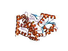
Horse LADH (Liver Alcohol Dehydrogenase)

The first-ever isolated alcohol dehydrogenase (ADH) was purified in 1937 from Saccharomyces cerevisiae (brewer's yeast). Many aspects of the catalytic mechanism for the horse liver ADH enzyme were investigated by Hugo Theorell and coworkers. ADH was also one of the first oligomeric enzymes that had its amino acid sequence and three-dimensional structure determined.

In early 1960, it was discovered in fruit flies of the genus Drosophila.

Properties

The alcohol dehydrogenases comprise a group of several isozymes that catalyse the oxidation of primary and secondary alcohols to aldehydes and ketones, respectively, and also can catalyse the reverse reaction. In mammals this is a redox (reduction/oxidation) reaction involving the coenzyme nicotinamide adenine dinucleotide (NAD+).

Mechanism of action in humans

Steps

  1. Binding of the coenzyme NAD+
  2. Binding of the alcohol substrate by coordination to zinc(II) ion
  3. Deprotonation of His-51
  4. Deprotonation of nicotinamide ribose
  5. Deprotonation of Thr-48
  6. Deprotonation of the alcohol
  7. Hydride transfer from the alkoxide ion to NAD+, leading to NADH and a zinc-bound aldehyde or ketone
  8. Release of aldehyde.

The mechanism in yeast and bacteria is the reverse of this reaction. These steps are supported through kinetic studies.

Involved subunits

The substrate is coordinated to the zinc and this enzyme has two zinc atoms per subunit. One is the active site, which is involved in catalysis. In the active site, the ligands are Cys-46, Cys-174, His-67, and one water molecule. The other subunit is involved with structure. In this mechanism, the hydride from the alcohol goes to NAD+. Crystal structures indicate that the His-51 deprotonates the nicotinamide ribose, which deprotonates Ser-48. Finally, Ser-48 deprotonates the alcohol, making it an aldehyde. From a mechanistic perspective, if the enzyme adds hydride to the re face of NAD+, the resulting hydrogen is incorporated into the pro-R position. Enzymes that add hydride to the re face are deemed Class A dehydrogenases.

Active site

The active site of alcohol dehydrogenase

The active site of human ADH1 (PDB:1HSO) consists of a zinc atom, His-67, Cys-174, Cys-46, Thr-48, His-51, Ile-269, Val-292, Ala-317, and Phe-319. In the commonly studied horse liver isoform, Thr-48 is a Ser, and Leu-319 is a Phe. The zinc coordinates the substrate (alcohol). The zinc is coordinated by Cys-46, Cys-174, and His-67. Leu-319, Ala-317, His-51, Ile-269 and Val-292 stabilize NAD+ by forming hydrogen bonds. His-51 and Ile-269 form hydrogen bonds with the alcohols on nicotinamide ribose. Phe-319, Ala-317 and Val-292 form hydrogen bonds with the amide on NAD+.

Structural zinc site

The structural zinc binding motif in alcohol dehydrogenase from an MD simulation

Mammalian alcohol dehydrogenases also have a structural zinc site. This Zn ion plays a structural role and is crucial for protein stability. The structures of the catalytic and structural zinc sites in horse liver alcohol dehydrogenase (HLADH) as revealed in crystallographic structures, which has been studied computationally with quantum chemistry as well as with classical molecular dynamics methods. The structural zinc site is composed of four closely spaced cysteine ligands (Cys97, Cys100, Cys103, and Cys111 in the amino acid sequence) positioned in an almost symmetric tetrahedron around the Zn ion. A recent study showed that the interaction between zinc and cysteine is governed by primarily an electrostatic contribution with an additional covalent contribution to the binding.

Types

Human

In humans, ADH exists in multiple forms as a dimer and is encoded by at least seven genes. Among the five classes (I-V) of alcohol dehydrogenase, the hepatic forms that are used primarily in humans are class 1. Class 1 consists of α, β, and γ subunits that are encoded by the genes ADH1A, ADH1B, and ADH1C. The enzyme is present at high levels in the liver and the lining of the stomach. It catalyzes the oxidation of ethanol to acetaldehyde (ethanal):

CH3CH2OH + NAD+ → CH3CHO + NADH + H+

This allows the consumption of alcoholic beverages, but its evolutionary purpose is probably the breakdown of alcohols naturally contained in foods or produced by bacteria in the digestive tract.

Another evolutionary purpose is reversible metabolism of retinol (vitamin A), an alcohol, to retinaldehyde, also known as retinal, which is then irreversibly converted into retinoic acid, which regulates expression of hundreds of genes.

alcohol dehydrogenase 1A,
α polypeptide
Identifiers
SymbolADH1A
Alt. symbolsADH1
NCBI gene124
HGNC249
OMIM103700
RefSeqNM_000667
UniProtP07327
Other data
EC number1.1.1.1
LocusChr. 4 q23

Search for
alcohol dehydrogenase 1B,
β polypeptide
Identifiers
SymbolADH1B
Alt. symbolsADH2
NCBI gene125
HGNC250
OMIM103720
RefSeqNM_000668
UniProtP00325
Other data
EC number1.1.1.1
LocusChr. 4 q23

Search for
alcohol dehydrogenase 1C,
γ polypeptide
Identifiers
SymbolADH1C
Alt. symbolsADH3
NCBI gene126
HGNC251
OMIM103730
RefSeqNM_000669
UniProtP00326
Other data
EC number1.1.1.1
LocusChr. 4 q23

Search for

Alcohol dehydrogenase is also involved in the toxicity of other types of alcohol: For instance, it oxidizes methanol to produce formaldehyde and ultimately formic acid. Humans have at least six slightly different alcohol dehydrogenases. Each is a dimer (i.e., consists of two polypeptides), with each dimer containing two zinc ions Zn2+. One of those ions is crucial for the operation of the enzyme: It is located at the catalytic site and holds the hydroxyl group of the alcohol in place.

Alcohol dehydrogenase activity varies between men and women, between young and old, and among populations from different areas of the world. For example, young women are unable to process alcohol at the same rate as young men because they do not express the alcohol dehydrogenase as highly, although the inverse is true among the middle-aged. The level of activity may not be dependent only on level of expression but also on allelic diversity among the population.

The human genes that encode class II, III, IV, and V alcohol dehydrogenases are ADH4, ADH5, ADH7, and ADH6, respectively.

Search for
alcohol dehydrogenase 5
(class III), χ polypeptide
Identifiers
SymbolADH5
NCBI gene128
HGNC253
OMIM103710
RefSeqNM_000671
UniProtP11766
Other data
EC number1.1.1.1
LocusChr. 4 q23

Search for
alcohol dehydrogenase 6
(class V)
Identifiers
SymbolADH6
NCBI gene130
HGNC255
OMIM103735
RefSeqNM_000672
UniProtP28332
Other data
EC number1.1.1.1
LocusChr. 4 q23

Search for
alcohol dehydrogenase 7
(class IV), μ or σ polypeptide
Identifiers
SymbolADH7
NCBI gene131
HGNC256
OMIM600086
RefSeqNM_000673
UniProtP40394
Other data
EC number1.1.1.1
LocusChr. 4 q23-q24

Search for

Yeast and bacteria

Unlike humans, yeast and bacteria (except lactic acid bacteria, and E. coli in certain conditions) do not ferment glucose to lactate. Instead, they ferment it to ethanol and CO2. The overall reaction can be seen below:

Glucose + 2 ADP + 2 Pi → 2 ethanol + 2 CO2 + 2 ATP + 2 H2O
Alcohol Dehydrogenase

In yeast and many bacteria, alcohol dehydrogenase plays an important part in fermentation: Pyruvate resulting from glycolysis is converted to acetaldehyde and carbon dioxide, and the acetaldehyde is then reduced to ethanol by an alcohol dehydrogenase called ADH1. The purpose of this latter step is the regeneration of NAD+, so that the energy-generating glycolysis can continue. Humans exploit this process to produce alcoholic beverages, by letting yeast ferment various fruits or grains. Yeast can produce and consume their own alcohol.

The main alcohol dehydrogenase in yeast is larger than the human one, consisting of four rather than just two subunits. It also contains zinc at its catalytic site. Together with the zinc-containing alcohol dehydrogenases of animals and humans, these enzymes from yeasts and many bacteria form the family of "long-chain"-alcohol dehydrogenases.

Brewer's yeast also has another alcohol dehydrogenase, ADH2, which evolved out of a duplicate version of the chromosome containing the ADH1 gene. ADH2 is used by the yeast to convert ethanol back into acetaldehyde, and it is expressed only when sugar concentration is low. Having these two enzymes allows yeast to produce alcohol when sugar is plentiful (and this alcohol then kills off competing microbes), and then continue with the oxidation of the alcohol once the sugar, and competition, is gone.

Plants

In plants, ADH catalyses the same reaction as in yeast and bacteria to ensure that there is a constant supply of NAD+. Maize has two versions of ADH - ADH1 and ADH2, Arabidopsis thaliana contains only one ADH gene. The structure of Arabidopsis ADH is 47%-conserved, relative to ADH from horse liver. Structurally and functionally important residues, such as the seven residues that provide ligands for the catalytic and noncatalytic zinc atoms, however, are conserved, suggesting that the enzymes have a similar structure. ADH is constitutively expressed at low levels in the roots of young plants grown on agar. If the roots lack oxygen, the expression of ADH increases significantly. Its expression is also increased in response to dehydration, to low temperatures, and to abscisic acid, and it plays an important role in fruit ripening, seedlings development, and pollen development. Differences in the sequences of ADH in different species have been used to create phylogenies showing how closely related different species of plants are. It is an ideal gene to use due to its convenient size (2–3 kb in length with a ≈1000 nucleotide coding sequence) and low copy number.

Iron-containing

Iron-containing alcohol dehydrogenase
PDB 1jqa EBI.jpg
bacillus stearothermophilus glycerol dehydrogenase complex with glycerol
 
Identifiers
SymbolFe-ADH
PfamPF00465
Pfam clanCL0224
InterProIPR001670
PROSITEPDOC00059
SCOP21jqa / SCOPe / SUPFAM

Available protein structures:

A third family of alcohol dehydrogenases, unrelated to the above two, are iron-containing ones. They occur in bacteria and fungi. In comparison to enzymes the above families, these enzymes are oxygen-sensitive. Members of the iron-containing alcohol dehydrogenase family include:

Other types

A further class of alcohol dehydrogenases belongs to quinoenzymes and requires quinoid cofactors (e.g., pyrroloquinoline quinone, PQQ) as enzyme-bound electron acceptors. A typical example for this type of enzyme is methanol dehydrogenase of methylotrophic bacteria.

Applications

In biotransformation, alcohol dehydrogenases are often used for the synthesis of enantiomerically pure stereoisomers of chiral alcohols. Often, high chemo- and enantioselectivity can be achieved. One example is the alcohol dehydrogenase from Lactobacillus brevis (LbADH), which is described to be a versatile biocatalyst. The high chemospecificity has been confirmed also in the case of substrates presenting two potential redox sites. For instance cinnamaldehyde presents both aliphatic double bond and aldehyde function. Unlike conventional catalysts, alcohol dehydrogenases are able to selectively act only on the latter, yielding exclusively cinnamyl alcohol.

In fuel cells, alcohol dehydrogenases can be used to catalyze the breakdown of fuel for an ethanol fuel cell. Scientists at Saint Louis University have used carbon-supported alcohol dehydrogenase with poly(methylene green) as an anode, with a nafion membrane, to achieve about 50 μA/cm2.

In 1949, E. Racker defined one unit of alcohol dehydrogenase activity as the amount that causes a change in optical density of 0.001 per minute under the standard conditions of assay. Recently, the international definition of enzymatic unit (E.U.) has been more common: one unit of Alcohol Dehydrogenase will convert 1.0 μmole of ethanol to acetaldehyde per minute at pH 8.8 at 25 °C.

Clinical significance

Alcoholism

There have been studies showing that variations in ADH that influence ethanol metabolism have an impact on the risk of alcohol dependence. The strongest effect is due to variations in ADH1B that increase the rate at which alcohol is converted to acetaldehyde. One such variant is most common in individuals from East Asia and the Middle East, another is most common in individuals from Africa. Both variants reduce the risk for alcoholism, but individuals can become alcoholic despite that. Researchers have tentatively detected a few other genes to be associated with alcoholism, and know that there must be many more remaining to be found. Research continues in order to identify the genes and their influence on alcoholism.

Drug dependence

Drug dependence is another problem associated with ADH, which researchers think might be linked to alcoholism. One particular study suggests that drug dependence has seven ADH genes associated with it, however, more research is necessary. Alcohol dependence and other drug dependence may share some risk factors, but because alcohol dependence is often comorbid with other drug dependences, the association of ADH with the other drug dependencies may not be causal.

Poisoning

Fomepizole, a drug that competitively inhibits alcohol dehydrogenase, can be used in the setting of acute methanol or ethylene glycol toxicity. This prevents the conversion of the methanol or ethylene glycol to its toxic metabolites (such as formic acid, formaldehyde, or glycolate). The same effect is also sometimes achieved with ethanol, again by competitive inhibition of ADH.

Drug metabolism

The drug hydroxyzine is broken into its active metabolite cetirizine by alcohol dehydrogenase. Other drugs with alcohol groups may be metabolized in a similar way as long as steric hindrance does not prevent the alcohol from reaching the active site

Alcohol and health

From Wikipedia, the free encyclopedia

Alcohol (also known as ethanol) has a number of effects on health. Short-term effects of alcohol consumption include intoxication and dehydration. Long-term effects of alcohol include changes in the metabolism of the liver and brain, several types of cancer and alcohol use disorder. Alcohol intoxication affects the brain, causing slurred speech, clumsiness, and delayed reflexes. Alcohol consumption can cause hypoglycemia in diabetics on certain medications, such as insulin or sulfonylurea, by blocking gluconeogenesis. There is an increased risk of developing an alcohol use disorder for teenagers while their brain is still developing. Adolescents who drink have a higher probability of injury including death.

Even light and moderate alcohol consumption have negative effects on health, such as by increasing a person's risk of developing several cancers. A 2014 World Health Organization report found that harmful alcohol consumption caused about 3.3 million deaths annually worldwide. Negative effects are related to the amount consumed with no safe lower limit seen. Some nations have introduced alcohol packaging warning messages that inform consumers about alcohol and cancer, as well as fetal alcohol syndrome.

The median lethal dose of alcohol in test animals is a blood alcohol content of 0.45%. This is about six times the level of ordinary intoxication (0.08%), but vomiting or unconsciousness may occur much sooner in people who have a low tolerance for alcohol. The high tolerance of chronic heavy drinkers may allow some of them to remain conscious at levels above 0.40%, although serious health hazards are incurred at this level.

Alcohol also limits the production of vasopressin (antidiuretic hormone) from the hypothalamus and the secretion of this hormone from the posterior pituitary gland. This is what causes severe dehydration when alcohol is consumed in large amounts. It also causes a high concentration of water in the urine and vomit, and the intense thirst that goes along with a hangover.

Short-term effects

The short-term effects of alcohol consumption range from a decrease in anxiety and motor skills at lower doses to unconsciousness, anterograde amnesia, and central nervous system depression at higher doses. Cell membranes are highly permeable to alcohol, so once alcohol is in the bloodstream it can diffuse into nearly every cell in the body.

The concentration of alcohol in blood is measured via blood alcohol content (BAC). The amount and circumstances of consumption play a large part in determining the extent of intoxication; for example, eating a heavy meal before alcohol consumption causes alcohol to absorb more slowly. Hydration also plays a role, especially in determining the extent of hangovers. After binge drinking, unconsciousness can occur and extreme levels of consumption can lead to alcohol poisoning and death (a concentration in the blood stream of 0.40% will kill half of those affected). Alcohol may also cause death indirectly, by asphyxiation from vomit.

Alcohol disrupts normal sleep patterns thereby reducing sleep quality and can greatly exacerbate sleep problems. During abstinence, residual disruptions in sleep regularity and sleep patterns are the greatest predictors of relapse.

Heavy alcohol consumption while in a hunger state can cause alcoholic ketoacidosis, a life-threatening metabolic derailment.

Long-term effects

According to the World Health Organization's 2018 Global Status Report on Alcohol and Health, there are more than 3 million people who die from the harmful effects of alcohol each year, which amounts to more than 5% of the burden of disease worldwide. The US National Institutes of Health similarly estimates that 3.3 million deaths (5.9% of all deaths) were believed to be due to alcohol each year.

Guidelines in the US and the UK advise that if people choose to drink, they should drink moderately.

Even light and moderate alcohol consumption increases a person's cancer risk, especially the risk of developing squamous cell carcinoma of the esophagus, cancers of the mouth and tongue, liver cancer, and breast cancer.

Health risks of alcohol consumption

A systematic analysis of data from the Global Burden of Disease Study, which was an observational study, found that long-term consumption of any amount of alcohol is associated with an increased risk of death in all people, and that even moderate consumption appears to be risky. Similar to prior analyses, it found an apparent benefit for older women in reducing the risks of death from ischemic heart disease and from diabetes mellitus, but unlike prior studies it found those risks cancelled by an apparent increased risk of death from breast cancer and other causes. A 2016 systematic review and meta-analysis found that moderate ethanol consumption brought no mortality benefit compared with lifetime abstention from ethanol consumption. Risk is greater in younger people due to heavy episodic drinking which may result in violence or accidents.

Long-term heavy use of alcohol damages nearly every organ and system in the body. Risks include alcohol use disorder, malnutrition, chronic pancreatitis, alcoholic liver disease (e.g., permanent liver scarring) and several types of cancer. In addition, damage to the central nervous system and peripheral nervous system (e.g., painful peripheral neuropathy) can occur from chronic alcohol misuse.

The developing adolescent brain is particularly vulnerable to the toxic effects of alcohol.

A systematic analysis found in 2022 that the level of alcohol consumption recommended by many existing guidelines is too high in particular for young people in all regions.

DNA damage

Acetaldehyde is produced when cells process ethanol. Acetaldehyde, is a DNA damaging metabolite that can interact with DNA to crosslink the two strands of the DNA duplex. The mechanisms the cells use for repairing these crosslinks are error prone, thus leading to mutations that in the long term can cause cancer.

Pregnancy

Medical organizations strongly discourage drinking alcohol during pregnancy. Alcohol passes easily from the mother's bloodstream through the placenta and into the bloodstream of the fetus, which interferes with brain and organ development. Alcohol can affect the fetus at any stage during pregnancy, but the level of risk depends on the amount and frequency of alcohol consumed. Regular heavy drinking and heavy episodic drinking (also called binge drinking), entailing four or more standard alcoholic drinks (a pint of beer or 50 ml drink of a spirit such as whisky corresponds to about two units of alcohol) on any one occasion, pose the greatest risk for harm, but lesser amounts can cause problems as well. There is no known safe amount or safe time to drink during pregnancy, and the U.S. Centers for Disease Control and Prevention recommends complete abstinence for women who are pregnant, trying to become pregnant, or are sexually active and not using birth control.

Prenatal alcohol exposure can lead to fetal alcohol spectrum disorders (FASDs). The most severe form of FASD is fetal alcohol syndrome (FAS). Problems associated with FASD include abnormal facial development, low birth weight, stunted growth, small head size, delayed or uncoordinated motor skills, hearing or vision problems, learning disabilities, behavior problems, and inappropriate social skills compared to same-age peers. Those affected are more likely to have trouble in school, legal problems, participate in high-risk behaviors, and develop substance use disorders like excessive drinking themselves.

Cardiovascular disease

In 2010, a systematic review reported that moderate consumption of alcohol does not cause harm to people with cardiovascular disease. However, the authors did not encourage people to start drinking alcohol in the hope of any benefit. In a 2018 study on 599,912 drinkers, a roughly linear association was found with alcohol consumption and a higher risk of stroke, coronary artery disease excluding myocardial infarction, heart failure, fatal hypertensive disease, and fatal aortic aneurysm, even for moderate drinkers. The American Heart Association states that people who are currently non-drinkers should not start drinking alcohol. Alcohol consumption also increases the risk of developing harmful abnormal heart rhythms such as atrial fibrillation, even with regular light to moderate alcohol use.

Breastfeeding

The UK National Health Service states that "an occasional drink is unlikely to harm" a breastfed baby, and recommends consumption of "no more than one or two units of alcohol once or twice a week" for breastfeeding mothers (where a pint of beer or 50 ml drink of a spirit such as whisky corresponds to about two units of alcohol). The NHS also recommends to wait for a couple of hours before breastfeeding or express the milk into a bottle before drinking. Researchers have shown that intoxicated breastfeeding reduces the average milk expression but poses no immediate threat to the child as the amount of transferred alcohol is insignificant.

Alcohol education

Alcohol education is the practice of disseminating information about the effects of alcohol on health, as well as society and the family unit. It was introduced into the public schools by temperance organizations such as the Woman's Christian Temperance Union in the late 19th century. Initially, alcohol education focused on how the consumption of alcoholic beverages affected society, as well as the family unit. In the 1930s, this came to also incorporate education pertaining to alcohol's effects on health. Organizations such as the National Institute on Alcohol Abuse and Alcoholism in the United States were founded to promulgate alcohol education alongside those of the temperance movement, such as the American Council on Alcohol Problems.

Alcohol expectations

Alcohol expectations are beliefs and attitudes that people have about the effects they will experience when drinking alcoholic beverages. They are just largely beliefs about alcohol's effects on a person's behaviors, abilities, and emotions. Some people believe that if alcohol expectations can be changed, then alcohol use disorders might be reduced. Men tend to become more aggressive in laboratory studies in which they are drinking only tonic water but believe that it contains alcohol. They also become less aggressive when they believe they are drinking only tonic water, but are actually drinking tonic water that contains alcohol.

The phenomenon of alcohol expectations recognizes that intoxication has real physiological consequences that alter a drinker's perception of space and time, reduce psychomotor skills, and disrupt equilibrium. The manner and degree to which alcohol expectations interact with the physiological short-term effects of alcohol, resulting in specific behaviors, is unclear.

A single study found that if a society believes that intoxication leads to sexual behavior, rowdy behavior, or aggression, then people tend to act that way when intoxicated. But if a society believes that intoxication leads to relaxation and tranquil behavior, then it usually leads to those outcomes. Alcohol expectations vary within a society, so these outcomes are not certain.

People tend to conform to social expectations, and some societies expect that drinking alcohol will cause disinhibition. However, in societies in which the people do not expect that alcohol will disinhibit, intoxication seldom leads to disinhibition and bad behavior.

Alcohol expectations can operate in the absence of actual consumption of alcohol. Research in the United States over a period of decades has shown that men tend to become more sexually aroused when they think they have been drinking alcohol—even when they have not been drinking it.

Drug treatment programs

Most addiction treatment programs encourage people with drinking problems to see themselves as having a chronic, relapsing disease that requires a lifetime of attendance at 12-step meetings to keep in check.

Alcohol use disorder

Addiction experts in psychiatry, chemistry, pharmacology, forensic science, epidemiology, and the police and legal services engaged in delphic analysis regarding 20 popular recreational drugs. Alcohol was ranked 6th in dependence, 11th in physical harm, and 2nd in social harm.

Alcohol misuse prevention programs

More than 200 injuries and disease conditions are caused due to alcohol misuse. It is a causative agent influencing maternal health and development, noncommunicable diseases (including cancer and cardiovascular diseases), injuries, violence, mental health, and infectious diseases such as tuberculosis and HIV/AIDS. Harmful use of alcohol has been identified as a global health issue, and its management is a priority in the 2030 Agenda for Sustainable Development. In 2018, WHO launched the initiative SAFER, to decrease the number of deaths, diseases and injuries caused by alcohol misuse. It has been developed to address the regional, country and global health and developmental goals using high-impact, cost-effective, evidence-based interventions. Using a set of WHO tools and resources SAFER will concentrate on the more cost-effective interventions to reduce and prevent alcohol misuse. The five WHO "best buys" for decreasing alcohol misuse are priority in this action plan:

  • Strengthen restrictions on alcohol availability.
  • Advance and enforce drink driving countermeasures.
  • Facilitate access to screening, brief interventions, and treatment.
  • Enforce bans or comprehensive restrictions on alcohol advertising, sponsorship, and promotion.
  • Raise prices on alcohol through excise taxes and pricing policies.

The promotion and success of the SAFER initiative is based on three key principles to implement, to monitor, and to protect.

Recommended maximum intake

Binge drinking is becoming a major problem in the UK. Advice on weekly consumption is avoided in United Kingdom.

Since 1995, the UK government has advised that regular consumption of three to four units (one unit equates to 10 mL of pure ethanol) a day for men and or two to three units for women, would not pose significant health risks. However, consistently drinking more than four units a day (for men) and three units (women) is not advisable.

Previously (from 1992 until 1995), the advice was that men should drink no more than 21 units per week, and women no more than 14. (The difference between the sexes was due to the typically lower weight and water-to-body-mass ratio of women.) This was changed because a government study showed that many people were in effect "saving up" their units and using them at the end of the week, a phenomenon referred to as binge drinking. The Times reported in October 2007 that these limits had been "plucked out of the air" and had no scientific basis.

Sobriety

A midshipman is subjected to a random breathalyzer test to determine sobriety.

Sobriety is the condition of not having any measurable levels, or effects from mood-altering drugs. According to WHO "Lexicon of alcohol and drug terms", sobriety is continued abstinence from psychoactive drug use. Sobriety is also considered to be the natural state of a human being given at a birth. In a treatment setting, sobriety is the achieved goal of independence from consuming or craving mind-altering substances. As such, sustained abstinence is a prerequisite for sobriety. Early in abstinence, residual effects of mind-altering substances can preclude sobriety. These effects are labeled post-acute-withdrawal syndrome (PAWS). Someone who abstains, but has a latent desire to resume use, is not considered truly sober. An abstainer may be subconsciously motivated to resume drug use, but for a variety of reasons, abstains (e.g. such as a medical or legal concern precluding use). Sobriety has more specific meanings within specific contexts, such as the culture of Alcoholics Anonymous, other 12 step programs, law enforcement, and some schools of psychology. In some cases, sobriety implies achieving "life balance".

Injury and deaths

Injury is defined as physical damage or harm that is done or sustained. The potential of injuring oneself or others can be increased after consuming alcohol due to the certain short term effects related to the substance such as lack of coordination, blurred vision, and slower reflexes to name a few. Due to these effects the most common injuries include head, fall, and vehicle-related injuries. A study was conducted of patients admitted to the Ulster Hospital in Northern Ireland with fall related injuries. They found that 113 of those patients admitted to that hospital during that had consumed alcohol recently and that the injury severity was higher for those that had consumed alcohol compared to those that had not. Another study showed that 21% of patients admitted to the Emergency Department of the Bristol Royal Infirmary had either direct or indirect alcohol related injuries. If these figures are extrapolated it shows that the estimated number of patients with alcohol related injuries are over 7,000 during the year at this emergency department alone.

In the United States alcohol resulted in about 88,000 deaths in 2010. The World Health Organization calculated that more than 3 million people, mostly men, died as a result of harmful use of alcohol in 2016. This was about 13.5% of the total deaths of people between 20 and 39. More than 5% of the global disease burden was caused by the harmful use of alcohol. There are even higher estimates for Europe.

Genetic differences

Alcohol flush and respiratory reactions

Alcohol flush reaction is a condition in which an individual's face or body experiences flushes (appears red) or blotches as a result of an accumulation of acetaldehyde, a metabolic byproduct of the catabolic metabolism of alcohol. It is best known as a condition that is experienced by people of Asian descent. According to the analysis by HapMap Project, the rs671 allele of the ALDH2 gene responsible for the flush reaction is rare among Europeans and Africans, and it is very rare among Mexican-Americans. 30% to 50% of people of Chinese and Japanese ancestry have at least one ALDH*2 allele. The rs671 form of ALDH2, which accounts for most incidents of alcohol flush reaction worldwide, is native to East Asia and most common in southeastern China. It most likely originated among Han Chinese in central China, and it appears to have been positively selected in the past. Another analysis correlates the rise and spread of rice cultivation in Southern China with the spread of the allele. The reasons for this positive selection are unknown, but the hypothesis that elevated concentrations of acetaldehyde may have conferred protection against certain parasitic infections, such as Entamoeba histolytica have been suggested. The same SNP allele of ALDH2, also termed glu487lys, and the abnormal accumulation of acetaldehyde following the drinking of alcohol, is associated with the alcohol-induced respiratory reactions of rhinitis and asthma that occur in Eastern Asian populations.

Metabolism of alcohol (ethanol) to acetaldehyde (ethanal)
and then to acetic acid (ethanoic acid)

Alcohol and Native Americans

Compared with the United States population in general, the Native American population is much more susceptible to alcohol use disorder and related diseases and deaths. From 2006 to 2010, alcohol-attributed deaths accounted for 11.7 percent of all Native American deaths, more than twice the rates of the general U.S. population. The median alcohol-attributed death rate for Native Americans (60.6 per 100,000) was twice as high as the rate for any other racial or ethnic group. Males are affected disproportionately more by alcohol-related conditions than females.

Native American and Native Alaskan youth are far more likely to experiment with alcohol at a younger age than non-Native youth. Low self-esteem and transgenerational trauma have been associated with substance use disorders among Native American teens in the U.S. and Canada.

Native American populations exhibit genetic differences in the alcohol-metabolizing enzymes alcohol dehydrogenase and ALDH, although evidence that these genetic factors are more prevalent in Native Americans than other ethnic groups has been a subject of debate. According to one 2013 review of academic literature on the issue, there is a "substantial genetic component in Native Americans" and that "most Native Americans lack protective variants seen in other populations." Many scientists have provided evidence of the genetic component of alcohol use disorder by the biopsychosocial model of alcohol use disorder. Molecular genetics research currently has not found one specific gene that is responsible for the rates of alcohol use disorder among Native Americans, implying the phenomenon may be due to an interplay of multiple genes and environmental factors. Research on alcohol use disorder in families suggests that learned behavior augments genetic factors in increasing the probability that children of people with alcohol use disorder will themselves have problems with alcohol misuse.

Genetics and amount of consumption

Having a particular genetic variant (A-allele of ADH1B rs1229984) is associated with non-drinking and lower alcohol consumption. This variant is also associated with favorable cardiovascular profile and a reduced risk of coronary artery disease compared to those without the genetic variant, but it is unknown whether this may be caused by differences in alcohol consumption or by additional confounding effects of the genetic variant itself.

Gender differences

Historically, according to the British Medical Journal, "men have been far more likely than women to drink alcohol and to drink it in quantities that damage their health, with some figures suggesting up to a 12-fold difference between the sexes." However, analysis of data collected over a century from multiple countries suggests that the gender gap in alcohol consumption is narrowing, and that young women (born after 1981) are consuming alcohol more than their male counterparts. Such findings have implications for the way in which alcohol-use prevention and intervention programs are designed and implemented.

Alcohol use disorder

Alcohol use disorder (AUD) is defined as a medical condition characterized by an impaired ability to stop or control alcohol use despite adverse social, occupational, or health consequences. Excessive alcohol use can lead to health-related illness and continuous alcohol engagement can ultimately lead to death. Behavioral factors of AUD include binge drinking and heavy alcohol use throughout one's day. AUD affects each culture differently, but African Americans are found to be the hardest impacted. Common health-related illnesses that stem from AUD but are prevalent in African American communities are liver disease, cirrhosis, hypertension, heart disease, oral cancer, stroke, and more. In 2020, heart disease ranked number 3 in the leading cause of death for African Americans ages 15-24. However, on the, contrary African Americans have been proven to consume less alcohol than other counterparts. According to American's Health Rankings, 15.4% of blacks reported excessive drinking, 19.4% of Hispanics, 19.2% of whites and 16.9% of Native Americans. In the United States, social economic status affects, one's ability to access basic necessities to support one's health, life, and survival. If one has a higher socioeconomic status, their income is higher, they are able to support their living needs and have better access to healthcare. Higher SES status reduces the risk of AUD for all individuals. However, those with a lower socioeconomic status majority of minorities are less fortunate. They are faced with poverty, low income, unemployment, and lack of access to healthier food options, which then contributes to poor health and higher AUD risk. The correlation between levels of socioeconomic status is prominent in alcohol-related health illnesses between cultures.

Sensitivity

Several biological factors make women more vulnerable to the effects of alcohol than men.

  • Body fat. Women tend to weigh less than men, and—pound for pound—a woman's body contains less water and more fatty tissue than a man's. Because fat retains alcohol while water dilutes it, alcohol remains at higher concentrations for longer periods of time in a woman's body, exposing her brain and other organs to more alcohol.
  • Enzymes. Women have lower levels of two enzymes—alcohol dehydrogenase and aldehyde dehydrogenase—that metabolize (break down) alcohol in the stomach and liver. As a result, women absorb more alcohol into their bloodstreams than men.
  • Hormones. Changes in hormone levels during the menstrual cycle may also affect how a woman metabolizes alcohol.

Metabolism

Females demonstrated a higher average rate of elimination (mean, 0.017; range, 0.014–0.021 g/210 L) than males (mean, 0.015; range, 0.013–0.017 g/210 L). Female subjects on average had a higher percentage of body fat (mean, 26.0; range, 16.7–36.8%) than males (mean, 18.0; range, 10.2–25.3%).

Depression

The link between alcohol consumption, depression, and gender was examined by the Centre for Addiction and Mental Health (Canada). The study found that women taking antidepressants consumed more alcohol than women who did not experience depression as well as men taking antidepressants. The researchers, Kathryn Graham and a PhD Student, Agnes Massak, analyzed the responses to a survey by 14,063 Canadian residents aged 18–76 years. The survey included measures of quantity, frequency of drinking, depression, and antidepressant use, over the period of a year. The researchers used data from the GENACIS Canada survey, part of an international collaboration to investigate the influence of cultural variation on gender differences in alcohol use and related problems. The purpose of the study was to examine whether, like in other studies already conducted on male depression and alcohol consumption, depressed women also consumed less alcohol when taking anti-depressants. According to the study, both men and women experiencing depression (but not on antidepressants) drank more than non-depressed counterparts. Men taking antidepressants consumed significantly less alcohol than depressed men who did not use antidepressants. Non-depressed men consumed 436 drinks per year, compared to 579 drinks for depressed men not using antidepressants, and 414 drinks for depressed men who used antidepressants. Alcohol consumption remained higher whether the depressed women were taking antidepressants or not. 179 drinks per year for non-depressed women, 235 drinks for depressed women not using antidepressants, and 264 drinks for depressed women who used antidepressants. The lead researcher argued that the study "suggests that the use of antidepressants is associated with lower alcohol consumption among men with depression. But this does not appear to be true for women."

Teenage Alcohol Abuse

While, most teens understand the negative impacts of drinking a lot of alcohol in one sitting, many believe that consuming some alcohol will not be that risky of a behavior. However, teens who drink alcohol on average consume more alcohol in one sitting than most adults, and nearly half of all teens who consumed some amount of alcohol in the past 30 days, had done so in excess. Not only are teen drinkers more likely to get drunk, but the effects of drunkenness are worse. The temporarily impaired judgment can lead to permanent consequences such as serious and crippling injury to oneself or others, unplanned pregnancy, or alcoholism later in life. Even if one were to avoid terrible events, they will still suffer irreversible damage to brain development and be far more likely to abuse other substances in the future.

Nicotinic acetylcholine receptor

From Wikipedia, the free encyclopedia
 

Nicotinic acetylcholine receptors, or nAChRs, are receptor polypeptides that respond to the neurotransmitter acetylcholine. Nicotinic receptors also respond to drugs such as the agonist nicotine. They are found in the central and peripheral nervous system, muscle, and many other tissues of many organisms. At the neuromuscular junction they are the primary receptor in muscle for motor nerve-muscle communication that controls muscle contraction. In the peripheral nervous system: (1) they transmit outgoing signals from the presynaptic to the postsynaptic cells within the sympathetic and parasympathetic nervous system, and (2) they are the receptors found on skeletal muscle that receive acetylcholine released to signal for muscular contraction. In the immune system, nAChRs regulate inflammatory processes and signal through distinct intracellular pathways. In insects, the cholinergic system is limited to the central nervous system.

The nicotinic receptors are considered cholinergic receptors, since they respond to acetylcholine. Nicotinic receptors get their name from nicotine which does not stimulate the muscarinic acetylcholine receptors but selectively binds to the nicotinic receptors instead. The muscarinic acetylcholine receptor likewise gets its name from a chemical that selectively attaches to that receptor — muscarine. Acetylcholine itself binds to both muscarinic and nicotinic acetylcholine receptors.

As ionotropic receptors, nAChRs are directly linked to ion channels. New evidence suggests that these receptors can also use second messengers (as metabotropic receptors do) in some cases. Nicotinic acetylcholine receptors are the best-studied of the ionotropic receptors.

Since nicotinic receptors help transmit outgoing signals for the sympathetic and parasympathetic systems, nicotinic receptor antagonists such as hexamethonium interfere with the transmission of these signals. Thus, for example, nicotinic receptor antagonists interfere with the baroreflex that normally corrects changes in blood pressure by sympathetic and parasympathetic stimulation of the heart.

Structure

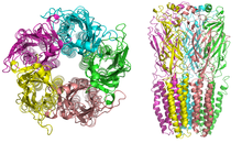
Nicotinic receptor structure

Nicotinic receptors, with a molecular mass of 290 kDa, are made up of five subunits, arranged symmetrically around a central pore. Each subunit comprises four transmembrane domains with both the N- and C-terminus located extracellularly. They possess similarities with GABAA receptors, glycine receptors, and the type 3 serotonin receptors (which are all ionotropic receptors), or the signature Cys-loop proteins.

In vertebrates, nicotinic receptors are broadly classified into two subtypes based on their primary sites of expression: muscle-type nicotinic receptors and neuronal-type nicotinic receptors. In the muscle-type receptors, found at the neuromuscular junction, receptors are either the embryonic form, composed of α1, β1, γ, and δ subunits in a 2:1:1:1 ratio ((α1)2β1γδ), or the adult form composed of α1, β1, δ, and ε subunits in a 2:1:1:1 ratio ((α1)2β1δε). The neuronal subtypes are various homomeric (all one type of subunit) or heteromeric (at least one α and one β) combinations of twelve different nicotinic receptor subunits: α2−α10 and β2−β4. Examples of the neuronal subtypes include: (α4)32)2, (α4)22)3, (α3)24)3, α4α6β32)2, (α7)5, and many others. In both muscle-type and neuronal-type receptors, the subunits are very similar to one another, especially in the hydrophobic regions.

A number of electron microscopy and x-ray crystallography studies have provided very high resolution structural information for muscle and neuronal nAChRs and their binding domains.

Binding

As with all ligand-gated ion channels, opening of the nAChR channel pore requires the binding of a chemical messenger. Several different terms are used to refer to the molecules that bind receptors, such as ligand, agonist, or transmitter. As well as the endogenous agonist acetylcholine, agonists of the nAChR include nicotine, epibatidine, and choline. Nicotinic antagonists that block the receptor include mecamylamine, dihydro-β-erythroidine, and hexamethonium.

In muscle-type nAChRs, the acetylcholine binding sites are located at the α and either ε or δ subunits interface. In neuronal nAChRs, the binding site is located at the interface of an α and a β subunit or between two α subunits in the case of α7 receptors. The binding site is located in the extracellular domain near the N terminus. When an agonist binds to the site, all present subunits undergo a conformational change and the channel is opened and a pore with a diameter of about 0.65 nm opens.

Channel opening

Nicotinic AChRs may exist in different interconvertible conformational states. Binding of an agonist stabilizes the open and desensitized states. In normal physiological conditions, the receptor needs exactly two molecules of ACh to open. Opening of the channel allows positively charged ions to move across it; in particular, sodium enters the cell and potassium exits. The net flow of positively charged ions is inward.

The nAChR is a non-selective cation channel, meaning that several different positively charged ions can cross through. It is permeable to Na+ and K+, with some subunit combinations that are also permeable to Ca2+. The amount of sodium and potassium the channels allow through their pores (their conductance) varies from 50–110 pS, with the conductance depending on the specific subunit composition as well as the permeant ion.

Many neuronal nAChRs can affect the release of other neurotransmitters. The channel usually opens rapidly and tends to remain open until the agonist diffuses away, which usually takes about 1 millisecond. AChRs can spontaneously open with no ligands bound or can spontaneously close with ligands bound, and mutations in the channel can shift the likelihood of either event. Therefore, ACh binding changes the probability of pore opening, which increases as more ACh binds.

The nAChR is unable to bind ACh when bound to any of the snake venom α-neurotoxins. These α-neurotoxins antagonistically bind tightly and noncovalently to nAChRs of skeletal muscles and in neurons, thereby blocking the action of ACh at the postsynaptic membrane, inhibiting ion flow and leading to paralysis and death. The nAChR contains two binding sites for snake venom neurotoxins. Progress in discovering the dynamics of binding action of these sites has proved difficult, although recent studies using normal mode dynamics have aided in predicting the nature of both the binding mechanisms of snake toxins and of ACh to nAChRs. These studies have shown that a twist-like motion caused by ACh binding is likely responsible for pore opening, and that one or two molecules of α-bungarotoxin (or other long-chain α-neurotoxin) suffice to halt this motion. The toxins seem to lock together neighboring receptor subunits, inhibiting the twist and therefore, the opening motion.

Effects

The activation of receptors by nicotine modifies the state of neurons through two main mechanisms. On one hand, the movement of cations causes a depolarization of the plasma membrane (which results in an excitatory postsynaptic potential in neurons) leading to the activation of voltage-gated ion channels. On the other hand, the entry of calcium acts, either directly or indirectly, on different intracellular cascades. This leads, for example, to the regulation of activity of some genes or the release of neurotransmitters.

Regulation

Desensitization

Ligand-bound desensitization of receptors was first characterized by Katz and Thesleff in the nicotinic acetylcholine receptor.

Prolonged or repeated exposure to a stimulus often results in decreased responsiveness of that receptor toward a stimulus, termed desensitization. nAChR function can be modulated by phosphorylation by the activation of second messenger-dependent protein kinases. PKA and PKC, as well as tyrosine kinases, have been shown to phosphorylate the nAChR resulting in its desensitization. It has been reported that, after prolonged receptor exposure to the agonist, the agonist itself causes an agonist-induced conformational change in the receptor, resulting in receptor desensitization.

Desensitized receptors can revert to a prolonged open state when an agonist is bound in the presence of a positive allosteric modulator, for example PNU-120,596. Also, there is evidence that indicates specific chaperone molecules have regulatory effects on these receptors.

Roles

The subunits of the nicotinic receptors belong to a multigene family (16 members in humans) and the assembly of combinations of subunits results in a large number of different receptors (for more information see the Ligand-Gated Ion Channel database). These receptors, with highly variable kinetic, electrophysiological and pharmacological properties, respond to nicotine differently, at very different effective concentrations. This functional diversity allows them to take part in two major types of neurotransmission. Classical synaptic transmission (wiring transmission) involves the release of high concentrations of neurotransmitter, acting on immediately neighboring receptors. In contrast, paracrine transmission (volume transmission) involves neurotransmitters released by axon terminals, which then diffuse through the extra-cellular medium until they reach their receptors, which may be distant. Nicotinic receptors can also be found in different synaptic locations; for example the muscle nicotinic receptor always functions post-synaptically. The neuronal forms of the receptor can be found both post-synaptically (involved in classical neurotransmission) and pre-synaptically where they can influence the release of multiple neurotransmitters.

Subunits

17 vertebrate nAChR subunits have been identified, which are divided into muscle-type and neuronal-type subunits. Although an α8 subunit/gene is present in avian species such as the chicken, it is not present in human or mammalian species.

The nAChR subunits have been divided into 4 subfamilies (I–IV) based on similarities in protein sequence. In addition, subfamily III has been further divided into 3 types.

Neuronal-type Muscle-type
I II III IV
α9, α10 α7, α8 1 2 3 α1, β1, δ, γ, ε
α2, α3, α4, α6 β2, β4 β3, α5

Neuronal nAChRs are transmembrane proteins that form pentameric structures assembled from a family of subunits composed of α2–α10 and β2–β4. These subunits were discovered from the mid-1980s through the early 1990s, when cDNAs for multiple nAChR subunits were cloned from rat and chicken brains, leading to the identification of eleven different genes (twelve in chickens) that code for neuronal nAChR subunits; The subunit genes identified were named α2–α108 only found in chickens) and β2–β4. It has also been discovered that various subunit combinations could form functional nAChRs that could be activated by acetylcholine and nicotine, and the different combinations of subunits generate subtypes of nAChRs with diverse functional and pharmacological properties. When expressed alone, α7, α8, α9, and α10 are able to form functional receptors, but other α subunits require the presence of β subunits to form functional receptors. In mammals, nAchR subunits have been found to be encoded by 17 genes, and of these, nine genes encoding α-subunits and three encoding β-subunits are expressed in the brain. β2 subunit-containing nAChRs (β2nAChRs) and α7nAChRs are widely expressed in the brain, whereas other nAChR subunits have more restricted expression. The pentameric assembly of nAChRs is subjected to the subunits that are produced in various cell types such as in the human lung where epithelial and muscular pentamers largely differ.

CHRNA5/A3/B4

An important nAchR gene cluster (CHRNA5/A3/B4) contains the genes encoding for the α5, α3 and β4 subunits. Genetic studies have identified single nucleotide polymorphisms (SNPs) in the chromosomal locus encoding these three nAChR genes as risk factors for nicotine dependence, lung cancer, chronic obstructive pulmonary disease, alcoholism, and peripheral arterial disease. The CHRNA5/A3/B4 nAChR subunit genes are found in a tight cluster in chromosomal region 15q24–25. The nAChR subunits encoded by this locus form the predominant nicotinic receptor subtypes expressed in the peripheral nervous system (PNS) and other key central nervous system (CNS) sites, such as the medial habenula, a structure between the limbic forebrain and midbrain involved in major cholinergic circuitry pathways. Further research of the CHRNA5/A3/B4 genes have revealed that “neuronal” nAChR genes are also expressed in non-neuronal cells where they are involved in various fundamental processes, such as inflammation. The CHRNA5/A3/B4 genes are co-expressed in many cell types and the transcriptional activities of the promoter regions of the three genes are regulated by many of the same transcription factors, demonstrating that their clustering may reflect control of gene expression.

CHRNA6/CHRNB3

CHRNB3 and CHRNA6 are also grouped in a gene cluster, located on 8p11. Multiple studies have shown that SNPS in the CHRNB3–CHRNA6 have been linked to nicotine dependence and smoking behavior, such as two SNPs in CHRNB3, rs6474413 and rs10958726. Genetic variation in this region also displays influence susceptibility to use drugs of abuse, including cocaine and alcohol consumption. Nicotinic receptors containing α6 or β3 subunits expressed in brain regions, especially in the ventral tegmental area and substantia nigra, are important for drug behaviors due to their role in dopamine release. Genetic variation in these genes can alter sensitivity to drugs of abuse in numerous ways, including changing the amino acid structure of the protein or cause alterations in transcriptional and translational regulation.

CHRNA4/CHRNB2

Other well studied nAChR genes include the CHRNA4 and CHRNB2, which have been associated as Autosomal Dominant Nocturnal Frontal Lobe Epilepsy (ADNFLE) genes. Both of these nAChR subunits are present in the brain and the occurrence of mutations in these two subunits cause a generalized type of epilepsy. Examples include the CHRNA4 insertion mutation 776ins3 that is associated with nocturnal seizures and psychiatric disorders, and the CHRNB2 mutation I312M that seems to cause not only epilepsy but also very specific cognitive deficits, such as deficits in learning and memory. There is naturally occurring genetic variation between these two genes and analysis of single nucleotide polymorphisms (SNPs) and other gene modifications show a higher variation in the CHRNA4 gene than in the CHRNB2 gene, implying that nAChR β2, the protein encoded by CHRNB2, associates with more subunits than α4. CHRNA2 has also been reported as a third candidate for nocturnal frontal lobe seizures.

CHRNA7

Several studies have reported an association between CHRNA7 and endophenotypes of psychiatric disorders and nicotine dependence, contributing to the significant clinical relevance of α7 and research being done on it. CHRNA7 was one of the first genes that had been considered to be involved with schizophrenia. Studies identified several CHRNA7 promoter polymorphisms that reduce the genes transcriptional activity to be associated with schizophrenia, which is consistent with the finding of reduced levels of a7 nAChRs in the brain of schizophrenic patients. Both nAChRs subtypes, α4β2 and α7, have been found to be significantly reduced in post-mortem studies of individuals with schizophrenia. Additionally, smoking rates are significantly higher in those with schizophrenia, implying that smoking nicotine may be a form of self-medicating.

Introduction to entropy

From Wikipedia, the free encyclopedia https://en.wikipedia.org/wiki/Introduct...