Search This Blog

Saturday, August 31, 2024

Amyloidosis

From Wikipedia, the free encyclopedia
https://en.wikipedia.org/wiki/Amyloidosis

Amyloidosis
Amyloidosis symptoms are often vague and require different physician specialists for diagnosis. Telltale symptoms may include an enlarged tongue (macroglossia) or bruising around the eyes (purpura)
SpecialtyInternal Medicine
SymptomsFeeling tired, weight loss, swelling of the legs, shortness of breath, bleeding, feeling light headed with standing
Usual onset55–65 years old
CausesGenetic or acquired
Diagnostic methodTissue biopsy
TreatmentSupportive care, directed at the underlying cause, dialysis, organ transplantation
PrognosisImproved with treatment
Frequency3–13 per million per year (AL amyloidosis)
Deaths1 per 1,000 people (developed world)

Amyloidosis is a group of diseases in which abnormal proteins, known as amyloid fibrils, build up in tissue. There are several non-specific and vague signs and symptoms associated with amyloidosis. These include fatigue, peripheral edema, weight loss, shortness of breath, palpitations, and feeling faint with standing. In AL amyloidosis, specific indicators can include enlargement of the tongue and periorbital purpura. In wild-type ATTR amyloidosis, non-cardiac symptoms include: bilateral carpal tunnel syndrome, lumbar spinal stenosis, biceps tendon rupture, small fiber neuropathy, and autonomic dysfunction.

There are about 36 different types of amyloidosis, each due to a specific protein misfolding. Within these 36 proteins, 19 are grouped into localized forms, 14 are grouped as systemic forms, and three proteins can identify as either. These proteins can become irregular due to genetic effects, as well as through acquired environmental factors. The four most common types of systemic amyloidosis are light chain (AL), inflammation (AA), dialysis-related (Aβ2M), and hereditary and old age (ATTR and wild-type transthyretin amyloid).

Diagnosis may be suspected when protein is found in the urine, organ enlargement is present, or problems are found with multiple peripheral nerves and it is unclear why. Diagnosis is confirmed by tissue biopsy. Due to the variable presentation, a diagnosis can often take some time to reach.

Treatment is geared towards decreasing the amount of the involved protein. This may sometimes be achieved by determining and treating the underlying cause. AL amyloidosis occurs in about 3–13 per million people per year and AA amyloidosis in about two per million people per year. The usual age of onset of these two types is 55 to 60 years old. Without treatment, life expectancy is between six months and four years. In the developed world about one per 1,000 deaths are from systemic amyloidosis. Amyloidosis has been described since at least 1639.

Signs and symptoms

Skin features of amyloidosis cutis dyschromica. Hyperpigmented and hypopigmented macules on (A) lower legs, (B) back and waist, (C) waist. (D) Individual blisters on upper arm.

The presentation of amyloidosis is broad and depends on the site of amyloid accumulation. The kidney and heart are the most common organs involved.

Kidneys

Amyloid deposition in the kidney often involve the glomerular capillaries and mesangial regions, affecting the organ's ability to filter and excrete waste and retain plasma protein. This can lead to high levels of protein in the urine (proteinuria) and nephrotic syndrome. Several types of amyloidosis, including the AL and AA types, are associated with nephrotic syndrome. Approximately 20% and 40–60% of people with AL and AA amyloidosis respectively progress to end-stage kidney disease requiring dialysis.

Heart

Amyloid deposition in the heart can cause both diastolic and systolic heart failure. EKG changes may be present, showing low voltage and conduction abnormalities like atrioventricular block or sinus node dysfunction.[medical citation needed] On echocardiography, the heart shows a restrictive filling pattern, with normal to mildly reduced systolic function. AA amyloidosis usually spares the heart. Cardiac amyloidosis can present with symptoms of heart failure including shortness of breath, fatigue, and edema. As cardiac amyloidosis progresses, the amyloid deposition can affect the heart's ability to pump and fill blood as well as its ability to maintain normal rhythm, which leads to worsening heart function and decline in people's quality of life.

Nervous system

People with amyloidosis may have central nervous system involvement, along with peripheral involvement which causes sensory and autonomic neuropathies. Sensory neuropathy develops in a symmetrical pattern and progresses in a distal to proximal manner. Autonomic neuropathy can present as orthostatic hypotension but may manifest more gradually with nonspecific gastrointestinal symptoms like constipation, nausea, or early satiety. Amyloidosis of the central nervous system can have more severe and systemic presentations that may include life-threatening arrhythmias, cardiac failure, malnutrition, infection, or death.

Neuropathic presentation can depend on the etiology of amyloidosis. People with amyloidosis may experience dysfunction in various organ systems depending on the location and extent of nervous system involvement. For example, peripheral neuropathy can cause erectile dysfunction, incontinence and constipation, pupillary dysfunction, and sensory loss depending on the distribution of amyloidosis along different peripheral nerves.

Gastrointestinal and accessory organs

Accumulation of amyloid proteins in the gastrointestinal system may be caused by a wide range of amyloid disorders and have different presentations depending on the degree of organ involvement. Potential symptoms include weight loss, diarrhea, abdominal pain, heartburn (gastrointestinal reflux), and GI bleeding. Amyloidosis may also affect accessory digestive organs including the liver, and may present with jaundice, fatty stool, anorexia, fluid buildup in the abdomen, and spleen enlargement.

Accumulation of amyloid proteins in the liver can lead to elevations in serum aminotransferases and alkaline phosphatase, two biomarkers of liver injury, which is seen in about one third of people. Liver enlargement is common. In contrast, spleen enlargement is rare, occurring in 5% of people. Splenic dysfunction, leading to the presence of Howell-Jolly bodies on blood smear, occurs in 24% of people with amyloidosis. Malabsorption is seen in 8.5% of AL amyloidosis and 2.4% of AA amyloidosis. One suggested mechanism for the observed malabsorption is that amyloid deposits in the tips of intestinal villi (fingerlike projections that increase the intestinal area available for absorption of food), begin to erode the functionality of the villi, presenting a sprue-like picture.

Glands

Both the thyroid and adrenal glands can be infiltrated. It is estimated that 10–20% of people with amyloidosis have hypothyroidism. Adrenal infiltration may be harder to appreciate given that its symptoms of orthostatic hypotension and low blood sodium concentration may be attributed to autonomic neuropathy and heart failure.

"Amyloid deposits occur in the pancreas of people who also have diabetes mellitus, although it is not known if this is functionally important. The major component of pancreatic amyloid is a 37-amino acid residue peptide known as islet amyloid polypeptide or 'amylin.' This is stored with insulin in secretory granules in [beta] cells and is co secreted with insulin." (Rang and Dale's Pharmacology, 2015.)

Musculoskeletal system

Amyloid proteins deposit most commonly inside the knee, followed by hands, wrists, elbow, hip, and ankle, causing joint pain. In males with advanced age (>80 years), there is significant risk of wild-type transthyretin amyloid deposition in synovial tissue of knee joint, but predominantly in old age deposition of wild type transthyretin is seen in cardiac ventricles. ATTR deposits have been found in ligamentum flavum of patients that underwent surgery for lumbar spinal stenosis.

In beta 2-microglobulin amyloidosis, males have high risk of getting carpal tunnel syndrome. Aβ2MG amyloidosis (Hemodialysis associated amyloidosis) tends to deposit in synovial tissue, causing chronic inflammation of the synovial tissue in knee, hip, shoulder and interphalangeal joints. Amyloid light chains deposition in shoulder joint causes enlarged shoulders, also known as "shoulder pad sign". Amyloid light chain depositions can also cause bilateral symmetric polyarthritis.

The deposition of amyloid proteins in the bone marrow without causing plasma cell dyscrasias is called amyloidoma. It is commonly found in cervical, lumbar, and sacral vertebrae. Those affected may be presented with bone pain due to bone lysis, lumbar paraparesis, and a variety of neurological symptoms. Vertebral fractures are also common.

Eyes

A rare development is amyloid purpura, a susceptibility to bleeding with bruising around the eyes, termed "raccoon-eyes". Amyloid purpura is caused by amyloid deposition in the blood vessels and reduced activity of thrombin and factor X, two clotting proteins that lose their function after binding with amyloid.

Oral cavity

Amyloid deposits in tissue can cause enlargement of structures. Twenty percent of people with AL amyloidosis have an enlarged tongue, that can lead to obstructive sleep apnea, difficulty swallowing, and altered taste. Tongue enlargement does not occur in ATTR or AA amyloidosis. Deposition of amyloid in the throat can cause hoarseness.

Pathogenesis

Amyloidoses can be considered protein misfolding diseases. The vast majority of proteins that have been found to form amyloid deposits are secreted proteins, so the misfolding and formation of amyloid occurs outside cells, in the extracellular space. Of the 37 proteins so far identified as being vulnerable to amyloid formation, only four are cytosolic. Most amyloid-forming proteins are relatively small, but otherwise there is currently no evidence of structural or functional similarities among proteins known to form disease-associated amyloids. One third of amyloid disease is hereditary, in which case there is normally an early age of onset. Half of amyloid-related diseases are sporadic and have a late age of onset – in these cases, the protein aggregation may be associated with aging-related decline in protein regulation. Some medical treatments are associated with amyloid disease, but this is rare.

Amyloid-forming proteins aggregate into distinctive fibrillar forms with a beta-sheet structure. The beta-sheet form of amyloid is proteolysis-resistant, meaning it can not be degraded or broken down. As a result, amyloid deposits into the body's extracellular space. The process of forming amyloid fibrils is thought to have intermediate oligomeric forms. Both the oligomers and amyloid fibrils can be toxic to cells and can interfere with proper organ function. The relative significance of different aggregation species may depend on the protein involved and the organ system affected.

Diagnosis

Diagnosis of amyloidosis generally requires tissue biopsy. The biopsy is assessed for evidence of characteristic amyloid deposits. The tissue is treated with various stains. The most useful stain in the diagnosis of amyloid is Congo red, which, combined with polarized light, makes the amyloid proteins appear apple-green on microscopy. Also, thioflavin T stain may be used. A number of imaging techniques such as a Nuclear Medicine PYP scan, DPD scan or SAP scan are also in use.

A sample of tissue can be biopsied or obtained directly from the affected internal organ, but the first-line site of biopsy is subcutaneous abdominal fat, known as a "fat pad biopsy", due to its ease of acquisition. An abdominal fat biopsy is not completely sensitive and may result in false negatives, which means a negative result does not exclude the diagnosis of amyloidosis. However, direct biopsy of the affected organ may still be unnecessary as other less invasive methods of biopsy can also be used, including rectal mucosa, salivary gland, lip, or bone marrow biopsy which can achieve a diagnosis in up to 85% of people.

In the amyloid deposition of the joints, there will be a decreased signal in both T1 and T2 weighted MRI images. In amyloidoma, there will be low T1 signal with gadolinium injection and low T2 signal.

The type of the amyloid protein can be determined in various ways: the detection of abnormal proteins in the bloodstream (on protein electrophoresis or light chain determination); binding of particular antibodies to the amyloid found in the tissue (immunohistochemistry); or extraction of the protein and identification of its individual amino acids. Immunohistochemistry can identify AA amyloidosis the majority of the time, but can miss many cases of AL amyloidosis. Laser microdissection with mass spectrometry is the most reliable method of identifying the different forms of amyloidosis.

AL was previously considered the most common form of amyloidosis, and a diagnosis often begins with a search for plasma cell dyscrasia, memory B cells producing aberrant immunoglobulins or portions of immunoglobulins. Immunofixation electrophoresis of urine or serum is positive in 90% of people with AL amyloidosis. Immunofixation electrophoresis is more sensitive than regular electrophoresis but may not be available in all centers. Alternatively immunohistochemical staining of a bone marrow biopsy looking for dominant plasma cells can be sought in people with a high clinical suspicion for AL amyloidosis but negative electrophoresis.

ATTR is now considered to be the most common form of amyloidosis. It may be either age related in wild-type ATTR (ATTRv) or familial transthyretin-associated amyloidosis, is suspected in people with family history of idiopathic neuropathies or heart failure who lack evidence of plasma cell dyscrasias. ATTR can be identified using isoelectric focusing which separates mutated forms of transthyretin. Findings can be corroborated by genetic testing to look for specific known mutations in transthyretin that predispose to amyloidosis.

AA is suspected on clinical grounds in individuals with longstanding infections or inflammatory diseases. AA can be identified by immunohistochemistry staining.

Classification

Historical classification systems were based on clinical factors. Until the early 1970s, the idea of a single amyloid substance predominated. Various descriptive classification systems were proposed based on the organ distribution of amyloid deposits and clinical findings. Most classification systems included primary (i.e., idiopathic) amyloidosis, in which no associated clinical condition was identified, and secondary amyloidosis (i.e., secondary to chronic inflammatory conditions). Some classification systems included myeloma-associated, familial, and localized amyloidosis.

The modern era of amyloidosis classification began in the late 1960s with the development of methods to make amyloid fibrils soluble. These methods permitted scientists to study the chemical properties of amyloids. Descriptive terms such as primary amyloidosis, secondary amyloidosis, and others (e.g., senile amyloidosis), which are not based on cause, provide little useful information and are no longer recommended.

The modern classification of amyloid disease tends to use an abbreviation of the protein that makes the majority of deposits, prefixed with the letter A. For example, amyloidosis caused by transthyretin is termed "ATTR". Deposition patterns vary between people but are almost always composed of just one amyloidogenic protein. Deposition can be systemic (affecting many different organ systems) or organ-specific. Many amyloidoses are inherited, due to mutations in the precursor protein.

Other forms are due to different diseases causing overabundant or abnormal protein production – such as with overproduction of immunoglobulin light chains (termed AL amyloidosis), or with continuous overproduction of acute phase proteins in chronic inflammation (which can lead to AA amyloidosis).

About 60 amyloid proteins have been identified so far. Of those, at least 36 have been associated with a human disease.

All amyloid fibril proteins start with the letter "A" followed by the protein suffix (and any applicable specification). See below for a list of amyloid fibril proteins which have been found in humans:

Fibril protein Precursor protein Target Organs Systemic and/or localized Acquired or hereditary
AL Immunoglobulin light chain All organs, usually except CNS S, L A, H
AH Immunoglobulin heavy chain All organs except CNS S, L A
AA (Apo) serum amyloid A All organs except CNS S A
ATTR Transthyretin, wild type

Transthyretin, variants

Heart mainly in males, lung, ligaments, tenosynovium

PNS, ANS, heart, eye, leptomeninges

S

S

A

H

Aβ2M β2-microglobulin, wild type

β2-microglobulin, variants

Musculoskeletal system

ANS

S

S

A

H

AApoAI Apolipoprotein A I, variants Heart, liver, kidney, PNS, testis, larynx (C

terminal variants), skin (C terminal variants)

S H
AApoAII Apolipoprotein A II, variants Kidney S H
AApoAIV Apolipoprotein A IV, wild type Kidney medulla and systemic S A
AApoCII Apolipoprotein C II, variants Kidney S H
AApoCIII Apolipoprotein C III, variants Kidney S H
AGel Gelsolin, variants Kidney, PNS, cornea S H
ALys Lysozyme, variants Kidney S H
ALECT2 Leukocyte chemotactic factor-2 Kidney, primarily S A
AFib Fibrinogen a, variants Kidney, primarily S H
ACys Cystatin C, variants CNS, PNS, skin S H
ABri ABriPP, variants CNS S H
ADanb ADanPP, variants CNS L H
Aβ protein precursor, wild type

Aβ protein precursor, variant

CNS

CNS

L

L

A

H

AαSyn α-Synuclein CNS L A
ATau Tau CNS L A
APrP Prion protein, wild type

Prion protein variants

Prion protein variant

CJD, fatal insomnia

CJD, GSS syndrome, fatal insomnia

PNS

L

L

S

A

H

H

ACal (Pro)calcitonin C-cell thyroid tumours

Kidney

L

S

A

A

AIAPP Islet amyloid polypeptidec Islets of Langerhans, insulinomas L A
AANF Atrial natriuretic factor Cardiac atria L A
APro Prolactin Pituitary prolactinomas, aging pituitary L A
AIns Insulin Iatrogenic, local injection L A
ASPCd Lung surfactant protein Lung L A
ACor Corneodesmosin Cornified epithelia, hair follicles L A
AMed Lactadherin Senile aortic, media L A
AKer Kerato-epithelin Cornea, hereditary L A
ALac Lactoferrin Cornea L A
AOAAP Odontogenic ameloblast-associated protein Odontogenic tumours L A
ASem1 Semenogelin 1 Vesicula seminalis L A
AEnf Enfurvitide Iatrogenic L A
ACatKe Cathepsin K Tumour associated L A
AEFEMP1e EGF-containing fibulin-like extracellular

matrix protein 1 (EFEMP1)

Portal veins, Aging associated L A

Alternative

An older clinical method of classification refers to amyloidoses as systemic or localised:

Another classification is primary or secondary.

Additionally, based on the tissues in which it is deposited, it is divided into mesenchymal (organs derived from mesoderm) or parenchymal (organs derived from ectoderm or endoderm).

Treatment

Treatment depends on the type of amyloidosis that is present. Treatment with high dose melphalan, a chemotherapy agent, followed by stem cell transplantation has shown promise in early studies and is recommended for stage I and II AL amyloidosis. However, only 20–25% of people are eligible for stem cell transplant. Chemotherapy treatment including cyclophosphamide-bortezomib-dexamethasone is currently the recommended treatment option for people with AL Amyloidosis not eligible for transplant.

In AA, symptoms may improve if the underlying condition is treated. In people who have inflammation caused by AA amyloidosis, tumour necrosis factor (TNF)-alpha inhibitors such as infliximab and etanercept are used for an average duration of 20 months. If TNF-alpha inhibitors are not effective, Interleukin-1 inhibitors (e.g., anakinra, canakinumab, rilonacept) and interleukin-6 inhibitors (e.g., tocilizumab) may be considered.

Management of ATTR amyloidosis will depend on its classification as wild type or variant. Both may be treated with tafamidis, a low toxicity oral agent that prevents destabilization of correctly folded protein. Studies showed tafamidis reduced mortality and hospitalization due to heart failure. Previously, for variant ATTR amyloidosis, liver transplant was the only effective treatment. New therapies include diflunisal, inotersen, and patisiran.

Diflunisal binds to misfolded mutant TTR protein to prevent its buildup, like how tafamidis works. Low-certainty evidence indicates that it mitigates worsening of peripheral neuropathy and disability from disease progression.

Inotersen blocks gene expression of both wild-type and mutant TTR, reducing amyloid precursor. Moderate-certainty evidence suggests that it mitigates worsening of peripheral neuropathy. Long-term efficacy and safety of inotersen use in people with mutant TTR-related amyloidosis is still be evaluated in a phase-III clinical trial as of 2021. Both diflunisal and inotersen may also mitigate declines in quality-of-life, though the evidence for this effect is unclear. For people with cardiac ATTR the effect of inotersen use is inconclusive and requires further investigation. In 2018, inotersen was approved by the European Medicines Agency to treat polyneuropathy in adults with hereditary transthyretin amyloidosis. It has since been approved for use in Canada, the European Union and in the USA.

Patisiran functions similarly to inotersen. Moderate-certainty evidence suggests that patisiran mitigates worsening of peripheral neuropathy and disability from disease progression. Additionally, low-certainty evidence suggests that patisiran mitigates decreases in quality-of-life and slightly reduces the rate of adverse events versus placebo. There is no evidence of an effect on mortality rate. A review of early data from use of patisiran in people with variant cardiac ATTR suggests that it may reduce mortality and hospitalization, however this is still being investigated and requires further investigation. In 2018, patisiran was not recommended by NICE in the UK for hereditary transthyretin-related amyloidosis. As of July 2019 further review however is occurring. It was approved for this use in the United States, however.

The roles of inotersen and patisiran in cardiac ATTR amyloidosis are still being investigated.

In 2021, in a clinical trial using the CRISPR gene-editing technique, several participants had an "80% to 96% drop in TTR levels, on par or better than the average of 81%" who were given patisiran.

Vutrisiran was approved by the U.S. Food and Drug Administration (FDA) in June 2022, for the treatment of the polyneuropathy of hereditary transthyretin-mediated (hATTR) amyloidosis in adults.

Support groups

People affected by amyloidosis are supported by organizations, including the Amyloidosis Research Consortium, Amyloidosis Foundation, Amyloidosis Support Groups, and Australian Amyloidosis Network.

Prognosis

Prognosis varies with the type of amyloidosis and the affected organ system. Prognosis for untreated AL cardiac amyloidosis is poor, with a median survival of six months. More specifically, AL amyloidosis can be classified as stage I, II or III based on cardiac biomarkers like Nt-proBNP and cardiac troponin. Survival diminishes with increasing stage, but recent advancements in treatments have improved median survival rates for stages I, II, and III, to 91.2, 60, and 7 months respectively.

Outcomes in a person with AA amyloidosis depend on the underlying disease, organ(s) affected, and correlate with the concentration of serum amyloid A protein.

People with ATTR, mutant ATTR and wild-type ATTR have a better prognosis when compared to people with AL and may survive for over a decade. Survival time is not associated with gender or age, however, some measures of reduced heart function are associated with a shorter survival time.

Senile systemic amyloidosis was determined to be the primary cause of death for 70% of people over 110 who have been autopsied.

Epidemiology

Amyloidosis has a combined estimated prevalence of 30 per 100,000 persons with the three most common forms being AL, ATTR, and AA. The median age at diagnosis is 64.

AL has the highest incidence at approximately 12 cases per million persons per year and an estimated prevalence of 30,000 to 45,000 cases in the US and European Union.

AA amyloidoses is the most common form in developing countries and can complicate longstanding infections with tuberculosis, osteomyelitis, and bronchiectasis. AA amyloidosis is caused by an increase in extracellular deposition of serum amyloid A (SAA) protein. SAA protein levels can rise in both direct and indirect manners, through infection, inflammation, and malignancies. The most common causes of AA amyloidosis in the West are rheumatoid arthritis, inflammatory bowel disease, psoriasis, and familial Mediterranean fever.

People undergoing long-term hemodialysis (14–15 years) can develop amyloidosis from accumulation of light chains of the HLA 1 complex which is normally filtered out by the kidneys.

Wild-type transthyretin (ATTR) amyloidosis is found in a quarter of elderly at postmortem. ATTR is found in 13–19% of people experiencing heart failure with preserved ejection fraction, making it a very common form of systemic amyloidosis.

Research

Treatments for ATTR-related neuropathy include TTR-specific oligonucleotides in the form of small interfering RNA (patisiran) or antisense inotersen, the former having recently received FDA approval. Research into treatments for ATTR amyloidosis have compared liver transplantation, oral drugs that stabilize the misfolding protein (including tafamidis and diflunisal), and newer therapeutic agents still being investigated (including patisiran).  Based on available research, liver transplant remains the most effective treatment option for advanced ATTR amyloidosis, protein stabilizing drugs may slow disease progression but were insufficient to justify delay of liver transplant, and newer agents such as patisiran require additional studies.

Pernicious anemia

From Wikipedia, the free encyclopedia
https://en.wikipedia.org/wiki/Pernicious_anemia
Pernicious anemia
Other namesVitamin B12 deficiency anemia, Biermer's anemia, Addison's anemia, Addison–Biermer anemia
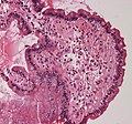
Micrograph showing nodular enterochromaffin-like cell hyperplasia, as demonstrated with chromogranin A immunostaining, in the body of the stomach. Parietal cells are not readily apparent. These changes are in keeping with autoimmune metaplastic atrophic gastritis, a histologic correlate of vitamin B12 deficiency anemia.
SpecialtyHematology
SymptomsFeeling tired, shortness of breath, pale skin, numbness in the hands and feet, confusion, poor reflexes
Usual onsetAny age, particularly those over 60 years old
CausesNot enough intrinsic factor
Diagnostic methodBlood tests, bone marrow tests
TreatmentVitamin B12 injections
PrognosisWith treatment a normal life
Frequency1 per 1000 people

Pernicious anemia is a disease where not enough red blood cells are produced due to a deficiency of vitamin B12. Those affected often have a gradual onset. The most common initial symptoms are feeling tired and weak. Other symptoms may include shortness of breath, feeling faint, a smooth red tongue, pale skin, chest pain, nausea and vomiting, loss of appetite, heartburn, numbness in the hands and feet, difficulty walking, memory loss, muscle weakness, poor reflexes, blurred vision, clumsiness, depression, and confusion. Without treatment, some of these problems may become permanent.

Pernicious anemia refers to a type of vitamin B12 deficiency anemia that results from lack of intrinsic factor. Lack of intrinsic factor is most commonly due to an autoimmune attack on the cells that create it in the stomach. It can also occur following the surgical removal of all or part of the stomach or small intestine; from an inherited disorder or illnesses that damage the stomach lining. When suspected, diagnosis is made by blood tests initially a complete blood count, and occasionally, bone marrow tests. Blood tests may show fewer but larger red blood cells, low numbers of young red blood cells, low levels of vitamin B12, and antibodies to intrinsic factor. Diagnosis is not always straightforward and can be challenging.

Because pernicious anemia is due to a lack of intrinsic factor, it is not preventable. Pernicious anemia can be treated with injections of vitamin B12. If the symptoms are serious, frequent injections are typically recommended initially. There are not enough studies that pills are effective in improving or eliminating symptoms. Often, treatment may be needed for life.

Pernicious anemia is the most common cause of clinically evident vitamin B12 deficiency worldwide. Pernicious anemia due to autoimmune problems occurs in about one per 1000 people in the US. Among those over the age of 60, about 2% have the condition. It more commonly affects people of northern European descent. Women are more commonly affected than men. With proper treatment, most people live normal lives. Due to a higher risk of stomach cancer, those with pernicious anemia should be checked regularly for this. The first clear description was by Thomas Addison in 1849. The term "pernicious" means "deadly", and this term came into use because, before the availability of treatment, the disease was often fatal.

Signs and symptoms

Pernicious anemia often presents slowly, and can cause harm insidiously and unnoticeably. Untreated, it can lead to neurological complications, and in serious cases, death. The onset may be vague and slow and the condition can be confused with other conditions, and there may be few to many symptoms without anemia. Pernicious anemia may be present without a person experiencing symptoms at first, over time, feeling tired and weak, lightheadedness, dizziness, headaches, rapid or irregular heartbeat, breathlessness, glossitis (a sore red tongue), poor ability to exercise, low blood pressure, cold hands and feet, pale or yellow skin, easy bruising and bleeding, low-grade fevers, tremor, cold sensitivity, chest pain, upset stomach, nausea, loss of appetite, heartburn, weight loss, diarrhea, constipation, severe joint pain, feeling abnormal sensations including tingling or numbness to the fingers and toes (pins and needles), and tinnitus, may occur. Anemia may present with a number of further common symptoms, including hair thinning and loss, early greying of the hair, mouth ulcers, bleeding gums, angular cheilitis, a look of exhaustion with pale and dehydrated or cracked lips and dark circles around the eyes, as well as brittle nails.

In more severe or prolonged cases of pernicious anemia, nerve cell damage may occur. This is may result in sense loss, difficulty in proprioception, neuropathic pain, difficulty walking, poor balance, loss of sensation in the feet, muscle weakness, blurred vision (either due to retinopathy or optic neuropathy), impaired urination, fertility problems, decreased sense of taste and smell, decreased level of consciousness, changes in reflexes, memory loss, mood swings, depression, irritability, cognitive impairment, confusion, anxiety, clumsiness, psychosis, and, in more severe cases, dementia. Anemia may also lead to cardiac murmurs and/or altered blood pressure (low or high). The deficiency may also present with thyroid disorders. In severe cases, the anemia may cause congestive heart failure. A complication of severe chronic PA is subacute combined degeneration of spinal cord, which leads to distal sensory loss (posterior column), absent ankle reflex, increased knee reflex response, and extensor plantar response. Other than anemia, hematological symptoms may include cytopenias, intramedullary hemolysis, and pseudothrombotic microangiopathy. Vitamin B12 deficiency, which is reversible, is occasionally confused with acute myeloid leukemia, which is an irreversible condition presenting with some of the same hematological symptoms, including hypercellular bone marrow with blastic differentiation and hypersegmented neutrophils. Pernicious anemia can cause osteoporosis and may lead to bone fractures. Pernicious anemia can contribute to a delay in physical growth in children, and may also be a cause for delay in puberty for adolescents.

Causes

Vitamin B12 cannot be produced by the human body, and must be obtained from the diet. When foods containing B12 are eaten, the vitamin is usually bound to protein and is released by proteases released by the pancreas into the small bowel. Following its release, most B12 is absorbed by the body in the small bowel (ileum) after binding to a protein known as intrinsic factor. Intrinsic factor is produced by parietal cells of the gastric mucosa (stomach lining) and the intrinsic factor-B12-complex is absorbed by enterocytes in the ileum's cubam receptors. PA is characterised by B12 deficiency caused by the absence of intrinsic factor. Other disorders that can disrupt the absorption of vitamin B12 in the small intestine include celiac disease, surgical removal of crohn's disease, and HIV.

Atrophic gastritis showing patchy atrophy of oxyntic mucosa

PA may be considered as an end stage of autoimmune atrophic gastritis, a disease characterised by stomach atrophy and the presence of antibodies to parietal cells and intrinsic factor. Autoimmune atrophic gastritis, is localised to the body of the stomach, where parietal cells are located. Antibodies to intrinsic factor and parietal cells cause the destruction of the oxyntic gastric mucosa, in which the parietal cells are located, leading to the subsequent loss of intrinsic factor synthesis. Without intrinsic factor, the ileum can no longer absorb the B12. Atrophic gastritis is often a precursor to gastric cancer.

Although the exact role of Helicobacter pylori infection in PA remains controversial, evidence indicates H. pylori is involved in the pathogenesis of the disease. A long-standing H. pylori infection may cause gastric autoimmunity by a mechanism known as molecular mimicry. Antibodies produced by the immune system can be cross-reactive and may bind to both H. pylori antigens and those found in the gastric mucosa. The antibodies are produced by activated B cells that recognise both pathogen and self-derived peptides. The autoantigens believed to cause the autoreactivity are the alpha and beta subunits of the sodium-potassium pump. In a study, B12 deficiency caused by Helicobacter pylori was positively correlated with CagA positivity and gastric inflammatory activity, rather than gastric atrophy. Less commonly, H. pylori and Zollinger-Ellison syndrome may cause a form of nonautoimmune gastritis that can lead to pernicious anemia.

Impaired B12 absorption can also occur following gastric removal (gastrectomy) or gastric bypass surgery. In these surgeries, either the parts of the stomach that produce gastric secretions are removed or they are bypassed. This means intrinsic factor, as well as other factors required for B12 absorption, are not available. However, B12 deficiency after gastric surgery does not usually become a clinical issue. This is probably because the body stores many years' worth of B12 in the liver and gastric surgery patients are adequately supplemented with the vitamin.

Although no specific PA susceptibility genes have been identified, a genetic factor likely is involved in the disease. Pernicious anemia is often found in conjunction with other autoimmune disorders, suggesting common autoimmune susceptibility genes may be a causative factor. In spite of that, previous family studies and case reports focusing on PA have suggested that there is a tendency of genetic heritance of PA in particular, and close relatives of the PA patients seem to have higher incidence of PA and associated PA conditions. Moreover, it was further indicated that the formation of antibodies to gastric cells was autosomal dominant gene determined, and the presence of antibodies to the gastric cells might not be necessarily related to the occurrence of atrophic gastritis related to PA.

Pathophysiology

Although the healthy body stores three to five years' worth of B12 in the liver, the usually undetected autoimmune activity in one's gut over a prolonged period of time leads to B12 depletion and the resulting anemia; pernicious anemia refers to one of the hematologic manifestations of chronic auto-immune gastritis, in which the immune system targets the parietal cells of the stomach or intrinsic factor itself, leading to decreased absorption of vitamin B12. The body needs enough intrinsic factor to absorb and reabsorb vitamin B12 from the bile, in which reduces the time needed to develop a deficiency.

B12 is required by enzymes for two reactions: the conversion of methylmalonyl-CoA to succinyl-CoA, and the conversion of homocysteine to methionine. In the latter reaction, the methyl group of levomefolic acid is transferred to homocysteine to produce tetrahydrofolate and methionine. This reaction is catalyzed by the enzyme methionine synthase with B12 as an essential cofactor. During B12 deficiency, this reaction cannot proceed, which leads to the accumulation of levomefolic acid. This accumulation depletes the other types of folate required for purine and thymidylate synthesis, which are required for the synthesis of DNA. Inhibition of DNA replication in maturing red blood cells results in the formation of large, fragile megaloblastic erythrocytes. The neurological aspects of the disease are thought to arise from the accumulation of methylmalonyl- CoA due to the requirement of B12 as a cofactor to the enzyme methylmalonyl-CoA mutase.

Diagnosis

Immunofluorescence staining pattern of gastric parietal cell antibodies on a stomach section

The insidious nature of PA may mean that diagnosis is delayed. Diagnosis is not always straightforward and can be challenging and can take up to several years to receive a diagnosis from the onset of symptoms and almost 60% of those affected are misdiagnosed or not initially diagnosed at all. PA may be suspected when a patient's blood smear shows large, fragile, immature erythrocytes, known as megaloblasts. A diagnosis of PA first requires demonstration of megaloblastic anemia by conducting a full blood count and blood smear, which evaluates the mean corpuscular volume (MCV), as well the mean corpuscular hemoglobin concentration (MCHC). PA is identified with a high MCV (macrocytic anemia) and a normal MCHC (normochromic anemia). Ovalocytes are also typically seen on the blood smear, and a pathognomonic feature of megaloblastic anemias (which include PA and others) is hypersegmented neutrophils. Neurological and other symptoms can occur without anemia.

Vitamin B12 serum levels are used to detect its deficiency, but do not distinguish its causes. Vitamin B12 levels can be falsely high or low and data for sensitivity and specificity vary widely. Normal serum levels may be found in cases of deficiency where myeloproliferative disorders, liver disease, transcobalamin II, or small intestinal bacterial overgrowth are present. Low levels of serum vitamin B12 may be caused by other factors than B12 deficiency, such as folate deficiency, pregnancy, oral contraceptive use, haptocorrin deficiency, and myeloma. High serum levels may caused by supplementing with vitamin B12, present of antibodies to intrinsic factor, or due to underlying condition.

The presence of antibodies to gastric parietal cells and intrinsic factor is common in PA. Parietal cell antibodies are found in other autoimmune disorders and also in up to 10% of healthy individuals. However, around 85% of PA patients have parietal cell antibodies, which means they are a sensitive marker for the disease. Intrinsic factor antibodies are much less sensitive than parietal cell antibodies, but they are much more specific. They are found in about half of PA patients and are very rarely found in other disorders. These antibody tests can distinguish between PA and food-B12 malabsorption.

A buildup of certain metabolites occurs in B12 deficiency due to its role in metabolic processes and cellular functions. Methylmalonic acid (MMA) can be measured in both the blood and urine, whereas homocysteine is only measured in the blood. An increase in both MMA and homocysteine distinguishes B12deficiency from folate deficiency because homocysteine alone increases in the latter.

Elevated gastrin levels can be found in around 80–90% of PA cases, but they may also be found in other forms of gastritis. Decreased pepsinogen I levels or a decreased pepsinogen I to pepsinogen II ratio may also be found, although these findings are less specific to PA and can be found in food-B12 malabsorption and other forms of gastritis.

The diagnosis of atrophic gastritis type A should be confirmed by gastroscopy and stepwise biopsy. About 90% of individuals with PA have antibodies for parietal cells; however, only 50% of all individuals in the general population with these antibodies have pernicious anemia.

Differential diagnosis

Forms of vitamin B12 deficiency other than PA must be considered in the differential diagnosis of megaloblastic anemia. For example, a B12-deficient state which causes megaloblastic anemia and which may be mistaken for classical PA may be caused by infection with the tapeworm Diphyllobothrium latum, possibly due to the parasite's competition with host for vitamin B12.

The classic test for PA, the Schilling test, is no longer widely used, as more efficient methods are available. This historic test consisted, in its first step, of taking an oral dose of radiolabelled vitamin B12, followed by quantitation of the vitamin in the patient's urine over a 24-hour period via measurement of the radioactivity. A second step of the test repeats the regimen and procedure of the first step, with the addition of oral intrinsic factor. A patient with PA presents lower than normal amounts of intrinsic factor; hence, addition of intrinsic factor in the second step results in an increase in vitamin B12 absorption (over the baseline established in the first). The Schilling test distinguished PA from other forms of B12 deficiency, specifically, from Imerslund–Gräsbeck syndrome, a B12-deficiency caused by mutations in CUBN that codes for cubilin the cobalamin receptor.

Vitamin B12 deficiency is also prevalent in patients having Crohn's disease (CD) so it should be differentiated.

Treatment

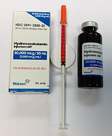
Hydroxocobalamin injection usp(1000 mcg/ml) is a clear red liquid solution of hydroxocobalamin which is available in a 30-ml brown glass multidose vial packaged in a paper box. Shown is 500 mcg B-12 (as 1/2 cc) drawn up in a 0.5-cc U-100 27 gauge x 1/2" insulin syringe, as prepared for subcutaneous injection.

Pernicious anemia is usually easily treated by providing the necessary level of vitamin B12 supplementation. Pernicious anemia can be treated with intramuscular injections of vitamin B12. Initially in high daily doses, followed by less frequent lower doses, as the condition improves. Activity may need to be limited during the course of treatment. As long as the body is saturated with vitamin B12 expected to result in cessation of anemia-related symptoms and there are no other symptoms, unless there are irreversible neurological complications. There are not enough studies on whether pills are as effective in improving or eliminating symptoms as parenteral treatment. Folate supplementation may affect the course and treatment of pernicious anemia if vitamin B12 not replaced. In some severe cases of anemia, a blood transfusion may be needed to resolve haematological effects. Treatment is lifelong.

The treatment of PA varies by country and area. Opinions vary over the efficacy of administration (parenteral/oral), the amount and time interval of the doses, or the forms of vitamin B12 (e.g. cyanocobalamin/hydroxocobalamin). More comprehensive studies are still needed in order to validate the feasibility of a particular therapeutic method for PA in clinical practices.

Prognosis

A person with well-treated PA can live a healthy life. Failure to diagnose and treat in time, however, may result in permanent neurological damage, excessive fatigue, depression, memory loss, and other complications. In severe cases, the neurological complications of pernicious anemia can lead to death – hence the name, "pernicious", meaning deadly.

There is an increased risk of gastric cancer in those with pernicious anemia linked to the common feature of atrophic gastritis.

Epidemiology

PA is estimated to affect 0.1% of the general population and 1.9% of those over 60, accounting for 20–50% of B12 deficiency in adults. A review of literature shows that the prevalence of PA is higher in Northern Europe, especially in Scandinavian countries, and among people of African descent, and that increased awareness of the disease and better diagnostic tools might play a role in apparently higher rates of incidence.

History

A case of anemia with a first recognition of associated atrophic gastritis a feature of pernicious anemia, was first described in 1824 by James Combe. This was fully investigated in 1849, by British physician Thomas Addison, from which it acquired the common name of Addison's anemia. In 1871, the first accurate description of the disease in continental Europe was made by Michael Anton Biermer, a German physician who noted the insidious course of the condition. Because it was untreatable and fatal at the time, he first referred to it as "pernicious" anemia. Russell coined the term subacute combined degeneration of spinal cord.

In 1907, Richard Clarke Cabot reported on a series of 1,200 patients with PA; their average survival was between one and three years. Pernicious anemia was a fatal disease before about the year 1920; until the importance of the liver in hematopoiesis was recognized, the treatment of pernicious anemia was unsuccessful and arbitrary. It may have motivated George Whipple, who had a keen interest in liver diseases, to investigate the liver's role in hematopoiesis. Whipple began evaluating the effects of treatments for anemia caused by chronic blood loss. Whipple, Huber, and Robchett studied the effects on hemoglobin and blood regeneration of a variety of treatments, among which only raw liver showed real promise. Serendipity is said to have played a role in this discovery. Whipple observed that blood regeneration was poor in dogs fed cooked liver after chronic blood loss. Had it not been that a lazy laboratory technician gave the dogs raw liver, the much more dramatic response might not have been discovered then.

1930 advert for liver extract to treat pernicious anemia

Around 1926, George Minot and William P. Murphy, who learned of Whipple's discovery, sought raw liver as a treatment for pernicious anemia. They later suggested a high-protein diet with high amounts of raw liver. This caused a rapid improvement in symptoms and a simultaneous rise in red blood cell counts. Fruit and iron were also part of the diet, and it appears that at this point, Minot and Murphy were not quite sure that the liver was a very important factor. It was thought that iron in liver tissue, not liver juice-soluble factor, cured hemorrhagic anemia in dogs. Thus, the discovery of liver juice as a treatment for pernicious anemia had been by coincidence. However, Minot, Murphy, and Whipple received the joint Nobel Prize for discovering a cure for a previously fatal disease of unknown cause in 1934, becoming the first Americans to be awarded the Nobel Prize in Physiology and Medicine.

It is not easy to eat uncooked liver, and extracts were developed as a concentrate of liver juice for intramuscular injection. In 1928, chemist Edwin Cohn prepared an extract that was 50 to 100 times stronger than obtained from raw liver. This became part of the standard management of pernicious anemia until the 1950s. The active ingredient in the liver remained unknown until 1948. The anti-pernicious anemia factor was only isolated from the liver by Smith, Rex, and others. The substance was cobalamin, which the discoverers called "vitamin B12". They showed that giving a few micrograms could prevent relapse in the disease. Dorothy Hodgkin and co-workers went on to use X-ray crystallography to elucidate the structure of cobalamin for which she, too, was awarded a Nobel Prize.

Understanding of the pathogenesis of pernicious anaemia increased over subsequent decades. It had long been known that the disease was associated with defects in the gastrointestinal tract: patients had chronic gastritis and lack of acid secretion (achlorhydria). It is known that transport of physiological amounts of vitamin B12 depends on the combined actions of gastric, ileal and pancreatic components. The gastric moiety was discovered and named 'intrinsic factor' by William Castle in 1930. A further important advance was made in the early 1960s by Doniach with the recognition that pernicious anemia is an autoimmune disease. Pernicious anemia is eventually treated with either injections or large oral doses of B12; injections are typically 1 mg every other day, or twice weekly, and oral doses are typically between 1 and 4 mg daily.

A medical author takes the view that Mary Todd Lincoln, the wife of American President Abraham Lincoln, had pernicious anemia for decades and died from it in 1882.

Research

Permeation enhancers

Treatment using oral drugs is an easier option in management but the bioavailabity of B12 is low. This is due to low absorption in the intestine, and breakdown by enzyme activity. Research continues to focus on the use of permeation enhancers or permeation absorbers in combination with the treatment. One of the better performing enhancers studied is salcoprozate sodium (SNAC). SNAC is able to form a noncovalent complex with cobalamin while preserving its chemical integrity and protect B12 from gastric acidity. This complex is much more lipophilic than the water-soluble vitamin B12, so is able to pass through cellular membranes with greater ease. Molecular dynamics are used in experiments to gain an understanding of the molecular interactions involved in the different molecules used and the degree of ease achieved in absorption across the gastric epithelium.

Hyperinsulinemia

From Wikipedia, the free encyclopedia
 
Hyperinsulinemia
Other namesHyperinsulinaemia
Insulin binding to its receptor

Hyperinsulinemia is a condition in which there are excess levels of insulin circulating in the blood relative to the level of glucose. While it is often mistaken for diabetes or hyperglycaemia, hyperinsulinemia can result from a variety of metabolic diseases and conditions, as well as non-nutritive sugars in the diet. While hyperinsulinemia is often seen in people with early stage type 2 diabetes mellitus, it is not the cause of the condition and is only one symptom of the disease (for opposing view see review in ). Type 1 diabetes only occurs when pancreatic beta-cell function is impaired. Hyperinsulinemia can be seen in a variety of conditions including diabetes mellitus type 2, in neonates and in drug-induced hyperinsulinemia. It can also occur in congenital hyperinsulinism, including nesidioblastosis.

Hyperinsulinemia is associated with hypertension, obesity, dyslipidemia, insulin resistance, and glucose intolerance. These conditions are collectively known as metabolic syndrome. This close association between hyperinsulinemia and conditions of metabolic syndrome suggest related or common mechanisms of pathogenicity. Hyperinsulinemia has been shown to "play a role in obese hypertension by increasing renal sodium retention".

In type 2 diabetes, the cells of the body become resistant to the effects of insulin as the receptors which bind to the hormone become less sensitive to insulin concentrations resulting in hyperinsulinemia and disturbances in insulin release. With a reduced response to insulin, the beta cells of the pancreas secrete increasing amounts of insulin in response to the continued high blood glucose levels resulting in hyperinsulinemia. In insulin resistant tissues, a threshold concentration of insulin is reached causing the cells to uptake glucose and therefore decreases blood glucose levels. Studies have shown that the high levels of insulin resulting from insulin resistance might enhance insulin resistance.

Studies on mice with genetically reduced circulating insulin suggest that hyperinsulinemia plays a causal role in high fat diet-induced obesity. In this study, mice with reduced insulin levels expended more energy and had fat cells that were reprogrammed to burn some energy as heat.

Hyperinsulinemia in neonates can be the result of a variety of environmental and genetic factors. If the mother of the infant is a diabetic and is not able to properly control her blood glucose levels, the hyperglycemic maternal blood can create a hyperglycemic environment in the fetus. To compensate for the increased blood glucose levels, fetal pancreatic beta cells can undergo hyperplasia. The rapid division of beta cells results in increased levels of insulin being secreted to compensate for the high blood glucose levels. Following birth, the hyperglycemic maternal blood is no longer accessible to the neonate resulting in a rapid drop in the newborn's blood glucose levels. As insulin levels are still elevated this may result in hypoglycemia. To treat the condition, high concentration doses of glucose are given to the neonate as required maintaining normal blood glucose levels. The hyperinsulinemia condition subsides after one to two days.

Symptoms and signs

A large abdomen is a strong indicator of Hyperinsulinemia, so measure waist to hip ratio. But the best way to know if you have hyperinsulinemia is to get insulin levels checked. It is important to note that in some people Insulin can be elevated in the presence of normal glucose for 10-20 years, so it is best not to rely on glucose levels without also measuring insulin. 

Some patients may experience a variety of symptoms when hypoglycemia is present, including:

If a person experiences any of these symptoms, a visit to a qualified medical practitioner is advised, and diagnostic blood testing, such as Fasting Insulin Levels, may be required.

Causes

Possible causes include:

Often, individuals get metabolically (internally) sick before showing signs of obesity. But visceral obesity can go undetected, and is extremely dangerous. Lifestyle choices including diet, exercise, and sleep deprivation play the largest role in Hyperinsulinemia & insulin resistance.

Belly fat is a strong predictor of high insulin levels. Obesity is characterized by an excess of adipose tissue – insulin increases the synthesis of fatty acids from glucose, facilitates the entry of glucose into adipocytes and inhibits breakdown of fat in adipocytes.

On the other hand, adipose tissue is known to secrete various metabolites, hormones and cytokines that may play a role in causing hyperinsulinemia. Specifically cytokines secreted by adipose tissue directly affect the insulin signalling cascade, and thus insulin secretion. Adiponectins are cytokines that are inversely related to percent body fat; that is people with a low body fat will have higher concentrations of adiponectins where as people with high body fat will have lower concentrations of adiponectins. In 2011, it was reported that hyperinsulinemia is strongly associated with low adiponectin concentrations in obese people, though whether low adiponectin has a causal role in hyperinsulinemia remains to be established.

Diagnosis

Fasting Insulin levels in blood may be measured as this can be elevated in the presence of normal glucose. Diagnosis is often made by checking normal levels of glucose that exceed 1.7 mmol/L (30 mg/dL) when 1 mg of glucagon is administered IM or IV. In addition, urine samples or blood samples are also used to check levels of ketones and low free fatty acids.

After diagnosis, most people are required to continue regular check ups for evaluations.

Differential diagnosis

  • Hyperinsulinemia is often mistaken for diabetes or hypoglycaemia. These are separate, albeit related, conditions. Adipocytes will generate triglycerides in the presence of insulin but refers to a liver condition rather than a pancreatic one.

Treatment

Treatment is typically achieved via diet and exercise, although metformin may be used to reduce insulin levels in some patients (typically where obesity is present). A referral to a dietician is beneficial. Another method used to lower excessively high insulin levels is cinnamon, specifically Ceylon cinnamon, as was demonstrated when supplemented in clinical human trials.

A healthy diet that is low in simple sugars and processed carbohydrates, and high in fiber, and vegetable protein is often recommended. This includes replacing white bread with whole-grain bread, reducing intake of foods composed primarily of starch such as potatoes, and increasing intake of legumes and green vegetables, particularly soy.

Regular monitoring of weight, blood sugar, and insulin are advised, as hyperinsulinemia may develop into diabetes mellitus type 2.

It has been shown in many studies that physical exercise improves insulin sensitivity. The mechanism of exercise on improving insulin sensitivity is not well understood however it is thought that exercise causes the glucose receptor GLUT4 to translocate to the membrane. As more GLUT4 receptors are present on the membrane more glucose is taken up into cells decreasing blood glucose levels which then causes decreased insulin secretion and some alleviation of hyperinsulinemia. Another proposed mechanism of improved insulin sensitivity by exercise is through AMPK activity. The beneficial effect of exercise on hyperinsulinemia was shown in a study in 2009, where they found that improving fitness through exercise significantly decreases blood insulin concentrations. Moreover, a diet that consists of high amounts of carbs have been linked to weight gain and obesity in rodents. Although this has not been tested in humans, it is assumed that it could aid with the prevention of weight gain in humans, and possibly obesity. Medications have also been studied to treat hyperinsulinemia, although these might have some side effects, these could be used as an alternative.

Pancreatic beta cell function

From Wikipedia, the free encyclopedia
 
Pancreatic beta cell function
Other namesGβ, HOMA-Beta, IGI, SPINA-GBeta
SpecialtyEndocrinology

Pancreatic beta cell function (synonyms Gβ or, if calculated from fasting concentrations of insulin and glucose, HOMA-Beta or SPINA-GBeta) is one of the preconditions of euglycaemia, i.e. normal blood sugar regulation. It is defined as insulin secretory capacity, i.e. the maximum amount of insulin to be produced by beta cells in a given unit of time.

Physiology and pathophysiology

Beta cells play a paramount role in glucose homeostasis. Progressive loss of insulin secretory capacity is a key defect associated with the transition from a healthy glycaemic state to hyperglycaemia, characteristic of untreated diabetes mellitus. In type 1 diabetes mellitus and pancreatogenic diabetes beta cell destruction is a primary event from the perspective of the feedback loop. In type 2 diabetes beta cell dysfunction is an essential constituent as well, but subsequent to the development of insulin resistance. Other mechanisms, including lipotoxicity, amyloid deposition, oxidative stress, mitochondrial dysfunction, ER stress and inflammation may be involved as well. The beta cell loss in type 2 diabetes is mainly caused by reduced beta cell number rather than size. Hyperglycaemia becomes clinically significant once insulin over-secretion can no longer compensate for the degree of insulin resistance.

It remains an unsolved question if impaired pancreatic beta cell function or hypersecretion of insulin represent the primary event in the pathogenesis of type 2 diabetes. Both scenarios may be cause and consequence, and it has been postulated that the direction of causality depends on the respective subtype of diabetes. Therefore, they may be part of a complex feedback loop involving glucose toxicity leading to a biphasic response, thereby preventing neoplastic effects of dynamical compensation by mutant takeover.

Assessing beta cell function

Measuring beta-cell function is a challenge, since insulin secretory capacity cannot be readily assessed. Therefore, indirect methods of measurement have been developed. They include dynamic and static function tests.

Single-point measurements

One-time measurements of certain hormones or metabolites provide some limited information. Examples are:

Although single-point measurements have the benefit of being convenient and inexpensive, they are generally not regareded as sufficiently informative for early diagnosis of impaired glucose homeostasis or early-stage type 1 diabetes.

Dynamic function tests

Dynamic function tests for beta-cell function include:

Static function tests

Static function tests for the assessment of beta-cell function comprise:

Challenges and limits

Measuring beta-cell function requires the rate of secretion to be interpreted in relation to the prevailing glucose concentration. Therefore, a mathematical model is needed that links the time courses of insulin secretion and glucose concentration as a mechanistic causal relationship.

Hyberbolic relationship between insulin sensitivity and beta cell function showing dynamical compensation in "healthy" insulin resistance (transition from A to B) and the evolution of type 2 diabetes mellitus (transition from A to C).
Hyberbolic relationship between insulin sensitivity and beta cell function showing dynamical compensation in "healthy" insulin resistance (transition from A to B) and the evolution of type 2 diabetes mellitus (transition from A to C). Changed from Cobelli et al. 2007 and Hannon et al. 2018

Additionally, beta-cell function has to be interpreted in light of the prevailing insulin sensitivity. This is necessary since the beta cell mass is adjusted as required by dynamical compensation, giving rise to a hyperbolic relationship between insulin sensitivity and beta cell function. In the state of insulin resistance beta cells proliferate and their secretory capacity subsequently rises. One possibility to address this relation is to resort to a normalization of beta cell function based on a disposition metric. The disposition index, calculated as product of insulin sensitivity and beta cell function, is assumed to be a constant during the development of insulin resistance. It is generally assumed that the glucose tolerance of an individual is related to the disposition index. In this model, different values of glucose tolerance are represented by different hyperbolas, so that within one hyperbola the product of insulin sensitivity and beta cell function remains a constant.

In summary, to provide a meaningful mechanistic explanation of insulin-glucose homeostasis, beta cell function and insulin sensitivity have to be assessed simultaneously and it is necessary to interpret all observations in the context of insulin sensitivity or resistance.

Historical materialism

From Wikipedia, the free encyclopedia https://en.wikipedia.org/wiki/Dialectic Historical materialism is Karl M...