Search This Blog

Wednesday, April 23, 2025

Animal languages

From Wikipedia, the free encyclopedia

Parrots (Australian ringneck)

Animal languages are forms of communication between animals that show similarities to human language. Animals communicate through a variety of signs, such as sounds and movements. Signing among animals may be considered a form of language if the inventory of signs is large enough, the signs are relatively arbitrary, and the animals seem to produce them with a degree of volition (as opposed to relatively automatic conditioned behaviors or unconditioned instincts, usually including facial expressions).

Many researchers argue that animal communication lacks a key aspect of human language, the creation of new patterns of signs under varied circumstances. Humans, by contrast, routinely produce entirely new combinations of words. Some researchers, including the linguist Charles Hockett, argue that human language and animal communication differ so much that the underlying principles are unrelated. Accordingly, linguist Thomas A. Sebeok has proposed to not use the term "language" for animal sign systems. However, other linguists and biologists, including Marc Hauser, Noam Chomsky, and W. Tecumseh Fitch, assert that an evolutionary continuum exists between the communication methods of animal and human language.

Aspects of human language

The Belgian conservationist Claudine André with a bonobo

Some experts argue the following properties separate human language from animal communication:

  • Arbitrariness: There is usually no rational relationship between a sound or sign and its meaning. For example, there is nothing intrinsically house-like about the word "house".
  • Discreteness: Language is composed of small, separate, and repeatable parts (discrete units, e.g. morphemes) that are used in combination to create meaning.
  • Displacement: Language can be used to communicate about things that are not in the immediate vicinity either spatially or temporally.
  • Duality of patterning: The smallest meaningful units (words or morphemes) consist of sequences of units without meaning (sounds or phonemes). This is also referred to as double articulation.
  • Productivity: Users can understand and create an indefinitely large number of utterances.
  • Semanticity: Specific signals have specific meanings.

Research with apes, like that of Francine Patterson with Koko (gorilla) or Allen and Beatrix Gardner with Washoe (chimpanzee), suggested that apes are capable of using language that meets some of these requirements, including arbitrariness, discreteness, and productivity.

In the wild, chimpanzees have been seen "talking" to each other when warning about approaching danger. For example, if one chimpanzee sees a snake, said chimpanzee may make a low, rumbling noise, signaling for all the other chimps to climb into nearby trees. In this case, the chimpanzees' communication does not indicate displacement, as it is entirely contained to an observable event.

Arbitrariness has been noted in meerkat calls; bee dances demonstrate elements of spatial displacement; and cultural transmission has possibly occurred through language between the bonobos named Kanzi and Panbanisha.

Claims that animals have language skills akin to humans, however, are extremely controversial. In his book The Language Instinct, Steven Pinker illustrates that claims of chimpanzees acquiring language are exaggerated and rest on very limited or specious evidence.

The American linguist Charles Hockett theorized that there are sixteen features of human language that distinguish human communication from that of animals. He called these the design features of language. The features mentioned below have so far been found in all spoken human languages, and at least one is missing from any other animal communication system.

  • Vocal-auditory channel: Sounds are emitted from the mouth and perceived by the auditory system. While this applies to many animal communication systems, there are many exceptions, such as those relying on visual communication. One example is cobras extending the ribs behind their heads to send the message of intimidation or of feeling threatened. In humans, sign languages provide many examples of fully formed languages that use a visual channel.
  • Broadcast transmission and directional reception: This requires that the recipient can tell the direction that the signal comes from and thus the originator of the signal.
  • Rapid fading (transitory nature): The signal lasts a short time. This is true of all systems involving sound. It does not take into account audio recording technology and is also not true for written language. It tends not to apply to animal signals involving chemicals and smells which often fade slowly. For example, a skunk's smell, produced in its glands, lingers to deter a predator from attacking.
  • Interchangeability: All utterances that are understood can be produced. This is different from some communication systems where, for example, males produce one set of behaviors and females another and they are unable to interchange these messages so that males use the female signal and vice versa. For example, Heliothine moths have differentiated communication: females are able to send a chemical to indicate preparedness to mate, while males cannot send the chemical.
  • Total feedback: The sender of a message is aware of the message being sent.
  • Specialization: The signal produced is intended for communication and is not due to another behavior. For example, dog panting is a natural reaction to being overheated, but is not produced to specifically relay a particular message.
  • Semanticity: There is some fixed relationship between a signal and a meaning.

Primates

Humans are able to distinguish real words from fake words based on the phonological order of the word itself. In a 2013 study, baboons were shown to have this skill as well. The discovery has led researchers to believe that reading is not as advanced a skill as previously believed, but instead based on the ability to recognize and distinguish letters from one another. The experimental setup consisted of six young adult baboons, and results were measured by allowing the animals to use a touch screen and select whether or not the displayed word was a real word, or a non-word such as "dran" or "telk". The study lasted for six weeks, with approximately 50,000 tests completed in that time. The researchers minimized common bigrams, or combinations of two letters, in non-words, and maximized them in real words. Further studies will attempt to teach baboons how to use an artificial alphabet.

In a 2016 study, a team of biologists from several universities concluded that macaques possess vocal tracts physically capable of speech, "but lack a speech-ready brain to control it".

Non-primates

Among the most studied examples of non-primate languages are:

Birds

  • Bird songs: Songbirds can be highly articulate. Grey parrots and macaws are well known for their ability to mimic human language. At least one specimen, Alex, appeared able to answer a number of simple questions about objects he was presented with, such as answering simple mathematical equations and identifying colors. Parrots, hummingbirds and songbirds display vocal learning patterns. Crows have been studied for their ability to understand recursion.

Insects

  • Bee dancing: Used to communicate the direction and distance of food source in many species of bees. In 2023, James C. Nieh, Associate Dean and Professor of Biology with the University of California, San Diego, performed an experiment to determine if the dances of bees were innate skills or if they were developed through observation of older bees within their hive. The research group determined that the dance bees performed was to some degree innate, but the consistency and accuracy of the dance was a skill passed down by older bees. Although the experimental hive that contained only workers of the same age developed better accuracy when conveying angle and direction as they got older, their ability to communicate distance never reached the level of the control beehives.

Mammals

  • African forest elephants: Cornell University's Elephant Listening Project began in 1999 when Katy Payne began studying the calls of African forest elephants in Dzanga National Park in the Central African Republic. Andrea Turkalo has continued Payne's work in Dzanga National Park by observing elephant communication. For nearly 20 years, Turkalo has used a spectrogram to record the noises that the elephants make. After extensive observation and research, she has been able to recognize elephants by their voices. Researchers hope to translate these voices into an elephant dictionary, but this will likely not occur for many years. Because elephant calls are often made at very low frequencies, the spectrogram is designed to detect lower frequencies than humans can perceive, allowing Turkalo to better understand the elephants' noise making. Cornell's research on African forest elephants has challenged the idea that humans are considerably better at using language than animals, and that animals only have a small set of information they can convey to others. As Turkalo explained, "many of their calls are in some ways similar to human speech." Elephants in captivity can be taught to remember tone, melody, and recognise more than 20 words.
  • Mustached bats: Since these animals spend most of their lives in the dark, they rely heavily on their auditory system to communicate, including via echolocation and using calls to locate each other. Studies have shown that mustached bats use a wide variety of calls to communicate with one another. These calls include 33 different sounds, or "syllables", that the bats either use alone or combine in various ways to form composite syllables.
  • Prairie dogs: Con Slobodchikoff studied prairie dog communication and discovered that they use different alarm calls and escape behaviors for different species of predators. Their calls transmit semantic information, which was demonstrated when playbacks of alarm calls in the absence of predators led to escape behavior appropriate for the types of predators associated with the calls. The alarm calls also contain descriptive information about the general size, color, and speed of the predator.

Aquatic mammals

  • Bottlenose dolphins: Dolphins can hear one another up to 6 miles apart underwater. Researchers observed a mother dolphin successfully communicating with her baby using a telephone. It appeared that both dolphins knew who they were speaking with and what they were speaking about. Not only do dolphins communicate via nonverbal cues, they also seem to chatter and respond to other dolphins' vocalizations.
Spectrogram of humpback whale vocalizations. Detail is shown for the first 24 seconds of the 37 second humpback whale "song" recording. The whale songs and echolocation "clicks" are visible as horizontal striations and vertical sweeps respectively.
  • Whales: Two groups of whales, the humpback whale and a subspecies of blue whale found in the Indian Ocean, are known to produce repeated sounds at varying frequencies, known as whale songs. Male humpback whales perform these vocalizations only during the mating season, and so it is surmised the purpose of songs is to aid sexual selection. Humpbacks also make a sound called a feeding call, which is five to ten seconds in length at a nearly constant frequency. Humpbacks generally feed cooperatively by gathering in groups, swimming underneath shoals of fish and lunging up vertically through the fish and out of the water together. Prior to these lunges, whales make their feeding call. The exact purpose of the call is not known, but research suggests that fish react to it. When the sound was played back to them, a group of herring responded to the sound by moving away from the call, even though no whale was present.
  • Sea lions: Since 1971, Ronald J. Schusterman and his research associates have studied sea lions' cognitive ability. They have discovered that sea lions are able to recognize relationships between stimuli based on similar functions or connections made with their peers, rather than only the stimuli's common features. This is called equivalence classification. This ability to recognize equivalence may be a precursor to language. Research is currently being conducted at the Pinniped Cognition & Sensory Systems Laboratory to determine how sea lions form these equivalence relations. Sea lions have also been proven to understand simple syntax and commands when taught an artificial sign language similar to one used with primates. The sea lions studied were able to learn and use a number of syntactic relations between the signs they were taught, such as how the signs should be arranged in relation to each other. However, the sea lions rarely used the signs semantically or logically. In the wild, it is thought that sea lions use reasoning skills associated with equivalence relations in order to make important decisions that can affect their survival, e.g. recognizing friends and relatives or avoiding enemies and predators. Sea lions use various postural positions and a range of barks, chirps, clicks, moans, growls, and squeaks to communicate. It has yet to be proven that sea lions use echolocation as a means of communication.

The effects of learning on auditory signaling in these animals is of interest to researchers. Several investigators have pointed out that some marine mammals appear to have the capacity to alter both the contextual and structural features of their vocalizations as a result of experience. Janik and Slater have stated that learning can modify vocalizations in one of two ways, by influencing the context in which a particular call is used, or by altering the acoustic structure of the call itself. Male California sea lions can learn to inhibit their barking in the presence of any male dominant to them, but vocalize normally when dominant males are absent. The different call types of gray seals can be selectively conditioned and controlled by different cues, and the use of food reinforcement can also modify vocal emissions. A captive male harbor seal named Hoover demonstrated a case of vocal mimicry, but similar observations have not been reported since. Still shows that under the right circumstances pinnipeds may use auditory experience in addition to environmental consequences such as food reinforcement and social feedback to modify their vocal emissions.

In a 1992 study, Robert Gisiner and Schusterman conducted experiments in which they attempted to teach syntax to a female California sea lion named Rocky. Rocky was taught signed words, then she was asked to perform various tasks dependent on word order after viewing a signed instruction. It was found that Rocky was able to determine relations between signs and words, and form basic syntax. A 1993 study by Schusterman and David Kastak found that the California sea lion was capable of understanding abstract concepts such as symmetry, sameness and transitivity. This suggests that equivalence relations can form without language.

The distinctive sounds of sea lions are produced both above and below water. To mark territory, sea lions "bark", with non-alpha males making more noise than alphas. Although females also bark, they do so less frequently and most often in connection with birthing pups or caring for their young. Females produce a highly directional bawling vocalization, the pup attraction call, which helps mothers and pups locate one another. As noted in Animal Behavior, their amphibious lifestyle has made them need acoustic communication for social organization while on land.

Sea lions can hear frequencies between 100 Hz and 40,000 Hz, and vocalize from 100 to 10,000 Hz.

Mollusks

  • Caribbean reef squid have been shown to communicate using a variety of color, shape, and texture changes. Squid are capable of rapid changes in skin color and pattern through nervous system control of chromatophores. In addition to camouflage and appearing larger in the face of a threat, squid use color, patterns, and flashing to communicate with one another in various courtship rituals. Caribbean reef squid can send one message via color patterns to a squid on their right, while they send another message to a squid on their left.

Fish

  • Freshwater Elephant Fish have been observed to have their own language.
  • Mexican Tetra have been observed communicating with a series of clicks, and have also been observed to have regional accents.

Comparison of "animal language" and "animal communication"

It is worth distinguishing "animal language" from "animal communication", although there is some comparative interchange in certain cases (e.g. Cheney & Seyfarth's vervet monkey call studies). Animal language typically does not include bee dancing, bird song, whale song, dolphin signature whistles, prairie dog alarm calls, or the communicative systems found in most social mammals. The features of language as listed above are a dated formulation by Hockett in 1960. Through this formulation Hockett made one of the earliest attempts to break down features of human language for the purpose of applying Darwinian gradualism. Although an influence on early animal language efforts (see below), it is no longer considered the key architecture at the core of animal language research.

Clever Hans, an Orlov Trotter horse that was claimed to have been able to perform arithmetic and other intellectual tasks

Animal language results are controversial for several reasons (for a related controversy, see also Clever Hans). Early chimpanzee work was executed using chimpanzee infants raised as if they were human; a test of the nature vs. nurture hypothesis. Chimpanzees have a laryngeal structure very different from that of humans, and it has been suggested that chimpanzees are not capable of voluntary control of their breathing, although better studies are needed to accurately confirm this. This combination is thought to make it very difficult for the chimpanzees to reproduce the vocal intonations required for human language. Researchers eventually moved towards a gestural (sign language) modality, as well as keyboard devices with buttons with symbols (known as "lexigrams") that the animals could press to produce artificial language. Other chimpanzees learned by observing human subjects performing the task. This latter group of researchers studying chimpanzee communication through symbol recognition (keyboard) as well as through the use of sign language (gestural), are on the forefront of communicative breakthroughs in the study of animal language, and they are familiar with their subjects on a first name basis: Sarah, Lana, Kanzi, Koko, Sherman, Austin and Chantek.

Perhaps the best known critic of animal language is Herbert Terrace. Terrace's 1979 criticism using his own research with the chimpanzee Nim Chimpsky] was scathing and spelled the end of animal language research in that era, most of which emphasized the production of language by animals. In short, he accused researchers of over-interpreting their results, especially as it is rarely parsimonious to ascribe true intentional "language production" when other simpler explanations for the behaviors (gestural hand signs) could be put forth. Additionally, his animals failed to show generalization of the concept of reference between the modalities of comprehension and production; this generalization is one of many fundamental ones that are trivial for human language use. The simpler explanation according to Terrace was that the animals had learned a sophisticated series of context-based behavioral strategies to obtain either primary (food) or social reinforcement, behaviors that could be over-interpreted as language use.

In 1984 Louis Herman published an account of artificial language found in the bottlenose dolphin in the journal Cognition. A major difference between Herman's work and previous research was his emphasis on a method of studying language comprehension only (rather than language comprehension and production by the animal(s)), which enabled rigorous controls and statistical tests, largely because he was limiting his research to evaluating the animals' physical behaviors (in response to sentences) with blinded observers, rather than attempting to interpret possible language utterances or productions. The dolphins' names here were Akeakamai and Phoenix. Irene Pepperberg used the vocal modality for language production and comprehension in a grey parrot named Alex in the verbal mode, and Sue Savage-Rumbaugh continues to study bonobos such as Kanzi and Panbanisha. R. Schusterman duplicated many of the dolphin results in his California sea lions ("Rocky"), and came from a more behaviorist tradition than Herman's cognitive approach. Schusterman's emphasis is on the importance on a learning structure known as equivalence classes.

However, overall, there has not been any meaningful dialog between the linguistics and animal language spheres, despite capturing the public's imagination in the popular press. Furthermore, the growing field of language evolution is another source of future interchange between these disciplines. Most primate researchers tend to show a bias toward a shared pre-linguistic ability between humans and chimpanzees, dating back to a common ancestor, while dolphin and parrot researchers stress the general cognitive principles underlying these abilities. More recent related controversies regarding animal abilities include the closely linked areas of theory of mind, Imitation (e.g. Nehaniv & Dautenhahn, 2002),[56] Animal Culture (e.g. Rendell & Whitehead, 2001), and Language Evolution (e.g. Christiansen & Kirby, 2003).

There has been a recent emergence in animal language research which has contested the idea that animal communication is less sophisticated than human communication. Denise Herzing has done research on dolphins in the Bahamas whereby she created a two-way conversation via a submerged keyboard. The keyboard allows divers to communicate with wild dolphins. By using sounds and symbols on each key the dolphins could either press the key with their nose or mimic the whistling sound emitted in order to ask humans for a specific prop. This ongoing experiment has shown that in non-linguistic creatures sophisticated and rapid thinking does occur despite our previous conceptions of animal communication. Further research done with Kanzi using lexigrams has strengthened the idea that animal communication is much more complex than once thought.

Tuesday, April 22, 2025

Eukaryote

From Wikipedia, the free encyclopedia
https://en.wikipedia.org/wiki/Eukaryote

The eukaryotes (/jˈkærits, -əts/ yoo-KARR-ee-ohts, -⁠əts) constitute the domain of Eukaryota or Eukarya, organisms whose cells have a membrane-bound nucleus. All animals, plants, fungi, seaweeds, and many unicellular organisms are eukaryotes. They constitute a major group of life forms alongside the two groups of prokaryotes: the Bacteria and the Archaea. Eukaryotes represent a small minority of the number of organisms, but given their generally much larger size, their collective global biomass is much larger than that of prokaryotes.

The eukaryotes emerged within the archaeal kingdom Promethearchaeati and its sole phylum Promethearchaeota. This implies that there are only two domains of life, Bacteria and Archaea, with eukaryotes incorporated among the Archaea. Eukaryotes first emerged during the Paleoproterozoic, likely as flagellated cells. The leading evolutionary theory is they were created by symbiogenesis between an anaerobic Promethearchaeati archaean and an aerobic proteobacterium, which formed the mitochondria. A second episode of symbiogenesis with a cyanobacterium created the plants, with chloroplasts.

Eukaryotic cells contain membrane-bound organelles such as the nucleus, the endoplasmic reticulum, and the Golgi apparatus. Eukaryotes may be either unicellular or multicellular. In comparison, prokaryotes are typically unicellular. Unicellular eukaryotes are sometimes called protists. Eukaryotes can reproduce both asexually through mitosis and sexually through meiosis and gamete fusion (fertilization).

Etymology

The word eukaryote is derived from the Greek words "eu" (εὖ) meaning "true" or "good" and "karyon" (κάρυον) meaning "nut" or "kernel", referring to the nucleus of a cell.

Diversity

Eukaryotes are organisms that range from microscopic single cells, such as picozoans under 3 micrometres across, to animals like the blue whale, weighing up to 190 tonnes and measuring up to 33.6 metres (110 ft) long, or plants like the coast redwood, up to 120 metres (390 ft) tall. Many eukaryotes are unicellular; the informal grouping called protists includes many of these, with some multicellular forms like the giant kelp up to 200 feet (61 m) long. The multicellular eukaryotes include the animals, plants, and fungi, but again, these groups too contain many unicellular species. Eukaryotic cells are typically much larger than those of prokaryotes—the bacteria and the archaea—having a volume of around 10,000 times greater. Eukaryotes represent a small minority of the number of organisms, but, as many of them are much larger, their collective global biomass (468 gigatons) is far larger than that of prokaryotes (77 gigatons), with plants alone accounting for over 81% of the total biomass of Earth.

The eukaryotes are a diverse lineage, consisting mainly of microscopic organisms. Multicellularity in some form has evolved independently at least 25 times within the eukaryotes. Complex multicellular organisms, not counting the aggregation of amoebae to form slime molds, have evolved within only six eukaryotic lineages: animals, symbiomycotan fungi, brown algae, red algae, green algae, and land plants. Eukaryotes are grouped by genomic similarities, so that groups often lack visible shared characteristics.

Distinguishing features

Nucleus

The defining feature of eukaryotes is that their cells have a well-defined, membrane-bound nuclei, distinguishing them from prokaryotes that lack such a structure. Eukaryotic cells have a variety of internal membrane-bound structures, called organelles, and a cytoskeleton which defines the cell's organization and shape. The nucleus stores the cell's DNA, which is divided into linear bundles called chromosomes; these are separated into two matching sets by a microtubular spindle during nuclear division, in the distinctively eukaryotic process of mitosis.

Biochemistry

Eukaryotes differ from prokaryotes in multiple ways, with unique biochemical pathways such as sterane synthesis. The eukaryotic signature proteins have no homology to proteins in other domains of life, but appear to be universal among eukaryotes. They include the proteins of the cytoskeleton, the complex transcription machinery, the membrane-sorting systems, the nuclear pore, and some enzymes in the biochemical pathways.

Internal membranes

Prokaryote, to same scale
Eukaryotic cell with endomembrane system
Eukaryotic cells are some 10,000 times larger than prokaryotic cells by volume, and contain membrane-bound organelles.

Eukaryote cells include a variety of membrane-bound structures, together forming the endomembrane system. Simple compartments, called vesicles and vacuoles, can form by budding off other membranes. Many cells ingest food and other materials through a process of endocytosis, where the outer membrane invaginates and then pinches off to form a vesicle. Some cell products can leave in a vesicle through exocytosis.

The nucleus is surrounded by a double membrane known as the nuclear envelope, with nuclear pores that allow material to move in and out. Various tube- and sheet-like extensions of the nuclear membrane form the endoplasmic reticulum, which is involved in protein transport and maturation. It includes the rough endoplasmic reticulum, covered in ribosomes which synthesize proteins; these enter the interior space or lumen. Subsequently, they generally enter vesicles, which bud off from the smooth endoplasmic reticulum. In most eukaryotes, these protein-carrying vesicles are released and further modified in stacks of flattened vesicles (cisternae), the Golgi apparatus.

Vesicles may be specialized; for instance, lysosomes contain digestive enzymes that break down biomolecules in the cytoplasm.

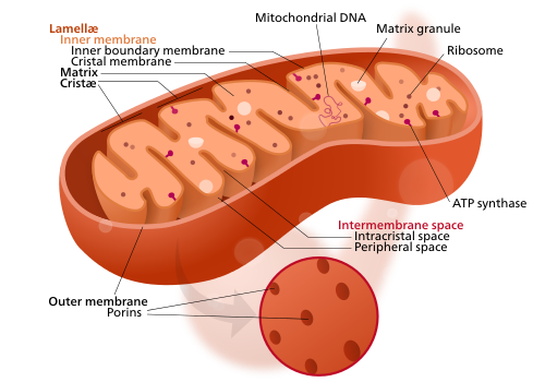
Mitochondria

Mitochondria are essentially universal in the eukaryotes, and with their own DNA somewhat resemble prokaryotic cells.

Mitochondria are organelles in eukaryotic cells. The mitochondrion is commonly called "the powerhouse of the cell", for its function providing energy by oxidising sugars or fats to produce the energy-storing molecule ATP. Mitochondria have two surrounding membranes, each a phospholipid bilayer, the inner of which is folded into invaginations called cristae where aerobic respiration takes place.

Mitochondria contain their own DNA, which has close structural similarities to bacterial DNA, from which it originated, and which encodes rRNA and tRNA genes that produce RNA which is closer in structure to bacterial RNA than to eukaryote RNA.

Some eukaryotes, such as the metamonads Giardia and Trichomonas, and the amoebozoan Pelomyxa, appear to lack mitochondria, but all contain mitochondrion-derived organelles, like hydrogenosomes or mitosomes, having lost their mitochondria secondarily. They obtain energy by enzymatic action in the cytoplasm. It is thought that mitochondria developed from prokaryotic cells which became endosymbionts living inside eukaryotes.

Plastids

The most common type of plastid is the chloroplast, which contains chlorophyll and produces organic compounds by photosynthesis.

Plants and various groups of algae have plastids as well as mitochondria. Plastids, like mitochondria, have their own DNA and are developed from endosymbionts, in this case cyanobacteria. They usually take the form of chloroplasts which, like cyanobacteria, contain chlorophyll and produce organic compounds (such as glucose) through photosynthesis. Others are involved in storing food. Although plastids probably had a single origin, not all plastid-containing groups are closely related. Instead, some eukaryotes have obtained them from others through secondary endosymbiosis or ingestion. The capture and sequestering of photosynthetic cells and chloroplasts, kleptoplasty, occurs in many types of modern eukaryotic organisms.

Cytoskeletal structures

The cytoskeleton. Actin filaments are shown in red, microtubules in green. (The nucleus is in blue.)

The cytoskeleton provides stiffening structure and points of attachment for motor structures that enable the cell to move, change shape, or transport materials. The motor structures are microfilaments of actin and actin-binding proteins, including α-actinin, fimbrin, and filamin are present in submembranous cortical layers and bundles. Motor proteins of microtubules, dynein and kinesin, and myosin of actin filaments, provide dynamic character of the network.

Many eukaryotes have long slender motile cytoplasmic projections, called flagella, or multiple shorter structures called cilia. These organelles are variously involved in movement, feeding, and sensation. They are composed mainly of tubulin, and are entirely distinct from prokaryotic flagella. They are supported by a bundle of microtubules arising from a centriole, characteristically arranged as nine doublets surrounding two singlets. Flagella may have hairs (mastigonemes), as in many stramenopiles. Their interior is continuous with the cell's cytoplasm.

Centrioles are often present, even in cells and groups that do not have flagella, but conifers and flowering plants have neither. They generally occur in groups that give rise to various microtubular roots. These form a primary component of the cytoskeleton, and are often assembled over the course of several cell divisions, with one flagellum retained from the parent and the other derived from it. Centrioles produce the spindle during nuclear division.

Cell wall

The cells of plants, algae, fungi and most chromalveolates, but not animals, are surrounded by a cell wall. This is a layer outside the cell membrane, providing the cell with structural support, protection, and a filtering mechanism. The cell wall also prevents over-expansion when water enters the cell.

The major polysaccharides making up the primary cell wall of land plants are cellulose, hemicellulose, and pectin. The cellulose microfibrils are linked together with hemicellulose, embedded in a pectin matrix. The most common hemicellulose in the primary cell wall is xyloglucan.

Sexual reproduction

Sexual reproduction requires a life cycle that alternates between a haploid phase, with one copy of each chromosome in the cell, and a diploid phase, with two copies. In eukaryotes, haploid gametes are produced by meiosis; two gametes fuse to form a diploid zygote.

Eukaryotes have a life cycle that involves sexual reproduction, alternating between a haploid phase, where only one copy of each chromosome is present in each cell, and a diploid phase, with two copies of each chromosome in each cell. The diploid phase is formed by fusion of two haploid gametes, such as eggs and spermatozoa, to form a zygote; this may grow into a body, with its cells dividing by mitosis, and at some stage produce haploid gametes through meiosis, a division that reduces the number of chromosomes and creates genetic variability. There is considerable variation in this pattern. Plants have both haploid and diploid multicellular phases. Eukaryotes have lower metabolic rates and longer generation times than prokaryotes, because they are larger and therefore have a smaller surface area to volume ratio.

The evolution of sexual reproduction may be a primordial characteristic of eukaryotes. Based on a phylogenetic analysis, Dacks and Roger have proposed that facultative sex was present in the group's common ancestor. A core set of genes that function in meiosis is present in both Trichomonas vaginalis and Giardia intestinalis, two organisms previously thought to be asexual. Since these two species are descendants of lineages that diverged early from the eukaryotic evolutionary tree, core meiotic genes, and hence sex, were likely present in the common ancestor of eukaryotes. Species once thought to be asexual, such as Leishmania parasites, have a sexual cycle. Amoebae, previously regarded as asexual, may be anciently sexual; while present-day asexual groups could have arisen recently.

Evolution

History of classification

In antiquity, the two lineages of animals and plants were recognized by Aristotle and Theophrastus. The lineages were given the taxonomic rank of kingdom by Linnaeus in the 18th century. Though he included the fungi with plants with some reservations, it was later realized that they are quite distinct and warrant a separate kingdom. The various single-cell eukaryotes were originally placed with plants or animals when they became known. In 1818, the German biologist Georg A. Goldfuss coined the word Protozoa to refer to organisms such as ciliates, and this group was expanded until Ernst Haeckel made it a kingdom encompassing all single-celled eukaryotes, the Protista, in 1866. The eukaryotes thus came to be seen as four kingdoms:

The protists were at that time thought to be "primitive forms", and thus an evolutionary grade, united by their primitive unicellular nature. Understanding of the oldest branchings in the tree of life only developed substantially with DNA sequencing, leading to a system of domains rather than kingdoms as top level rank being put forward by Carl Woese, Otto Kandler, and Mark Wheelis in 1990, uniting all the eukaryote kingdoms in the domain "Eucarya", stating, however, that "'eukaryotes' will continue to be an acceptable common synonym". In 1996, the evolutionary biologist Lynn Margulis proposed to replace kingdoms and domains with "inclusive" names to create a "symbiosis-based phylogeny", giving the description "Eukarya (symbiosis-derived nucleated organisms)".

Phylogeny

Tree of eukaryotes showing major subgroups and thumbnail diagrams of representative members of each group, based on 2023 phylogenomic reconstructions.

By 2014, a rough consensus started to emerge from the phylogenomic studies of the previous two decades. The majority of eukaryotes can be placed in one of two large clades dubbed Amorphea (similar in composition to the unikont hypothesis) and the Diphoda (formerly bikonts), which includes plants and most algal lineages. A third major grouping, the Excavata, has been abandoned as a formal group as it was found to be paraphyletic. The proposed phylogeny below includes two groups of excavates (Discoba and Metamonada), and incorporates the 2021 proposal that picozoans are close relatives of rhodophytes. The Provora are a group of microbial predators discovered in 2022.

One view of the great kingdoms and their stem groups. The Metamonada are hard to place, being sister possibly to Discoba or to Malawimonada or being a paraphyletic group external to all other eukaryotes.

Origin of eukaryotes

In the theory of symbiogenesis, a merger of an archaean and an aerobic bacterium created the eukaryotes, with aerobic mitochondria; a second merger added chloroplasts, creating the green plants.

The origin of the eukaryotic cell, or eukaryogenesis, is a milestone in the evolution of life, since eukaryotes include all complex cells and almost all multicellular organisms. The last eukaryotic common ancestor (LECA) is the hypothetical origin of all living eukaryotes, and was most likely a biological population, not a single individual. The LECA is believed to have been a protist with a nucleus, at least one centriole and flagellum, facultatively aerobic mitochondria, sex (meiosis and syngamy), a dormant cyst with a cell wall of chitin or cellulose, and peroxisomes.

An endosymbiotic union between a motile anaerobic archaean and an aerobic alphaproteobacterium gave rise to the LECA and all eukaryotes, with mitochondria. A second, much later endosymbiosis with a cyanobacterium gave rise to the ancestor of plants, with chloroplasts.

The presence of eukaryotic biomarkers in archaea points towards an archaeal origin. The genomes of Promethearchaeati archaea have plenty of eukaryotic signature protein genes, which play a crucial role in the development of the cytoskeleton and complex cellular structures characteristic of eukaryotes. In 2022, cryo-electron tomography demonstrated that Promethearchaeati archaea have a complex actin-based cytoskeleton, providing the first direct visual evidence of the archaeal ancestry of eukaryotes.

Fossils

The timing of the origin of eukaryotes is hard to determine, but the discovery of Qingshania magnificia, the earliest multicelluar eukaryote from North China which lived 1.635 billion years ago, suggests that the crown group eukaryotes originated from the late Paleoproterozoic (Statherian). The earliest unequivocal unicellular eukaryotes, Tappania plana, Shuiyousphaeridium macroreticulatum, Dictyosphaera macroreticulata, Germinosphaera alveolata, and Valeria lophostriata from North China, lived approximately 1.65 billion years ago.

Some acritarchs are known from at least 1.65 billion years ago, and a fossil, Grypania, which may be an alga, is as much as 2.1 billion years old. The "problematic" fossil Diskagma has been found in paleosols 2.2 billion years old.

Reconstruction of the problematic Diskagma buttonii, a terrestrial fossil less than 1mm high, from rocks around 2.2 billion years old

The Neoarchean fossil Thuchomyces shares similarities with eukaryotes, specifically fungi. It especially resembles the problematic fossil Diskagma, with hyphae and multiple differentiated layers. However, it is over 600 million years older than all other possible eukaryotes, and many of its "eukaryote features" are not specific to the clade, meaning it is almost certainly a microbial mat instead.

Structures proposed to represent "large colonial organisms" have been found in the black shales of the Palaeoproterozoic such as the Francevillian B Formation, in Gabon, dubbed the "Francevillian biota" which is dated at 2.1 billion years old. However, the status of these structures as fossils is contested, with other authors suggesting that they might represent pseudofossils. The oldest fossils that can unambiguously be assigned to eukaryotes are from the Ruyang Group of China, dating to approximately 1.8-1.6 billion years ago. Fossils that are clearly related to modern groups start appearing an estimated 1.2 billion years ago, in the form of red algae, though recent work suggests the existence of fossilized filamentous algae in the Vindhya basin dating back perhaps to 1.6 to 1.7 billion years ago.

The presence of steranes, eukaryotic-specific biomarkers, in Australian shales previously indicated that eukaryotes were present in these rocks dated at 2.7 billion years old, but these Archaean biomarkers have been rebutted as later contaminants. The oldest valid biomarker records are only around 800 million years old. In contrast, a molecular clock analysis suggests the emergence of sterol biosynthesis as early as 2.3 billion years ago. The nature of steranes as eukaryotic biomarkers is further complicated by the production of sterols by some bacteria.

Whenever their origins, eukaryotes may not have become ecologically dominant until much later; a massive increase in the zinc composition of marine sediments 800 million years ago has been attributed to the rise of substantial populations of eukaryotes, which preferentially consume and incorporate zinc relative to prokaryotes, approximately a billion years after their origin (at the latest).

Positron emission tomography

 
Positron emission tomography
Cardiac PET scanner

Positron emission tomography
(PET) is a functional imaging technique that uses radioactive substances known as radiotracers to visualize and measure changes in metabolic processes, and in other physiological activities including blood flow, regional chemical composition, and absorption. Different tracers are used for various imaging purposes, depending on the target process within the body.

For example:

PET is a common imaging technique, a medical scintillography technique used in nuclear medicine. A radiopharmaceutical—a radioisotope attached to a drug—is injected into the body as a tracer. When the radiopharmaceutical undergoes beta plus decay, a positron is emitted, and when the positron interacts with an ordinary electron, the two particles annihilate and two gamma rays are emitted in opposite directions. These gamma rays are detected by two gamma cameras to form a three-dimensional image.

PET scanners can incorporate a computed tomography scanner (CT) and are known as PET–CT scanners. PET scan images can be reconstructed using a CT scan performed using one scanner during the same session.

One of the disadvantages of a PET scanner is its high initial cost and ongoing operating costs.

Uses

PET is both a medical and research tool used in pre-clinical and clinical settings. It is used heavily in the imaging of tumors and the search for metastases within the field of clinical oncology, and for the clinical diagnosis of certain diffuse brain diseases such as those causing various types of dementias. PET is a valuable research tool to learn and enhance our knowledge of the normal human brain, heart function, and support drug development. PET is also used in pre-clinical studies using animals. It allows repeated investigations into the same subjects over time, where subjects can act as their own control and substantially reduces the numbers of animals required for a given study. This approach allows research studies to reduce the sample size needed while increasing the statistical quality of its results.

Physiological processes lead to anatomical changes in the body. Since PET is capable of detecting biochemical processes as well as expression of some proteins, PET can provide molecular-level information much before any anatomic changes are visible. PET scanning does this by using radiolabelled molecular probes that have different rates of uptake depending on the type and function of tissue involved. Regional tracer uptake in various anatomic structures can be visualized and relatively quantified in terms of injected positron emitter within a PET scan.

PET imaging is best performed using a dedicated PET scanner. It is also possible to acquire PET images using a conventional dual-head gamma camera fitted with a coincidence detector. The quality of gamma-camera PET imaging is lower, and the scans take longer to acquire. However, this method allows a low-cost on-site solution to institutions with low PET scanning demand. An alternative would be to refer these patients to another center or relying on a visit by a mobile scanner.

Alternative methods of medical imaging include single-photon emission computed tomography (SPECT), computed tomography (CT), magnetic resonance imaging (MRI) and functional magnetic resonance imaging (fMRI), and ultrasound. SPECT is an imaging technique similar to PET that uses radioligands to detect molecules in the body. SPECT is less expensive and provides inferior image quality than PET.

Oncology

Whole-body PET scan using 18F-FDG (fluorodeoxyglucose). The normal brain and kidneys are labeled, and radioactive urine from breakdown of the FDG is seen in the bladder. In addition, a large metastatic tumor mass from colon cancer is seen in the liver.

PET scanning with the radiotracer [18F]fluorodeoxyglucose (FDG) is widely used in clinical oncology. FDG is a glucose analog that is taken up by glucose-using cells and phosphorylated by hexokinase (whose mitochondrial form is significantly elevated in rapidly growing malignant tumors). Metabolic trapping of the radioactive glucose molecule allows the PET scan to be utilized. The concentrations of imaged FDG tracer indicate tissue metabolic activity as it corresponds to the regional glucose uptake. FDG is used to explore the possibility of cancer spreading to other body sites (cancer metastasis). These FDG PET scans for detecting cancer metastasis are the most common in standard medical care (representing 90% of current scans). The same tracer may also be used for the diagnosis of types of dementia. Less often, other radioactive tracers, usually but not always labelled with fluorine-18 (18F), are used to image the tissue concentration of different kinds of molecules of interest inside the body.

A typical dose of FDG used in an oncological scan has an effective radiation dose of 7.6 mSv. Because the hydroxy group that is replaced by fluorine-18 to generate FDG is required for the next step in glucose metabolism in all cells, no further reactions occur in FDG. Furthermore, most tissues (with the notable exception of liver and kidneys) cannot remove the phosphate added by hexokinase. This means that FDG is trapped in any cell that takes it up until it decays, since phosphorylated sugars, due to their ionic charge, cannot exit from the cell. This results in intense radiolabeling of tissues with high glucose uptake, such as the normal brain, liver, kidneys, and most cancers, which have a higher glucose uptake than most normal tissue due to the Warburg effect. As a result, FDG-PET can be used for diagnosis, staging, and monitoring treatment of cancers, particularly in Hodgkin lymphoma, non-Hodgkin lymphoma, and lung cancer.

A 2020 review of research on the use of PET for Hodgkin lymphoma found evidence that negative findings in interim PET scans are linked to higher overall survival and progression-free survival; however, the certainty of the available evidence was moderate for survival, and very low for progression-free survival.

A few other isotopes and radiotracers are slowly being introduced into oncology for specific purposes. For example, 11C-labelled metomidate (11C-metomidate) has been used to detect tumors of adrenocortical origin. Also, fluorodopa (FDOPA) PET/CT (also called F-18-DOPA PET/CT) has proven to be a more sensitive alternative to finding and also localizing pheochromocytoma than the iobenguane (MIBG) scan.

Neuroimaging

Neurology

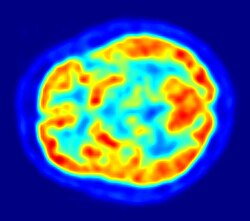
PET scan of the human brain

PET imaging with oxygen-15 indirectly measures blood flow to the brain. In this method, increased radioactivity signal indicates increased blood flow which is assumed to correlate with increased brain activity. Because of its two-minute half-life, oxygen-15 must be piped directly from a medical cyclotron for such uses, which is difficult.

PET imaging with FDG takes advantage of the fact that the brain is normally a rapid user of glucose. Standard FDG PET of the brain measures regional glucose use and can be used in neuropathological diagnosis.

Brain pathologies such as Alzheimer's disease (AD) greatly decrease brain metabolism of both glucose and oxygen in tandem. Therefore FDG PET of the brain may also be used to successfully differentiate Alzheimer's disease from other dementing processes, and also to make early diagnoses of Alzheimer's disease. The advantage of FDG PET for these uses is its much wider availability. Some fluorine-18 based radioactive tracers used for Alzheimer's include florbetapir, flutemetamol, Pittsburgh compound B (PiB) and florbetaben, which are all used to detect amyloid-beta plaques, a potential biomarker for Alzheimer's in the brain.

PET imaging with FDG can also be used for localization of "seizure focus". A seizure focus will appear as hypometabolic during an interictal scan. Several radiotracers (i.e. radioligands) have been developed for PET that are ligands for specific neuroreceptor subtypes such as [11C]raclopride, [18F]fallypride and [18F]desmethoxyfallypride for dopamine D2/D3 receptors; [11C]McN5652 and [11C]DASB for serotonin transporters; [18F]mefway for serotonin 5HT1A receptors; and [18F]nifene for nicotinic acetylcholine receptors or enzyme substrates (e.g. 6-FDOPA for the AADC enzyme). These agents permit the visualization of neuroreceptor pools in the context of a plurality of neuropsychiatric and neurologic illnesses.

PET may also be used for the diagnosis of hippocampal sclerosis, which causes epilepsy. FDG, and the less common tracers flumazenil and MPPF have been explored for this purpose. If the sclerosis is unilateral (right hippocampus or left hippocampus), FDG uptake can be compared with the healthy side. Even if the diagnosis is difficult with MRI, it may be diagnosed with PET.

The development of a number of novel probes for non-invasive, in-vivo PET imaging of neuroaggregate in human brain has brought amyloid imaging close to clinical use. The earliest amyloid imaging probes included [18F]FDDNP, developed at the University of California, Los Angeles, and Pittsburgh compound B (PiB), developed at the University of Pittsburgh. These probes permit the visualization of amyloid plaques in the brains of Alzheimer's patients and could assist clinicians in making a positive clinical diagnosis of AD pre-mortem and aid in the development of novel anti-amyloid therapies. [11C]polymethylpentene (PMP) is a novel radiopharmaceutical used in PET imaging to determine the activity of the acetylcholinergic neurotransmitter system by acting as a substrate for acetylcholinesterase. Post-mortem examination of AD patients has shown decreased levels of acetylcholinesterase. [11C]PMP is used to map the acetylcholinesterase activity in the brain, which could allow for premortem diagnoses of AD and help to monitor AD treatments. Avid Radiopharmaceuticals has developed and commercialized a compound called florbetapir that uses the longer-lasting radionuclide fluorine-18 to detect amyloid plaques using PET scans.

Neuropsychology or cognitive neuroscience

To examine links between specific psychological processes or disorders and brain activity.

Psychiatry

Numerous compounds that bind selectively to neuroreceptors of interest in biological psychiatry have been radiolabeled with C-11 or F-18. Radioligands that bind to dopamine receptors (D1, D2, reuptake transporter), serotonin receptors (5HT1A, 5HT2A, reuptake transporter), opioid receptors (mu and kappa), cholinergic receptors (nicotinic and muscarinic) and other sites have been used successfully in studies with human subjects. Studies have been performed examining the state of these receptors in patients compared to healthy controls in schizophrenia, substance abuse, mood disorders and other psychiatric conditions.

Stereotactic surgery and radiosurgery

PET can also be used in image guided surgery for the treatment of intracranial tumors, arteriovenous malformations and other surgically treatable conditions.

Cardiology

Cardiology, atherosclerosis and vascular disease study: FDG PET can help in identifying hibernating myocardium. However, the cost-effectiveness of PET for this role versus SPECT is unclear. FDG PET imaging of atherosclerosis to detect patients at risk of stroke is also feasible. Also, it can help test the efficacy of novel anti-atherosclerosis therapies.

Infectious diseases

Imaging infections with molecular imaging technologies can improve diagnosis and treatment follow-up. Clinically, PET has been widely used to image bacterial infections using FDG to identify the infection-associated inflammatory response. Three different PET contrast agents have been developed to image bacterial infections in vivo are [18F]maltose, [18F]maltohexaose, and [18F]2-fluorodeoxysorbitol (FDS). FDS has the added benefit of being able to target only Enterobacteriaceae.

Bio-distribution studies

In pre-clinical trials, a new drug can be radiolabeled and injected into animals. Such scans are referred to as biodistribution studies. The information regarding drug uptake, retention and elimination over time can be obtained quickly and cost-effectively compare to the older technique of killing and dissecting the animals. Commonly, drug occupancy at a purported site of action can be inferred indirectly by competition studies between unlabeled drug and radiolabeled compounds to bind with specificity to the site. A single radioligand can be used this way to test many potential drug candidates for the same target. A related technique involves scanning with radioligands that compete with an endogenous (naturally occurring) substance at a given receptor to demonstrate that a drug causes the release of the natural substance.

Small animal imaging

A miniature animal PET has been constructed that is small enough for a fully conscious rat to be scanned. This RatCAP (rat conscious animal PET) allows animals to be scanned without the confounding effects of anesthesia. PET scanners designed specifically for imaging rodents, often referred to as microPET, as well as scanners for small primates, are marketed for academic and pharmaceutical research. The scanners are based on microminiature scintillators and amplified avalanche photodiodes (APDs) through a system that uses single-chip silicon photomultipliers.

In 2018 the UC Davis School of Veterinary Medicine became the first veterinary center to employ a small clinical PET scanner as a scanner for clinical (rather than research) animal diagnosis. Because of cost as well as the marginal utility of detecting cancer metastases in companion animals (the primary use of this modality), veterinary PET scanning is expected to be rarely available in the immediate future.

Musculo-skeletal imaging

PET imaging has been used for imaging muscles and bones. FDG is the most commonly used tracer for imaging muscles, and NaF-F18 is the most widely used tracer for imaging bones.

Muscles

PET is a feasible technique for studying skeletal muscles during exercise. Also, PET can provide muscle activation data about deep-lying muscles (such as the vastus intermedialis and the gluteus minimus) compared to techniques like electromyography, which can be used only on superficial muscles directly under the skin. However, a disadvantage is that PET provides no timing information about muscle activation because it has to be measured after the exercise is completed. This is due to the time it takes for FDG to accumulate in the activated muscles.

Bones

Together with [18F]sodium floride, PET for bone imaging has been in use for 60 years for measuring regional bone metabolism and blood flow using static and dynamic scans. Researchers have recently started using [18F]sodium fluoride to study bone metastasis as well.

Safety

PET scanning is non-invasive, but it does involve exposure to ionizing radiation. FDG, which is now the standard radiotracer used for PET neuroimaging and cancer patient management, has an effective radiation dose of 14 mSv.

The amount of radiation in FDG is similar to the effective dose of spending one year in the American city of Denver, Colorado (12.4 mSv/year). For comparison, radiation dosage for other medical procedures range from 0.02 mSv for a chest X-ray and 6.5–8 mSv for a CT scan of the chest. Average civil aircrews are exposed to 3 mSv/year, and the whole body occupational dose limit for nuclear energy workers in the US is 50 mSv/year. For scale, see Orders of magnitude (radiation).

For PET–CT scanning, the radiation exposure may be substantial—around 23–26 mSv (for a 70 kg person—dose is likely to be higher for higher body weights).

Operation

Radionuclides and radiotracers

Schematic view of a detector block and ring of a PET scanner
Isotopes used in PET scans
Isotope 11C 13N 15O 18F 68Ga 64Cu 52Mn 55Co 89Zr 82Rb
Half-life 20 min 10 min 2 min 110 min 67.81 min 12.7 h 5.6 d 17.5 h 78.4 h 1.3 min

Radionuclides are incorporated either into compounds normally used by the body such as glucose (or glucose analogues), water, or ammonia, or into molecules that bind to receptors or other sites of drug action. Such labelled compounds are known as radiotracers. PET technology can be used to trace the biologic pathway of any compound in living humans (and many other species as well), provided it can be radiolabeled with a PET isotope. Thus, the specific processes that can be probed with PET are virtually limitless, and radiotracers for new target molecules and processes are continuing to be synthesized. As of this writing there are already dozens in clinical use and hundreds applied in research. In 2020 by far the most commonly used radiotracer in clinical PET scanning is the carbohydrate derivative FDG. This radiotracer is used in essentially all scans for oncology and most scans in neurology, thus makes up the large majority of radiotracer (>95%) used in PET and PET–CT scanning.

Due to the short half-lives of most positron-emitting radioisotopes, the radiotracers have traditionally been produced using a cyclotron in close proximity to the PET imaging facility. The half-life of fluorine-18 is long enough that radiotracers labeled with fluorine-18 can be manufactured commercially at offsite locations and shipped to imaging centers. Recently rubidium-82 generators have become commercially available. These contain strontium-82, which decays by electron capture to produce positron-emitting rubidium-82.

The use of positron-emitting isotopes of metals in PET scans has been reviewed, including elements not listed above, such as lanthanides.

Immuno-PET

The isotope 89Zr has been applied to the tracking and quantification of molecular antibodies with PET cameras (a method called "immuno-PET").

The biological half-life of antibodies is typically on the order of days, see daclizumab and erenumab by way of example. To visualize and quantify the distribution of such antibodies in the body, the PET isotope 89Zr is well suited because its physical half-life matches the typical biological half-life of antibodies, see table above.

Emission

Schema of a PET acquisition process

To conduct the scan, a short-lived radioactive tracer isotope is injected into the living subject (usually into blood circulation). Each tracer atom has been chemically incorporated into a biologically active molecule. There is a waiting period while the active molecule becomes concentrated in tissues of interest. Then the subject is placed in the imaging scanner. The molecule most commonly used for this purpose is FDG, a sugar, for which the waiting period is typically an hour. During the scan, a record of tissue concentration is made as the tracer decays.

As the radioisotope undergoes positron emission decay (also known as positive beta decay), it emits a positron, an antiparticle of the electron with opposite charge. The emitted positron travels in tissue for a short distance (typically less than 1 mm, but dependent on the isotope), during which time it loses kinetic energy, until it decelerates to a point where it can interact with an electron. The encounter annihilates both electron and positron, producing a pair of annihilation (gamma) photons moving in approximately opposite directions. These are detected when they reach a scintillator in the scanning device, creating a burst of light which is detected by photomultiplier tubes or silicon avalanche photodiodes (Si APD). The technique depends on simultaneous or coincident detection of the pair of photons moving in approximately opposite directions (they would be exactly opposite in their center of mass frame, but the scanner has no way to know this, and so has a built-in slight direction-error tolerance). Photons that do not arrive in temporal "pairs" (i.e. within a timing-window of a few nanoseconds) are ignored.

Localization of the positron annihilation event

The most significant fraction of electron–positron annihilations results in two 511 keV gamma photons being emitted at almost 180 degrees to each other. Hence, it is possible to localize their source along a straight line of coincidence (also called the line of response, or LOR). In practice, the LOR has a non-zero width as the emitted photons are not exactly 180 degrees apart. If the resolving time of the detectors is less than 500 picoseconds rather than about 10 nanoseconds, it is possible to localize the event to a segment of a chord, whose length is determined by the detector timing resolution. As the timing resolution improves, the signal-to-noise ratio (SNR) of the image will improve, requiring fewer events to achieve the same image quality. This technology is not yet common, but it is available on some new systems.

Image reconstruction

The raw data collected by a PET scanner are a list of 'coincidence events' representing near-simultaneous detection (typically, within a window of 6 to 12 nanoseconds of each other) of annihilation photons by a pair of detectors. Each coincidence event represents a line in space connecting the two detectors along which the positron emission occurred (i.e., the line of response (LOR)).

Analytical techniques, much like the reconstruction of computed tomography (CT) and single-photon emission computed tomography (SPECT) data, are commonly used, although the data set collected in PET is much poorer than CT, so reconstruction techniques are more difficult. Coincidence events can be grouped into projection images, called sinograms. The sinograms are sorted by the angle of each view and tilt (for 3D images). The sinogram images are analogous to the projections captured by CT scanners, and can be reconstructed in a similar way. The statistics of data thereby obtained are much worse than those obtained through transmission tomography. A normal PET data set has millions of counts for the whole acquisition, while the CT can reach a few billion counts. This contributes to PET images appearing "noisier" than CT. Two major sources of noise in PET are scatter (a detected pair of photons, at least one of which was deflected from its original path by interaction with matter in the field of view, leading to the pair being assigned to an incorrect LOR) and random events (photons originating from two different annihilation events but incorrectly recorded as a coincidence pair because their arrival at their respective detectors occurred within a coincidence timing window).

In practice, considerable pre-processing of the data is required – correction for random coincidences, estimation and subtraction of scattered photons, detector dead-time correction (after the detection of a photon, the detector must "cool down" again) and detector-sensitivity correction (for both inherent detector sensitivity and changes in sensitivity due to angle of incidence).

Filtered back projection (FBP) has been frequently used to reconstruct images from the projections. This algorithm has the advantage of being simple while having a low requirement for computing resources. Disadvantages are that shot noise in the raw data is prominent in the reconstructed images, and areas of high tracer uptake tend to form streaks across the image. Also, FBP treats the data deterministically – it does not account for the inherent randomness associated with PET data, thus requiring all the pre-reconstruction corrections described above.

Statistical, likelihood-based approaches: Statistical, likelihood-based iterative expectation-maximization algorithms such as the Shepp–Vardi algorithm are now the preferred method of reconstruction. These algorithms compute an estimate of the likely distribution of annihilation events that led to the measured data, based on statistical principles. The advantage is a better noise profile and resistance to the streak artifacts common with FBP, but the disadvantage is greater computer resource requirements. A further advantage of statistical image reconstruction techniques is that the physical effects that would need to be pre-corrected for when using an analytical reconstruction algorithm, such as scattered photons, random coincidences, attenuation and detector dead-time, can be incorporated into the likelihood model being used in the reconstruction, allowing for additional noise reduction. Iterative reconstruction has also been shown to result in improvements in the resolution of the reconstructed images, since more sophisticated models of the scanner physics can be incorporated into the likelihood model than those used by analytical reconstruction methods, allowing for improved quantification of the radioactivity distribution.

Research has shown that Bayesian methods that involve a Poisson likelihood function and an appropriate prior probability (e.g., a smoothing prior leading to total variation regularization or a Laplacian distribution leading to -based regularization in a wavelet or other domain), such as via Ulf Grenander's Sieve estimator or via Bayes penalty methods or via I.J. Good's roughness method may yield superior performance to expectation-maximization-based methods which involve a Poisson likelihood function but do not involve such a prior.

Attenuation correction: Quantitative PET Imaging requires attenuation correction. In these systems attenuation correction is based on a transmission scan using 68Ge rotating rod source.

Transmission scans directly measure attenuation values at 511 keV. Attenuation occurs when photons emitted by the radiotracer inside the body are absorbed by intervening tissue between the detector and the emission of the photon. As different LORs must traverse different thicknesses of tissue, the photons are attenuated differentially. The result is that structures deep in the body are reconstructed as having falsely low tracer uptake. Contemporary scanners can estimate attenuation using integrated x-ray CT equipment, in place of earlier equipment that offered a crude form of CT using a gamma ray (positron emitting) source and the PET detectors.

While attenuation-corrected images are generally more faithful representations, the correction process is itself susceptible to significant artifacts. As a result, both corrected and uncorrected images are always reconstructed and read together.

2D/3D reconstruction: Early PET scanners had only a single ring of detectors, hence the acquisition of data and subsequent reconstruction was restricted to a single transverse plane. More modern scanners now include multiple rings, essentially forming a cylinder of detectors.

There are two approaches to reconstructing data from such a scanner:

  1. Treat each ring as a separate entity, so that only coincidences within a ring are detected, the image from each ring can then be reconstructed individually (2D reconstruction), or
  2. Allow coincidences to be detected between rings as well as within rings, then reconstruct the entire volume together (3D).

3D techniques have better sensitivity (because more coincidences are detected and used) hence less noise, but are more sensitive to the effects of scatter and random coincidences, as well as requiring greater computer resources. The advent of sub-nanosecond timing resolution detectors affords better random coincidence rejection, thus favoring 3D image reconstruction.

Time-of-flight (TOF) PET: For modern systems with a higher time resolution (roughly 3 nanoseconds) a technique called "time-of-flight" is used to improve the overall performance. Time-of-flight PET makes use of very fast gamma-ray detectors and data processing system which can more precisely decide the difference in time between the detection of the two photons. It is impossible to localize the point of origin of the annihilation event exactly (currently within 10 cm). Therefore, image reconstruction is still needed. TOF technique gives a remarkable improvement in image quality, especially signal-to-noise ratio.

Combination of PET with CT or MRI

Complete body PET–CT fusion image
Brain PET–MRI fusion image

PET scans are increasingly read alongside CT or MRI scans, with the combination (co-registration) giving both anatomic and metabolic information (i.e., what the structure is, and what it is doing biochemically). Because PET imaging is most useful in combination with anatomical imaging, such as CT, modern PET scanners are now available with integrated high-end multi-detector-row CT scanners (PET–CT). Because the two scans can be performed in immediate sequence during the same session, with the patient not changing position between the two types of scans, the two sets of images are more precisely registered, so that areas of abnormality on the PET imaging can be more perfectly correlated with anatomy on the CT images. This is very useful in showing detailed views of moving organs or structures with higher anatomical variation, which is more common outside the brain.

At the Jülich Institute of Neurosciences and Biophysics, the world's largest PET–MRI device began operation in April 2009. A 9.4-tesla magnetic resonance tomograph (MRT) combined with a PET. Presently, only the head and brain can be imaged at these high magnetic field strengths.

For brain imaging, registration of CT, MRI and PET scans may be accomplished without the need for an integrated PET–CT or PET–MRI scanner by using a device known as the N-localizer.

Limitations

The minimization of radiation dose to the subject is an attractive feature of the use of short-lived radionuclides. Besides its established role as a diagnostic technique, PET has an expanding role as a method to assess the response to therapy, in particular, cancer therapy, where the risk to the patient from lack of knowledge about disease progress is much greater than the risk from the test radiation. Since the tracers are radioactive, the elderly and pregnant are unable to use it due to risks posed by radiation.

Limitations to the widespread use of PET arise from the high costs of cyclotrons needed to produce the short-lived radionuclides for PET scanning and the need for specially adapted on-site chemical synthesis apparatus to produce the radiopharmaceuticals after radioisotope preparation. Organic radiotracer molecules that will contain a positron-emitting radioisotope cannot be synthesized first and then the radioisotope prepared within them, because bombardment with a cyclotron to prepare the radioisotope destroys any organic carrier for it. Instead, the isotope must be prepared first, then the chemistry to prepare any organic radiotracer (such as FDG) accomplished very quickly, in the short time before the isotope decays. Few hospitals and universities are capable of maintaining such systems, and most clinical PET is supported by third-party suppliers of radiotracers that can supply many sites simultaneously. This limitation restricts clinical PET primarily to the use of tracers labelled with fluorine-18, which has a half-life of 110 minutes and can be transported a reasonable distance before use, or to rubidium-82 (used as rubidium-82 chloride) with a half-life of 1.27 minutes, which is created in a portable generator and is used for myocardial perfusion studies. In recent years a few on-site cyclotrons with integrated shielding and "hot labs" (automated chemistry labs that are able to work with radioisotopes) have begun to accompany PET units to remote hospitals. The presence of the small on-site cyclotron promises to expand in the future as the cyclotrons shrink in response to the high cost of isotope transportation to remote PET machines. In recent years the shortage of PET scans has been alleviated in the US, as rollout of radiopharmacies to supply radioisotopes has grown 30%/year.

Because the half-life of fluorine-18 is about two hours, the prepared dose of a radiopharmaceutical bearing this radionuclide will undergo multiple half-lives of decay during the working day. This necessitates frequent recalibration of the remaining dose (determination of activity per unit volume) and careful planning with respect to patient scheduling.

History

A PET scanner released in 2003

The concept of emission and transmission tomography was introduced by David E. Kuhl, Luke Chapman and Roy Edwards in the late 1950s. Their work would lead to the design and construction of several tomographic instruments at Washington University School of Medicine and later at the University of Pennsylvania. In the 1960s and 70s tomographic imaging instruments and techniques were further developed by Michel Ter-Pogossian, Michael E. Phelps, Edward J. Hoffman and others at Washington University School of Medicine.

Work by Gordon Brownell, Charles Burnham and their associates at the Massachusetts General Hospital beginning in the 1950s contributed significantly to the development of PET technology and included the first demonstration of annihilation radiation for medical imaging. Their innovations, including the use of light pipes and volumetric analysis, have been important in the deployment of PET imaging. In 1961, James Robertson and his associates at Brookhaven National Laboratory built the first single-plane PET scan, nicknamed the "head-shrinker."

One of the factors most responsible for the acceptance of positron imaging was the development of radiopharmaceuticals. In particular, the development of labeled 2-fluorodeoxy-D-glucose (FDG-firstly synthethized and described by two Czech scientists from Charles University in Prague in 1968) by the Brookhaven group under the direction of Al Wolf and Joanna Fowler was a major factor in expanding the scope of PET imaging. The compound was first administered to two normal human volunteers by Abass Alavi in August 1976 at the University of Pennsylvania. Brain images obtained with an ordinary (non-PET) nuclear scanner demonstrated the concentration of FDG in that organ. Later, the substance was used in dedicated positron tomographic scanners, to yield the modern procedure.

The logical extension of positron instrumentation was a design using two 2-dimensional arrays. PC-I was the first instrument using this concept and was designed in 1968, completed in 1969 and reported in 1972. The first applications of PC-I in tomographic mode as distinguished from the computed tomographic mode were reported in 1970. It soon became clear to many of those involved in PET development that a circular or cylindrical array of detectors was the logical next step in PET instrumentation. Although many investigators took this approach, James Robertson and Zang-Hee Cho were the first to propose a ring system that has become the prototype of the current shape of PET. The first multislice cylindrical array PET scanner was completed in 1974 at the Mallinckrodt Institute of Radiology by the group led by Ter-Pogossian.

The PET–CT scanner, attributed to David Townsend and Ronald Nutt, was named by Time as the medical invention of the year in 2000.

Cost

As of August 2008, Cancer Care Ontario reports that the current average incremental cost to perform a PET scan in the province is CA$1,000–1,200 per scan. This includes the cost of the radiopharmaceutical and a stipend for the physician reading the scan.

In the United States, a PET scan is estimated to be US$1,500-$5,000.

In England, the National Health Service reference cost (2015–2016) for an adult outpatient PET scan is £798.

In Australia, as of July 2018, the Medicare Benefits Schedule Fee for whole body FDG PET ranges from A$953 to A$999, depending on the indication for the scan.

Quality control

The overall performance of PET systems can be evaluated by quality control tools such as the Jaszczak phantom.

Clinical trial

From Wikipedia, the free encyclopedia https://en.wikipedia.org/wiki/Clinical_...