Search This Blog

Wednesday, October 3, 2018

Endocannabinoid system

From Wikipedia, the free encyclopedia
 
The endocannabinoid system (ECS) is a biological system composed of endocannabinoids, which are endogenous lipid-based retrograde neurotransmitters that bind to cannabinoid receptors, and cannabinoid receptor proteins that are expressed throughout the mammalian central nervous system (including the brain) and peripheral nervous system. The endocannabinoid system is involved in regulating a variety of physiological and cognitive processes including fertility, pregnancy, during pre- and postnatal development, appetite, pain-sensation, mood, and memory, and in mediating the pharmacological effects of cannabis. The ECS is also involved in mediating some of the physiological and cognitive effects of voluntary physical exercise in humans and other animals, such as contributing to exercise-induced euphoria as well as modulating locomotor activity and motivational salience for rewards. In humans, the plasma concentration of certain endocannabinoids (i.e., anandamide) have been found to rise during physical activity; since endocannabinoids can effectively penetrate the blood–brain barrier, it has been suggested that anandamide, along with other euphoriant neurochemicals, contributes to the development of exercise-induced euphoria in humans, a state colloquially referred to as a runner's high.
 
Two primary endocannabinoid receptors have been identified: CB1, first cloned in 1990; and CB2, cloned in 1993. CB1 receptors are found predominantly in the brain and nervous system, as well as in peripheral organs and tissues, and are the main molecular target of the endocannabinoid ligand (binding molecule), anandamide, as well as its mimetic phytocannabinoid, THC. One other main endocannabinoid is 2-arachidonoylglycerol (2-AG) which is active at both cannabinoid receptors, along with its own mimetic phytocannabinoid, CBD. 2-AG and CBD are involved in the regulation of appetite, immune system functions and pain management.

Basic overview

The endocannabinoid system, broadly speaking, includes:
The neurons, neural pathways, and other cells where these molecules, enzymes, and one or both cannabinoid receptor types are all colocalized collectively comprise the endocannabinoid system.
The endocannabinoid system has been studied using genetic and pharmacological methods. These studies have revealed that cannabinoids act as neuromodulators for a variety of processes, including motor learning, appetite, and pain sensation, among other cognitive and physical processes. The localization of the CB1 receptor in the endocannabinoid system has a very large degree of overlap with the orexinergic projection system, which mediates many of the same functions, both physical and cognitive. Moreover, CB1 is colocalized on orexin projection neurons in the lateral hypothalamus and many output structures of the orexin system, where the CB1 and orexin receptor 1 (OX1) receptors physically and functionally join together to form the CB1–OX1 receptor heterodimer.

Expression of receptors

Cannabinoid binding sites exist throughout the central and peripheral nervous systems. The two most relevant receptors for cannabinoids are the CB1 and CB2 receptors, which are expressed predominantly in the brain and immune system respectively. Density of expression varies based on species and correlates with the efficacy that cannabinoids will have in modulating specific aspects of behavior related to the site of expression. For example, in rodents, the highest concentration of cannabinoid binding sites are in the basal ganglia and cerebellum, regions of the brain involved in the initiation and coordination of movement. In humans, cannabinoid receptors exist in much lower concentration in these regions, which helps explain why cannabinoids possess a greater efficacy in altering rodent motor movements than they do in humans.

A recent analysis of cannabinoid binding in CB1 and CB2 receptor knockout mice found cannabinoid responsiveness even when these receptors were not being expressed, indicating that an additional binding receptor may be present in the brain. Binding has been demonstrated by 2-arachidonoylglycerol (2-AG) on the TRPV1 receptor suggesting that this receptor may be a candidate for the established response.

In addition to CB1 and CB2, certain orphan receptors are known to bind endocannabinoids as well, including GPR18, GPR55 (a regulator of neuroimmune function), and GPR119. CB1 has also been noted to form a functional human receptor heterodimer in orexin neurons with OX1, the CB1–OX1 receptor, which mediates feeding behavior and certain physical processes such as cannabinoid-induced pressor responses which are known to occur through signaling in the rostral ventrolateral medulla.

Endocannabinoid synthesis, release, and degradation

During neurotransmission, the pre-synaptic neuron releases neurotransmitters into the synaptic cleft which bind to cognate receptors expressed on the post-synaptic neuron. Based upon the interaction between the transmitter and receptor, neurotransmitters may trigger a variety of effects in the post-synaptic cell, such as excitation, inhibition, or the initiation of second messenger cascades. Based on the cell, these effects may result in the on-site synthesis of endogenous cannabinoids anandamide or 2-AG by a process that is not entirely clear, but results from an elevation in intracellular calcium. Expression appears to be exclusive, so that both types of endocannabinoids are not co-synthesized. This exclusion is based on synthesis-specific channel activation: a recent study found that in the bed nucleus of the stria terminalis, calcium entry through voltage-sensitive calcium channels produced an L-type current resulting in 2-AG production, while activation of mGluR1/5 receptors triggered the synthesis of anandamide.

Evidence suggests that the depolarization-induced influx of calcium into the post-synaptic neuron causes the activation of an enzyme called transacylase. This enzyme is suggested to catalyze the first step of endocannabinoid biosynthesis by converting phosphatidylethanolamine, a membrane-resident phospholipid, into N-acyl-phosphatidylethanolamine (NAPE). Experiments have shown that phospholipase D cleaves NAPE to yield anandamide. This process is mediated by bile acids. In NAPE-phospholipase D (NAPEPLD)-knockout mice, cleavage of NAPE is reduced in low calcium concentrations, but not abolished, suggesting multiple, distinct pathways are involved in anandamide synthesis. The synthesis of 2-AG is less established and warrants further research.

Once released into the extracellular space by a putative endocannabinoid transporter, messengers are vulnerable to glial cell inactivation. Endocannabinoids are taken up by a transporter on the glial cell and degraded by fatty acid amide hydrolase (FAAH), which cleaves anandamide into arachidonic acid and ethanolamine or monoacylglycerol lipase (MAGL), and 2-AG into arachidonic acid and glycerol. While arachidonic acid is a substrate for leukotriene and prostaglandin synthesis, it is unclear whether this degradative byproduct has unique functions in the central nervous system. Emerging data in the field also points to FAAH being expressed in postsynaptic neurons complementary to presynaptic neurons expressing cannabinoid receptors, supporting the conclusion that it is major contributor to the clearance and inactivation of anandamide and 2-AG after endocannabinoid reuptake. A neuropharmacological study demonstrated that an inhibitor of FAAH (URB597) selectively increases anandamide levels in the brain of rodents and primates. Such approaches could lead to the development of new drugs with analgesic, anxiolytic-like and antidepressant-like effects, which are not accompanied by overt signs of abuse liability.

Binding and intracellular effects

Cannabinoid receptors are G-protein coupled receptors located on the pre-synaptic membrane. While there have been some papers that have linked concurrent stimulation of dopamine and CB1 receptors to an acute rise in cyclic adenosine monophosphate (cAMP) production, it is generally accepted that CB1 activation via cannabinoids causes a decrease in cAMP concentration by inhibition of adenylyl cyclase and a rise in the concentration of mitogen-activated protein kinase (MAP kinase). The relative potency of different cannabinoids in inhibition of adenylyl cyclase correlates with their varying efficacy in behavioral assays. This inhibition of cAMP is followed by phosphorylation and subsequent activation of not only a suite of MAP kinases (p38/p42/p44), but also the PI3/PKB and MEK/ERK pathway (Galve-Roperh et al., 2002; Davis et al., 2005; Jones et al., 2005; Graham et al., 2006). Results from rat hippocampal gene chip data after acute administration of tetrahydrocannabinol (THC) showed an increase in the expression of transcripts encoding myelin basic protein, endoplasmic proteins, cytochrome oxidase, and two cell adhesion molecules: NCAM, and SC1; decreases in expression were seen in both calmodulin and ribosomal RNAs (Kittler et al., 2000). In addition, CB1 activation has been demonstrated to increase the activity of transcription factors like c-Fos and Krox-24 (Graham et al., 2006).

Binding and neuronal excitability

The molecular mechanisms of CB1-mediated changes to the membrane voltage have also been studied in detail. Cannabinoids reduce calcium influx by blocking the activity of voltage-dependent N-, P/Q- and L-type calcium channels. In addition to acting on calcium channels, activation of Gi/o and Gs, the two most commonly coupled G-proteins to cannabinoid receptors, has been shown to modulate potassium channel activity. Recent studies have found that CB1 activation specifically facilitates potassium ion flux through GIRKs, a family of potassium channels. Immunohistochemistry experiments demonstrated that CB1 is co-localized with GIRK and Kv1.4 potassium channels, suggesting that these two may interact in physiological contexts.

In the central nervous system, CB1 receptors influence neuronal excitability, reducing the incoming synaptic input. This mechanism, known as presynaptic inhibition, occurs when a postsynaptic neuron releases endocannabinoids in retrograde transmission, which then bind to cannabinoid receptors on the presynaptic terminal. CB1 receptors then reduce the amount of neurotransmitter released, so that subsequent excitation in the presynaptic neuron results in diminished effects on the postsynaptic neuron. It is likely that presynaptic inhibition uses many of the same ion channel mechanisms listed above, although recent evidence has shown that CB1 receptors can also regulate neurotransmitter release by a non-ion channel mechanism, i.e. through Gi/o-mediated inhibition of adenylyl cyclase and protein kinase A. Direct effects of CB1 receptors on membrane excitability have been reported, and strongly impact the firing of cortical neurons.[42] A series of behavioral experiments demonstrated that NMDAR, an ionotropic glutamate receptor, and the metabotropic glutamate receptors (mGluRs) work in concert with CB1 to induce analgesia in mice, although the mechanism underlying this effect is unclear.

Functions of the endocannabinoid system

Memory

Mice treated with tetrahydrocannabinol (THC) show suppression of long-term potentiation in the hippocampus, a process that is essential for the formation and storage of long-term memory. These results concur with anecdotal evidence suggesting that smoking cannabis impairs short-term memory. Consistent with this finding, mice without the CB1 receptor show enhanced memory and long-term potentiation indicating that the endocannabinoid system may play a pivotal role in the extinction of old memories. One study found that the high-dose treatment of rats with the synthetic cannabinoid HU-210 over several weeks resulted in stimulation of neural growth in the rats' hippocampus region, a part of the limbic system playing a part in the formation of declarative and spatial memories, but did not investigate the effects on short-term or long-term memory. Taken together, these findings suggest that the effects of endocannabinoids on the various brain networks involved in learning and memory may vary.

Role in hippocampal neurogenesis

In the adult brain, the endocannabinoid system facilitates the neurogenesis of hippocampal granule cells. In the subgranular zone of the dentate gyrus, multipotent neural progenitors (NP) give rise to daughter cells that, over the course of several weeks, mature into granule cells whose axons project to and synapse onto dendrites on the CA3 region. NPs in the hippocampus have been shown to possess fatty acid amide hydrolase (FAAH) and express CB1 and utilize 2-AG. Intriguingly, CB1 activation by endogenous or exogenous cannabinoids promote NP proliferation and differentiation; this activation is absent in CB1 knockouts and abolished in the presence of antagonist.

Induction of synaptic depression

The inhibitory effects of cannabinoid receptor stimulation on neurotransmitter release have caused this system to be connected to various forms of depressant plasticity. A recent study conducted with the bed nucleus of the stria terminalis found that the endurance of the depressant effects was mediated by two different signaling pathways based on the type of receptor activated. 2-AG was found to act on presynaptic CB1 receptors to mediate retrograde short-term depression (STD) following activation of L-type calcium currents, while anandamide was synthesized after mGluR5 activation and triggered autocrine signalling onto postsynapic TRPV1 receptors that induced long-term depression (LTD). Similar post-synaptic receptor dependencies were found in the striatum, but here both effects relied on presynaptic CB1 receptors. These findings provide the brain a direct mechanism to selectively inhibit neuronal excitability over variable time scales. By selectively internalizing different receptors, the brain may limit the production of specific endocannabinoids to favor a time scale in accordance with its needs.

Appetite

Evidence for the role of the endocannabinoid system in food-seeking behavior comes from a variety of cannabinoid studies. Emerging data suggests that THC acts via CB1 receptors in the hypothalamic nuclei to directly increase appetite. It is thought that hypothalamic neurons tonically produce endocannabinoids that work to tightly regulate hunger. The amount of endocannabinoids produced is inversely correlated with the amount of leptin in the blood. For example, mice without leptin not only become massively obese but express abnormally high levels of hypothalamic endocannabinoids as a compensatory mechanism. Similarly, when these mice were treated with an endocannabinoid inverse agonists, such as rimonabant, food intake was reduced. When the CB1 receptor is knocked out in mice, these animals tend to be leaner and less hungry than wild-type mice. A related study examined the effect of THC on the hedonic (pleasure) value of food and found enhanced dopamine release in the nucleus accumbens and increased pleasure-related behavior after administration of a sucrose solution. A related study found that endocannabinoids affect taste perception in taste cells In taste cells, endocannabinoids were shown to selectively enhance the strength of neural signaling for sweet tastes, whereas leptin decreased the strength of this same response. While there is need for more research, these results suggest that cannabinoid activity in the hypothalamus and nucleus accumbens is related to appetitive, food-seeking behavior.

Energy balance and metabolism

The endocannabinoid system has been shown to have a homeostatic role by controlling several metabolic functions, such as energy storage and nutrient transport. It acts on peripheral tissues such as adipocytes, hepatocytes, the gastrointestinal tract, the skeletal muscles and the endocrine pancreas. It has also been implied in modulating insulin sensitivity. Through all of this, the endocannabinoid system may play a role in clinical conditions, such as obesity, diabetes, and atherosclerosis, which may also give it a cardiovascular role.

Stress response

While the secretion of glucocorticoids in response to stressful stimuli is an adaptive response necessary for an organism to respond appropriately to a stressor, persistent secretion may be harmful. The endocannabinoid system has been implicated in the habituation of the hypothalamic-pituitary-adrenal axis (HPA axis) to repeated exposure to restraint stress. Studies have demonstrated differential synthesis of anandamide and 2-AG during tonic stress. A decrease of anandamide was found along the axis that contributed to basal hypersecretion of corticosterone; in contrast, an increase of 2-AG was found in the amygdala after repeated stress, which was negatively correlated to magnitude of the corticosterone response. All effects were abolished by the CB1 antagonist AM251, supporting the conclusion that these effects were cannabinoid-receptor dependent. These findings show that anandamide and 2-AG divergently regulate the HPA axis response to stress: while habituation of the stress-induced HPA axis via 2-AG prevents excessive secretion of glucocorticoids to non-threatening stimuli, the increase of basal corticosterone secretion resulting from decreased anandamide allows for a facilitated response of the HPA axis to novel stimuli.

Exploration, social behavior, and anxiety

These contrasting effects reveal the importance of the endocannabinoid system in regulating anxiety-dependent behavior. Results suggest that glutamatergic cannabinoid receptors are not only responsible for mediating aggression, but produce an anxiolytic-like function by inhibiting excessive arousal: excessive excitation produces anxiety that limited the mice from exploring both animate and inanimate objects. In contrast, GABAergic neurons appear to control an anxiogenic-like function by limiting inhibitory transmitter release. Taken together, these two sets of neurons appear to help regulate the organism's overall sense of arousal during novel situations.

Immune function

Evidence suggests that endocannabinoids may function as both neuromodulators and immunomodulators in the immune system. Here, they seem to serve an autoprotective role to ameliorate muscle spasms, inflammation, and other symptoms of multiple sclerosis and skeletal muscle spasms. Functionally, the activation of cannabinoid receptors has been demonstrated to play a role in the activation of GTPases in macrophages, neutrophils, and BM cells. These receptors have also been implicated in the proper migration of B cells into the marginal zone (MZ) and the regulation of healthy IgM levels. Some disorders seem to trigger an upregulation of cannabinoid receptors selectively in cells or tissues related to symptom relief and inhibition of disease progression, such as in that rodent neuropathic pain model, where receptors are increased in the spinal cord microglia, dorsal root ganglion, and thalamic neurons.

Multiple sclerosis

Historical records from ancient China and Greece suggest that preparations of Cannabis indica were commonly prescribed to ameliorate multiple sclerosis-like symptoms such as tremors and muscle pain. Modern research has confirmed these effects in a study on diseased mice, wherein both endogenous and exogenous agonists showed ameliorating effects on tremor and spasticity. It remains to be seen whether pharmaceutical preparations such as dronabinol have the same effects in humans. Due to increasing use of medical Cannabis and rising incidence of multiple sclerosis patients who self-medicate with the drug, there has been much interest in exploiting the endocannabinoid system in the cerebellum to provide a legal and effective relief. In mouse models of multiple sclerosis, there is a profound reduction and reorganization of CB1 receptors in the cerebellum. Serial sections of cerebellar tissue subjected to immunohistochemistry revealed that this aberrant expression occurred during the relapse phase but returned to normal during the remitting phase of the disease. Other studies suggest that CB1 agonists promote the survival of oligodendrocytes in vitro in the absence of growth and trophic factors; in addition, these agonist have been shown to promote mRNA expression of myelin lipid protein. (Kittler et al., 2000; Mollna-Holgado et al., 2002). Taken together, these studies point to the exciting possibility that cannabinoid treatment may not only be able to attenuate the symptoms of multiple sclerosis but also improve oligodendrocyte function (reviewed in Pertwee, 2001; Mollna-Holgado et al., 2002). 2-AG stimulates proliferation of a microglial cell line by a CB2 receptor dependent mechanism, and the number of microglial cells is increased in multiple sclerosis.

Female reproduction

The developing embryo expresses cannabinoid receptors early in development that are responsive to anandamide secreted in the uterus. This signaling is important in regulating the timing of embryonic implantation and uterine receptivity. In mice, it has been shown that anandamide modulates the probability of implantation to the uterine wall. For example, in humans, the likelihood of miscarriage increases if uterine anandamide levels are too high or low. These results suggest that intake of exogenous cannabinoids (e.g. cannabis) can decrease the likelihood for pregnancy for women with high anandamide levels, and alternatively, it can increase the likelihood for pregnancy in women whose anandamide levels were too low.

Autonomic nervous system

Peripheral expression of cannabinoid receptors led researchers to investigate the role of cannabinoids in the autonomic nervous system. Research found that the CB1 receptor is expressed presynaptically by motor neurons that innervate visceral organs. Cannabinoid-mediated inhibition of electric potentials results in a reduction in noradrenaline release from sympathetic nervous system nerves. Other studies have found similar effects in endocannabinoid regulation of intestinal motility, including the innervation of smooth muscles associated with the digestive, urinary, and reproductive systems.

Analgesia

At the spinal cord, cannabinoids suppress noxious-stimulus-evoked responses of neurons in the dorsal horn, possibly by modulating descending noradrenaline input from the brainstem. As many of these fibers are primarily GABAergic, cannabinoid stimulation in the spinal column results in disinhibition that should increase noradrenaline release and attenuation of noxious-stimuli-processing in the periphery and dorsal root ganglion.

The endocannabinoid most researched in pain is palmitoylethanolamide. Palmitoylethanolamide is a fatty amine related to anandamide, but saturated and although initially it was thought that palmitoylethanolamide would bind to the CB1 and the CB2 receptor, later it was found that the most important receptors are the PPAR-alpha receptor, the TRPV receptor and the GPR55 receptor. Palmitoylethanolamide has been evaluated for its analgesic actions in a great variety of pain indications and found to be safe and effective. Basically these data are proof of concept for endocannabinoids and related fatty amines to be therapeutically useful analgesics; palmitoylethanolamide is available under the brand names Normast and PeaPure as nutraceuticals.
Endocannabinoids are involved in placebo induced analgesia responses.

Thermoregulation

Anandamide and N-arachidonoyl dopamine (NADA) have been shown to act on temperature-sensing TRPV1 channels, which are involved in thermoregulation. TRPV1 is activated by the exogenous ligand capsaicin, the active component of chili peppers, which is structurally similar to endocannabinoids. NADA activates the TRPV1 channel with an EC50 of approximately of 50 nM.  The high potency makes it the putative endogenous TRPV1 agonist. Anandamide has also been found to activate TRPV1 on sensory neuron terminals, and subsequently cause vasodilation. TRPV1 may also be activated by methanandamide and arachidonyl-2'-chloroethylamide (ACEA).

Sleep

Increased endocannabinoid signaling within the central nervous system promotes sleep-inducing effects. Intercerebroventricular administration of anandamide in rats has been shown to decrease wakefulness and increase slow-wave sleep and REM sleep. Administration of anandamide into the basal forebrain of rats has also been shown to increase levels of adenosine, which plays a role in promoting sleep and suppressing arousal. REM sleep deprivation in rats has been demonstrated to increase CB1 receptor expression in the central nervous system. Furthermore, anandamide levels possess a circadian rhythm in the rat, with levels being higher in the light phase of the day, which is when rats are usually asleep or less active, since they are nocturnal.

Physical exercise

Anandamide is an endogenous cannabinoid neurotransmitter that binds to cannabinoid receptors. It has been shown that aerobic exercise causes an increase in plasma anandamide levels, where the magnitude of this increase is highest at moderate exercise intensity (i.e., exercising at ~70–80% maximum heart rate). Increases in plasma anandamide levels are associated with psychoactive effects because anandamide is able to cross the blood–brain barrier and act within the central nervous system. Thus, because anandamide is a euphoriant and aerobic exercise is associated with euphoric effects, it has been proposed that anandamide partly mediates the short-term mood-lifting effects of exercise (e.g., the euphoria of a runner's high) via exercise-induced increases in its synthesis.

In mice it was demonstrated that certain features of a runner's high depend on cannabinoid receptors. Pharmacological or genetic disruption of cannabinoid signaling via cannabinoid receptors prevents the analgesic and anxiety-reducing effects of running.
Experimental use of CB1 -/- phenotype

Neuroscientists often utilize transgenic CB1 knockout mice to discern novel roles for the endocannabinoid system. While CB1 knockout mice are healthy and live into adulthood, there are significant differences between CB1 knockout and wild-type mice. When subjected to a high-fat diet, CB1 knockout mice tend to be about sixty percent leaner and slightly less hungry than wildtype. Compared to wildtype, CB1 knockout mice exhibit severe deficits in motor learning, memory retrieval, and increased difficulty in completing the Morris water maze. There is also evidence indicating that these knockout animals have an increased incidence and severity of stroke and seizure.

Endocannabinoids in plants

The endocannabinoid system is by molecular phylogenetic distribution of apparently ancient lipids in the plant kingdom, indicative of biosynthetic plasticity and potential physiological roles of endocannabinoid-like lipids in plants, and detection of arachidonic acid (AA) indicates chemotaxonomic connections between monophyletic groups with common ancestor dates to around 500 million years ago (silurian; devonian). The phylogenetic distribution of these lipids may be a consequence of interactions/adaptations to the surrounding conditions such as chemical plant-pollinator interactions, communication and defense mechanisms. The two novel EC-like molecules derived from the eicosatetraenoic acid juniperonic acid, an omega-3 structural isomer of AA, namely juniperoyl ethanolamide and 2-juniperoyl glycerol (1/2-AG) in gymnosperms, lycophytes and few monilophytes, show AA is an evolutionarily conserved signalling molecule that acts in plants in response to stress similar to that in animal systems.

Central nervous system effects from radiation exposure during spaceflight

From Wikipedia, the free encyclopedia
Acute and late radiation damage to the central nervous system (CNS) may lead to changes in motor function and behavior or neurological disorders. Radiation and synergistic effects of radiation with other space flight factors may affect neural tissues, which in turn may lead to changes in function or behavior. Data specific to the spaceflight environment must be compiled to quantify the magnitude of this risk. If this is identified as a risk of high enough magnitude then appropriate protection strategies should be employed.
— Human Research Program Requirements Document, HRP-47052, Rev. C, dated Jan 2009.
A vigorous ground-based cellular and animal model research program will help quantify the risk to the CNS from space radiation exposure on future long distance space missions and promote the development of optimized countermeasures.

Possible acute and late risks to the CNS from galactic cosmic rays (GCRs) and solar proton events (SPEs) are a documented concern for human exploration of our solar system. In the past, the risks to the CNS of adults who were exposed to low to moderate doses of ionizing radiation (0 to 2 Gy (Gray) (Gy = 100 rad)) have not been a major consideration. However, the heavy ion component of space radiation presents distinct biophysical challenges to cells and tissues as compared to the physical challenges that are presented by terrestrial forms of radiation. Soon after the discovery of cosmic rays, the concern for CNS risks originated with the prediction of the light flash phenomenon from single HZE nuclei traversals of the retina; this phenomenon was confirmed by the Apollo astronauts in 1970 and 1973. HZE nuclei are capable of producing a column of heavily damaged cells, or a microlesion, along their path through tissues, thereby raising concern over serious impacts on the CNS. In recent years, other concerns have arisen with the discovery of neurogenesis and its impact by HZE nuclei, which have been observed in experimental models of the CNS.

Human epidemiology is used as a basis for risk estimation for cancer, acute radiation risks, and cataracts. This approach is not viable for estimating CNS risks from space radiation, however. At doses above a few Gy, detrimental CNS changes occur in humans who are treated with radiation (e.g., gamma rays and protons) for cancer. Treatment doses of 50 Gy are typical, which is well above the exposures in space even if a large SPE were to occur. Thus, of the four categories of space radiation risks (cancer, CNS, degenerative, and acute radiation syndromes), the CNS risk relies most extensively on experimental data with animals for its evidence base. Understanding and mitigating CNS risks requires a vigorous research program that will draw on the basic understanding that is gained from cellular and animal models, and on the development of approaches to extrapolate risks and the potential benefits of countermeasures for astronauts.

Several experimental studies, which use heavy ion beams simulating space radiation, provide constructive evidence of the CNS risks from space radiation. First, exposure to HZE nuclei at low doses ( less than 50 cGy) significantly induces neurocognitive deficits, such as learning and behavioral changes as well as operant reactions in the mouse and rat. Exposures to equal or higher doses of low-LET radiation (e.g., gamma or X rays) do not show similar effects. The threshold of performance deficit following exposure to HZE nuclei depends on both the physical characteristics of the particles, such as linear energy transfer (LET), and the animal age at exposure. A performance deficit has been shown to occur at doses that are similar to the ones that will occur on a Mars mission (less than 0.5 Gy). The neurocognitive deficits with the dopaminergic nervous system are similar to aging and appear to be unique to space radiation. Second, exposure to HZE disrupts neurogenesis in mice at low doses (less than 1 Gy), showing a significant dose-related reduction of new neurons and oligodendrocytes in the subgranular zone (SGZ) of the hippocampal dentate gyrus. Third, reactive oxygen species (ROS) in neuronal precursor cells arise following exposure to HZE nuclei and protons at low dose, and can persist for several months. Antioxidants and anti-inflammatory agents can possibly reduce these changes. Fourth, neuroinflammation arises from the CNS following exposure to HZE nuclei and protons. In addition, age-related genetic changes increase the sensitivity of the CNS to radiation.

Research with animal models that are irradiated with HZE nuclei has shown that important changes to the CNS occur with the dose levels that are of concern to NASA. However, the significance of these results on the morbidity to astronauts has not been elucidated. One model of late tissue effects  suggests that significant effects will occur at lower doses, but with increased latency. It is to be noted that the studies that have been conducted to date have been carried out with relatively small numbers of animals (less than 10 per dose group); therefore, testing of dose threshold effects at lower doses (less than 0.5 Gy) has not been carried out sufficiently at this time. As the problem of extrapolating space radiation effects in animals to humans will be a challenge for space radiation research, such research could become limited by the population size that is used in animal studies. Furthermore, the role of dose protraction has not been studied to date. An approach to extrapolate existing observations to possible cognitive changes, performance degradation, or late CNS effects in astronauts has not been discovered. New approaches in systems biology offer an exciting tool to tackle this challenge. Recently, eight gaps were identified for projecting CNS risks. Research on new approaches to risk assessment may be needed to provide the necessary data and knowledge to develop risk projection models of the CNS from space radiation.

Introduction

Both GCRs and SPEs are of concern for CNS risks. The major GCRs are composed of protons, α-particles, and particles of HZE nuclei with a broad energy spectra ranging from a few tens to above 10,000 MeV/u. In interplanetary space, GCR organ dose and dose-equivalent of more than 0.2 Gy or 0.6 Sv per year, respectively, are expected. The high energies of GCRs allow them to penetrate to hundreds of centimeters of any material, thus precluding radiation shielding as a plausible mitigation measure to GCR risks on the CNS. For SPEs, the possibility exists for an absorbed dose of over 1 Gy from an SPE if crew members are in a thinly shielded spacecraft or performing a spacewalk. The energies of SPEs, although substantial (tens to hundreds of MeV), do not preclude radiation shielding as a potential countermeasure. However, the costs of shielding may be high to protect against the largest events.

The fluence of charged particles hitting the brain of an astronaut has been estimated several times in the past. One estimate is that during a 3-year mission to Mars at solar minimum (assuming the 1972 spectrum of GCR), 20 million out of 43 million hippocampus cells and 230 thousand out of 1.3 million thalamus cell nuclei will be directly hit by one or more particles with charge Z> 15.These numbers do not include the additional cell hits by energetic electrons (delta rays) that are produced along the track of HZE nuclei  or correlated cellular damage. The contributions of delta rays from GCR and correlated cellular damage increase the number of damaged cells two- to three-fold from estimates of the primary track alone and present the possibility of heterogeneously damaged regions, respectively. The importance of such additional damage is poorly understood.

At this time, the possible detrimental effects to an astronaut’s CNS from the HZE component of GCR have yet to be identified. This is largely due to the lack of a human epidemiological basis with which to estimate risks and the relatively small number of published experimental studies with animals. RBE factors are combined with human data to estimate cancer risks for low-LET radiation exposure. Since this approach is not possible for CNS risks, new approaches to risk estimation will be needed. Thus, biological research is required to establish risk levels and risk projection models and, if the risk levels are found to be significant, to design countermeasures.

Description of central nervous system risks of concern to NASA

Acute and late CNS risks from space radiation are of concern for Exploration missions to the moon or Mars. Acute CNS risks include: altered cognitive function, reduced motor function, and behavioral changes, all of which may affect performance and human health. Late CNS risks are possible neurological disorders such as Alzheimer’s disease, dementia, or premature aging. The effect of the protracted exposure of the CNS to the low dose-rate (< 50 mGy h–1) of proton, HZE particles, and neutrons of the relevant energies for doses up to 2 Gy is of concern.

Current NASA permissible exposure limits

PELs for short-term and career astronaut exposure to space radiation have been approved by the NASA Chief Health and Medical Officer. The PELs set requirements and standards for mission design and crew selection as recommended in NASA-STD-3001, Volume 1. NASA has used dose limits for cancer risks and the non-cancer risks to the BFOs, skin, and lens since 1970. For Exploration mission planning, preliminary dose limits for the CNS risks are based largely on experimental results with animal models. Further research is needed to validate and quantify these risks, however, and to refine the values for dose limits. The CNS PELs, which correspond to the doses at the region of the brain called the hippocampus, are set for time periods of 30 days or 1 year, or for a career with values of 500, 1,000, and 1,500 mGy-Eq, respectively. Although the unit mGy-Eq is used, the RBE for CNS effects is largely unknown; therefore, the use of the quality factor function for cancer risk estimates is advocated. For particles with charge Z>10, an addition PEL requirement limits the physical dose (mGy) for 1 year and the career to 100 and 250 mGy, respectively. NASA uses computerized anatomical geometry models to estimate the body self-shielding at the hippocampus.

Evidence

Review of human data

Evidence of the effects of terrestrial forms of ionizing radiation on the CNS has been documented from radiotherapy patients, although the dose is higher for these patients than would be experienced by astronauts in the space environment. CNS behavioral changes such as chronic fatigue and depression occur in patients who are undergoing irradiation for cancer therapy. Neurocognitive effects, especially in children, are observed at lower radiation doses. A recent review on intelligence and the academic achievement of children after treatment for brain tumors indicates that radiation exposure is related to a decline in intelligence and academic achievement, including low intelligence quotient (IQ) scores, verbal abilities, and performance IQ; academic achievement in reading, spelling, and mathematics; and attention functioning. Mental retardation was observed in the children of the atomic-bomb survivors in Japan who were exposed to radiation prenatally at moderate doses (<2 15="" 8="" at="" but="" earlier="" gy="" later="" not="" or="" p="" post-conception="" prenatal="" times.="" to="" weeks="">
Radiotherapy for the treatment of several tumors with protons and other charged particle beams provides ancillary data for considering radiation effects for the CNS. NCRP Report No. 153 notes charge particle usage “for treatment of pituitary tumors, hormone-responsive metastatic mammary carcinoma, brain tumors, and intracranial arteriovenous malformations and other cerebrovascular diseases.” In these studies are found associations with neurological complications such as impairments in cognitive functioning, language acquisition, visual spatial ability, and memory and executive functioning, as well as changes in social behaviors. Similar effects did not appear in patients who were treated with chemotherapy. In all of these examples, the patients were treated with extremely high doses that were below the threshold for necrosis. Since cognitive functioning and memory are closely associated with the cerebral white volume of the prefrontal/frontal lobe and cingulate gyrus, defects in neurogenesis may play a critical role in neurocognitive problems in irradiated patients.

Review of space flight issues

The first proposal concerning the effect of space radiation on the CNS was made by Cornelius Tobias in his 1952 description of light flash phenomenon caused by single HZE nuclei traversals of the retina. Light flashes, such as those described by Tobias, were observed by the astronauts during the early Apollo missions as well as in dedicated experiments that were subsequently performed on Apollo and Skylab missions. More recently, studies of light flashes were made on the Russian Mir space station and the ISS. A 1973 report by the NAS considered these effects in detail. This phenomenon, which is known as a Phosphene, is the visual perception of flickering light. It is considered a subjective sensation of light since it can be caused by simply applying pressure on the eyeball. The traversal of a single, highly charged particle through the occipital cortex or the retina was estimated to be able to cause a light flash. Possible mechanisms for HZE-induced light flashes include direction ionization and Cerenkov radiation within the retina.

The observation of light flashes by the astronauts brought attention to the possible effects of HZE nuclei on brain function. The microlesion concept, which considered the effects of the column of damaged cells surrounding the path of an HZE nucleus traversing critical regions of the brain, originated at this time. An important task that still remains is to determine whether and to what extent such particle traversals contribute to functional degradation within the CNS.

The possible observation of CNS effects in astronauts who were participating in past NASA missions is highly unlikely for several reasons. First, the lengths of past missions are relatively short and the population sizes of astronauts are small. Second, when astronauts are traveling in LEO, they are partially protected by the magnetic field and the solid body of the Earth, which together reduce the GCR dose-rate by about two-thirds from its free space values. Furthermore, the GCR in LEO has lower LET components compared to the GCR that will be encountered in transit to Mars or on the lunar surface because the magnetic field of the Earth repels nuclei with energies that are below about 1,000 MeV/u, which are of higher LET. For these reasons, the CNS risks are a greater concern for long-duration lunar missions or for a Mars mission than for missions on the ISS.

Radiobiology studies of central nervous system risks for protons, neutrons, and high-Z high-energy nuclei

Both GCR and SPE could possibly contribute to acute and late CNS risks to astronaut health and performance. This section presents a description of the studies that have been performed on the effects of space radiation in cell, tissue, and animal models.

Effects in neuronal cells and the central nervous system

Neurogenesis
The CNS consists of neurons, astrocytes, and oligodendrocytes that are generated from multipotent stem cells. NCRP Report No. 153 provides the following excellent and short introduction to the composition and cell types of interest for radiation studies of the CNS: “The CNS consists of neurons differing markedly in size and number per unit area. There are several nuclei or centers that consist of closely packed neuron cell bodies (e.g., the respiratory and cardiac centers in the floor of the fourth ventricle). In the cerebral cortex the large neuron cell bodies, such as Betz cells, are separated by a considerable distance. Of additional importance are the neuroglia which are the supporting cells and consist of astrocytes, oligodendroglia, and microglia. These cells permeate and support the nervous tissue of the CNS, binding it together like a scaffold that also supports the vasculature. The most numerous of the neuroglia are Type I astrocytes, which make up about half the brain, greatly outnumbering the neurons. Neuroglia retain the capability of cell division in contrast to neurons and, therefore, the responses to radiation differ between the cell types. A third type of tissue in the brain is the vasculature which exhibits a comparable vulnerability for radiation damage to that found elsewhere in the body. Radiation-induced damage to oligodendrocytes and endothelial cells of the vasculature accounts for major aspects of the pathogenesis of brain damage that can occur after high doses of low-LET radiation.” Based on studies with low-LET radiation, the CNS is considered a radioresistant tissue. For example: in radiotherapy, early brain complications in adults usually do not develop if daily fractions of 2 Gy or less are administered with a total dose of up to 50 Gy. The tolerance dose in the CNS, as with other tissues, depends on the volume and the specific anatomical location in the human brain that is irradiated.

In recent years, studies with stem cells uncovered that neurogenesis still occurs in the adult hippocampus, where cognitive actions such as memory and learning are determined. This discovery provides an approach to understand mechanistically the CNS risk of space radiation. Accumulating data indicate that radiation not only affects differentiated neural cells, but also the proliferation and differentiation of neuronal precursor cells and even adult stem cells. Recent evidence points out that neuronal progenitor cells are sensitive to radiation. Studies on low-LET radiation show that radiation stops not only the generation of neuronal progenitor cells, but also their differentiation into neurons and other neural cells. NCRP Report No. 153  notes that cells in the SGZ of the dentate gyrus undergo dose-dependent apoptosis above 2 Gy of X-ray irradiation, and the production of new neurons in young adult male mice is significantly reduced by relatively low (>2 Gy) doses of X rays. NCRP Report No. 153  also notes that: “These changes are observed to be dose dependent. In contrast there were no apparent effects on the production of new astrocytes or oligodendrocytes. Measurements of activated microglia indicated that changes in neurogenesis were associated with a significant dose-dependent inflammatory response even 2 months after irradiation. This suggests that the pathogenesis of long-recognized radiation-induced cognitive injury may involve loss of neural precursor cells from the SGZ of the hippocampal dentate gyrus and alterations in neurogenesis.”

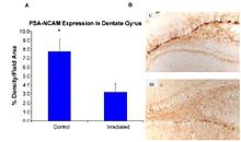
Recent studies provide evidence of the pathogenesis of HZE nuclei in the CNS. The authors of one of these studies  were the first to suggest neurodegeneration with HZE nuclei, as shown in figure 6-1(a). These studies demonstrate that HZE radiation led to the progressive loss of neuronal progenitor cells in the SGZ at doses of 1 to 3 Gy in a dosedependent manner. NCRP Report No. 153  notes that “Mice were irradiated with 1 to 3 Gy of 12C or 56Fe-ions and 9 months later proliferating cells and immature neurons in the dentate SGZ were quantified. The results showed that reductions in these cells were dependent on the dose and LET. Loss of precursor cells was also associated with altered neurogenesis and a robust inflammatory response, as shown in figures 6-1(a) and 6-1(b). These results indicate that high-LET radiation has a significant and long-lasting effect on the neurogenic population in the hippocampus that involves cell loss and changes in the microenvironment. The work has been confirmed by other studies. These investigators noted that these changes are consistent with those found in aged subjects, indicating that heavy-particle irradiation is a possible model for the study of aging.”

Figure 6-1(a). (Panel A) Expression of polysialic acid form of neural cell adhesion molecule (PSA-NCAM) in the hippocampus of rats that were irradiated (IR) with 2.5 Gy of 56Fe high-energy radiation and control subjects as measured by % density/field area measured. (Panel B) PSA-NCAM staining in the dentate gyrus of representative irradiated (IR) and control (C) subjects at 5x magnification.
Figure 6-1(b). Numbers of proliferating cells (left panel) and immature neurons (right panel) in the dentate SGZ are significantly decreased 48 hours after irradiation. Antibodies against Ki-67 and doublecortin (Dcx) were used to detect proliferating cells and immature neurons, respectively. Doses from 2 to 10 Gy significantly (p < 0.05) reduced the numbers of proliferating cells. Immature neurons were also reduced in a dose-dependent fashion (p<0 .001="" an="" and="" animals="" average="" bar="" bars="" div="" each="" error.="" error="" four="" of="" represents="" standard="">
Oxidative damage
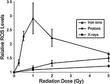
Recent studies indicate that adult rat neural precursor cells from the hippocampus show an acute, dose-dependent apoptotic response that was accompanied by an increase in ROS. Low-LET protons are also used in clinical proton beam radiation therapy, at an RBE of 1.1 relative to megavoltage X rays at a high dose. NCRP Report No. 153 notes that: “Relative ROS levels were increased at nearly all doses (1 to 10 Gy) of Bragg-peak 250 MeV protons at post-irradiation times (6 to 24 hours) compared to unirradiated controls. The increase in ROS after proton irradiation was more rapid than that observed with X rays and showed a well-defined dose response at 6 and 24 hours, increasing about 10-fold over controls at a rate of 3% per Gy. However, by 48 hours post-irradiation, ROS levels fell below controls and coincided with minor reductions in mitochondrial content. Use of the antioxidant alpha-lipoic acid (before or after irradiation) was shown to eliminate the radiation-induced rise in ROS levels. These results corroborate the earlier studies using X rays and provide further evidence that elevated ROS are integral to the radioresponse of neural precursor cells.” Furthermore, high-LET radiation led to significantly higher levels of oxidative stress in hippocampal precursor cells as compared to lower-LET radiations (X rays, protons) at lower doses (≤1 Gy) (figure 6-2). The use of the antioxidant lipoic acid was able to reduce ROS levels below background levels when added before or after 56Fe-ion irradiation. These results conclusively show that low doses of 56Fe-ions can elicit significant levels of oxidative stress in neural precursor cells at a low dose.

Figure 6-2. Dose response for oxidative stress after 56Fe-ion irradiation. Hippocampal precursors that are subjected to 56Fe-ion irradiation were analyzed for oxidative stress 6 hours after exposure. At doses ≤1 Gy a linear dose response for the induction of oxidative stress was observed. At higher 56Fe doses, oxidative stress fell to values that were found using lower-LET irradiations (X rays, protons). Experiments, which represent a minimum of three independent measurements (±SE), were normalized against unirradiated controls set to unity. ROS levels induced after 56Fe irradiation were significantly (P < 0.05) higher than controls.
Neuroinflammation
Neuroinflammation, which is a fundamental reaction to brain injury, is characterized by the activation of resident microglia and astrocytes and local expression of a wide range of inflammatory mediators. Acute and chronic neuroinflammation has been studied in the mouse brain following exposure to HZE. The acute effect of HZE is detectable at 6 and 9 Gy; no studies are available at lower doses. Myeloid cell recruitment appears by 6 months following exposure. The estimated RBE value of HZE irradiation for induction of an acute neuroinflammatory response is three compared to that of gamma irradiation. COX-2 pathways are implicated in neuroinflammatory processes that are caused by low-LET radiation. COX-2 up-regulation in irradiated microglia cells leads to prostaglandin E2 production, which appears to be responsible for radiation-induced gliosis (overproliferation of astrocytes in damaged areas of the CNS).

Behavioral effects

As behavioral effects are difficult to quantitate, they consequently are one of the most uncertain of the space radiation risks. NCRP Report No. 153  notes that: “The behavioral neurosciences literature is replete with examples of major differences in behavioral outcome depending on the animal species, strain, or measurement method used. For example, compared to unirradiated controls, X-irradiated mice show hippocampal-dependent spatial learning and memory impairments in the Barnes maze, but not in the Morris water maze  which, however, can be used to demonstrate deficits in rats. Particle radiation studies of behavior have been accomplished with rats and mice, but with some differences in the outcome depending on the endpoint measured.”

The following studies provide evidence that space radiation affects the CNS behavior of animals in a somewhat dose- and LET-dependent manner.
Sensorimotor effects
Sensorimotor deficits and neurochemical changes were observed in rats that were exposed to low doses of 56Fe-ions. Doses that are below 1 Gy reduce performance, as tested by the wire suspension test. Behavioral changes were observed as early as 3 days after radiation exposure and lasted up to 8 months. Biochemical studies showed that the K+-evoked release of dopamine was significantly reduced in the irradiated group, together with an alteration of the nerve signaling pathways. A negative result was reported by Pecaut et al., in which no behavioral effects were seen in female C57/BL6 mice in a 2- to 8-week period following their exposure to 0, 0.1, 0.5 or 2 Gy accelerated 56Fe-ions (1 GeV/u56Fe) as measured by open-field, rotorod, or acoustic startle habituation.
Radiation-induced changes in conditioned taste aversion
There is evidence that deficits in conditioned taste aversion (CTA) are induced by low doses of heavy ions. The CTA test is a classical conditioning paradigm that assesses the avoidance behavior that occurs when the ingestion of a normally acceptable food item is associated with illness. This is considered a standard behavioral test of drug toxicity. NCRP Report No. 153 notes that: “The role of the dopaminergic system in radiation-induced changes in CTA is suggested by the fact that amphetamine-induced CTA, which depends on the dopaminergic system, is affected by radiation, whereas lithium chloride-induced CTA, which does not involve the dopaminergic system, is not affected by radiation. It was established that the degree of CTA due to radiation is LET-dependent ([figure 6-3]) and that 56Fe-ions are the most effective of the various low and high LET radiation types that have been tested. Doses as low as ~0.2 Gy of 56Fe-ions appear to have an effect on CTA.”

The RBE of different types of heavy particles on CNS function and cognitive/behavioral performance was studied in Sprague-Dawley rats. The relationship between the thresholds for the HZE particle-induced disruption of amphetamine-induced CTA learning is shown in figure 6-4; and for the disruption of operant responding is shown in figure 6-5. These figures show a similar pattern of responsiveness to the disruptive effects of exposure to either 56Fe or 28Si particles on both CTA learning and operant responding. These results suggest that the RBE of different particles for neurobehavioral dysfunction cannot be predicted solely on the basis of the LET of the specific particle.

Figure 6-3. ED50 for CTA as a function of LET for the following radiation sources: 40Ar = argon ions, 60Co = Cobalt-60 gamma rays, e = electrons, 56FE = iron ions, 4He = helium ions, n0 = neutrons, 20Ne = neon ions.
Figure 6-4. Radiation-induced disruption in CTA. This figure shows the relationship between exposure to different energies of 56FE and 28Si particles and the threshold dose for the disruption of amphetamine-induced CTA learning. Only a single energy of 48Ti particles was tested. The threshold dose (cGy) for the disruption of the response is plotted against particle LET (keV/μm).
Figure 6-5.jpg High-LET radiation effects on operant response. This figure shows the relationship between the exposure to different energies of 56Fe and 28Si particles and the threshold dose for the disruption of performance on a food-reinforced operant response. Only a single energy of 48Ti particles was tested. The threshold dose (cGy) for the disruption of the response is plotted against particle LET (keV/μm).
Radiation affect on operant conditioning
Operant conditioning uses several consequences to modify a voluntary behavior. Recent studies by Rabin et al. have examined the ability of rats to perform an operant order to obtain food reinforcement using an ascending fixed ratio (FR) schedule. They found that 56Fe-ion doses that are above 2 Gy affect the appropriate responses of rats to increasing work requirements. NCRP Report No. 153  notes that "The disruption of operant response in rats was tested 5 and 8 months after exposure, but maintaining the rats on a diet containing strawberry, but not blueberry, extract were shown to prevent the disruption. When tested 13 and 18 months after irradiation, there were no differences in performance between the irradiated rats maintained on control, strawberry or blueberry diets. These observations suggest that the beneficial effects of antioxidant diets may be age dependent."
Spatial learning and memory
The effects of exposure to HZE nuclei on spatial learning, memory behavior, and neuronal signaling have been tested, and threshold doses have also been considered for such effects. It will be important to understand the mechanisms that are involved in these deficits to extrapolate the results to other dose regimes, particle types, and, eventually, astronauts. Studies on rats were performed using the Morris water maze test 1 month after whole-body irradiation with 1.5 Gy of 1 GeV/u 56Fe-ions. Irradiated rats demonstrated cognitive impairment that was similar to that seen in aged rats. This leads to the possibility that an increase in the amount of ROS may be responsible for the induction of both radiation- and age-related cognitive deficits.

NCRP Report No. 153 notes that: “Denisova et al. exposed rats to 1.5 Gy of 1 GeV/u56Feions and tested their spatial memory in an eight-arm radial maze. Radiation exposure impaired the rats’ cognitive behavior, since they committed more errors than control rats in the radial maze and were unable to adopt a spatial strategy to solve the maze. To determine whether these findings related to brain-region specific alterations in sensitivity to oxidative stress, inflammation or neuronal plasticity, three regions of the brain, the striatum, hippocampus and frontal cortex that are linked to behavior, were isolated and compared to controls. Those that were irradiated were adversely affected as reflected through the levels of dichlorofluorescein, heat shock, and synaptic proteins (for example, synaptobrevin and synaptophysin). Changes in these factors consequently altered cellular signaling (for example, calciumdependent protein kinase C and protein kinase A). These changes in brain responses significantly correlated with working memory errors in the radial maze. The results show differential brain-region-specific sensitivity induced by 56Fe irradiation ([figure 6-6]). These findings are similar to those seen in aged rats, suggesting that increased oxidative stress and inflammation may be responsible for the induction of both radiation and age-related cognitive deficits.”

Figure 6-6. Brain-region-specific calcium-dependent protein kinase C expression was assessed in control and irradiated rats using standard Western blotting procedures. Values are means ± SEM (standard error of mean).

Acute central nervous system risks

In addition to the possible in-flight performance and motor skill changes that were described above, the immediate CNS effects (i.e., within 24 hours following exposure to low-LET radiation) are anorexia and nausea. These prodromal risks are dose-dependent and, as such, can provide an indicator of the exposure dose. Estimates are ED50 = 1.08 Gy for anorexia, ED50 = 1.58 Gy for nausea, and ED50=2.40 Gy for emesis. The relative effectiveness of different radiation types in producing emesis was studied in ferrets and is illustrated in figure 6-7. High-LET radiation at doses that are below 0.5 Gy show greater relative biological effectiveness compared to low-LET radiation. The acute effects on the CNS, which are associated with increases in cytokines and chemokines, may lead to disruption in the proliferation of stem cells or memory loss that may contribute to other degenerative diseases.

Figure 6-7. LET dependence of RBE of radiation in producing emesis or retching in a ferret. B = bremsstrahlung; e = electrons; P = protons; 60Co = cobalt gamma rays; n0 = neutrons; and 56Fe = iron.

Computer models and systems biology analysis of central nervous system risks

Since human epidemiology and experimental data for CNS risks from space radiation are limited, mammalian models are essential tools for understanding the uncertainties of human risks. Cellular, tissue, and genetic animal models have been used in biological studies on the CNS using simulated space radiation. New technologies, such as three-dimensional cell cultures, microarrays, proteomics, and brain imaging, are used in systematic studies on CNS risks from different radiation types. According to biological data, mathematical models can be used to estimate the risks from space radiation.

Systems biology approaches to Alzheimer’s disease that consider the biochemical pathways that are important in CNS disease evolution have been developed by research that was funded outside NASA. Figure 6-8 shows a schematic of the biochemical pathways that are important in the development of Alzheimer’s disease. The description of the interaction of space radiation within these pathways would be one approach to developing predictive models of space radiation risks. For example, if the pathways that were studied in animal models could be correlated with studies in humans who are suffering from Alzheimer’s disease, an approach to describe risk that uses biochemical degrees-of-freedom could be pursued. Edelstein-Keshet and Spiros  have developed an in silico model of senile plaques that are related to Alzheimer’s disease. In this model, the biochemical interactions among TNF, IL-1B, and IL-6 are described within several important cell populations, including astrocytes, microglia, and neurons. Further, in this model soluble amyloid causes microglial chemotaxis and activates IL-1B secretion. Figure 6-9 shows the results of the Edelstein-Keshet and Spiros model simulating plaque formation and neuronal death. Establishing links between space radiation-induced changes to the changes that are described in this approach can be pursued to develop an in silico model of Alzheimer’s disease that results from space radiation.

Figure 6-8.Molecular pathways important in Alzheimer’s disease. From Kyoto Encyclopedia of Genes and Genomes. Copyrighted image located at http://www.genome.jp/kegg/pathway/hsa/hsa05010.html
 
Figure 6-9. Model of plaque formation and neuronal death in Alzheimer’s disease. From Edelstein-Keshet and Spiros, 2002 : Top row: Formation of a plaque and death of neurons in the absence of glial cells, when fibrous amyloid is the only injurious influence. The simulation was run with no astrocytes or microglia, and the health of neurons was determined solely by the local fibrous amyloid. Shown above is a time sequence (left to right) of three stages in plaque development, at early, intermediate, and advanced stages. Density of fibrous deposit is represented by small dots and neuronal health by shading from white (healthy) to black (dead). Note radial symmetry due to simple diffusion. Bottom row: Effect of microglial removal of amyloid on plaque morphology. Note that microglia (small star-like shapes) are seen approaching the plaque (via chemotaxis to soluble amyloid, not shown). At a later stage, they have congregated at the plaque center, where they adhere to fibers. As a result of the removal of soluble and fibrous amyloid, the microglia lead to irregular plaque morphology. Size scale: In this figure, the distance between the small single dots (representing low-fiber deposits) is 10 mm. Similar results were obtained for a 10-fold scaling in the time scale of neuronal health dynamics.
Other interesting candidate pathways that may be important in the regulation of radiation-induced degenerative CNS changes are signal transduction pathways that are regulated by Cdk5. Cdk5 is a kinase that plays a key role in neural development; its aberrant expression and activation are associated with neurodegenerative processes, including Alzheimer’s disease. This kinase is up-regulated in neural cells following ionizing radiation exposure.

Risks in context of exploration mission operational scenarios

Projections for space missions

Reliable projections of CNS risks for space missions cannot be made from the available data. Animal behavior studies indicate that high-HZE radiation has a high RBE, but the data are not consistent. Other uncertainties include: age at exposure, radiation quality, and dose-rate effects, as well as issues regarding genetic susceptibility to CNS risk from space radiation exposure. More research is required before CNS risk can be estimated.

Potential for biological countermeasures

The goal of space radiation research is to estimate and reduce uncertainties in risk projection models and, if necessary, develop countermeasures and technologies to monitor and treat adverse outcomes to human health and performance that are relevant to space radiation for short-term and career exposures, including acute or late CNS effects from radiation exposure. The need for the development of countermeasures to CNS risks is dependent on further understanding of CNS risks, especially issues that are related to a possible dose threshold, and if so, which NASA missions would likely exceed threshold doses. As a result of animal experimental studies, antioxidant and anti-inflammation are expected to be effective countermeasures for CNS risks from space radiation. Diets of blueberries and strawberries were shown to reduce CNS risks after heavy-ion exposure. Estimating the effects of diet and nutritional supplementation will be a primary goal of CNS research on countermeasures.

A diet that is rich in fruit and vegetables significantly reduces the risk of several diseases. Retinoids and vitamins A, C, and E are probably the most well-known and studied natural radioprotectors, but hormones (e.g., melatonin), glutathione, superoxide dismutase, and phytochemicals from plant extracts (including green tea and cruciferous vegetables), as well as metals (especially selenium, zinc, and copper salts) are also under study as dietary supplements for individuals, including astronauts, who have been overexposed to radiation. Antioxidants should provide reduced or no protection against the initial damage from densely ionizing radiation such as HZE nuclei, because the direct effect is more important than the free-radical-mediated indirect radiation damage at high LET. However, there is an expectation that some benefits should occur for persistent oxidative damage that is related to inflammation and immune responses. Some recent experiments suggest that, at least for acute high-dose irradiation, an efficient radioprotection by dietary supplements can be achieved, even in the case of exposure to high-LET radiation. Although there is evidence that dietary antioxidants (especially strawberries) can protect the CNS from the deleterious effects of high doses of HZE particles, because the mechanisms of biological effects are different at low dose-rates compared to those of acute irradiation, new studies for protracted exposures will be needed to understand the potential benefits of biological countermeasures.

Concern about the potential detrimental effects of antioxidants was raised by a recent meta-study of the effects of antioxidant supplements in the diet of normal subjects. The authors of this study did not find statistically significant evidence that antioxidant supplements have beneficial effects on mortality. On the contrary, they concluded that β-carotene, vitamin A, and vitamin E seem to increase the risk of death. Concerns are that the antioxidants may allow rescue of cells that still sustain DNA mutations or altered genomic methylation patterns following radiation damage to DNA, which can result in genomic instability. An approach to target damaged cells for apoptosis may be advantageous for chronic exposures to GCR.

Individual risk factors

Individual factors of potential importance are genetic factors, prior radiation exposure, and previous head injury, such as concussion. Apolipoprotein E (ApoE) has been shown to be an important and common factor in CNS responses. ApoE controls the redistribution of lipids among cells and is expressed at high levels in the brain. New studies are considering the effects of space radiation for the major isoforms of ApoE, which are encoded by distinct alleles (ε2, ε3, and ε4). The isoform ApoE ε4 has been shown to increase the risk of cognitive impairments and to lower the age for Alzheimer’s disease. It is not known whether the interaction of radiation sensitivity or other individual risks factors is the same for high- and low-LET radiation. Other isoforms of ApoE confer a higher risk for other diseases. People who carry at least one copy of the ApoE ε4 allele are at increased risk for atherosclerosis, which is also suspected to be a risk increased by radiation. People who carry two copies of the ApoE ε2 allele are at risk for a condition that is known as hyperlipoproteinemia type III. It will therefore be extremely challenging to consider genetic factors in a multipleradiation-risk paradigm.

Conclusion

Reliable projections for CNS risks from space radiation exposure cannot be made at this time due to a paucity of data on the subject. Existing animal and cellular data do suggest that space radiation can produce neurological and behavioral effects; therefore, it is possible that mission operations will be impacted. The significance of these results on the morbidity to astronauts has not been elucidated, however. It is to be noted that studies, to date, have been carried out with relatively small numbers of animals (

Selfish brain theory

From Wikipedia, the free encyclopedia
 
The “Selfish Brain” theory describes the characteristic of the human brain to cover its own, comparably high energy requirements with the utmost of priorities when regulating energy fluxes in the organism. The brain behaves selfishly in this respect. The "Selfish brain" theory amongst other things provides a possible explanation for the origin of obesity, the severe and pathological form of overweight. The Luebeck obesity and diabetes specialist Achim Peters developed the fundamentals of this theory between 1998 and 2004. The interdisciplinary “Selfish Brain: brain glucose and metabolic syndrome” research group headed by Peters and supported by the German Research Foundation (DFG) at the University of Luebeck has in the meantime been able to reinforce the basics of the theory through experimental research.

The explanatory power of the Selfish Brain theory

Investigative approach of the Selfish Brain theory

The brain performs many functions for the human organism. Most are of a cognitive nature or concern the regulation of the motor system. A previously lesser investigated aspect of brain activity was the regulation of energy metabolism. The "Selfish Brain" theory shed new light on this function. It states that the brain behaves selfishly by controlling energy fluxes in such a way that it allocates energy to itself before the needs of the other organs are satisfied. The internal energy consumption of the brain is very high. Although its mass constitutes only 2% of the entire body weight, it consumes 20% of the carbohydrates ingested over a 24-hour period. This corresponds to 100 g of glucose per day, or half the daily requirement for a human being. A 30-year-old office worker with a body weight of 75 kg and a height of 1.85 m consumes approx. 200 g glucose per day.

Before now the scientific community assumed that the energy needs of the brain, the muscles and the organs were all met in parallel. The hypothalamus, an area of the upper brainstem, was thought to play a central role in regulating two feedback loops within narrow limits.
  • The "lipostatic theory" established by Gordon C Kennedy in 1953 describes the fat deposition feedback system. The hypothalamus receives signals from circulating metabolic products or hormones about how much adipose tissue there is in the body as well as its prevailing metabolic status. Using these signals the hypothalamus can adapt the absorption of nutrients so that the body’s fat depots remain constant, i.e. a "lipostasis" is achieved.
  • The "glucostatic theory" developed in the same year by Jean Mayer describes the blood glucose feedback system. According to this theory the hypothalamus controls the absorption of nutrients via receptors that measure the glucose level in the blood. In this way a certain glucose concentration is set by adjusting the intake of nutrients. Mayer also included the brain in his calculations. Although he considered that food intake served to safeguard the energy homoeostasis of the central nervous system, he did imply that the energy flux from the body to the brain was a passive process.
On the basis of these theories a number of international research groups still position the origin of obesity in a disorder in one of the two above described feedback systems. However, there are scenarios in weight regulation that can not be explained in this way. For example, upon inanition of the body (e.g. during fasting) almost all the organs such as the heart, liver, spleen and kidneys dramatically lose weight (approx. 40%) and the blood glucose concentration falls. During this time, however, the brain mass hardly changes (less than 2% on average). A further example illustrates the inherent conflict between these two explanatory approaches: although large amounts of the appetite suppressing hormone leptin are released in obese individuals, they are still afflicted with a ravenous hunger once their blood glucose falls.

The "Selfish Brain" theory links in seamlessly with the traditions of the lipo- and glucostatic theories. What is new is that the “Selfish Brain” theory assumes there is another feedback control system that is supraordinate to the blood glucose and fat feedback control systems.

A feedback system is meant here in which the cerebral hemispheres, the integrating organ for the entire central nervous system, control the ATP concentration (adenosine-triposphate - a form of energy currency for the organism) of the neurons (see 3). In this way the cerebral hemispheres ensure the primacy of the brain’s energy supply and are therefore considered in the "Selfish Brain" theory as wings of a central authority that governs energy metabolism. Whenever required the cerebral hemispheres direct an energy flux from the body to the brain to maintain its energy status. In contrast to the ideas of Jean Mayer, the "Selfish Brain" theory assumes an active "Energy on Demand" process. It is controlled by cerebral ATP sensors that react sensitively to changes in ATP in neurons over the entire brain.

The "Selfish Brain" theory combines the theories of Kennedy and Mayer, considering blood glucose and fat feedback control systems as a complex. This regulates the energy flux from the environment to the body, i.e. the intake of nutrients. It is regulated by a hypothalamic nucleus. Here as well there are sensors that record changes in both blood glucose and fat depots, and which activate biochemical processes that maintain a certain body weight.

For achieving their goal of maintaining energy homeostasis in the brain, the cerebral hemispheres depend on subordinate feedback loops, since these loops send signals for energy procurement to their control organ. If these signals are not processed correctly, e.g. due to impairments in the amygdala or hippocampus, the energy supply to the brain will not be endangered, but anomalies such as obesity can still result. The origin of this is not to be found in the blood glucose or fat feedback control systems, but much rather in the regulating instances within the cerebral hemispheres.

Energy procurement by the brain

The brain can cover its energy needs (particularly those of the cerebral hemispheres) either by allocation or nutrient intake. The corresponding signal to the subordinate regulatory system originates in the cerebral hemispheres. The most phylogenetically recent part of the brain is characterized by a high plasticity and a high capacity to learn with this process. It is always able to adapt its regulatory processes by processing responses from the periphery, memorizing the results of individual feedback loops and behaviors, and anticipating any possible build-ups.

Energy procurement by the brain is complicated by three factors. Firstly, the brain always requests energy whenever it is needed. It can only store energy in a very restricted form. Peters therefore refers to this as an "energy on demand" system. Secondly, the brain is almost exclusively dependent on glucose as an ATP-substrate. Lactate and betahydroxybutyric acid can also be considered as substrates, but only under certain conditions, e.g. with considerable stress levels or malnutrition. Thirdly, the brain is separated from the rest of the body’s circulation by the blood-brain-barrier. The blood glucose has to be brought there via a special, insulin-independent transporter.

The healthy and the diseased brain: energy supply through allocation or food intake

Allocation represents the way a healthy brain secures its energy supply when acutely needed. It diverts blood glucose from the periphery and leads it across the blood-brain-barrier. An important role here is played by the stress system, whose neural pathways lead directly to the organs (heart, muscle, adipose tissue, liver, pancreas, etc.) and which also acts indirectly on these organs via the bloodstream by the stress hormones adrenaline and cortisol. This system ensures that the glucose is transported to the brain, and that uptake by the musculature and the adipose tissue is reduced. In order to achieve that, the release of insulin and its effect on organs is halted.

The acute supply of energy to the brain from the intake of nutrients presents problems for the organism. In the event of an emergency food intake is only activated if allocation is insufficient, and must be taken as a sign of disease. In this case the required energy can not be requested from the body, and it can only be taken directly from the environment. This pathology is due to defects lying within the control centers of the brain such as the hippocampus, amygdala and hypothalamus. These may be due to mechanical (tumors, injuries), genetic defects (lacking brain-derived neurotrophic factor (BDNF) receptors or leptin receptors), faulty programming (post-traumatic stress disorder, conditioning of eating behavior, advertising for sweets) or false signals may arise due to the influence of antidepressants, drugs, alcohol, pesticides, saccharin or viruses.

Such disorders can have a negative impact on a number of behavioral types:
  • Eating behavior (eating, drinking)
  • Social behavior (e.g. dealing with conflicts, sexuality)
  • Behavior during food procurement (movement, orientation)
Diseases can then result. The "Selfish Brain” research group has concentrated above all on obesity as a pathology.

The following applies irrespective of the nature of energy provision: the brain never gives up on being selfish. Peters therefore differentiates the healthy from the diseased brain through its ability to compete for its energy requirements even under adverse conditions where there are excessive demands from the body. He contraposes the "selfish brain with high fitness" that can tap the bodies energy reserves even in times of short food supply at the expense of the body mass, and the "selfish brain with low fitness", that is unable to do this, and which instead takes in additional food and bears the risk of developing obesity.

Obesity - a build-up in the supply chain

The "Selfish Brain" theory can be considered as a new way to understand obesity. Disorders in the control centers of the brain such as the hippocampus, amygdala and hypothalamus are thought to underlie this, as outlined above. Whatever the type of disruption that exists, it entails that the energy procurement for the brain is accomplished less by allocation and more by the intake of nutrients even though the muscles have no additional energy requirement. If one imagines the energy supply of the human organism as a supply-chain that passes from the outside world with its numerous options for nutrient intake via the body to the brain as the end user and control organ, then obesity can be considered as being caused by a build-up in this supply-chain. This is characterized by an excessive accumulation of energy in the adipose tissue or blood. An allocation failure is expressed as a weakening of the sympathetic nervous system (SNS). The result is that energy intended for the brain mainly enters buffer storage areas, i.e. the adipose tissue and the musculature. Only a small proportion reaches the brain. In order to cover its huge energy needs the brain commands the individual to consume more food. The accumulation process escalates, and the buffer storage areas are continuously filled up. This leads to the development of obesity. In many cases, at a time which is dependent on an affected individual's personal disposition, obesity can also be overlain by a diabetes mellitus. In such a situation the adipose tissue and musculature can no longer accept any energy, and the energy then accumulates in the blood so that hyperglycemia results.

Work on the "Selfish Brain" theory

The basics of the theory

In 1998 Achim Peters drafted the basic version of the “Selfish Brain" theory and formulated its axioms. In his explanation of the “Selfish Brain” theory he referred to approx. 5000 published citations from classical endocrinology and diabetology and the modern neurosciences, but argued both mathematically (using differential equations) and system theoretically. That was a novel methodological approach for diabetology. The regulation of adenosine tripophoshate content plays a central role (a type of energy currency for the organism) in the brain.

Peters assumes a double feedback structure, where the ATP content in the neurons of the brain is stabilized by measurements from two sensors of differing sensitivity that produce the raw energy request signals. The more sensitive sensor records ATP deficits and induces an allocation signal for glucose that is compensated for by requests from the body. The other less sensitive sensor is only activated with glucose excesses and conveys a signal to halt the brain glucose allocation. The optimal ATP quantity is determined by the balance between these receptor signals.

Peters considers that the stress system also operates according to this double feedback structure, which is also closely related to the supply of glucose to the brain. If an individual is confronted with a stress-inducing stimulus, it responds with an increased central-nervous information processing and along with that an increased glucose requirement in the brain. The hormone cortisol, important for regulating stress reactions, and the hormone adrenaline, important for glucose procurement, are released from the adrenal glands. The amount of cortisol that is released is also determined by a balance between a sensitive and a less sensitive sensor, just as is the case with the control of ATP content. This process is terminated if the stress system returns to a resting state.

This model underlies the axioms for the “Selfish Brain" theory as developed by Peters:
  1. The ATP content in the brain is held constant within tight limits, irrespective of the state of the body
  2. The stress system strives to return to a resting state

Integrative power of the “Selfish Brain" theory

The "Selfish Brain" theory is an integrated concept, since from a methodological standpoint it can be seen as a union of two separate research directions. On the one hand it integrates peripheral metabolism research which investigates how energy metabolism functions through intake of nutrients into the organs of the body. On the other it incorporates the results of the brain metabolism expert Luc Pellerin from the University of Lausanne, who found that the neurons in the brain are supplied with energy via their neighboring astrocytes whenever required. This requirement oriented principle for the nerve cells is termed "Energy on demand".

With this approach the "Selfish Brain" theory recognizes the description of two ends of a supply chain. The brain doesn’t just control the supply chain, but it is also its end consumer, and not the body through which the supply chain passes. The priority of the brain implies that the regulation of energy supply in a human organism is accomplished by the demand rather than the supply principle: Energy is ordered when it is needed.
Fig. 1: Energy supply chain of the "Selfish Brain".
If the ATP concentration drops in the nerve cells of the brain, a cerebral mechanism is (pull 1) set in motion which increases the energy flux directed from the body to the brain according to the "Energy on demand" principle. (solid arrows show stimulation, interrupted arrows inhibition; yellow means: "belongs to the controlling brain parts "). If the energy content in the body falls (blood, adipose tissue), the falling glucose and the falling adipose tissue hormone leptin induce another cerebral mechanism (pull 2). This entails that more energy is absorbed from the immediate environment into the body (ingestion behavior). When the available supplies in the immediate vicinity disappear, a further cerebral mechanism (pull 3) initiates moving and exploration, i.e. foraging for food. The glucostatic and the lipostatic theories describe the second step in this supply chain (area with dark grey background). The "Selfish Brain" theory links to the two traditional theories and expands them by considering the brain as an end consumer in a continuous supply chain (light gray)

The founding of the "Selfish Brain" research group

After the axioms were formulated in 1998 Achim Peters sought experts in other specialties to develop his "Selfish Brain" theory further. Already at an early stage he had matched up his ideas with the views of other leading international scientists. Amongst them was the Swiss brain metabolism specialist Luc Pellerin, the renowned obesity expert Denis G. Baskin, the internationally famous stress researcher Mary Dallman and the renowned neurobiologist Larry W. Swanson. At the University of Luebeck Achim Peters compared his findings with the well-known neuroendocrinologist Prof. Dr. Horst Lorenz Fehm. A year later in 1999 an intensive collaboration was started with the psychiatrist and psychotherapist Prof. Dr. Ulrich Schweiger who also worked at the University of Luebeck.

In 2004 the interdisciplinary research group: "Selfish Brain: brain glucose and metabolic syndrome" supported by the German Research Foundation (DFG) was officially founded. Achim Peters was appointed to a professorship that was especially created for the group. He also succeeded in winning over additional reputable scientists for the project, including Prof. Dr. Rolf Hilgenfeld, an eminent SARS expert and the developer of one of the first inhibitors of the virus. At this time the research group consists of 18 scientific subproject investigators from a number of specialties including internal medicine, psychiatry, neurobiology, molecular medicine and mathematics. The advisory committee includes Professors Luc Pellerin, Denis Baskin and Mary Dallman under its ranks.

"Train the brain": a therapy of obesity based on the "Selfish Brain" theory

According to the “Selfish Brain” theory obesity can also be attributed to psychological causes. Poor coping strategies in stress situations represent one of these. An association was found between the tendency to evade conflict, and the habit of reducing psychological stress by immediately consuming sweets. The direct supply of glucose circumvents the glucose procurement from the body that would otherwise occur with a normal allocation process following the release of the stress hormone adrenaline. An existing allocation problem with obesity can be made even worse by such bad behavior. The stress system can also be weakened further because it may forget how to react autonomously.

These relationships have led to the development of an innovative multidisciplinary psychiatric and internal medical program at the University of Luebeck for obesity therapy. Prof. Dr. Ulrich Schweiger of the Clinic for Psychiatry and Psychotherapy led by Prof. Dr. F. Hohagen has been a key player in this development. In close cooperation with Schweiger, the internist Achim Peters derived a therapeutic concept from the “Selfish Brain” theory that was fixed on both feelings and coordinated behavior emanating from the brain. The aim of this therapy is to modify the settings and behaviors coded in the emotional memory centers of the brain that have become habit. "Train the Brain" is the catchphrase describing these therapeutic measures that may be enabled by the unusual plasticity and learning-capacity of the brain. It might just simply involve the practicing of eating behaviors that can be tolerated from a health perspective, and combining this with a reduction in detrimental habits. However, it could also involve the modification of behaviors associated with the handling of conflicts and other stress situations. According to the view of the “Selfish Brain” research group, if defective allocation is compensated for chronically by immediately consuming foodstuffs, a risk arises that eating will become the only reaction to a situation that requires a considerably more complex social behavior. The therapy of obesity therefore has both a physiological and a psychological component: It is not just the ability to allocate that must be restored, but actions and behaviors in everyday life.

Experimental evidence─ the theory’s scope of validity

In the first DFG funding period from 2004 to 2007 researchers from the Clinical Research Group “Selfish Brain: brain glucose and metabolic syndrome" expanded the scope of validity of the “Selfish Brain" theory in central aspects by carrying out experiments on healthy and diseased test subjects. The researchers in Luebeck found the following key results regarding the axioms of the theory:
  • The brain maintains its own glucose content "selfishly"
  • The brain is always supplied with a greater energy share than the body in extreme stress situations
  • In overweight individuals the brain’s energy distribution mechanism is disrupted
  • With chronic stress loads the energy flux between the brain and the body is diverted, a phenomenon that leads to the development of overweight
  • Nerve cells record their ATP content using two sensors of differing sensitivity
  • The resting state of the stress system is fine-tuned with the help of two cortisol receptors of differing sensitivity
The special position of the brain during inanition (due to fasting or tumor disease) was already confirmed experimentally over 80 years ago: The body mass reduces, but the mass of the brain hardly reduces, if at all (see 3). Recently this axiom of the selfish brain theory was supported by work at the University of Luebeck involving state-of-the-art magnetic resonance procedures, e.g. during metabolic stress. The ATP content in the brain and musculature of test subjects was examined by a magnetic resonance technique while either an energy deficit or surplus was induced in the blood by insulin or glucose injection. In both situations a sufficiently high ATP-concentration was measured in the brain. The measured high-energy-rich substances changed throughout to the benefit of the brain and to the disadvantage of the body cells. The glucose-supply of the brain had priority despite the physical stress that was being endured.

Some of the results were presented at the international congress organized by the "Selfish Brain” research group at the 23 and 24 February 2006 in Luebeck as well as at a press conference aimed at both specialists and the wider public.

In the second funding period that has been running since the end of 2007, the clarification of the following questions has now become the focus of interest:
  • How does the reward system of the "Selfish Brain" function and how does it lead amongst obese individuals to a faulty programming of energy management?
  • How can the redirection of metabolic fluxes be learned and trained?
  • How does "comfort feeding" affect stress reactions?
  • How is the glucose requirement of the brain increased in stress situations?
  • What does the molecular supply chain with which brain cells request glucose when needed look like?
  • Can viruses block this supply chain for the brain cells?

Year On

From Wikipedia, the free encyclopedia https://en.wikipedia.org/wiki/Year_On T...