Search This Blog

Friday, September 17, 2021

Automated insulin delivery systems

From Wikipedia, the free encyclopedia
https://en.wikipedia.org/wiki/Automated_insulin_delivery_systems

Automated insulin delivery systems are automated (or semi-automated) systems designed to assist people with diabetes, primarily type 1, by automatically adjusting insulin delivery to help them control their blood glucose levels. Currently available systems (as of October, 2020) can only deliver (and regulate delivery of) a single hormone- insulin. Other systems currently in development aim to improve on current systems by adding one or more additional hormones that can be delivered as needed, providing something closer to the endocrine functionality of a healthy pancreas.

The endocrine functionality of the pancreas is provided by islet cells which produce the hormones insulin and glucagon. Artificial pancreatic technology mimics the secretion of these hormones into the bloodstream in response to the body's changing blood glucose levels. Maintaining balanced blood sugar levels is crucial to the function of the brain, liver, and kidneys. Therefore, for type 1 patients, it is necessary that the levels be kept balanced when the body cannot produce insulin itself.

Automated insulin delivery systems are often referred to using the term artificial pancreas, but the term has no precise, universally accepted definition. For uses other than automated insulin delivery, see Artificial pancreas (disambiguation).

General Overview

History

The first automated insulin delivery system was known as the Biostator.

Classes of AID systems

Currently available AID systems fall into four broad classes based on their capabilities. The first systems released- suspend systems- can only halt insulin delivery. Loop systems can modulate delivery both up and down.

Threshold suspend

Threshold suspend systems are the simplest form of insulin delivery automation. They halt the constant flow of insulin from a pump (known as basal insulin) when a connected CGM reports a glucose level below a pre-set threshold. Halting basal delivery stops the normal preprogrammed rate of delivery, but it cannot remove insulin that has already been infused, so the overall efficacy of threshold suspend systems is limited due to the relatively slow pharmacokinetics of insulin delivered subcutaneously.

Predictive Low Glucose Suspend

A step forward from threshold suspend systems, Predictive Low Glucose Suspend (PLGS) systems use a mathematical model to extrapolate predicted future blood sugar levels based on recent past readings from a CGM. This allows the system to halt insulin delivery as much as 30 minutes prior to a predicted hypoglycemic event, allowing addition time for the slow pharmacokinetics of insulin to reflect that delivery has been halted.

Hybrid Closed Loop

Hybrid Closed Loop (HCL) systems further expand on the capabilities of PGLS systems by adjusting basal insulin delivery rates both up and down in response to values from a continuous glucose monitor. Through this modulation of basal insulin, the system is able to reduce the magnitude and duration both hyperglycemic and hypoglycemic events.

Advanced Hybrid Closed Loop

In addition to modulating basal insulin, Advanced Hybrid Closed Loop systems have the ability to deliver boluses of insulin to correct for elevated blood sugar.

Required Components

An automated insulin delivery system consists of three distinct components: a continuous glucose monitor to determine blood sugar levels, a pump to deliver insulin, and an algorithm that uses the data from the CGM to send commands to the pump. In the United States, the Food and Drug Administration (FDA) allows each component to be approved independently, allowing for more rapid approvals and incremental innovation. Each component is discussed in greater detail below.

Continuous Glucose Monitor (CGM)

Artificial pancreas feedback system

Continuous glucose monitors (CGMs) are medical devices which extrapolate an estimate of the glucose concentration in a patient's blood based on the level of glucose present in the subcutaneous interstitial fluid. A thin, biocompatible sensor wire coated with a glucose-reactive enzyme is inserted into the skin, allowing the system to read the voltage generated, and based on it, estimate blood glucose. The biggest advantage of a CGM over a traditional fingerstick blood glucose meter is that the CGM can take a new reading as often as every 60 seconds (although most only take a reading every 5 minutes), allowing for a sampling frequency that is able to provide not just a current blood sugar level, but a record of past measurements; allowing computer systems to project past short-term trends into the future, showing patients where their blood sugar levels are likely headed.

Early CGMs were not particularly accurate, but were still useful for observing and recording overall trends and provide warnings in the event of rapid changes in blood glucose readings.

Continuous blood glucose monitors are one of the set of devices that make up an artificial pancreas device system, the other being an insulin pump, and a glucose meter to calibrate the device. Continuous glucose monitors are a more recent breakthrough and have begun to hit the markets for patient use after approval from the FDA. Both the traditional and the continuous monitor require manual insulin delivery or carbohydrate intake depending on the readings from the devices. While the traditional blood glucose meters require the user to prick their finger every few hours to obtain data, continuous monitors use sensors placed just under the skin on the arm or abdomen to deliver blood sugar level data to receivers or smartphone apps as often as every few minutes. The sensors can be used for up to fourteen days. A number of different continuous monitors are currently approved by the FDA.

The first continuous glucose monitor (CGM) was approved in December 2016. Developed by Dexcom, the G5 Mobile Continuous Monitoring System requires users to prick their fingers twice a day (as opposed to the typical average 8 times daily with the traditional meters) in order to calibrate the sensors. The sensors last up to seven days. The device uses Bluetooth technology to warn the user either through a handheld receiver or app on a smartphone if blood glucose levels reach below a certain point. The cost for this device excluding any co-insurance is an estimated $4,800 a year.

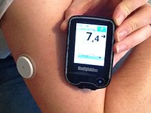
The white sensor is fixed to the upper arm and scanned with the reader. The reader is showing (top to bottom) days to replacement of sensor (11), current BG (7,4) & change (-> i.e. steady) and a diagram of the latest BG levels.
Blood glucose meter FreeStyle Libre from Abbott.

Abbott Laboratories' FreeStyle Libre CGM was approved in September 2017. Recently, the technology was modified to support smartphone use through the LibreLink app. This device does not require finger pricks at all and the sensor, placed on the upper arm, lasts 14 days. The estimated cost for this monitor is $1,300 a year.

Dexcom's next G6 model CGM was approved in March 2018, which can last up to ten days and does not need finger prick calibration. Like Medtronic's monitor, it can predict glucose level trends. It is compatible for integration into insulin pumps.

Control Algorithm

Insulin Pump

Currently Available Systems

Do-It-Yourself

Commercial

MiniMed 670G

In September 2016, the FDA approved the Medtronic MiniMed 670G, which was the first approved hybrid closed loop system. The device senses a diabetic person's basal insulin requirement and automatically adjusts its delivery to the body. It is made up of a continuous glucose monitor, an insulin pump, and a glucose meter for calibration. It automatically functions to modify the level of insulin delivery based on the detection of blood glucose levels by continuous monitor. It does this by sending the blood glucose data through an algorithm that analyzes and makes the subsequent adjustments. The system has two modes. Manual mode lets the user choose the rate at which basal insulin is delivered. Auto mode regulates basal insulin levels from the continuous monitor's readings every five minutes.

The device was originally available only to those aged 14 or older, and in June 2018 was approved by the FDA for use in children aged 7–14. Families have reported better sleep quality from use of the new system, as they do not have to worry about manually checking blood glucose levels during the night. The full cost of the system is $3700, but patients have the opportunity to get it for less.

Systems in Development

Ilet Bionic Pancreas

A team at Boston University working in collaboration with Massachusetts General Hospital on a dual hormone artificial pancreas system began clinical trials on their device called the Bionic Pancreas in 2008. In 2016, the Public Benefit Corporation Beta Bionics was formed. In conjunction with the formation of the company, Beta Bionics changed the preliminary name for their device from the Bionic Pancreas to the iLet. The device uses a closed-loop system to deliver both insulin and glucagon in response to sensed blood glucose levels. While not yet approved for public use, the 4th generation iLet prototype, presented in 2017, is around the size of an iPhone, with a touchscreen interface. It contains two chambers for both insulin and glucagon, and the device is configurable for use with only one hormone, or both. While trials continue to be run, the iLet has a projected final approval for the insulin-only system in 2020.

Inreda Diabetic

In collaboration with the Academic Medical Centre (AMC) in Amsterdam, Inreda is developing a closed loop system with insulin and glucagon. The initiator, Robin Koops, started to develop the device in 2004 and ran the first tests on himself. After several highly successful trials it received the European EC license in 2016. The product is expected to market in the second half of 2020. A smaller improved version is scheduled for 2023.

Approaches

Medical equipment

The medical equipment approach involves combining a continuous glucose monitor and an implanted insulin pump that can function together with a computer-controlled algorithm to replace the normal function of the pancreas. The development of continuous glucose monitors has led to the progress in artificial pancreas technology using this integrated system.

Closed-loop systems

Unlike the continuous sensor alone, the closed-loop system requires no user input in response to reading from the monitor; the monitor and insulin pump system automatically delivers the correct amount of hormone calculated from the readings transmitted. The system is what makes up the artificial pancreas device.

Current studies

Four studies on different artificial pancreas systems are being conducted starting in 2017 and going into the near future. The projects are funded by the National Institute of Diabetes and Digestive and Kidney Diseases, and are the final part of testing the devices before applying for approval for use. Participants in the studies are able to live their lives at home while using the devices and being monitored remotely for safety, efficacy, and a number of other factors.

The International Diabetes Closed-Loop trial, led by researchers from the University of Virginia, is testing a closed-loop system called inControl, which has a smartphone user interface. 240 people of ages 14 and up are participating for 6 months.

A full-year trial led by researchers from the University of Cambridge started in May 2017 and has enrolled an estimated 150 participants of ages 6 to 18 years. The artificial pancreas system being studied uses a smartphone and has a low glucose feature to improve glucose level control.

The International Diabetes Center in Minneapolis, Minnesota, in collaboration with Schneider Children's Medical Center of Israel, are planning a 6-month study that will begin in early 2019 and will involve 112 adolescents and young adults, ages 14 to 30. The main object of the study is to compare the current Medtronic 670G system to a new Medtronic-developed system. The new system has programming that aims to improve glucose control around mealtime, which is still a big challenge in the field.

The current 6-month study lead by the Bionic Pancreas team started in mid-2018 and enrolled 312 participants of ages 18 and above.

Physiological

The Bio-artificial pancreas: this diagram shows a cross section of bio-engineered tissue with encapsulated islet cells which deliver endocrine hormones in response to glucose.

The biotechnical company Defymed, based in France, is developing an implantable bio-artificial device called MailPan which features a bio-compatible membrane with selective permeability to encapsulate different cell types, including pancreatic beta cells. The implantation of the device does not require conjunctive immuno-suppressive therapy because the membrane prevents antibodies of the patient from entering the device and damaging the encapsulated cells. After being surgically implanted, the membrane sheet will be viable for years. The cells that the device holds can be produced from stem cells rather than human donors, and may also be replaced over time using input and output connections without surgery. Defymed is partially funded by JDRF, formerly known as the Juvenile Diabetes Research Foundation, but is now defined as an organization for all ages and all stages of type 1 diabetes.

In November 2018, it was announced that Defymed would partner with the Israel-based Kadimastem, a bio-pharmaceutical company developing stem-cell based regenerative therapies, to receive a two-year grant worth approximately $1.47 million for the development of a bio-artificial pancreas that would treat type 1 diabetes. Kadimastem's stem cell technology uses differentiation of human embryonic stem cells to obtain pancreatic endocrine cells. These include insulin-producing beta cells, as well as alpha cells, which produce glucagon. Both cells arrange in islet-like clusters, mimicking the structure of the pancreas. The aim of the partnership is to combine both technologies in a bio-artificial pancreas device, which releases insulin in response to blood glucose levels, to bring to clinical trial stages.

The San Diego, California based biotech company ViaCyte has also developed a product aiming to provide a solution for type 1 diabetes which uses an encapsulation device made of a semi-permeable immune reaction-protective membrane. The device contains pancreatic progenitor cells that have been differentiated from embryonic stem cells. After surgical implantation in an outpatient procedure, the cells mature into endocrine cells which arrange in islet-like clusters and mimic the function of the pancreas, producing insulin and glucagon. The technology advanced from pre-clinical studies to FDA approval for phase 1 clinical trials in 2014, and presented two-year data from the trial in June 2018. They reported that their product, called PEC-Encap, has so far been safe and well tolerated in patients at a dose below therapeutic levels. The encapsulated cells were able to survive and mature after implantation, and immune system rejection was decreased due to the protective membrane. The second phase of the trial will evaluate the efficacy of the product. ViaCyte has also been receiving financial support from JDRF on this project.

Initiatives around the globe

In the United States in 2006, JDRF (formerly the Juvenile Diabetes Research Foundation) launched a multi-year initiative to help accelerate the development, regulatory approval, and acceptance of continuous glucose monitoring and artificial pancreas technology.

Grassroots efforts to create and commercialize a fully automated artificial pancreas system have also arisen directly from patient advocates and the diabetes community. Bigfoot Biomedical, a company founded by parents of children with T1D have created algorithms and are developing a closed loop device that monitor blood sugar and appropriately provide insulin.

Sexual motivation and hormones

From Wikipedia, the free encyclopedia

Sexual motivation is influenced by hormones such as testosterone, estrogen, progesterone, oxytocin, and vasopressin. In most mammalian species, sex hormones control the ability and motivation to engage in sexual behaviours.

Measuring sexual motivation

Sexual motivation can be measured using a variety of different techniques. Self-report measures, such as the Sexual Desire Inventory, are commonly used to detect levels of sexual motivation in humans. Self-report techniques such as the bogus pipeline can be used to ensure individuals do not falsify their answers to represent socially desirable results. Sexual motivation can also be implicitly examined through frequency of sexual behaviour, including masturbation.

Hormones

Testosterone

According to data from the Journal of Zhejiang University–Science, male testosterone levels exhibit a rhythm that corresponds to recent sexual activity.

Testosterone appears to be a major contributing factor to sexual motivation in male primates, including humans. The elimination of testosterone in adulthood has been shown to reduce sexual motivation in both male humans and male primates. Male humans who had their testicular function suppressed with a GnRH antagonist displayed decreases in sexual desire and masturbation two weeks following the procedure. Research from male rhesus monkeys suggests testosterone functions to increase sexual motivation, thereby motivating males to compete for access to sexual partners. It is postulated that the motivating effects of testosterone in male rhesus monkeys promotes successful sexual competition and may be particularly important motivating tools for low ranking males. It is important to note that elimination of testosterone in primates does not reduce the ability to copulate; rather, it reduces the motivation to copulate.

Testosterone levels in males have been shown to vary according to the ovulating state of females. Males who were exposed to scents of ovulating women recorded higher testosterone levels than males who were exposed to scents of nonovulating women. Being exposed to female ovulating cues may increase testosterone, which in turn may increase males' motivation to engage in, and initiate, sexual behaviour. Ultimately, these higher levels of testosterone may increase the reproductive success of males exposed to female ovulation cues.

The relationship between testosterone and female sexual motivation is somewhat ambiguous. Research suggests androgens, such as testosterone, are not sufficient by themselves to prompt sexual motivation in females. In particular, studies with rhesus macaques have observed testosterone was not significantly associated with variations in level of sexual motivation in females. However, some research with nonhuman primates suggests a role for androgens in female sexual behaviour. Adrenalectomized female rhesus monkeys displayed diminished female sexual receptivity. Later studies revealed this diminished sexual receptivity was specific to the elimination of androgens that can be converted to estrogen.

It is also suggested that levels of testosterone are related to the type of relationship in which one is involved. Men involved in polyamorous relationships display higher levels of testosterone than men involved in either a single partner relationship or single men. Polyamorous women have both higher levels of testosterone and score higher on measures of sexual desire than women who are single or women who are in single-partner relationships.

Estrogens and progesterone

Estrogens and progesterone typically regulate motivation to engage in sexual behaviour for females in mammalian species, though the relationship between hormones and female sexual motivation is not as well understood. In particular, estrogens have been shown to correlate positively with increases in female sexual motivation, and progesterone has been associated with decreases in female sexual motivation. The periovulatory period of the female menstrual cycle is often associated with increased female receptivity and sexual motivation. During this stage in the cycle, estrogens are elevated in the female and progesterone levels are low. At this time, mating is more likely to result in female pregnancy.

Females at different stages of their menstrual cycle have been shown to display differences in sexual attraction. Heterosexual females not using birth control pills who are ovulating (high levels of estrogens) have a preference for the scent of males with low levels of fluctuating asymmetry. Ovulating heterosexual females also display preferences toward masculine faces and report greater sexual attraction to males other than their current partner. From an evolutionary perspective, increases in estrogens during fertile periods in females may direct sexual motivation toward males with preferential genes (the good genes hypothesis).

Following natural or surgically induced menopause, many women experience declines in sexual motivation. Menopause is associated with a rapid decline of estrogen, as well as a steady rate of decline of androgens. The decline of estrogen and androgen levels is believed to account for the lowered levels of sexual desire and motivation in postmenopausal women, although the direct relationship is not well understood.

In her memoir She's Not There: A Life in Two Genders, transgender woman Jennifer Finney Boylan wrote that taking estrogens and antiandrogens profoundly diminished her libido, and in transgender woman Julia Serano's memoir Whipping Girl: A Transsexual Woman on Sexism and the Scapegoating of Femininity, Serano wrote, in a section of her book she described as limited to hormonal changes that she said are experienced by many trans women she has spoken with, that a sharp decrease in her sex drive was the first thing she noticed when she started taking estrogens and antiandrogens.

Oxytocin and vasopressin

The hormones oxytocin and vasopressin are implicated in regulating both male and female sexual motivation. Oxytocin is released at orgasm and is associated with both sexual pleasure and the formation of emotional bonds. Based on the pleasure model of sexual motivation, the increased sexual pleasure that occurs following oxytocin release may encourage motivation to engage in future sexual activities. Emotional closeness can be an especially strong predictor of sexual motivation in females and insufficient oxytocin release may subsequently diminish sexual arousal and motivation in females.

High levels of vasopressin can lead to decreases in sexual motivation for females. A link between vasopressin release and aggression has been observed in females, which may impair female sexual arousal and sexual motivation by leading to feelings of neglect and hostility toward a sexual partner. In males, vasopressin is involved in the arousal phase. Vasopressin levels have been shown to increase during erectile response in male sexual arousal, and decrease back to baseline following ejaculation. The increase of vasopressin during erectile response may be directly associated with increased motivation to engage in sexual behaviour.

Nonprimate species

The hormonal influences of sexual motivation are much more clearly understood for nonprimate females. Suppression of estrogen receptors in the ventromedial nucleus of the hypothalamus in female rats has been observed to reduce female proceptivity and receptivity. Proceptivity and receptivity in the female rat are indicators of sexual motivation, thus indicating a direct relationship between estrogen levels and sexual motivation. In addition, female rats receiving doses of estrogen and progesterone were more likely to exert effort at gaining sexual attention from a male rat. The willingness of the female rats to access males was considered a direct measure of the females' levels of sexual motivation.

An increase in vasopressin has been observed in female rats which have just given birth. Vasopressin is associated with aggressive and hostile behaviours, and is postulated to decrease sexual motivation in females. Vasopressin administered in the female rat brain has been observed to result in an immediate decrease in sexual motivation.

Sexual orientation

Little research has been conducted on the effect of hormones on sexual motivation for same-sex sexual contact. One study observed the relationship between sexual motivation in lesbian and bisexual women and period-related changes in circulating estrogen concentrations. Lesbian women who were at the estrogen peak of their fertile cycle reported increased sexual motivation for sexual contact with women, whereas bisexual women reported only a slight increase in same-sex motivated sexual contact during peak estrogen levels.

Both lesbian and bisexual women showed decreases in sexual motivation for other-sex sexual contact at peak estrogen levels, with greater changes in the bisexual group than the lesbian group.

Clinical research

Men

  • Testosterone is critical for sexual desire, function, and arousal in men. Aromatization of testosterone into the estrogen estradiol appears to be partially responsible for the effects of testosterone on sexual desire and function in men. 5α-Reduction of testosterone into the more potent androgen dihydrotestosterone (DHT) may have a small contribution to the effects of testosterone on sexual desire and function in men. Based on animal research, metabolites of DHT including the neurosteroids and weak estrogens 3α-androstanediol and 3β-androstanediol may be involved in sexual function in men.
  • Men experience sexual dysfunction at testosterone levels of below 300 ng/dL, with men that have levels of testosterone of approximately 200 ng/dL often experiencing such problems. Complete loss of testicular testosterone production resulting in testosterone levels within the castrate range (95% decrease, to 15 ng/dL on average) with surgical or medical castration causes profound sexual dysfunction in men. Combined marked suppression of testicular testosterone production resulting in testosterone levels of just above the castrate/female range (70 to 80% decrease, to 100 ng/dL on average) and marked androgen receptor antagonism with high-dosage cyproterone acetate monotherapy causes profound sexual dysfunction in men. Treatment of men with medical castration and add-back of multiple dosages of testosterone to restore testosterone levels (to a range of about 200 to 900 ng/dL) showed that testosterone dose-dependently restored sexual desire and erectile function in men. High-dosage monotherapy with an androgen receptor antagonist such as bicalutamide or enzalutamide, which preserves testosterone and estradiol levels, has a minimal to moderate negative effect on sexual desire and erectile function in men in spite of strong blockade of the androgen receptor.
  • Estradiol supplementation maintains greater sexual desire in men with surgical or medical castration. High-dose estrogen therapy, which results in marked or complete suppression of testicular testosterone production such that testosterone levels are within the castrate range (95% decrease, to less than 50 ng/dL), causes decreased sexual desire and function. However, sexual function and activity appear to be significantly better with high-dose estrogen therapy than with surgical castration. Treatment of men with medical castration and add-back testosterone to restore testosterone levels, with or without the aromatase inhibitor anastrozole, showed that prevention of the conversion of testosterone into estradiol partially prevented restoration of sexual desire and erectile dysfunction by testosterone in men. However, this was not the case in another study with a similar design that used the aromatase inhibitor testolactone. Men with aromatase deficiency and estrogen insensitivity syndrome, and hence estrogen deficiency, appear to have normal sexual desire, function, and activity. However, estradiol supplementation in some men with aromatase deficiency increased sexual desire and activity but not in other men with aromatase deficiency. Treatment with the antiestrogenic selective estrogen receptor modulator (SERM) tamoxifen has been found to decrease sexual desire in men treated with it for male breast cancer. However, other studies have not found or reported decreased sexual function in men treated with SERMs including tamoxifen, clomifene, raloxifene, and toremifene.
  • 5α-Reductase inhibitors, which block the conversion of testosterone into DHT, result in a slightly increased risk of sexual dysfunction with an incidence of decreased libido and erectile dysfunction of about 3 to 16%. Treatment of healthy men with multiple dosages of testosterone enanthate, with or without the 5α-reductase inhibitor dutasteride, showed that dutasteride did not significantly influence changes in sexual desire and function. Treatment of men with high-dosage bicalutamide therapy, with or without the 5α-reductase inhibitor dutasteride, showed that dutasteride did not significantly influence sexual function. Combined high-dosage bicalutamide therapy plus dutasteride showed less sexual dysfunction than medical castration similarly to high-dosage bicalutamide monotherapy.
  • Treatment of men with very high-dosage DHT (a non-aromatizable androgen), which resulted in an increase in DHT levels by approximately 10-fold and complete suppression of testosterone and estradiol levels, showed that none of the measures of sexual function were significantly changed with the exception of a mild but significant decrease in sexual desire. Treatment of hypogonadal men with the aromatizable testosterone undecanoate and the non-aromatizable mesterolone showed that testosterone undecanoate produced better improvements in mood, libido, erection, and ejaculation than did mesterolone. However, the dosage of mesterolone could have been suboptimal.

Women

  • Estradiol seems to be the most important hormone for sexual desire in women. Periovulatory levels of estradiol increase sexual desire in women. Based on animal research, progesterone may also be involved in sexual function in women. Very limited clinical research suggests that progesterone does not increase sexual desire and may decrease it. There is little support for the notion that physiological levels of testosterone are important for sexual desire in women, although supraphysiological levels of testosterone can increase sexual desire in women similarly to the high levels in men.
  • There is little to no correlation between total testosterone levels within the normal physiological range and sexual desire in premenopausal women. Sexual desire is not increased in women with polycystic ovary syndrome (PCOS) in spite of high testosterone levels. Women with PCOS actually experience an improvement in sexual desire following treatment of their condition, likely due improved psychological functioning (e.g., body image).
  • Sexual desire is not decreased in women with complete androgen insensitivity syndrome (CAIS) relative to unaffected women in spite of a completely non-functional androgen receptor. Sexual desire is increased or unchanged in most women taking a combined birth control pill. This is in spite of the fact that almost all combined birth control pills contain the potently hepatotropic estrogen ethinylestradiol, and the typical doses of ethinylestradiol present in combined birth control pills increase sex hormone-binding globulin (SHBG) levels by 2- to 4-fold and consequently decrease free testosterone levels by 40 to 80%. However, there are some conflicting reports on the effects of combined birth control pills on sexual function in women. Progestogen-only birth control, such as with depot medroxyprogesterone acetate or the etonogestrel birth control implant, has shown mixed effects on sexual desire and function. Androgen receptor antagonists such as flutamide and bicalutamide cause little to no decrease in sexual desire in women.
  • Low dosages of testosterone that result in physiological levels of testosterone (< 50 ng/dL) do not increase sexual desire in women. High dosages of testosterone that result in supraphysiological levels of testosterone (> 50 ng/dL) significantly increase sexual desire in women, with levels of testosterone of 80 to 150 ng/dL "slightly" increasing sexual desire. Further higher dosages of testosterone may result in greater effects on sexual desire in women. High dosages of testosterone (with levels of > 50 ng/dL) have a risk of masculinization (e.g., acne, hair growth, voice changes) with long-term therapy in women. High dosages of testosterone but not low dosages of testosterone enhance the effects of low dosages of estrogens on sexual desire. Tibolone, a combined estrogen, progestin, and androgen, may increase sex drive to a greater extent than standard estrogen–progestogen therapy in postmenopausal women.

Transgender individuals

  • Testosterone therapy increases sexual desire and arousal in transgender men. Estradiol and antiandrogen therapy decreases sexual desire and arousal in transgender women. However, treatment with estradiol in transgender women who have undergone surgical castration appears to maintain significantly greater sexual desire and activity than would be expected for surgical castration alone.

See also


Endocrine system

From Wikipedia, the free encyclopedia
 
Endocrine system
Endocrine English.svg
Main glands of the endocrine system
Details
Identifiers
LatinSystema endocrinum
MeSHD004703
FMA9668

The endocrine system is a messenger system comprising feedback loops of the hormones released by internal glands of an organism directly into the circulatory system, regulating distant target organs. In vertebrates, the hypothalamus is the neural control center for all endocrine systems. In humans, the major endocrine glands are the thyroid gland and the adrenal glands. The study of the endocrine system and its disorders is known as endocrinology.

Glands that signal each other in sequence are often referred to as an axis, such as the hypothalamic-pituitary-adrenal axis. In addition to the specialized endocrine organs mentioned above, many other organs that are part of other body systems have secondary endocrine functions, including bone, kidneys, liver, heart and gonads. For example, the kidney secretes the endocrine hormone erythropoietin. Hormones can be amino acid complexes, steroids, eicosanoids, leukotrienes, or prostaglandins.

The endocrine system can be contrasted to both exocrine glands, which secrete hormones to the outside of the body, and paracrine signalling between cells over a relatively short distance. Endocrine glands have no ducts, are vascular, and commonly have intracellular vacuoles or granules that store their hormones. In contrast, exocrine glands, such as salivary glands, sweat glands, and glands within the gastrointestinal tract, tend to be much less vascular and have ducts or a hollow lumen. Endocrinology is a branch of internal medicine.

Structure

Major endocrine systems

The human endocrine system consists of several systems that operate via feedback loops. Several important feedback systems are mediated via the hypothalamus and pituitary.

Glands

Endocrine glands are glands of the endocrine system that secrete their products, hormones, directly into interstitial spaces and then absorbed into blood rather than through a duct. The major glands of the endocrine system include the pineal gland, pituitary gland, pancreas, ovaries, testes, thyroid gland, parathyroid gland, hypothalamus and adrenal glands. The hypothalamus and pituitary gland are neuroendocrine organs.

The hypothalamus and the anterior pituitary are two out of the three endocrine glands that are important in cell signaling. They are both part of the HPA axis which is known to play a role in cell signaling in the nervous system.

Hypothalamus: The hypothalamus is a key regulator of the autonomic nervous system. The endocrine system has three sets of endocrine outputs which include the magnocellular system, the parvocellular system, and autonomic intervention. The magnocellular is involved in the expression of oxytocin or vasopressin. The parvocellular is involved in controlling the secretion of hormones from the anterior pituitary.

Anterior Pituitary: The main role of the anterior pituitary gland is to produce and secret tropic hormones. Some examples of tropic hormones secreted by the anterior pituitary gland include TSH, ACTH, GH, LH, and FSH.

Cells

There are many types of cells that make up the endocrine system and these cells typically make up larger tissues and organs that function within and outside of the endocrine system.

Development

The fetal endocrine system is one of the first systems to develop during prenatal development.

Adrenal glands

The fetal adrenal cortex can be identified within four weeks of gestation. The adrenal cortex originates from the thickening of the intermediate mesoderm. At five to six weeks of gestation, the mesonephros differentiates into a tissue known as the gonadal ridge. The gonadal ridge produces the steroidogenic cells for both the gonads and the adrenal cortex. The adrenal medulla is derived from ectodermal cells. Cells that will become adrenal tissue move retroperitoneally to the upper portion of the mesonephros. At seven weeks of gestation, the adrenal cells are joined by sympathetic cells that originate from the neural crest to form the adrenal medulla. At the end of the eighth week, the adrenal glands have been encapsulated and have formed a distinct organ above the developing kidneys. At birth, the adrenal glands weight approximately eight to nine grams (twice that of the adult adrenal glands) and are 0.5% of the total body weight. At 25 weeks, the adult adrenal cortex zone develops and is responsible for the primary synthesis of steroids during the early postnatal weeks.

Thyroid gland

The thyroid gland develops from two different clusterings of embryonic cells. One part is from the thickening of the pharyngeal floor, which serves as the precursor of the thyroxine (T4) producing follicular cells. The other part is from the caudal extensions of the fourth pharyngobranchial pouches which results in the parafollicular calcitonin-secreting cells. These two structures are apparent by 16 to 17 days of gestation. Around the 24th day of gestation, the foramen cecum, a thin, flask-like diverticulum of the median anlage develops. At approximately 24 to 32 days of gestation the median anlage develops into a bilobed structure. By 50 days of gestation, the medial and lateral anlage have fused together. At 12 weeks of gestation, the fetal thyroid is capable of storing iodine for the production of TRH, TSH, and free thyroid hormone. At 20 weeks, the fetus is able to implement feedback mechanisms for the production of thyroid hormones. During fetal development, T4 is the major thyroid hormone being produced while triiodothyronine (T3) and its inactive derivative, reverse T3, are not detected until the third trimester.

Parathyroid glands

A lateral and ventral view of an embryo showing the third (inferior) and fourth (superior) parathyroid glands during the 6th week of embryogenesis

Once the embryo reaches four weeks of gestation, the parathyroid glands begins to develop. The human embryo forms five sets of endoderm-lined pharyngeal pouches. The third and fourth pouch are responsible for developing into the inferior and superior parathyroid glands, respectively. The third pharyngeal pouch encounters the developing thyroid gland and they migrate down to the lower poles of the thyroid lobes. The fourth pharyngeal pouch later encounters the developing thyroid gland and migrates to the upper poles of the thyroid lobes. At 14 weeks of gestation, the parathyroid glands begin to enlarge from 0.1 mm in diameter to approximately 1 – 2 mm at birth. The developing parathyroid glands are physiologically functional beginning in the second trimester.

Studies in mice have shown that interfering with the HOX15 gene can cause parathyroid gland aplasia, which suggests the gene plays an important role in the development of the parathyroid gland. The genes, TBX1, CRKL, GATA3, GCM2, and SOX3 have also been shown to play a crucial role in the formation of the parathyroid gland. Mutations in TBX1 and CRKL genes are correlated with DiGeorge syndrome, while mutations in GATA3 have also resulted in a DiGeorge-like syndrome. Malformations in the GCM2 gene have resulted in hypoparathyroidism. Studies on SOX3 gene mutations have demonstrated that it plays a role in parathyroid development. These mutations also lead to varying degrees of hypopituitarism.

Pancreas

The human fetal pancreas begins to develop by the fourth week of gestation. Five weeks later, the pancreatic alpha and beta cells have begun to emerge. Reaching eight to ten weeks into development, the pancreas starts producing insulin, glucagon, somatostatin, and pancreatic polypeptide. During the early stages of fetal development, the number of pancreatic alpha cells outnumbers the number of pancreatic beta cells. The alpha cells reach their peak in the middle stage of gestation. From the middle stage until term, the beta cells continue to increase in number until they reach an approximate 1:1 ratio with the alpha cells. The insulin concentration within the fetal pancreas is 3.6 pmol/g at seven to ten weeks, which rises to 30 pmol/g at 16–25 weeks of gestation. Near term, the insulin concentration increases to 93 pmol/g. The endocrine cells have dispersed throughout the body within 10 weeks. At 31 weeks of development, the islets of Langerhans have differentiated.

While the fetal pancreas has functional beta cells by 14 to 24 weeks of gestation, the amount of insulin that is released into the bloodstream is relatively low. In a study of pregnant women carrying fetuses in the mid-gestation and near term stages of development, the fetuses did not have an increase in plasma insulin levels in response to injections of high levels of glucose. In contrast to insulin, the fetal plasma glucagon levels are relatively high and continue to increase during development. At the mid-stage of gestation, the glucagon concentration is 6 μg/g, compared to 2 μg/g in adult humans. Just like insulin, fetal glucagon plasma levels do not change in response to an infusion of glucose. However, a study of an infusion of alanine into pregnant women was shown to increase the cord blood and maternal glucagon concentrations, demonstrating a fetal response to amino acid exposure.

As such, while the fetal pancreatic alpha and beta islet cells have fully developed and are capable of hormone synthesis during the remaining fetal maturation, the islet cells are relatively immature in their capacity to produce glucagon and insulin. This is thought to be a result of the relatively stable levels of fetal serum glucose concentrations achieved via maternal transfer of glucose through the placenta. On the other hand, the stable fetal serum glucose levels could be attributed to the absence of pancreatic signaling initiated by incretins during feeding. In addition, the fetal pancreatic islets cells are unable to sufficiently produce cAMP and rapidly degrade cAMP by phosphodiesterase necessary to secrete glucagon and insulin.

During fetal development, the storage of glycogen is controlled by fetal glucocorticoids and placental lactogen. Fetal insulin is responsible for increasing glucose uptake and lipogenesis during the stages leading up to birth. Fetal cells contain a higher amount of insulin receptors in comparison to adults cells and fetal insulin receptors are not downregulated in cases of hyperinsulinemia. In comparison, fetal haptic glucagon receptors are lowered in comparison to adult cells and the glycemic effect of glucagon is blunted. This temporary physiological change aids the increased rate of fetal development during the final trimester. Poorly managed maternal diabetes mellitus is linked to fetal macrosomia, increased risk of miscarriage, and defects in fetal development. Maternal hyperglycemia is also linked to increased insulin levels and beta cell hyperplasia in the post-term infant. Children of diabetic mothers are at an increased risk for conditions such as: polycythemia, renal vein thrombosis, hypocalcemia, respiratory distress syndrome, jaundice, cardiomyopathy, congenital heart disease, and improper organ development.

Gonads

The reproductive system begins development at four to five weeks of gestation with germ cell migration. The bipotential gonad results from the collection of the medioventral region of the urogenital ridge. At the five-week point, the developing gonads break away from the adrenal primordium. Gonadal differentiation begins 42 days following conception.

Male gonadal development

For males, the testes form at six fetal weeks and the sertoli cells begin developing by the eight week of gestation. SRY, the sex-determining locus, serves to differentiate the Sertoli cells. The Sertoli cells are the point of origin for anti-Müllerian hormone. Once synthesized, the anti-Müllerian hormone initiates the ipsilateral regression of the Müllerian tract and inhibits the development of female internal features. At 10 weeks of gestation, the Leydig cells begin to produce androgen hormones. The androgen hormone dihydrotestosterone is responsible for the development of the male external genitalia.

The testicles descend during prenatal development in a two-stage process that begins at eight weeks of gestation and continues through the middle of the third trimester. During the transabdominal stage (8 to 15 weeks of gestation), the gubernacular ligament contracts and begins to thicken. The craniosuspensory ligament begins to break down. This stage is regulated by the secretion of insulin-like 3 (INSL3), a relaxin-like factor produced by the testicles, and the INSL3 G-coupled receptor, LGR8. During the transinguinal phase (25 to 35 weeks of gestation), the testicles descend into the scrotum. This stage is regulated by androgens, the genitofemoral nerve, and calcitonin gene-related peptide. During the second and third trimester, testicular development concludes with the diminution of the fetal Leydig cells and the lengthening and coiling of the seminiferous cords.

Female gonadal development

For females, the ovaries become morphologically visible by the 8th week of gestation. The absence of testosterone results in the diminution of the Wolffian structures. The Müllerian structures remain and develop into the fallopian tubes, uterus, and the upper region of the vagina. The urogenital sinus develops into the urethra and lower region of the vagina, the genital tubercle develops into the clitoris, the urogenital folds develop into the labia minora, and the urogenital swellings develop into the labia majora. At 16 weeks of gestation, the ovaries produce FSH and LH/hCG receptors. At 20 weeks of gestation, the the cell precursors are present and oogonia mitosis is occurring. At 25 weeks of gestation, the ovary is morphologically defined and folliculogenesis can begin.

Studies of gene expression show that a specific complement of genes, such as follistatin and multiple cyclin kinase inhibitors are involved in ovarian development. An assortment of genes and proteins - such as WNT4, RSPO1, FOXL2, and various estrogen receptors - have been shown to prevent the development of testicles or the lineage of male-type cells.

Pituitary gland

The pituitary gland is formed within the rostral neural plate. The Rathke’s pouch, a cavity of ectodermal cells of the oropharynx, forms between the fourth and fifth week of gestation and upon full development, it gives rise to the anterior pituitary gland. By seven weeks of gestation, the anterior pituitary vascular system begins to develop. During the first 12 weeks of gestation, the anterior pituitary undergoes cellular differentiation. At 20 weeks of gestation, the hypophyseal portal system has developed. The Rathke’s pouch grows towards the third ventricle and fuses with the diverticulum. This eliminates the lumen and the structure becomes Rathke’s cleft. The posterior pituitary lobe is formed from the diverticulum. Portions of the pituitary tissue may remain in the nasopharyngeal midline. In rare cases this results in functioning ectopic hormone-secreting tumors in the nasopharynx.

The functional development of the anterior pituitary involves spatiotemporal regulation of transcription factors expressed in pituitary stem cells and dynamic gradients of local soluble factors. The coordination of the dorsal gradient of pituitary morphogenesis is dependent on neuroectodermal signals from the infundibular bone morphogenetic protein 4 (BMP4). This protein is responsible for the development of the initial invagination of the Rathke’s pouch. Other essential proteins necessary for pituitary cell proliferation are Fibroblast growth factor 8 (FGF8), Wnt4, and Wnt5. Ventral developmental patterning and the expression of transcription factors is influenced by the gradients of BMP2 and sonic hedgehog protein (SHH). These factors are essential for coordinating early patterns of cell proliferation.

Six weeks into gestation, the corticotroph cells can be identified. By seven weeks of gestation, the anterior pituitary is capable of secreting ACTH. Within eight weeks of gestation, somatotroph cells begin to develop with cytoplasmic expression of human growth hormone. Once a fetus reaches 12 weeks of development, the thyrotrophs begin expression of Beta subunits for TSH, while gonadotrophs being to express beta-subunits for LH and FSH. Male fetuses predominately produced LH-expressing gonadotrophs, while female fetuses produce an equal expression of LH and FSH expressing gonadotrophs. At 24 weeks of gestation, prolactin-expressing lactotrophs begin to emerge.

Function

Hormones

A hormone is any of a class of signaling molecules produced by cells in glands in multicellular organisms that are transported by the circulatory system to target distant organs to regulate physiology and behaviour. Hormones have diverse chemical structures, mainly of 3 classes: eicosanoids, steroids, and amino acid/protein derivatives (amines, peptides, and proteins). The glands that secrete hormones comprise the endocrine system. The term hormone is sometimes extended to include chemicals produced by cells that affect the same cell (autocrine or intracrine signalling) or nearby cells (paracrine signalling).

Hormones are used to communicate between organs and tissues for physiological regulation and behavioral activities, such as digestion, metabolism, respiration, tissue function, sensory perception, sleep, excretion, lactation, stress, growth and development, movement, reproduction, and mood.

Hormones affect distant cells by binding to specific receptor proteins in the target cell resulting in a change in cell function. This may lead to cell type-specific responses that include rapid changes to the activity of existing proteins, or slower changes in the expression of target genes. Amino acid–based hormones (amines and peptide or protein hormones) are water-soluble and act on the surface of target cells via signal transduction pathways; steroid hormones, being lipid-soluble, move through the plasma membranes of target cells to act within their nuclei.

Cell signalling

The typical mode of cell signalling in the endocrine system is endocrine signaling, that is, using the circulatory system to reach distant target organs. However, there are also other modes, i.e., paracrine, autocrine, and neuroendocrine signaling. Purely neurocrine signaling between neurons, on the other hand, belongs completely to the nervous system.

Autocrine

Autocrine signaling is a form of signaling in which a cell secretes a hormone or chemical messenger (called the autocrine agent) that binds to autocrine receptors on the same cell, leading to changes in the cells.

Paracrine

Some endocrinologists and clinicians include the paracrine system as part of the endocrine system, but there is not consensus. Paracrines are slower acting, targeting cells in the same tissue or organ. An example of this is somatostatin which is released by some pancreatic cells and targets other pancreatic cells.

Juxtacrine

Juxtacrine signaling is a type of intercellular communication that is transmitted via oligosaccharide, lipid, or protein components of a cell membrane, and may affect either the emitting cell or the immediately adjacent cells.

It occurs between adjacent cells that possess broad patches of closely opposed plasma membrane linked by transmembrane channels known as connexons. The gap between the cells can usually be between only 2 and 4 nm.

Clinical significance

Disease

Disability-adjusted life year for endocrine disorders per 100,000 inhabitants in 2002.
  no data
  less than 80
  80–160
  160–240
  240–320
  320–400
  400–480
  480–560
  560–640
  640–720
  720–800
  800–1000
  more than 1000

Diseases of the endocrine system are common, including conditions such as diabetes mellitus, thyroid disease, and obesity. Endocrine disease is characterized by misregulated hormone release (a productive pituitary adenoma), inappropriate response to signaling (hypothyroidism), lack of a gland (diabetes mellitus type 1, diminished erythropoiesis in chronic kidney failure), or structural enlargement in a critical site such as the thyroid (toxic multinodular goitre). Hypofunction of endocrine glands can occur as a result of loss of reserve, hyposecretion, agenesis, atrophy, or active destruction. Hyperfunction can occur as a result of hypersecretion, loss of suppression, hyperplastic or neoplastic change, or hyperstimulation.

Endocrinopathies are classified as primary, secondary, or tertiary. Primary endocrine disease inhibits the action of downstream glands. Secondary endocrine disease is indicative of a problem with the pituitary gland. Tertiary endocrine disease is associated with dysfunction of the hypothalamus and its releasing hormones.

As the thyroid, and hormones have been implicated in signaling distant tissues to proliferate, for example, the estrogen receptor has been shown to be involved in certain breast cancers. Endocrine, paracrine, and autocrine signaling have all been implicated in proliferation, one of the required steps of oncogenesis.

Other common diseases that result from endocrine dysfunction include Addison's disease, Cushing's disease and Graves' disease. Cushing's disease and Addison's disease are pathologies involving the dysfunction of the adrenal gland. Dysfunction in the adrenal gland could be due to primary or secondary factors and can result in hypercortisolism or hypocortisolism . Cushing's disease is characterized by the hypersecretion of the adrenocorticotropic hormone (ACTH) due to a pituitary adenoma that ultimately causes endogenous hypercortisolism by stimulating the adrenal glands. Some clinical signs of Cushing's disease include obesity, moon face, and hirsutism. Addison's disease is an endocrine disease that results from hypocortisolism caused by adrenal gland insufficiency. Adrenal insufficiency is significant because it is correlated with decreased ability to maintain blood pressure and blood sugar, a defect that can prove to be fatal.

Graves' disease involves the hyperactivity of the thyroid gland which produces the T3 and T4 hormones. Graves' disease effects range from excess sweating, fatigue, heat intolerance and high blood pressure to swelling of the eyes that causes redness, puffiness and in rare cases reduced or double vision.

Other animals

A neuroendocrine system has been observed in all animals with a nervous system and all vertebrates have a hypothalamus-pituitary axis. All vertebrates have a thyroid, which in amphibians is also crucial for transformation of larvae into adult form. All vertebrates have adrenal gland tissue, with mammals unique in having it organized into layers. All vertebrates have some form of a renin–angiotensin axis, and all tetrapods have aldosterone as a primary mineralocorticoid.

Additional images


Equality (mathematics)

From Wikipedia, the free encyclopedia https://en.wikipedia.org/wiki/Equality_...