Search This Blog

Friday, March 27, 2020

Influenza A virus subtype H5N1

From Wikipedia, the free encyclopedia
  
Influenza A virus subtype H5N1
Virus classification e
(unranked): Virus
Realm: Riboviria
Phylum: Negarnaviricota
Class: Insthoviricetes
Order: Articulavirales
Family: Orthomyxoviridae
Genus: Alphainfluenzavirus
Species: Influenza A virus
Serotype: Influenza A virus subtype H5N1
Notable strains

Influenza A virus subtype H5N1 (A/H5N1) is a subtype of the influenza A virus which can cause illness in humans and many other animal species. A bird-adapted strain of H5N1, called HPAI A(H5N1) for highly pathogenic avian influenza virus of type A of subtype H5N1, is the highly pathogenic causative agent of H5N1 flu, commonly known as avian influenza ("bird flu"). It is enzootic (maintained in the population) in many bird populations, especially in Southeast Asia. One strain of HPAI A(H5N1) is spreading globally after first appearing in Asia. It is epizootic (an epidemic in nonhumans) and panzootic (affecting animals of many species, especially over a wide area), killing tens of millions of birds and spurring the culling of hundreds of millions of others to stem its spread. Many references to "bird flu" and H5N1 in the popular media refer to this strain.

According to the World Health Organization (WHO) and the United Nations Food and Agriculture Organization, H5N1 pathogenicity is gradually continuing to rise in endemic areas, but the avian influenza disease situation in farmed birds is being held in check by vaccination, and there is "no evidence of sustained human-to-human transmission" of the virus. Eleven outbreaks of H5N1 were reported worldwide in June 2008, in five countries (China, Egypt, Indonesia, Pakistan and Vietnam) compared to 65 outbreaks in June 2006, and 55 in June 2007. The global HPAI situation significantly improved in the first half of 2008, but the FAO reports that imperfect disease surveillance systems mean that occurrence of the virus remains underestimated and underreported. In July 2013, the WHO announced a total of 630 confirmed human cases which resulted in the deaths of 375 people since 2003.

Several H5N1 vaccines have been developed and approved, and stockpiled by a number of countries, including the United States (in its National Stockpile), Britain, France, Canada, and Australia, for use in an emergency.

Research has shown that a highly contagious strain of H5N1, one that might allow airborne transmission between mammals, can be reached in only a few mutations, raising concerns about a pandemic and bioterrorism.

Overview

HPAI A(H5N1) is considered an avian disease, although there is some evidence of limited human-to-human transmission of the virus. A risk factor for contracting the virus is handling of infected poultry, but transmission of the virus from infected birds to humans has been characterized as inefficient. Still, around 60% of humans known to have been infected with the Asian strain of HPAI A(H5N1) have died from it, and H5N1 may mutate or reassort into a strain capable of efficient human-to-human transmission. In 2003, world-renowned virologist Robert G. Webster published an article titled "The world is teetering on the edge of a pandemic that could kill a large fraction of the human population" in American Scientist. He called for adequate resources to fight what he sees as a major world threat to possibly billions of lives. On September 29, 2005, David Nabarro, the newly appointed Senior United Nations System Coordinator for Avian and Human Influenza, warned the world that an outbreak of avian influenza could kill anywhere between 5 million and 150 million people. Experts have identified key events (creating new clades, infecting new species, spreading to new areas) marking the progression of an avian flu virus towards becoming pandemic, and many of those key events have occurred more rapidly than expected.

Due to the high lethality and virulence of HPAI A(H5N1), its endemic presence, its increasingly large host reservoir, and its significant ongoing mutations, in 2006, the H5N1 virus has been regarded to be the world's largest pandemic threat, and billions of dollars are being spent researching H5N1 and preparing for a potential influenza pandemic. At least 12 companies and 17 governments are developing prepandemic influenza vaccines in 28 different clinical trials that, if successful, could turn a deadly pandemic infection into a nondeadly one. Full-scale production of a vaccine that could prevent any illness at all from the strain would require at least three months after the virus's emergence to begin, but it is hoped that vaccine production could increase until one billion doses were produced by one year after the initial identification of the virus.

H5N1 may cause more than one influenza pandemic, as it is expected to continue mutating in birds regardless of whether humans develop herd immunity to a future pandemic strain. Influenza pandemics from its genetic offspring may include influenza A virus subtypes other than H5N1. While genetic analysis of the H5N1 virus shows that influenza pandemics from its genetic offspring can easily be far more lethal than the Spanish flu pandemic, planning for a future influenza pandemic is based on what can be done and there is no higher Pandemic Severity Index level than a Category 5 pandemic which, roughly speaking, is any pandemic as bad as the Spanish flu or worse; and for which all intervention measures are to be used.

Signs and symptoms

The different sites of infection (shown in red) of seasonal H1N1 versus avian H5N1 influences their lethality and ability to spread.
 
In general, humans who catch a humanized influenza A virus (a human flu virus of type A) usually have symptoms that include fever, cough, sore throat, muscle aches, conjunctivitis, and, in severe cases, breathing problems and pneumonia that may be fatal. The severity of the infection depends in large part on the state of the infected persons' immune systems and whether they had been exposed to the strain before (in which case they would be partially immune). No one knows if these or other symptoms will be the symptoms of a humanized H5N1 flu.

The avian influenza hemagglutinin binds alpha 2-3 sialic acid receptors, while human influenza hemagglutinins bind alpha 2-6 sialic acid receptors. This means when the H5N1 strain infects humans, it will replicate in the lower respiratory tract, and consequently will cause viral pneumonia. There is as yet no human form of H5N1, so all humans who have caught it so far have caught avian H5N1.

The reported mortality rate of highly pathogenic H5N1 avian influenza in a human is high; WHO data indicate 60% of cases classified as H5N1 resulted in death. However, there is some evidence the actual mortality rate of avian flu could be much lower, as there may be many people with milder symptoms who do not seek treatment and are not counted.

In one case, a boy with H5N1 experienced diarrhea followed rapidly by a coma without developing respiratory or flu-like symptoms. There have been studies of the levels of cytokines in humans infected by the H5N1 flu virus. Of particular concern is elevated levels of tumor necrosis factor-alpha, a protein associated with tissue destruction at sites of infection and increased production of other cytokines. Flu virus-induced increases in the level of cytokines is also associated with flu symptoms, including fever, chills, vomiting and headache. Tissue damage associated with pathogenic flu virus infection can ultimately result in death. The inflammatory cascade triggered by H5N1 has been called a 'cytokine storm' by some, because of what seems to be a positive feedback process of damage to the body resulting from immune system stimulation. H5N1 induces higher levels of cytokines than the more common flu virus types.

In birds

Clinical signs of H5N1 in birds range from mild—decrease in egg production, nasal discharge, coughing and sneezing—to severe, including loss of coordination, energy, and appetite; soft-shelled or misshapen eggs; purple discoloration of the wattles, head, eyelids, combs, and hocks; and diarrhea. Sometimes the first noticeable sign is sudden death.

Genetics

The H in H5N1 stands for "hemagglutinin", as depicted in this molecular model

The first known strain of HPAI A(H5N1) (called A/chicken/Scotland/59) killed two flocks of chickens in Scotland in 1959, but that strain was very different from the highly pathogenic strain of H5N1. The dominant strain of HPAI A(H5N1) in 2004 evolved from 1999 to 2002 creating the Z genotype. It has also been called "Asian lineage HPAI A(H5N1)".

Asian lineage HPAI A(H5N1) is divided into two antigenic clades. "Clade 1 includes human and bird isolates from Vietnam, Thailand, and Cambodia and bird isolates from Laos and Malaysia. Clade 2 viruses were first identified in bird isolates from China, Indonesia, Japan, and South Korea before spreading westward to the Middle East, Europe, and Africa. The clade 2 viruses have been primarily responsible for human H5N1 infections that have occurred during late 2005 and 2006, according to WHO. Genetic analysis has identified six subclades of clade 2, three of which have a distinct geographic distribution and have been implicated in human infections: Map
  • Subclade 1, Indonesia
  • Subclade 2, Europe, Middle East, and Africa (called EMA)
  • Subclade 3, China"
A 2007 study focused on the EMA subclade has shed further light on the EMA mutations. "The 36 new isolates reported here greatly expand the amount of whole-genome sequence data available from recent avian influenza (H5N1) isolates. Before our project, GenBank contained only 5 other complete genomes from Europe for the 2004–2006 period, and it contained no whole genomes from the Middle East or northern Africa. Our analysis showed several new findings. First, all European, Middle Eastern, and African samples fall into a clade that is distinct from other contemporary Asian clades, all of which share common ancestry with the original 1997 Hong Kong strain. Phylogenetic trees built on each of the 8 segments show a consistent picture of 3 lineages, as illustrated by the HA tree shown in Figure 1. Two of the clades contain exclusively Vietnamese isolates; the smaller of these, with 5 isolates, we label V1; the larger clade, with 9 isolates, is V2. The remaining 22 isolates all fall into a third, clearly distinct clade, labeled EMA, which comprises samples from Europe, the Middle East, and Africa. Trees for the other 7 segments display a similar topology, with clades V1, V2, and EMA clearly separated in each case. Analyses of all available complete influenza (H5N1) genomes and of 589 HA sequences placed the EMA clade as distinct from the major clades circulating in People's Republic of China, Indonesia, and Southeast Asia."

Terminology

H5N1 isolates are identified like this actual HPAI A(H5N1) example, A/chicken/Nakorn-Patom/Thailand/CU-K2/04(H5N1):
  • A stands for the genus of influenza (A, B or C).
  • chicken is the animal species the isolate was found in (note: human isolates lack this component term and are thus identified as human isolates by default)
  • Nakorn-Patom/Thailand is the place this specific virus was isolated
  • CU-K2 is the laboratory reference number that identifies it from other influenza viruses isolated at the same place and year
  • 04 represents the year of isolation 2004
  • H5 stands for the fifth of several known types of the protein hemagglutinin.
  • N1 stands for the first of several known types of the protein neuraminidase.
Other examples include: A/duck/Hong Kong/308/78(H5N3), A/avian/NY/01(H5N2), A/chicken/Mexico/31381-3/94(H5N2), and A/shoveler/Egypt/03(H5N2).

As with other avian flu viruses, H5N1 has strains called "highly pathogenic" (HP) and "low-pathogenic" (LP). Avian influenza viruses that cause HPAI are highly virulent, and mortality rates in infected flocks often approach 100%. LPAI viruses have negligible virulence, but these viruses can serve as progenitors to HPAI viruses. The strain of H5N1 responsible for the deaths of birds across the world is an HPAI strain; all other strains of H5N1, including a North American strain that causes no disease at all in any species, are LPAI strains. All HPAI strains identified to date have involved H5 and H7 subtypes. The distinction concerns pathogenicity in poultry, not humans. Normally, a highly pathogenic avian virus is not highly pathogenic to either humans or nonpoultry birds. This deadly strain of H5N1 is unusual in being deadly to so many species, including some, like domestic cats, never previously susceptible to any influenza virus.

Genetic structure and related subtypes

The N in H5N1 stands for "Neuraminidase", the protein depicted in this ribbon diagram

H5N1 is a subtype of the species Influenza A virus of the genus Alphainfluenzavirus of the family Orthomyxoviridae. Like all other influenza A subtypes, the H5N1 subtype is an RNA virus. It has a segmented genome of eight negative sense, single-strands of RNA, abbreviated as PB2, PB1, PA, HA, NP, NA, MP and NS.

HA codes for hemagglutinin, an antigenic glycoprotein found on the surface of the influenza viruses and is responsible for binding the virus to the cell that is being infected. NA codes for neuraminidase, an antigenic glycosylated enzyme found on the surface of the influenza viruses. It facilitates the release of progeny viruses from infected cells. The hemagglutinin (HA) and neuraminidase (NA) RNA strands specify the structure of proteins that are most medically relevant as targets for antiviral drugs and antibodies. HA and NA are also used as the basis for the naming of the different subtypes of influenza A viruses. This is where the H and N come from in H5N1.

Influenza A viruses are significant for their potential for disease and death in humans and other animals. Influenza A virus subtypes that have been confirmed in humans, in order of the number of known human pandemic deaths that they have caused, include:
  • H1N1, which caused the 1918 flu pandemic ("Spanish flu") and the 2009 flu pandemic ("swine flu") and is causing seasonal human flu
  • H2N2, which caused "Asian flu"
  • H3N2, which caused "Hong Kong flu" and causes seasonal human flu
  • H5N1, ("bird flu"), which is noted for having a strain (Asian-lineage HPAI H5N1) that kills over half the humans it infects, infecting and killing species that were never known to suffer from influenza viruses before (e.g. cats), being unable to be stopped by culling all involved poultry—some think due to being endemic in wild birds, and causing billions of dollars to be spent in flu pandemic preparation and preventiveness
  • H7N7, which has unusual zoonotic potential and killed one person
  • H1N2, which is endemic in humans and pigs and causes seasonal human flu
  • H9N2, which has infected three people
  • H7N2, which has infected two people
  • H7N3, which has infected two people
  • H10N7, which has infected two people
  • H7N9, which as of Feb 2014 has infected 309 people, and lead to 70 deaths

Low pathogenic H5N1

Low pathogenic avian influenza H5N1 (LPAI H5N1) also called "North American" H5N1 commonly occurs in wild birds. In most cases, it causes minor sickness or no noticeable signs of disease in birds. It is not known to affect humans at all. The only concern about it is that it is possible for it to be transmitted to poultry and in poultry mutate into a highly pathogenic strain.
  • 1966 – LPAI H5N1 A/Turkey/Ontario/6613/1966(H5N1) was detected in a flock of infected turkeys in Ontario, Canada
  • 1975 – LPAI H5N1 was detected in a wild mallard duck and a wild blue goose in Wisconsin.
  • 1981 and 1985 – LPAI H5N1 was detected in ducks by the University of Minnesota conducting a sampling procedure in which sentinel ducks were monitored in cages placed in the wild for a short period of time.
  • 1983 – LPAI H5N1 was detected in ring-billed gulls in Pennsylvania.
  • 1986 – LPAI H5N1 was detected in a wild mallard duck in Ohio.
  • 2005 – LPAI H5N1 was detected in ducks in Manitoba, Canada.
  • 2008 – LPAI H5N1 was detected in ducks in New Zealand.
  • 2009 – LPAI H5N1 was detected in commercial poultry in British Columbia.
"In the past, there was no requirement for reporting or tracking LPAI H5 or H7 detections in wild birds so states and universities tested wild bird samples independently of USDA. Because of this, the above list of previous detections might not be all inclusive of past LPAI H5N1 detections. However, the World Organization for Animal Health (OIE) recently changed its requirement of reporting detections of avian influenza. Effective in 2006, all confirmed LPAI H5 and H7 AI subtypes must be reported to the OIE because of their potential to mutate into highly pathogenic strains. Therefore, USDA now tracks these detections in wild birds, backyard flocks, commercial flocks and live bird markets."

High mutation rate

Influenza viruses have a relatively high mutation rate that is characteristic of RNA viruses. The segmentation of its genome facilitates genetic recombination by segment reassortment in hosts infected with two different strains of influenza viruses at the same time. A previously uncontagious strain may then be able to pass between humans, one of several possible paths to a pandemic.

The ability of various influenza strains to show species-selectivity is largely due to variation in the hemagglutinin genes. Genetic mutations in the hemagglutinin gene that cause single amino acid substitutions can significantly alter the ability of viral hemagglutinin proteins to bind to receptors on the surface of host cells. Such mutations in avian H5N1 viruses can change virus strains from being inefficient at infecting human cells to being as efficient in causing human infections as more common human influenza virus types. This doesn't mean that one amino acid substitution can cause a pandemic, but it does mean that one amino acid substitution can cause an avian flu virus that is not pathogenic in humans to become pathogenic in humans.

Influenza A virus subtype H3N2 is endemic in pigs in China, and has been detected in pigs in Vietnam, increasing fears of the emergence of new variant strains. The dominant strain of annual flu virus in January 2006 was H3N2, which is now resistant to the standard antiviral drugs amantadine and rimantadine. The possibility of H5N1 and H3N2 exchanging genes through reassortment is a major concern. If a reassortment in H5N1 occurs, it might remain an H5N1 subtype, or it could shift subtypes, as H2N2 did when it evolved into the Hong Kong Flu strain of H3N2.

Both the H2N2 and H3N2 pandemic strains contained avian influenza virus RNA segments. "While the pandemic human influenza viruses of 1957 (H2N2) and 1968 (H3N2) clearly arose through reassortment between human and avian viruses, the influenza virus causing the 'Spanish flu' in 1918 appears to be entirely derived from an avian source".

Prevention

Vaccine

There are several H5N1 vaccines for several of the avian H5N1 varieties, but the continual mutation of H5N1 renders them of limited use to date: while vaccines can sometimes provide cross-protection against related flu strains, the best protection would be from a vaccine specifically produced for any future pandemic flu virus strain. Daniel R. Lucey, co-director of the Biohazardous Threats and Emerging Diseases graduate program at Georgetown University has made this point, "There is no H5N1 pandemic so there can be no pandemic vaccine". However, "pre-pandemic vaccines" have been created; are being refined and tested; and do have some promise both in furthering research and preparedness for the next pandemic. Vaccine manufacturing companies are being encouraged to increase capacity so that if a pandemic vaccine is needed, facilities will be available for rapid production of large amounts of a vaccine specific to a new pandemic strain.

Public health

"The United States is collaborating closely with eight international organizations, including the World Health Organization (WHO), the Food and Agriculture Organization of the United Nations (FAO), the World Organization for Animal Health (OIE), and 88 foreign governments to address the situation through planning, greater monitoring, and full transparency in reporting and investigating avian influenza occurrences. The United States and these international partners have led global efforts to encourage countries to heighten surveillance for outbreaks in poultry and significant numbers of deaths in migratory birds and to rapidly introduce containment measures. The U.S. Agency for International Development (USAID) and the U.S. Department of State, the U.S. Department of Health and Human Services (HHS), and Agriculture (USDA) are coordinating future international response measures on behalf of the White House with departments and agencies across the federal government".

Together steps are being taken to "minimize the risk of further spread in animal populations", "reduce the risk of human infections", and "further support pandemic planning and preparedness".

Ongoing detailed mutually coordinated onsite surveillance and analysis of human and animal H5N1 avian flu outbreaks are being conducted and reported by the USGS National Wildlife Health Center, the Centers for Disease Control and Prevention, the World Health Organization, the European Commission, and others.

Treatment

There is no highly effective treatment for H5N1 flu, but oseltamivir (commercially marketed by Roche as Tamiflu), can sometimes inhibit the influenza virus from spreading inside the user's body. This drug has become a focus for some governments and organizations trying to prepare for a possible H5N1 pandemic. On April 20, 2006, Roche AG announced that a stockpile of three million treatment courses of Tamiflu are waiting at the disposal of the World Health Organization to be used in case of a flu pandemic; separately Roche donated two million courses to the WHO for use in developing nations that may be affected by such a pandemic but lack the ability to purchase large quantities of the drug.

However, WHO expert Hassan al-Bushra has said:
"Even now, we remain unsure about Tamiflu's real effectiveness. As for a vaccine, work cannot start on it until the emergence of a new virus, and we predict it would take six to nine months to develop it. For the moment, we cannot by any means count on a potential vaccine to prevent the spread of a contagious influenza virus, whose various precedents in the past 90 years have been highly pathogenic".
Animal and lab studies suggest that Relenza (zanamivir), which is in the same class of drugs as Tamiflu, may also be effective against H5N1. In a study performed on mice in 2000, "zanamivir was shown to be efficacious in treating avian influenza viruses H9N2, H6N1, and H5N1 transmissible to mammals". In addition, mice studies suggest the combination of zanamivir, celecoxib and mesalazine looks promising producing a 50% survival rate compared to no survival in the placebo arm. While no one knows if zanamivir will be useful or not on a yet to exist pandemic strain of H5N1, it might be useful to stockpile zanamivir as well as oseltamivir in the event of an H5N1 influenza pandemic. Neither oseltamivir nor zanamivir can be manufactured in quantities that would be meaningful once efficient human transmission starts. In September, 2006, a WHO scientist announced that studies had confirmed cases of H5N1 strains resistant to Tamiflu and Amantadine. Tamiflu-resistant strains have also appeared in the EU, which remain sensitive to Relenza.

Epidemiology

The earliest infections of humans by H5N1 coincided with an epizootic (an epidemic in nonhumans) of H5N1 influenza in Hong Kong's poultry population in 1997. This panzootic (a disease affecting animals of many species, especially over a wide area) outbreak was stopped by the killing of the entire domestic poultry population within the territory. However, the disease has continued to spread; outbreaks were reported in Asia again in 2003. On December 21, 2009 the WHO announced a total of 447 cases which resulted in the deaths of 263.

Contagiousness

Highly pathogenic H5N1
  Countries with humans, poultry and wild birds killed by H5N1
  Countries with poultry or wild birds killed by H5N1 and has reported human cases of H5N1
  Countries with poultry or wild birds killed by H5N1

H5N1 is easily transmissible between birds, facilitating a potential global spread of H5N1. While H5N1 undergoes mutation and reassortment, creating variations which can infect species not previously known to carry the virus, not all of these variant forms can infect humans. H5N1 as an avian virus preferentially binds to a type of galactose receptors that populate the avian respiratory tract from the nose to the lungs and are virtually absent in humans, occurring only in and around the alveoli, structures deep in the lungs where oxygen is passed to the blood. Therefore, the virus is not easily expelled by coughing and sneezing, the usual route of transmission.

H5N1 is mainly spread by domestic poultry, both through the movements of infected birds and poultry products and through the use of infected poultry manure as fertilizer or feed. Humans with H5N1 have typically caught it from chickens, which were in turn infected by other poultry or waterfowl. Migrating waterfowl (wild ducks, geese and swans) carry H5N1, often without becoming sick. Many species of birds and mammals can be infected with HPAI A(H5N1), but the role of animals other than poultry and waterfowl as disease-spreading hosts is unknown.

According to a report by the World Health Organization, H5N1 may be spread indirectly. The report stated the virus may sometimes stick to surfaces or get kicked up in fertilizer dust to infect people.

Virulence

H5N1 has mutated into a variety of strains with differing pathogenic profiles, some pathogenic to one species but not others, some pathogenic to multiple species. Each specific known genetic variation is traceable to a virus isolate of a specific case of infection. Through antigenic drift, H5N1 has mutated into dozens of highly pathogenic varieties divided into genetic clades which are known from specific isolates, but all belong to genotype Z of avian influenza virus H5N1, now the dominant genotype. H5N1 isolates found in Hong Kong in 1997 and 2001 were not consistently transmitted efficiently among birds and did not cause significant disease in these animals. In 2002, new isolates of H5N1 were appearing within the bird population of Hong Kong. These new isolates caused acute disease, including severe neurological dysfunction and death in ducks. This was the first reported case of lethal influenza virus infection in wild aquatic birds since 1961.

Genotype Z emerged in 2002 through reassortment from earlier highly pathogenic genotypes of H5N1 that first infected birds in China in 1996, and first infected humans in Hong Kong in 1997. Genotype Z is endemic in birds in Southeast Asia, has created at least two clades that can infect humans, and is spreading across the globe in bird populations. Mutations occurring within this genotype are increasing their pathogenicity. Birds are also able to shed the virus for longer periods of time before their death, increasing the transmissibility of the virus.

Transmission and host range

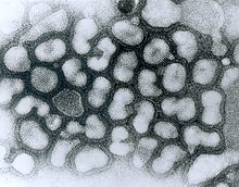
Transmission electron micrograph (TEM) of negatively stained Influenza A virus particles (small and white) attached to host cells (large and irregular) (late passage). (Source: Dr. Erskine Palmer, Centers for Disease Control and Prevention Public Health Image Library)

Infected birds transmit H5N1 through their saliva, nasal secretions, feces and blood. Other animals may become infected with the virus through direct contact with these bodily fluids or through contact with surfaces contaminated with them. H5N1 remains infectious after over 30 days at 0 °C (32 °F) (over one month at freezing temperature) or 6 days at 37 °C (99 °F) (one week at human body temperature); at ordinary temperatures it lasts in the environment for weeks. In Arctic temperatures, it does not degrade at all.

Because migratory birds are among the carriers of the highly pathogenic H5N1 virus, it is spreading to all parts of the world. H5N1 is different from all previously known highly pathogenic avian flu viruses in its ability to be spread by animals other than poultry.

In October 2004, researchers discovered H5N1 is far more dangerous than was previously believed. Waterfowl were revealed to be directly spreading this highly pathogenic strain to chickens, crows, pigeons, and other birds, and the virus was increasing its ability to infect mammals, as well. From this point on, avian flu experts increasingly referred to containment as a strategy that can delay, but not ultimately prevent, a future avian flu pandemic.

"Since 1997, studies of influenza A (H5N1) indicate that these viruses continue to evolve, with changes in antigenicity and internal gene constellations; an expanded host range in avian species and the ability to infect felids; enhanced pathogenicity in experimentally infected mice and ferrets, in which they cause systemic infections; and increased environmental stability."

The New York Times, in an article on transmission of H5N1 through smuggled birds, reports Wade Hagemeijer of Wetlands International stating, "We believe it is spread by both bird migration and trade, but that trade, particularly illegal trade, is more important".

On September 27, 2007, researchers reported the H5N1 bird flu virus can also pass through a pregnant woman's placenta to infect the fetus. They also found evidence of what doctors had long suspected—the virus not only affects the lungs, but also passes throughout the body into the gastrointestinal tract, the brain, liver, and blood cells.

In May 2013, North Korea confirmed a H5N1 bird flu outbreak that forced authorities to kill over 160,000 ducks in Pyongyang.

H5N1 transmission studies in ferrets (2011)

Novel, contagious strains of H5N1 were created by Ron Fouchier of the Erasmus Medical Center in Rotterdam, the Netherlands, who first presented his work to the public at an influenza conference in Malta in September 2011. Three mutations were introduced into the H5N1 virus genome, and the virus was then passed from the noses of infected ferrets to the noses of uninfected ones, which was repeated 10 times. After these 10 passages the H5N1 virus had acquired the ability of transmission between ferrets via aerosols or respiratory droplets.

After Fouchier offered an article describing this work to the leading academic journal Science, the US National Science Advisory Board for Biosecurity (NSABB) recommended against publication of the full details of the study, and the one submitted to Nature by Yoshihiro Kawaoka of the University of Wisconsin describing related work. However, after additional consultations at the World Health Organization and by the NSABB, the NSABB reversed its position and recommended publication of revised versions of the two papers. However, then the Dutch government declared that this type of manuscripts required Fouchier to apply for an export permit in the light of EU directive 428/2009 on dual use goods. After much controversy surrounding the publishing of his research, Fouchier complied (under formal protest) with Dutch government demands to obtain a special permit for submitting his manuscript, and his research appeared in a special issue of the journal Science devoted to H5N1. The papers by Fouchier and Kawaoka conclude that it is entirely possible that a natural chain of mutations could lead to an H5N1 virus acquiring the capability of airborne transmission between mammals, and that a H5N1 influenza pandemic would not be impossible.

In May 2013, it was reported that scientists at the Harbin Veterinary Research Institute in Harbin, China had created H5N1 strains which passed between guinea pigs.

Society and culture

H5N1 has had a significant effect on human society, especially the financial, political, social, and personal responses to both actual and predicted deaths in birds, humans, and other animals. Billions of U.S. dollars are being raised and spent to research H5N1 and prepare for a potential avian influenza pandemic. Over $10 billion have been spent and over 200 million birds have been killed to try to contain H5N1.

People have reacted by buying less chicken, causing poultry sales and prices to fall. Many individuals have stockpiled supplies for a possible flu pandemic. International health officials and other experts have pointed out that many unknown questions still hover around the disease.

Dr. David Nabarro, Chief Avian Flu Coordinator for the United Nations, and former Chief of Crisis Response for the World Health Organization has described himself as "quite scared" about H5N1's potential impact on humans. Nabarro has been accused of being alarmist before, and on his first day in his role for the United Nations, he proclaimed the avian flu could kill 150 million people. In an interview with the International Herald Tribune, Nabarro compares avian flu to AIDS in Africa, warning that underestimations led to inappropriate focus for research and intervention.

Disease surveillance in China

From Wikipedia, the free encyclopedia

Surveillance for communicable diseases is the main public health surveillance activity in China. Currently, the disease surveillance system in China has three major components:
  • National Disease Reporting System (NDRS): The system covers the entire population (1.3 billion persons) living in all the provinces, prefectures, and counties that make up mainland China. Thirty-five communicable diseases are reportable under this system.
  • Nationwide Disease Surveillance Points (DSPs): This surveillance system, comprising 145 reporting sites selected by stratified cluster random sampling, covers a 1% representative sample of China's population.
  • Surveillance system for specific infectious diseases, occupational diseases, food poisoning, etc.
There are 35 notifiable infectious diseases, which are divided into Classes A, B, and C. The functions of the surveillance include explaining the natural history of infectious diseases, describing the distribution of case occurrence, triggering disease-control effort, monitoring epidemic of infectious diseases during natural disasters, predicting and controlling epidemics and providing the base of policy adjustment.

Data collected through the disease surveillance network serve as the basis for formulating health policies and devising strategies for preventing disease. A computerized reporting system for notifiable diseases has been established that links China's 30 provinces, autonomous regions, and municipalities. Mechanisms for providing timely feedback to units that report data and for systematically assessing the quality of those data are important attributes of this system.

National Disease Reporting System (NDRS)

In 1959, a system for reporting infectious diseases was established. Data collected at the village level are reported to prevention units in township hospitals. From the prevention units, data are transmitted through county health and epidemic-prevention stations to provincial centers and then on to the Chinese Academy of Preventive Medicine. Since 1977, the Ministry of Public Health has convened annual meetings to analyze these data on the morbidity and mortality associated with infectious diseases.

In 1987, a Nationwide Antiepidemic Computer Telecommunication Network (NATCN) was established as an official information system for the National Disease Reporting System (NDRS). The Ministry of Public Health and the provincial centers of health and epidemic prevention support this network, which monitors disease epidemics at various levels within the public health system. As technical facilities of the network improve, use of the NACTN will expand into all aspects of public health surveillance.

Computer Network Development

After receiving approval from the Ministry of Public Health in 1986, the Chinese Academy of Preventive Medicine (CAPM) began to establish a nationwide microcomputer communication network. The purpose was to link all the country's provincial centers of health and epidemic prevention in an effort to improve the system for preventing epidemics. After a year was spent establishing and modifying the system, a network that connected the capitals of 30 provinces, autonomous regions, and municipalities began operating in 1987. The primary function of the network was to collect data on the morbidity and mortality associated with reportable communicable diseases, to obtain information on outbreaks of other types of disease, and to provide monthly and annual reports to local and national health authorities.

Morbidity and Mortality Monthly Reports (MMMR)

Each month, all provinces transmit county-level summaries of the numbers of cases and deaths associated with 35 notifiable communicable diseases to the Academy of Preventive Medicine. Reports are sent on the 13th to 15th day of each month via the NACTN. At the central node of NACTN, the Academy's Center of Computer Science and Health Statistics compiles and analyzes the data, provides feedback to the provinces, and creates national summaries within one week. Copies of the MMMR are distributed regularly to health authorities at various levels.

Morbidity and Mortality Annual Report (MMAR)

Each January, all provinces provide supplementary reports to revise and update the monthly reports submitted during the previous year. Age- and occupation-specific reports of mortality and morbidity are also submitted at this time. In April, after the surveillance data have been reviewed at the national meeting on epidemic diseases, the MMAR and other analytical reports are distributed.

Computer Telecommunication of Surveillance Data: Technical Issues

Until the 1980s, no public digital communications system was available in China. In establishing the nationwide communication network, modems with common analog telephone lines had to be used. Making this large communication system run successfully posed major challenges. With some of the problems in mind, the system was designed to have strong fault-tolerant redundancy - with the capability for self-correction - to overcome the myriad of problems caused by poor-quality telephone lines and cumbersome telephone exchange systems. 

The NACTN was enhanced by incorporating the following functions.
  • Breakpoint recording with resumption of operations: When telephone lines break during data transmission, the system is designed to record the break-point status for every case. This allows data transmission to resume automatically when line connections are reestablished.
  • Automatic node scanning and re-circling: This feature allows the system to scan the status of all network nodes to allocate telephone lines and thereby optimize the strategy for maintaining line connections. This important mechanism improves the efficiency of the system and makes data transmission more successful.
  • Automatic sorting/batching, rescheduling, and executing of transmissions: The system can execute all necessary network commands to carry out the communication task arranged by command files of the MMMR/MMAR system. The system adjusts the path as needed in order to complete transmissions that have been delayed because of problems in the system.
  • Data compression and security: Before transmission, data are processed by a "two-phase compressing" procedure. Data file size can be compressed more than 90%, resulting in shorter online transmission times. Thus, receiving data from the 30 provincial reporting centers on the network requires only about an hour of online operation. Compression also makes data transmission more secure.

System Support for NACTN

  • Personnel: In each province, the computer divisions have selected one or two persons who are dedicated to operating the province's nodes of the NACTN. In 1987, a working group convened to coordinate computer applications and activities. Several times each year, persons from provincial centers meet to discuss network problems and to devise solutions.
  • Hardware and software: Special software has been developed: YQS for collecting and processing information and producing reports and TXS for managing network communications.

Future Developments of the NACTN

  • Accumulating information
  • Updating techniques
  • Establishing subnetworks within provinces
In collaboration with the NACTN, a few provinces have established subnetworks to facilitate local communication.

Existing Databases

  • National report on infectious disease
  • National disease surveillance
  • National report on occupational disease
  • National report on outbreaks of food poisoning
  • National survey data bases: Drinking water quality; Human-parasite infections; Nutritional surveys; Nutrition for the elderly; Child-nutrition surveillance; Diarrheal disease of children; Smoking and health.

National Disease Surveillance Points (DSPs)

In the period 1980–1989, the network of DSPs covered 29 provinces, autonomous regions, and municipalities that had a combined population of 10 million persons (<1 1978="" a="" china="" class="mw-redirect" href="https://en.wikipedia.org/wiki/Random_sample" in="" it="" network="" not="" obtain="" of="" population-based="" population="" possible="" proposed="" s="" the="" title="Random sample" to="" was="" when="">random sample
. Because participation in the network was voluntary, the data collected were biased, even after attempts were made to adjust the sample to improve national representativeness. Persons covered by the DSPs tended to be from the upper-middle socioeconomic stratum.

In 1989, efforts were begun to select a new sample of surveillance points. We used stratified cluster random sampling to select 145 DSPs in 30 provinces, autonomous regions, and municipalities, which have a combined population structure similar to that shown in the national census. Data on individual births and deaths, as well as on infectious diseases and certain types of behaviors (e.g., tobacco smoking), are recorded. At the household level, information on socioeconomic indicators, health-care conditions, and environmental factors is collected.

Information obtained from the DSPs is compared with data obtained from the National Disease Reporting System to enable policymakers to estimate more accurately the burden of morbidity and mortality associated with infectious disease. More importantly, policy makers can evaluate information from the DSPs in relation to the economic development, cultural background, and health-care-service use by the population covered by this surveillance system.

Data collected at DSPs:
  • Household information: includes data on number of members, income, health-care situation, water supply, and toilet facilities.
  • Individual information: includes data on occupation, education, births, deaths, episodes of infectious diseases, pregnancy, lactation, feeding, and vaccination status.
Each month, data collected by the township hospitals and village prevention units are submitted to the country, which then conveys the information through the provincial centers to the Chinese Academy of Preventive Medicine. The Academy distributes monthly reports to the Ministry of Public Health, to provincial health authorities, and to all DSPs. An annual report is also published and distributed.

Surveys and Investigations

Surveys and investigations are conducted by the DSPs to improve the quality and promote the use of data collected. These surveys and investigations are designed to generate information that can be used by policymakers. At present, the following activities are being undertaken:
  • Characterizing risk factors and patterns of death among adults;
  • Identifying factors that influence the quality of data collected by DSPs;
  • Developing methods to monitor Chronic diseases in China;
  • Devising approaches to promote use of data from DSPs by policymakers.

Development of the Network of Disease Surveillance Points

Samples of persons already monitored under existing DSPs will be used for data collection to address important and emerging public health issues. Issues to be addressed include a prospective study of the health consequences of smoking, an assessment of drinking-water quality and disease occurrence, an evaluation of the national "Expanded Program on Immunization", and an epidemiologic study of hepatitis. By selecting samples in this fashion, investigators can correlate data from these special studies with data routinely collected under the system of DSPs.

Surveillance of Specific Infectious diseases

The following are examples of surveillance for specific infectious diseases.
  • For each 10-day period during the summer, cases of cholera - diagnosed by microbiologic or clinical criteria - from all the provinces, autonomous regions, and municipalities are reported to the national level; this information is compiled and conveyed back to the provincial reporting sources.
  • In eight provinces, surveillance among subgroups of the population with elevated risk for infection with human immunodeficiency virus (HIV) is conducted at the national diagnostic laboratories by using immediate reports and confirmatory testing.
  • A surveillance network for epidemic hemorrhagic fever has been established for immediate reporting of cases. During the peak season, surveillance for disease among rodents is conducted to provide an early-warning system at the local level.

Quality control of data collection

Quality control (QC) in association with data collection (DC) has been an important component of disease-surveillance activities in China.
  • In November of each year, the NDRS actively surveys hospitals and households to identify the proportion of notifiable diseases that went unreported. During a recent year, for example, the proportion of class A and B infectious diseases that was unreported was 27%; this proportion was used to correct the total annual estimate of morbidity attributable to infectious diseases.
  • Disease Surveillance Points (DSPs) are surveyed annually to estimate underreporting of births, deaths, and morbidity due to infectious diseases. From 1990 to 1991, for example, reporting of morbidity from infectious diseases improved. The proportions of unreported births, age-specific deaths, and disease-specific deaths are also reported. In 1991, a team from the Chinese Academy of Preventive Medicine evaluated the quality of data reported from 18 DSPs located in nine provinces. In their study, the evaluation team identified factors that influenced data quality.

Use of surveillance data for control of disease

Surveillance data have been used to implement and evaluate public health programs.
Monitoring morbidity from infectious disease during heavy flooding in 1991
When six provinces around the Yangtze River were heavily flooded in 1991, the central government expressed serious concern about disease-prevention activities in these provinces. In response, experts were dispatched to the flooded area, and prevention guidelines were developed and distributed to the affected provinces. Simultaneously, a system for collecting daily reports of disease activity was established. Every 3 days, DSP data on infectious disease morbidity were compared with data from previous years to identify potential outbreaks. For example, rates of hepatitis during the flood were compared with rates for the comparable time periods from the preceding 2 years. Data collected from June to October 1991 indicated that infectious diseases had been controlled effectively during the flood.
Forecasting the epidemiologic transition in China
In a study sponsored by the World Bank, data collected in DSPs in the period 1986-1989 have been used to study the epidemiologic transition in China. Mortality from leading causes of death was projected for 2010 and 2030. After risk factors were assessed and the impact of preventive programs on these chronic diseases was estimated, mortality rates were recalculated. These analyses were used to develop recommendations for program planning to the Ministry of Health.
Prediction and control of meningitis
After surveillance data on morbidity from meningitis in China were reviewed by empirical analysis and Boyer's Theorem, it was predicted that morbidity from this disease would peak in 1984 or 1985. Additional analyses suggested that the vaccination program that had been conducted for several years, which provided vaccination only for children <12 a="" adequate="" adopted="" age="" all="" areas="" be="" children="" class="mw-redirect" control="" coverage="" data="" disease.="" expanded="" href="https://en.wikipedia.org/wiki/Blood_serum" in="" including="" months="" new="" not="" of="" predicted="" program="" surveillance="" that="" the="" therefore="" title="Blood serum" to="" upsurge="" vaccination="" was="" which="" would="" years="">serum
epidemiologic data) identified a high risk of meningitis outbreaks. The results in Henan Province suggested that the intensified vaccination coverage was successful in decreasing rates of meningitis.
Strategy for vaccination for poliomyelitis
After data from 1988 to 1989 on rates of poliomyelitis and vaccination coverage were reviewed, high-risk areas were identified. In these areas, persons received supplementary vaccination in 1989–1990. By 1991, rates of poliomyelitis had begun to fall.

Disease surveillance

From Wikipedia, the free encyclopedia
 
Disease surveillance is an epidemiological practice by which the spread of disease is monitored in order to establish patterns of progression. The main role of disease surveillance is to predict, observe, and minimize the harm caused by outbreak, epidemic, and pandemic situations, as well as increase knowledge about which factors contribute to such circumstances. A key part of modern disease surveillance is the practice of disease case reporting.

In modern times, reporting incidences of disease outbreaks has been transformed from manual record keeping, to instant worldwide internet communication.

The number of cases could be gathered from hospitals - which would be expected to see most of the occurrences - collated, and eventually made public. With the advent of modern communication technology, this has changed dramatically. Organizations like the World Health Organization (WHO) and the Centers for Disease Control and Prevention (CDC) now can report cases and deaths from significant diseases within days - sometimes within hours - of the occurrence. Further, there is considerable public pressure to make this information available quickly and accurately. 

Mandatory reporting

Formal reporting of notifiable infectious diseases is a requirement placed upon health care providers by many regional and national governments, and upon national governments by the World Health Organization to monitor spread as a result of the transmission of infectious agents. Since 1969, WHO has required that all cases of the following diseases be reported to the organization: cholera, plague, yellow fever, smallpox, relapsing fever and typhus. In 2005, the list was extended to include polio and SARS. Regional and national governments typically monitor a larger set of (around 80 in the U.S.) communicable diseases that can potentially threaten the general population. Tuberculosis, HIV, botulism, hantavirus, anthrax, and rabies are examples of such diseases. The incidence counts of diseases are often used as health indicators to describe the overall health of a population. World Health Organization.

The World Health Organization is the lead agency for coordinating global response to major diseases. The WHO maintains Web sites for a number of diseases, and has active teams in many countries where these diseases occur. 

During the SARS outbreak in early 2004, for example, the Beijing staff of the WHO produced updates every few days for the duration of the outbreak. Beginning in January 2004, the WHO has produced similar updates for H5N1.[2] These results are widely reported and closely watched.

WHO's Epidemic and Pandemic Alert and Response (EPR) to detect, verify rapidly and respond appropriately to epidemic-prone and emerging disease threats covers the following diseases:

Political challenges

As the lead organization in global public health, the WHO occupies a delicate role in global politics. It must maintain good relationships with each of the many countries in which it is active. As a result, it may only report results within a particular country with the agreement of the country's government. Because some governments regard the release of any information on disease outbreaks as a state secret, this can place the WHO in a difficult position.

The WHO coordinated International Outbreak Alert and Response is designed to ensure "outbreaks of potential international importance are rapidly verified and information is quickly shared within the Network" but not necessarily by the public; integrate and coordinate "activities to support national efforts" rather than challenge national authority within that nation in order to "respect the independence and objectivity of all partners". The commitment that "All Network responses will proceed with full respect for ethical standards, human rights, national and local laws, cultural sensitivities and tradition" ensures each nation that its security, financial, and other interests will be given full weight.

Technical challenges

Testing for a disease can be expensive, and distinguishing between two diseases can be prohibitively difficult in many countries. One standard means of determining if a person has had a particular disease is to test for the presence of antibodies that are particular to this disease. In the case of H5N1, for example, there is a low pathogenic H5N1 strain in wild birds in North America that a human could conceivably have antibodies against. It would be extremely difficult to distinguish between antibodies produced by this strain, and antibodies produced by Asian lineage HPAI A(H5N1). Similar difficulties are common, and make it difficult to determine how widely a disease may have spread.

There is currently little available data on the spread of H5N1 in wild birds in Africa and Asia. Without such data, predicting how the disease might spread in the future is difficult. Information that scientists and decision makers need to make useful medical products and informed decisions for health care, but currently lack include:
  • Surveillance of wild bird populations
  • Cell cultures of particular strains of diseases

H5N1

Surveillance of H5N1 in humans, poultry, wild birds, cats and other animals remains very weak in many parts of Asia and Africa. Much remains unknown about the exact extent of its spread.

H5N1 in China is less than fully reported. Blogs have described many discrepancies between official China government announcements concerning H5N1 and what people in China see with their own eyes. Many reports of total H5N1 cases have excluded China due to widespread disbelief in China's official numbers.

"Only half the world's human bird flu cases are being reported to the World Health Organization within two weeks of being detected, a response time that must be improved to avert a pandemic, a senior WHO official said Saturday. Shigeru Omi, WHO's regional director for the Western Pacific, said it is estimated that countries would have only two to three weeks to stamp out, or at least slow, a pandemic flu strain after it began spreading in humans."

David Nabarro, chief avian flu coordinator for the United Nations, says avian flu has too many unanswered questions.

CIDRAP reported on August 25, 2006 on a new US government Web site that allows the public to view current information about testing of wild birds for H5N1 avian influenza which is part of a national wild-bird surveillance plan that "includes five strategies for early detection of highly pathogenic avian influenza. Sample numbers from three of these will be available on HEDDS: live wild birds, subsistence hunter-killed birds, and investigations of sick and dead wild birds. The other two strategies involve domestic bird testing and environmental sampling of water and wild-bird droppings. [...] A map on the new USGS site shows that 9,327 birds from Alaska have been tested so far this year, with only a few from most other states. Last year officials tested just 721 birds from Alaska and none from most other states, another map shows. The goal of the surveillance program for 2006 is to collect 75,000 to 100,000 samples from wild birds and 50,000 environmental samples, officials have said."

Thursday, March 26, 2020

Orthomyxoviridae (flu virus family)

From Wikipedia, the free encyclopedia

Orthomyxovirus
Virus classification e
(unranked): Virus
Realm: Riboviria
Phylum: Negarnaviricota
Class: Insthoviricetes
Order: Articulavirales
Family: Orthomyxoviridae
Genera

Orthomyxoviridae (ὀρθός, orthós, Greek for "straight"; μύξα, mýxa, Greek for "mucus") is a family of RNA viruses. It includes seven genera: Influenzavirus A, Influenzavirus B, Influenzavirus C, Influenzavirus D, Isavirus, Thogotovirus, and Quaranjavirus. The first four genera contain viruses that cause influenza in vertebrates, including birds, humans, and other mammals. Isaviruses infect salmon; the thogotoviruses are arboviruses, infecting vertebrates and invertebrates, such as ticks and mosquitoes.

The four genera of Influenza virus that infect vertebrates, which are identified by antigenic differences in their nucleoprotein and matrix protein, are as follows:

Classification

Influenza virus
 
In a phylogenetic-based taxonomy, the category "RNA virus" includes the category "negative-sense ssRNA virus", which includes the Order "Mononegavirales", and the Family "Orthomyxovirus" (among others). The genera-associated species and serotypes of Orthomyxovirus are shown in the following table. 

Orthomyxovirus Genera, Species, and Serotypes
Genus Species (* indicates type species) Serotypes or Subtypes Hosts
Influenza virus A Influenza A virus* H1N1, H1N2, H2N2, H3N1, H3N2, H3N8, H5N1, H5N2, H5N3, H5N8, H5N9, H7N1, H7N2, H7N3, H7N4, H7N7, H7N9, H9N2, H10N7 Human, pig, bird, horse, bat
Influenza virus B Influenza B virus* Victoria, Yamagata[5] Human, seal
Influenza virus C Influenza C virus*
Human, pig, dog
Influenza virus D Influenza D virus*
Pig, cattle
Isavirus Infectious salmon anemia virus*
Atlantic salmon
Thogotovirus Thogotovirus*
Tick, mosquito, mammal (including human)
Dhori virus Batken virus, Bourbon virus, Jos virus
Quaranjavirus[6]
Quaranfil virus,* Johnston Atoll virus

Types

There are four genera of influenza virus, each containing only a single species, or type. Influenza A and C infect a variety of species, while influenza B almost exclusively infects humans, and influenza D infects cattle and pigs.

Influenza A

Influenza A viruses are further classified, based on the viral surface proteins hemagglutinin (HA or H) and neuraminidase (NA or N). Sixteen H subtypes (or serotypes) and nine N subtypes of influenza A virus have been identified. 

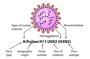
Diagram of influenza nomenclature

Further variation exists; thus, specific influenza strain isolates are identified by a standard nomenclature specifying virus type, geographical location where first isolated, sequential number of isolation, year of isolation, and HA and NA subtype.

Examples of the nomenclature are:
  1. A/Brisbane/59/2007 (H1N1)
  2. A/Moscow/10/99 (H3N2).
The type A viruses are the most virulent human pathogens among the three influenza types and cause the most severe disease. The serotypes that have been confirmed in humans, ordered by the number of known human pandemic deaths, are:
Known flu pandemics
Name of pandemic Date Deaths Case fatality rate Subtype involved Pandemic Severity Index
1889–1890 flu pandemic
(Asiatic or Russian Flu)
1889–1890 1 million 0.15% possibly H3N8
or H2N2
NA
1918 flu pandemic
(Spanish flu)
1918–1920 20 to 100 million 2% H1N1 5
Asian Flu 1957–1958 1 to 1.5 million 0.13% H2N2 2
Hong Kong Flu 1968–1969 0.75 to 1 million <0 .1="" font=""> H3N2 2
Russian flu 1977–1978 no accurate count N/A H1N1 N/A
2009 flu pandemic 2009–2010 105,700–395,600 0.03% H1N1 NA

Influenza B

Influenza B virus is almost exclusively a human pathogen, and is less common than influenza A. The only other animal known to be susceptible to influenza B infection is the seal. This type of influenza mutates at a rate 2–3 times lower than type A and consequently is less genetically diverse, with only one influenza B serotype. As a result of this lack of antigenic diversity, a degree of immunity to influenza B is usually acquired at an early age. However, influenza B mutates enough that lasting immunity is not possible. This reduced rate of antigenic change, combined with its limited host range (inhibiting cross species antigenic shift), ensures that pandemics of influenza B do not occur.

Influenza C

The influenza C virus infects humans and pigs, and can cause severe illness and local epidemics. However, influenza C is less common than the other types and usually causes mild disease in children.

Influenza D

This is a genus that was classified in 2016, the members of which were first isolated in 2011. This genus appears to be most closely related to Influenza C, from which it diverged several hundred years ago. There are at least two strains of this genus in extant. The main hosts appear to be cattle, but this virus has seen to infect pigs as well.

Morphology

Structure of the influenza virion. The hemagglutinin (HA) and neuraminidase (NA) proteins are shown on the surface of the particle. The viral RNAs that make up the genome are shown as red coils inside the particle and bound to Ribonuclear Proteins (RNPs).

The virion is pleomorphic; the envelope can occur in spherical and filamentous forms. In general, the virus's morphology is ellipsoidal with particles 80 to 120 nm in diameter, or filamentous virions 80–120 nm in diameter and up to 20 µm long. There are some 500 distinct spike-like surface projections of the envelope each projecting 10 to 14 nm from the surface with varying surface densities.

The major glycoprotein (HA) is interposed irregularly by clusters of neuraminidase (NA), with a ratio of HA to NA of about 4–5 to 1.

Cholesterol-laden membranes with protruding glycoproteins enclose the nucleocapsids; nucleoproteins of different size classes with a loop at each end; the arrangement within the virion is uncertain. The ribonuclear proteins are filamentous and fall in the range of 50 to 130 nm long and 9 to 15 nm in diameter. They have a helical symmetry.

Genome

Viruses of this family contain 6 to 8 segments of linear negative-sense single stranded RNA.

The total genome length is 12000–15000 nucleotides (nt). The size of each segment is as follows:

segment protein size (nt) protein size (aa)
PB1 polymerase 2300–2500 757+87 (F2)
PB2 polymerase 2300–2500 759
PA polymerase 2200–2300 716
HA Hemagglutinin 1700–1800 550
NP nucleoprotein 1500–1600 498
NA Neuraminidase 1400–1500 454
M Membrane protein(s) 1000–1100 252+97
NS non-structural protein(s) 800–900 230+121

The Genome sequence has terminal repeated sequences; repeated at both ends. Terminal repeats at the 5'-end 12–13 nucleotides long. Nucleotide sequences of 3'-terminus identical; the same in genera of same family; most on RNA (segments), or on all RNA species. Terminal repeats at the 3'-end 9–11 nucleotides long. Encapsidated nucleic acid is solely genomic. Each virion may contain defective interfering copies. In Influenza A (H1N1) PB1-F2 is produced from an alternative reading frame in PB1. The M and NS genes produce 2 different genes via alternative splicing.

Structure

The following applies for Influenza A viruses, although other influenza strains are very similar in structure:

The influenza A virus particle or virion is 80–120 nm in diameter, usually producing both ellipsoidal, baciliform, and filamentous particles. Unusually for a virus, the influenza A genome is not a single piece of nucleic acid; instead, it contains eight pieces of segmented negative-sense RNA (13.5 kilobases total), which encode 11 proteins (HA, NA, NP, M1, M2, NS1, NEP, PA, PB1, PB1-F2, PB2). The best-characterised of these viral proteins are hemagglutinin and neuraminidase, two large glycoproteins found on the outside of the viral particles. Neuraminidase is an enzyme involved in the release of progeny virus from infected cells, by cleaving sugars that bind the mature viral particles. By contrast, hemagglutinin is a lectin that mediates binding of the virus to target cells and entry of the viral genome into the target cell. The hemagglutinin (H) and neuraminidase (N) proteins are targets for antiviral drugs. These proteins are also recognised by antibodies, i.e. they are antigens. The responses of antibodies to these proteins are used to classify the different serotypes of influenza A viruses, hence the H and N in H5N1.

Replication cycle

Invasion and replication of the influenza virus. The steps in this process are discussed in the text.

Typically, influenza is transmitted from infected mammals through the air by coughs or sneezes, creating aerosols containing the virus, and from infected birds through their droppings. Influenza can also be transmitted by saliva, nasal secretions, feces and blood. Infections occur through contact with these bodily fluids or with contaminated surfaces. Out of a host, flu viruses can remain infectious for about one week at human body temperature, over 30 days at 0 °C (32 °F), and indefinitely at very low temperatures (such as lakes in northeast Siberia). They can be inactivated easily by disinfectants and detergents.

The viruses bind to a cell through interactions between its hemagglutinin glycoprotein and sialic acid sugars on the surfaces of epithelial cells in the lung and throat (Stage 1 in infection figure). The cell imports the virus by endocytosis. In the acidic endosome, part of the haemagglutinin protein fuses the viral envelope with the vacuole's membrane, releasing the viral RNA (vRNA) molecules, accessory proteins and RNA-dependent RNA polymerase into the cytoplasm (Stage 2). These proteins and vRNA form a complex that is transported into the cell nucleus, where the RNA-dependent RNA polymerase begins transcribing complementary positive-sense cRNA (Steps 3a and b). The cRNA is either exported into the cytoplasm and translated (step 4), or remains in the nucleus. Newly synthesised viral proteins are either secreted through the Golgi apparatus onto the cell surface (in the case of neuraminidase and hemagglutinin, step 5b) or transported back into the nucleus to bind vRNA and form new viral genome particles (step 5a). Other viral proteins have multiple actions in the host cell, including degrading cellular mRNA and using the released nucleotides for vRNA synthesis and also inhibiting translation of host-cell mRNAs.

Negative-sense vRNAs that form the genomes of future viruses, RNA-dependent RNA transcriptase, and other viral proteins are assembled into a virion. Hemagglutinin and neuraminidase molecules cluster into a bulge in the cell membrane. The vRNA and viral core proteins leave the nucleus and enter this membrane protrusion (step 6). The mature virus buds off from the cell in a sphere of host phospholipid membrane, acquiring hemagglutinin and neuraminidase with this membrane coat (step 7). As before, the viruses adhere to the cell through hemagglutinin; the mature viruses detach once their neuraminidase has cleaved sialic acid residues from the host cell. After the release of new influenza virus, the host cell dies.

Orthomyxoviridae viruses are one of two RNA viruses that replicate in the nucleus (the other being retroviridae). This is because the machinery of orthomyxo viruses cannot make their own mRNAs. They use cellular RNAs as primers for initiating the viral mRNA synthesis in a process known as cap snatching. Once in the nucleus, the RNA Polymerase Protein PB2 finds a cellular pre-mRNA and binds to its 5' capped end. Then RNA Polymerase PA cleaves off the cellular mRNA near the 5' end and uses this capped fragment as a primer for transcribing the rest of the viral RNA genome in viral mRNA. This is due to the need of mRNA to have a 5' cap in order to be recognized by the cell's ribosome for translation.

Since RNA proofreading enzymes are absent, the RNA-dependent RNA transcriptase makes a single nucleotide insertion error roughly every 10 thousand nucleotides, which is the approximate length of the influenza vRNA. Hence, nearly every newly manufactured influenza virus will contain a mutation in its genome. The separation of the genome into eight separate segments of vRNA allows mixing (reassortment) of the genes if more than one variety of influenza virus has infected the same cell (superinfection). The resulting alteration in the genome segments packaged into viral progeny confers new behavior, sometimes the ability to infect new host species or to overcome protective immunity of host populations to its old genome (in which case it is called an antigenic shift).

Viability and disinfection

Mammalian influenza viruses tend to be labile, but can survive several hours in mucus. Avian influenza virus can survive for 100 days in distilled water at room temperature, and 200 days at 17 °C (63 °F). The avian virus is inactivated more quickly in manure, but can survive for up to 2 weeks in feces on cages. Avian influenza viruses can survive indefinitely when frozen. Influenza viruses are susceptible to bleach, 70% ethanol, aldehydes, oxidizing agents, and quaternary ammonium compounds. They are inactivated by heat of 133 °F (56 °C) for minimum of 60 minutes, as well as by low pH <2 .="" p="">

Vaccination and prophylaxis

Vaccines and drugs are available for the prophylaxis and treatment of influenza virus infections. Vaccines are composed of either inactivated or live attenuated virions of the H1N1 and H3N2 human influenza A viruses, as well as those of influenza B viruses. Because the antigenicities of the wild viruses evolve, vaccines are reformulated annually by updating the seed strains.

When the antigenicities of the seed strains and wild viruses do not match, vaccines fail to protect the vaccinees. In addition, even when they do match, escape mutants are often generated.

Drugs available for the treatment of influenza include Amantadine and Rimantadine, which inhibit the uncoating of virions by interfering with M2, and Oseltamivir (marketed under the brand name Tamiflu), Zanamivir, and Peramivir, which inhibit the release of virions from infected cells by interfering with NA. However, escape mutants are often generated for the former drug and less frequently for the latter drug.

Hydrogen-like atom

From Wikipedia, the free encyclopedia https://en.wikipedia.org/wiki/Hydrogen-like_atom ...