Search This Blog

Monday, June 25, 2018

Embryonic stem cell

From Wikipedia, the free encyclopedia

Human embryonic stem cells in cell culture

Pluripotent: Embryonic stem cells are able to develop into any type of cell, excepting those of the placenta. Only embryonic stem cells of the morula are totipotent: able to develop into any type of cell, including those of the placenta.

Embryonic stem cells (ES cells or ESCs) are pluripotent stem cells derived from the inner cell mass of a blastocyst, an early-stage pre-implantation embryo.[1][2] Human embryos reach the blastocyst stage 4–5 days post fertilization, at which time they consist of 50–150 cells. Isolating the embryoblast, or inner cell mass (ICM) results in destruction of the blastocyst, a process which raises ethical issues, including whether or not embryos at the pre-implantation stage should be have the same moral considerations as embryos in the post-implantation stage of development.[3][4] Researchers are currently focusing heavily on the therapeutic potential of embryonic stem cells, with clinical use being the goal for many labs. These cells are being studied to be used as clinical therapies, models of genetic disorders, and cellular/DNA repair. However, adverse effects in the research and clinical processes have also been reported.

Properties


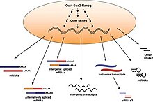
The transcriptome of embryonic stem cells

Embryonic stem cells (ESCs), derived from the blastocyst stage of early mammalian embryos, are distinguished by their ability to differentiate into any cell type and by their ability to propagate. It is these traits that makes them valuable in the scientific/medical fields. ESC are also described as having a normal karyotype, maintaining high telomerase activity, and exhibiting remarkable long-term proliferative potential.[5]

Pluripotent

Embryonic stem cells of the inner cell mass are pluripotent, meaning they are able to differentiate to generate primitive ectoderm, which ultimately differentiates during gastrulation into all derivatives of the three primary germ layers: ectoderm, endoderm, and mesoderm. These include each of the more than 220 cell types in the adult human body. Pluripotency distinguishes embryonic stem cells from adult stem cells, which are multipotent and can only produce a limited number of cell types.

Propagation

Under defined conditions, embryonic stem cells are capable of propagating indefinitely in an undifferentiated state. Conditions must either prevent the cells from clumping, or maintain an environment that supports an unspecialized state.[2] While being able to remain undifferentiated, ESCs also have the capacity, when provided with the appropriate signals, to differentiate (presumably via the initial formation of precursor cells) into nearly all mature cell phenotypes.[6]

Uses

Due to their plasticity and potentially unlimited capacity for self-renewal, embryonic stem cell therapies have been proposed for regenerative medicine and tissue replacement after injury or disease. Pluripotent stem cells have shown potential in treating a number of varying conditions, including but not limited to: spinal cord injuries, age related macular degeneration, diabetes, neurodegenerative disorders (such as Parkinson's disease), AIDS, etc.[7] In addition to their potential in regenerative medicine, embryonic stem cells provide an alternative source of tissue/organs which serves as a possible solution to the donor shortage dilemma. Not only that, but tissue/organs derived from ESCs can be made immunocompatible with the recipient. Aside from these uses, embryonic stem cells can also serve as tools for the investigation of early human development, study of genetic disease and as in vitro systems for toxicology testing.[5]

Utilizations

According to a 2002 article in PNAS, "Human embryonic stem cells have the potential to differentiate into various cell types, and, thus, may be useful as a source of cells for transplantation or tissue engineering."[8]


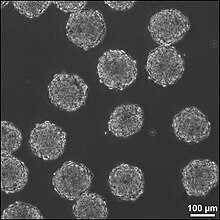
Embryoid bodies 24 hours after formation.

However, embryonic stem cells are not limited to cell/tissue engineering.

Cell Replacement Therapies

Current research focuses on differentiating ESCs into a variety of cell types for eventual use as cell replacement therapies (CRTs). Some of the cell types that have or are currently being developed include cardiomyocytes (CM), neurons, hepatocytes, bone marrow cells, islet cells and endothelial cells.[9] However, the derivation of such cell types from ESCs is not without obstacles, therefore current research is focused on overcoming these barriers. For example, studies are underway to differentiate ESCs in to tissue specific CMs and to eradicate their immature properties that distinguish them from adult CMs.[10]

Clinical Potential

  • Researchers have differentiated ESCs into dopamine-producing cells with the hope that these neurons could be used in the treatment of Parkinson’s disease.[11][12]
  • ESCs have been differentiated to natural killer (NK) cells and bone tissue.[13]
  • Studies involving ESCs are underway to provide an alternative treatment for diabetes. For example, D’Amour et al. were able to differentiate ESCs into insulin producing cells[14] and researchers at Harvard University were able to produce large quantities of pancreatic beta cells from ES.[15]
  • An article published in the European Heart Journal describes a translational process of generating human embryonic stem cell-derived cardiac progenitor cells to be used in clinical trials of patients with severe heart failure.[16]

Drug Discovery

Besides becoming an important alternative to organ transplants, ESCs are also being used in field of toxicology and as cellular screens to uncover new chemical entities (NCEs) that can be developed as small molecule drugs. Studies have shown that cardiomyocytes derived from ESCs are validated in vitro models to test drug responses and predict toxicity profiles.[9] ES derived cardiomyocytes have been shown to respond to pharmacological stimuli and hence can be used to assess cardiotoxicity like Torsades de Pointes.[17]

ESC-derived hepatocytes are also useful models that could be used in the preclinical stages of drug discovery. However, the development of hepatocytes from ESCs has proven to be challenging and this hinders the ability to test drug metabolism. Therefore, current research is focusing on establishing fully functional ESC-derived hepatocytes with stable phase I and II enzyme activity.[18]

Models of Genetic Disorder

Several new studies have started to address the concept of modeling genetic disorders with embryonic stem cells. Either by genetically manipulating the cells, or more recently, by deriving diseased cell lines identified by prenatal genetic diagnosis (PGD), modeling genetic disorders is something that has been accomplished with stem cells. This approach may very well prove invaluable at studying disorders such as Fragile-X syndrome, Cystic fibrosis, and other genetic maladies that have no reliable model system.

Yury Verlinsky, a Russian-American medical researcher who specialized in embryo and cellular genetics (genetic cytology), developed prenatal diagnosis testing methods to determine genetic and chromosomal disorders a month and a half earlier than standard amniocentesis. The techniques are now used by many pregnant women and prospective parents, especially couples who have a history of genetic abnormalities or where the woman is over the age of 35 (when the risk of genetically related disorders is higher). In addition, by allowing parents to select an embryo without genetic disorders, they have the potential of saving the lives of siblings that already had similar disorders and diseases using cells from the disease free offspring.[19]

Scientists have discovered a new technique for deriving human embryonic stem cell (ESC). Normal ESC lines from different sources of embryonic material including morula and whole blastocysts have been established. These findings allows researchers to construct ESC lines from embryos that acquire different genetic abnormalities; therefore, allowing for recognition of mechanisms in the molecular level that are possibly blocked that could impede the disease progression. The ESC lines originating from embryos with genetic and chromosomal abnormalities provide the data necessary to understand the pathways of genetic defects.[20]

A donor patient acquires one defective gene copy and one normal, and only one of these two copies is used for reproduction. By selecting egg cell derived from embryonic stem cells that have two normal copies, researchers can find variety of treatments for various diseases. To test this theory Dr. McLaughlin and several of his colleagues looked at whether parthenogenesis תשרקץצףפעסןembryonic stem cells can be used in a mouse model that has thalassemia intermedia. This disease is described as an inherited blood disorder in which there is a lack of hemoglobin leading to anemia. The mouse model used, had one defective gene copy. Embryonic stem cells from an unfertilized egg of the diseased mice were gathered and those stem cells that contained only healthy hemoglobin genes were identified. The healthy embryonic stem cell lines were then converted into cells transplanted into the carrier mice. After five weeks, the test results from the transplant illustrated that these carrier mice now had a normal blood cell count and hemoglobin levels.[21]

Repair of DNA damage

Differentiated somatic cells and ES cells use different strategies for dealing with DNA damage. For instance, human foreskin fibroblasts, one type of somatic cell, use non-homologous end joining (NHEJ), an error prone DNA repair process, as the primary pathway for repairing double-strand breaks (DSBs) during all cell cycle stages.[22] Because of its error-prone nature, NHEJ tends to produce mutations in a cell’s clonal descendants.

ES cells use a different strategy to deal with DSBs.[23] Because ES cells give rise to all of the cell types of an organism including the cells of the germ line, mutations arising in ES cells due to faulty DNA repair are a more serious problem than in differentiated somatic cells. Consequently, robust mechanisms are needed in ES cells to repair DNA damages accurately, and if repair fails, to remove those cells with un-repaired DNA damages. Thus, mouse ES cells predominantly use high fidelity homologous recombinational repair (HRR) to repair DSBs.[23] This type of repair depends on the interaction of the two sister chromosomes formed during S phase and present together during the G2 phase of the cell cycle. HRR can accurately repair DSBs in one sister chromosome by using intact information from the other sister chromosome. Cells in the G1 phase of the cell cycle (i.e. after metaphase/cell division but prior the next round of replication) have only one copy of each chromosome (i.e. sister chromosomes aren’t present). Mouse ES cells lack a G1 checkpoint and do not undergo cell cycle arrest upon acquiring DNA damage.[24] Rather they undergo programmed cell death (apoptosis) in response to DNA damage.[25] Apoptosis can be used as a fail-safe strategy to remove cells with un-repaired DNA damages in order to avoid mutation and progression to cancer.[26] Consistent with this strategy, mouse ES stem cells have a mutation frequency about 100-fold lower than that of isogenic mouse somatic cells.[27]

Clinical trial

On January 23, 2009, Phase I clinical trials for transplantation of oligodendrocytes (a cell type of the brain and spinal cord) derived from human ES cells into spinal cord-injured individuals received approval from the U.S. Food and Drug Administration (FDA), marking it the world's first human ES cell human trial.[28] The study leading to this scientific advancement was conducted by Hans Keirstead and colleagues at the University of California, Irvine and supported by Geron Corporation of Menlo Park, CA, founded by Michael D. West, PhD. A previous experiment had shown an improvement in locomotor recovery in spinal cord-injured rats after a 7-day delayed transplantation of human ES cells that had been pushed into an oligodendrocytic lineage.[29] The phase I clinical study was designed to enroll about eight to ten paraplegics who have had their injuries no longer than two weeks before the trial begins, since the cells must be injected before scar tissue is able to form. The researchers emphasized that the injections were not expected to fully cure the patients and restore all mobility. Based on the results of the rodent trials, researchers speculated that restoration of myelin sheathes and an increase in mobility might occur. This first trial was primarily designed to test the safety of these procedures and if everything went well, it was hoped that it would lead to future studies that involve people with more severe disabilities.[30] The trial was put on hold in August 2009 due to FDA concerns regarding a small number of microscopic cysts found in several treated rat models but the hold was lifted on July 30, 2010.[31]

In October 2010 researchers enrolled and administered ESTs to the first patient at Shepherd Center in Atlanta.[32] The makers of the stem cell therapy, Geron Corporation, estimated that it would take several months for the stem cells to replicate and for the GRNOPC1 therapy to be evaluated for success or failure.

In November 2011 Geron announced it was halting the trial and dropping out of stem cell research for financial reasons, but would continue to monitor existing patients, and was attempting to find a partner that could continue their research.[33] In 2013 BioTime (AMEXBTX), led by CEO Dr. Michael D. West, acquired all of Geron's stem cell assets, with the stated intention of restarting Geron's embryonic stem cell-based clinical trial for spinal cord injury research.[34]

BioTime company Asterias Biotherapeutics (NYSE MKT: AST) was granted a $14.3 million Strategic Partnership Award by the California Institute for Regenerative Medicine (CIRM) to re-initiate the world’s first embryonic stem cell-based human clinical trial, for spinal cord injury. Supported by California public funds, CIRM is the largest funder of stem cell-related research and development in the world.[35]

The award provides funding for Asterias to reinitiate clinical development of AST-OPC1 in subjects with spinal cord injury and to expand clinical testing of escalating doses in the target population intended for future pivotal trials.[35]

AST-OPC1 is a population of cells derived from human embryonic stem cells (hESCs) that contains oligodendrocyte progenitor cells (OPCs). OPCs and their mature derivatives called oligodendrocytes provide critical functional support for nerve cells in the spinal cord and brain. Asterias recently presented the results from phase 1 clinical trial testing of a low dose of AST-OPC1 in patients with neurologically-complete thoracic spinal cord injury. The results showed that AST-OPC1 was successfully delivered to the injured spinal cord site. Patients followed 2–3 years after AST-OPC1 administration showed no evidence of serious adverse events associated with the cells in detailed follow-up assessments including frequent neurological exams and MRIs. Immune monitoring of subjects through one year post-transplantation showed no evidence of antibody-based or cellular immune responses to AST-OPC1. In four of the five subjects, serial MRI scans performed throughout the 2–3 year follow-up period indicate that reduced spinal cord cavitation may have occurred and that AST-OPC1 may have had some positive effects in reducing spinal cord tissue deterioration. There was no unexpected neurological degeneration or improvement in the five subjects in the trial as evaluated by the International Standards for Neurological Classification of Spinal Cord Injury (ISNCSCI) exam.[35]

The Strategic Partnership III grant from CIRM will provide funding to Asterias to support the next clinical trial of AST-OPC1 in subjects with spinal cord injury, and for Asterias’ product development efforts to refine and scale manufacturing methods to support later-stage trials and eventually commercialization. CIRM funding will be conditional on FDA approval for the trial, completion of a definitive agreement between Asterias and CIRM, and Asterias’ continued progress toward the achievement of certain pre-defined project milestones.[35]

Adverse effect

The major concern with the possible transplantation of ESC into patients as therapies is their ability to form tumors including teratoma.[36] Safety issues prompted the FDA to place a hold on the first ESC clinical trial (see below), however no tumors were observed.

The main strategy to enhance the safety of ESC for potential clinical use is to differentiate the ESC into specific cell types (e.g. neurons, muscle, liver cells) that have reduced or eliminated ability to cause tumors. Following differentiation, the cells are subjected to sorting by flow cytometry for further purification. ESC are predicted to be inherently safer than IPS cells because they are not genetically modified with genes such as c-Myc that are linked to cancer. Nonetheless, ESC express very high levels of the iPS inducing genes and these genes including Myc are essential for ESC self-renewal and pluripotency,[37] and potential strategies to improve safety by eliminating c-Myc expression are unlikely to preserve the cells' "stemness". However, N-myc and L-myc have been identified to induce iPS cells instead of c-myc with similar efficiency.[38]

History

  • 1964: Lewis Kleinsmith and G. Barry Pierce Jr. isolated a single type of cell from a teratocarcinoma, a tumor now known to be derived from a germ cell.[39] These cells isolated from the teratocarcinoma replicated and grew in cell culture as a stem cell and are now known as embryonal carcinoma (EC) cells.[40] Although similarities in morphology and differentiating potential (pluripotency) led to the use of EC cells as the in vitro model for early mouse development,[41] EC cells harbor genetic mutations and often abnormal karyotypes that accumulated during the development of the teratocarcinoma. These genetic aberrations further emphasized the need to be able to culture pluripotent cells directly from the inner cell mass.

Martin Evans revealed a new technique for culturing the mouse embryos in the uterus to allow for the derivation of ES cells from these embryos.
  • 1981: Embryonic stem cells (ES cells) were independently first derived from mouse embryos by two groups. Martin Evans and Matthew Kaufman from the Department of Genetics, University of Cambridge published first in July, revealing a new technique for culturing the mouse embryos in the uterus to allow for an increase in cell number, allowing for the derivation of ES cells from these embryos.[42] Gail R. Martin, from the Department of Anatomy, University of California, San Francisco, published her paper in December and coined the term “Embryonic Stem Cell”.[43] She showed that embryos could be cultured in vitro and that ES cells could be derived from these embryos. In 1998, a breakthrough occurred when researchers, led by James Thomson at the University of Wisconsin-Madison, first developed a technique to isolate and grow human embryonic stem cells in cell culture.[44]
  • 1989: Mario R. Cappechi, Martin J. Evans, and Oliver Smithies publish their research which details their isolation and genetic modifications of embryonic stem cells, creating the first "knockout mice". [45] In creating knockout mice, this publication provided scientists with an entirely new way to study disease.
  • 1998: A paper titled "Embryonic Stem Cell Lines Derived From Human Blastocysts" is published by a team from the University of Wisconsin, Madison. The researchers behind this study not only create the first embryonic stem cells, but recognize their pluripotency, as well as their capacity for self-renewal. The abstract of the paper notes the significance of the discovery with regards to the fields of developmental biology and drug discovery. [46]

Techniques and conditions for derivation and culture

Derivation from humans

In vitro fertilization generates multiple embryos. The surplus of embryos is not clinically used or is unsuitable for implantation into the patient, and therefore may be donated by the donor with consent. Human embryonic stem cells can be derived from these donated embryos or additionally they can also be extracted from cloned embryos using a cell from a patient and a donated egg.[47] The inner cell mass (cells of interest), from the blastocyst stage of the embryo, is separated from the trophectoderm, the cells that would differentiate into extra-embryonic tissue. Immunosurgery, the process in which antibodies are bound to the trophectoderm and removed by another solution, and mechanical dissection are performed to achieve separation. The resulting inner cell mass cells are plated onto cells that will supply support. The inner cell mass cells attach and expand further to form a human embryonic cell line, which are undifferentiated. These cells are fed daily and are enzymatically or mechanically separated every four to seven days. For differentiation to occur, the human embryonic stem cell line is removed from the supporting cells to form embryoid bodies, is co-cultured with a serum containing necessary signals, or is grafted in a three-dimensional scaffold to result.[48]

Derivation from other animals

Embryonic stem cells are derived from the inner cell mass of the early embryo, which are harvested from the donor mother animal. Martin Evans and Matthew Kaufman reported a technique that delays embryo implantation, allowing the inner cell mass to increase. This process includes removing the donor mother's ovaries and dosing her with progesterone, changing the hormone environment, which causes the embryos to remain free in the uterus. After 4–6 days of this intrauterine culture, the embryos are harvested and grown in in vitro culture until the inner cell mass forms “egg cylinder-like structures,” which are dissociated into single cells, and plated on fibroblasts treated with mitomycin-c (to prevent fibroblast mitosis). Clonal cell lines are created by growing up a single cell. Evans and Kaufman showed that the cells grown out from these cultures could form teratomas and embryoid bodies, and differentiate in vitro, all of which indicating that the cells are pluripotent.[42]

Gail Martin derived and cultured her ES cells differently. She removed the embryos from the donor mother at approximately 76 hours after copulation and cultured them overnight in a medium containing serum. The following day, she removed the inner cell mass from the late blastocyst using microsurgery. The extracted inner cell mass was cultured on fibroblasts treated with mitomycin-c in a medium containing serum and conditioned by ES cells. After approximately one week, colonies of cells grew out. These cells grew in culture and demonstrated pluripotent characteristics, as demonstrated by the ability to form teratomas, differentiate in vitro, and form embryoid bodies. Martin referred to these cells as ES cells.[43]

It is now known that the feeder cells provide leukemia inhibitory factor (LIF) and serum provides bone morphogenetic proteins (BMPs) that are necessary to prevent ES cells from differentiating.[49][50] These factors are extremely important for the efficiency of deriving ES cells. Furthermore, it has been demonstrated that different mouse strains have different efficiencies for isolating ES cells.[51] Current uses for mouse ES cells include the generation of transgenic mice, including knockout mice. For human treatment, there is a need for patient specific pluripotent cells. Generation of human ES cells is more difficult and faces ethical issues. So, in addition to human ES cell research, many groups are focused on the generation of induced pluripotent stem cells (iPS cells).[52]

Potential method for new cell line derivation

On August 23, 2006, the online edition of Nature scientific journal published a letter by Dr. Robert Lanza (medical director of Advanced Cell Technology in Worcester, MA) stating that his team had found a way to extract embryonic stem cells without destroying the actual embryo.[53] This technical achievement would potentially enable scientists to work with new lines of embryonic stem cells derived using public funding in the USA, where federal funding was at the time limited to research using embryonic stem cell lines derived prior to August 2001. In March, 2009, the limitation was lifted.[54]

Induced pluripotent stem cells

The iPSC technology was pioneered by Shinya Yamanaka’s lab in Kyoto, Japan, who showed in 2006 that the introduction of four specific genes encoding transcription factors could convert adult cells into pluripotent stem cells.[55] He was awarded the 2012 Nobel Prize along with Sir John Gurdon "for the discovery that mature cells can be reprogrammed to become pluripotent." [56]

In 2007 it was shown that pluripotent stem cells highly similar to embryonic stem cells can be generated by the delivery of three genes (Oct4, Sox2, and Klf4) to differentiated cells.[57] The delivery of these genes "reprograms" differentiated cells into pluripotent stem cells, allowing for the generation of pluripotent stem cells without the embryo. Because ethical concerns regarding embryonic stem cells typically are about their derivation from terminated embryos, it is believed that reprogramming to these "induced pluripotent stem cells" (iPS cells) may be less controversial. Both human and mouse cells can be reprogrammed by this methodology, generating both human pluripotent stem cells and mouse pluripotent stem cells without an embryo.[58]

This may enable the generation of patient specific ES cell lines that could potentially be used for cell replacement therapies. In addition, this will allow the generation of ES cell lines from patients with a variety of genetic diseases and will provide invaluable models to study those diseases.

However, as a first indication that the induced pluripotent stem cell (iPS) cell technology can in rapid succession lead to new cures, it was used by a research team headed by Rudolf Jaenisch of the Whitehead Institute for Biomedical Research in Cambridge, Massachusetts, to cure mice of sickle cell anemia, as reported by Science journal's online edition on December 6, 2007.[59][60]

On January 16, 2008, a California-based company, Stemagen, announced that they had created the first mature cloned human embryos from single skin cells taken from adults. These embryos can be harvested for patient matching embryonic stem cells.[61]

Contamination by reagents used in cell culture

The online edition of Nature Medicine published a study on January 24, 2005, which stated that the human embryonic stem cells available for federally funded research are contaminated with non-human molecules from the culture medium used to grow the cells.[62] It is a common technique to use mouse cells and other animal cells to maintain the pluripotency of actively dividing stem cells. The problem was discovered when non-human sialic acid in the growth medium was found to compromise the potential uses of the embryonic stem cells in humans, according to scientists at the University of California, San Diego.[63]

However, a study published in the online edition of Lancet Medical Journal on March 8, 2005 detailed information about a new stem cell line that was derived from human embryos under completely cell- and serum-free conditions. After more than 6 months of undifferentiated proliferation, these cells demonstrated the potential to form derivatives of all three embryonic germ layers both in vitro and in teratomas. These properties were also successfully maintained (for more than 30 passages) with the established stem cell lines.[64]

Stem-cell therapy

From Wikipedia, the free encyclopedia

Stem-cell therapy is the use of stem cells to treat or prevent a disease or condition.[1]

Bone marrow transplant is the most widely used stem-cell therapy, but some therapies derived from umbilical cord blood are also in use. Research is underway to develop various sources for stem cells, as well as to apply stem-cell treatments for neurodegenerative diseases and conditions such as diabetes and heart disease, among others.

Stem-cell therapy has become controversial following developments such as the ability of scientists to isolate and culture embryonic stem cells, to create stem cells using somatic cell nuclear transfer and their use of techniques to create induced pluripotent stem cells. This controversy is often related to abortion politics and to human cloning. Additionally, efforts to market treatments based on transplant of stored umbilical cord blood have been controversial.

Medical uses

For over 30 years, bone marrow has been used to treat cancer patients with conditions such as leukaemia and lymphoma; this is the only form of stem-cell therapy that is widely practiced.[2][3][4] During chemotherapy, most growing cells are killed by the cytotoxic agents. These agents, however, cannot discriminate between the leukaemia or neoplastic cells, and the hematopoietic stem cells within the bone marrow. It is this side effect of conventional chemotherapy strategies that the stem-cell transplant attempts to reverse; a donor's healthy bone marrow reintroduces functional stem cells to replace the cells lost in the host's body during treatment. The transplanted cells also generate an immune response that helps to kill off the cancer cells; this process can go too far, however, leading to graft vs host disease, the most serious side effect of this treatment.[5]

Another stem-cell therapy called Prochymal, was conditionally approved in Canada in 2012 for the management of acute graft-vs-host disease in children who are unresponsive to steroids.[6] It is an allogenic stem therapy based on mesenchymal stem cells (MSCs) derived from the bone marrow of adult donors. MSCs are purified from the marrow, cultured and packaged, with up to 10,000 doses derived from a single donor. The doses are stored frozen until needed.[7]

The FDA has approved five hematopoietic stem-cell products derived from umbilical cord blood, for the treatment of blood and immunological diseases.[8]

In 2014, the European Medicines Agency recommended approval of limbal stem cells for people with severe limbal stem cell deficiency due to burns in the eye.[9]

Research


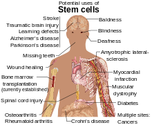
Diseases and conditions where stem cell treatment is promising or emerging.

Stem cells are being studied for a number of reasons. The molecules and exosomes released from stem cells are also being studied in an effort to make medications.[10] The paracrine soluble factors produced by stem cells, known as the stem cell secretome, has been found to be the predominant mechanism by which stem cell-based therapies mediate their effects in degenerative, auto-immune and inflammatory diseases.[11]

Applications

Neurodegeneration

Research has been conducted on the effects of stem cells on animal models of brain degeneration, such as in Parkinson's, Amyotrophic lateral sclerosis, and Alzheimer's disease.[12][13][14] There have been preliminary studies related to multiple sclerosis.[15][16]

Healthy adult brains contain neural stem cells which divide to maintain general stem-cell numbers, or become progenitor cells. In healthy adult laboratory animals, progenitor cells migrate within the brain and function primarily to maintain neuron populations for olfaction (the sense of smell).

Pharmacological activation of endogenous neural stem cells has been reported to induce neuroprotection and behavioral recovery in adult rat models of neurological disorder.[17][18][19]

Brain and spinal cord injury

Stroke and traumatic brain injury lead to cell death, characterized by a loss of neurons and oligodendrocytes within the brain. Clinical and animal studies have been conducted into the use of stem cells in cases of spinal cord injury.[20][21][22]

Heart

Stems cells are studied in people with severe heart disease.[23] The work by Bodo-Eckehard Strauer[24] was discredited by identifying hundreds of factual contradictions.[25] Among several clinical trials reporting that adult stem cell therapy is safe and effective, actual evidence of benefit has been reported from only a few studies.[26] Some preliminary clinical trials achieved only modest improvements in heart function following use of bone marrow stem cell therapy.[27][28]

Stem-cell therapy for treatment of myocardial infarction usually makes use of autologous bone marrow stem cells, but other types of adult stem cells may be used, such as adipose-derived stem cells.[29]

Possible mechanisms of recovery include:[12]
  • Generation of heart muscle cells
  • Stimulating growth of new blood vessels to repopulate damaged heart tissue
  • Secretion of growth factors

Criticisms

In 2013, studies of autologous bone marrow stem cells on ventricular function were found to contain "hundreds" of discrepancies.[30] Critics report that of 48 reports there seemed to be just five underlying trials, and that in many cases whether they were randomized or merely observational accepter-versus-rejecter, was contradictory between reports of the same trial. One pair of reports of identical baseline characteristics and final results, was presented in two publications as, respectively, a 578 patient randomized trial and as a 391 patient observational study. Other reports required (impossible) negative standard deviations in subsets of patients, or contained fractional patients, negative NYHA classes. Overall there were many more patients published as having receiving stem cells in trials, than the number of stem cells processed in the hospital's laboratory during that time. A university investigation, closed in 2012 without reporting, was reopened in July 2013.[31]

In 2014, a meta-analysis on stem cell therapy using bone marrow stem cells for heart disease revealed discrepancies in published clinical trial reports, whereby studies with a higher number of discrepancies showed an increase in effect sizes.[32] Another meta-analysis based on the intra-subject data of 12 randomised trials was unable to find any significant benefits of stem cell therapy on primary endpoints, such as major adverse events or increase in heart function measures, concluding there was no benefit.[33]

The TIME trial, which used a randomised, double blind, placebo-controlled trial design, concluded that "bone marrow mononuclear cells administration did not improve recovery of LV function over 2 years" in people who had a myocardial infarction.[34] Accordingly, the BOOST-2 trial conducted in 10 medical centres in Germany and Norway reported that the trial result "does not support the use of nucleated BMCs in patients with STEMI and moderately reduced LVEF".[35] Furthermore, the trial also did not meet any other secondary MRI endpoints,[36] leading to a conclusion that intracoronary bone marrow stem cell therapy does not offer a functional or clinical benefit.[37]

Blood-cell formation

The specificity of the human immune-cell repertoire is what allows the human body to defend itself from rapidly adapting antigens. However, the immune system is vulnerable to degradation upon the pathogenesis of disease, and because of the critical role that it plays in overall defense, its degradation is often fatal to the organism as a whole. Diseases of hematopoietic cells are diagnosed and classified via a subspecialty of pathology known as hematopathology. The specificity of the immune cells is what allows recognition of foreign antigens, causing further challenges in the treatment of immune disease. Identical matches between donor and recipient must be made for successful transplantation treatments, but matches are uncommon, even between first-degree relatives. Research using both hematopoietic adult stem cells and embryonic stem cells has provided insight into the possible mechanisms and methods of treatment for many of these ailments.[citation needed]

Fully mature human red blood cells may be generated ex vivo by hematopoietic stem cells (HSCs), which are precursors of red blood cells. In this process, HSCs are grown together with stromal cells, creating an environment that mimics the conditions of bone marrow, the natural site of red-blood-cell growth. Erythropoietin, a growth factor, is added, coaxing the stem cells to complete terminal differentiation into red blood cells.[38] Further research into this technique should have potential benefits to gene therapy, blood transfusion, and topical medicine.

Regrowing teeth

In 2004, scientists at King's College London discovered a way to cultivate a complete tooth in mice[39] and were able to grow bioengineered teeth stand-alone in the laboratory. Researchers are confident that the tooth regeneration technology can be used to grow live teeth in human patients.

In theory, stem cells taken from the patient could be coaxed in the lab turning into a tooth bud which, when implanted in the gums, will give rise to a new tooth, and would be expected to be grown in a time over three weeks.[40] It will fuse with the jawbone and release chemicals that encourage nerves and blood vessels to connect with it. The process is similar to what happens when humans grow their original adult teeth. Many challenges remain, however, before stem cells could be a choice for the replacement of missing teeth in the future.[41][42]

Cochlear hair cell regrowth

Heller has reported success in re-growing cochlea hair cells with the use of embryonic stem cells.[43]

Blindness and vision impairment

Since 2003, researchers have successfully transplanted corneal stem cells into damaged eyes to restore vision. "Sheets of retinal cells used by the team are harvested from aborted fetuses, which some people find objectionable." When these sheets are transplanted over the damaged cornea, the stem cells stimulate renewed repair, eventually restore vision.[44] The latest such development was in June 2005, when researchers at the Queen Victoria Hospital of Sussex, England were able to restore the sight of forty patients using the same technique. The group, led by Sheraz Daya, was able to successfully use adult stem cells obtained from the patient, a relative, or even a cadaver. Further rounds of trials are ongoing.[45]

Pancreatic beta cells

Diabetes patients lose the function of insulin-producing beta cells within the pancreas.[46] In recent experiments, scientists have been able to coax embryonic stem cell to turn into beta cells in the lab. In theory if the beta cell is transplanted successfully, they will be able to replace malfunctioning ones in a diabetic patient.[47]

Orthopaedics

Clinical case reports in the treatment orthopaedic conditions have been reported. To date, the focus in the literature for musculoskeletal care appears to be on mesenchymal stem cells. Centeno et al. have published MRI evidence of increased cartilage and meniscus volume in individual human subjects. The results of trials that include a large number of subjects, are yet to be published. However, a published safety study conducted in a group of 227 patients over a 3-4-year period shows adequate safety and minimal complications associated with mesenchymal cell transplantation.[50]

Wakitani has also published a small case series of nine defects in five knees involving surgical transplantation of mesenchymal stem cells with coverage of the treated chondral defects.[51]

Wound healing

Stem cells can also be used to stimulate the growth of human tissues. In an adult, wounded tissue is most often replaced by scar tissue, which is characterized in the skin by disorganized collagen structure, loss of hair follicles and irregular vascular structure. In the case of wounded fetal tissue, however, wounded tissue is replaced with normal tissue through the activity of stem cells.[52] A possible method for tissue regeneration in adults is to place adult stem cell "seeds" inside a tissue bed "soil" in a wound bed and allow the stem cells to stimulate differentiation in the tissue bed cells. This method elicits a regenerative response more similar to fetal wound-healing than adult scar tissue formation.[52] Researchers are still investigating different aspects of the "soil" tissue that are conducive to regeneration.[52]

Infertility

Culture of human embryonic stem cells in mitotically inactivated porcine ovarian fibroblasts (POF) causes differentiation into germ cells (precursor cells of oocytes and spermatozoa), as evidenced by gene expression analysis.[53]

Human embryonic stem cells have been stimulated to form Spermatozoon-like cells, yet still slightly damaged or malformed.[54] It could potentially treat azoospermia.

In 2012, oogonial stem cells were isolated from adult mouse and human ovaries and demonstrated to be capable of forming mature oocytes.[55] These cells have the potential to treat infertility.

HIV/AIDS

Destruction of the immune system by the HIV is driven by the loss of CD4+ T cells in the peripheral blood and lymphoid tissues. Viral entry into CD4+ cells is mediated by the interaction with a cellular chemokine receptor, the most common of which are CCR5 and CXCR4. Because subsequent viral replication requires cellular gene expression processes, activated CD4+ cells are the primary targets of productive HIV infection.[56] Recently scientists have been investigating an alternative approach to treating HIV-1/AIDS, based on the creation of a disease-resistant immune system through transplantation of autologous, gene-modified (HIV-1-resistant) hematopoietic stem and progenitor cells (GM-HSPC).[57]

Clinical trials

Regenerative treatment models

Stem cells are thought to mediate repair via five primary mechanisms: 1) providing an anti-inflammatory effect, 2) homing to damaged tissues and recruiting other cells, such as endothelial progenitor cells, that are necessary for tissue growth, 3) supporting tissue remodeling over scar formation, 4) inhibiting apoptosis, and 5) differentiating into bone, cartilage, tendon, and ligament tissue.[58][59]

To further enrich blood supply to the damaged areas, and consequently promote tissue regeneration, platelet-rich plasma could be used in conjunction with stem cell transplantation.[60][61] The efficacy of some stem cell populations may also be affected by the method of delivery; for instance, to regenerate bone, stem cells are often introduced in a scaffold where they produce the minerals necessary for generation of functional bone.[60][61][62][63]

Stem cells have also been shown to have a low immunogenicity due to the relatively low number of MHC molecules found on their surface. In addition, they have been found to secrete chemokines that alter the immune response and promote tolerance of the new tissue. This allows for allogeneic treatments to be performed without a high rejection risk.[64]

Drug discovery and biomedical research

The ability to grow up functional adult tissues indefinitely in culture through Directed differentiation creates new opportunities for drug research. Researchers are able to grow up differentiated cell lines and then test new drugs on each cell type to examine possible interactions in vitro before performing in vivo studies. This is critical in the development of drugs for use in veterinary research because of the possibilities of species specific interactions. The hope is that having these cell lines available for research use will reduce the need for research animals used because effects on human tissue in vitro will provide insight not normally known before the animal testing phase.[65]
With the advent of induced pluripotent stem cells (iPSC), treatments being explored and created for the used in endangered low production animals possible. Rather than needing to harvest embryos or eggs, which are limited, the researchers can remove mesenchymal stem cells with greater ease and greatly reducing the danger to the animal due to noninvasive techniques. This allows the limited eggs to be put to use for reproductive purposes only.[65]

Conservation

Stem cells are being explored for use in conservation efforts. Spermatogonial stem cells have been harvested from a rat and placed into a mouse host and fully mature sperm were produced with the ability to produce viable offspring. Currently research is underway to find suitable hosts for the introduction of donor spermatogonial stem cells. If this becomes a viable option for conservationists, sperm can be produced from high genetic quality individuals who die before reaching sexual maturity, preserving a line that would otherwise be lost.[66]

Sources for stem cells

Most stem cells intended for regenerative therapy are generally isolated either from the patient's bone marrow or from adipose tissue.[61][63] Mesenchymal stem cells can differentiate into the cells that make up bone, cartilage, tendons, and ligaments, as well as muscle, neural and other progenitor tissues, they have been the main type of stem cells studied in the treatment of diseases affecting these tissues.[67][68] The number of stem cells transplanted into damaged tissue may alter efficacy of treatment. Accordingly, stem cells derived from bone marrow aspirates, for instance, are cultured in specialized laboratories for expansion to millions of cells.[61][63] Although adipose-derived tissue also requires processing prior to use, the culturing methodology for adipose-derived stem cells is not as extensive as that for bone marrow-derived cells.[69][70] While it is thought that bone-marrow derived stem cells are preferred for bone, cartilage, ligament, and tendon repair, others believe that the less challenging collection techniques and the multi-cellular microenvironment already present in adipose-derived stem cell fractions make the latter the preferred source for autologous transplantation.[60]

New sources of mesenchymal stem cells are being researched, including stem cells present in the skin and dermis which are of interest because of the ease at which they can be harvested with minimal risk to the animal.[71] Hematopoetic stem cells have also been discovered to be travelling in the blood stream and possess equal differentiating ability as other mesenchymal stem cells, again with a very non-invasive harvesting technique.[72]
There has been more recent interest in the use of extra embryonic mesenchymal stem cells. Research is underway to examine the differentiating capabilities of stem cells found in the umbilical cord, yolk sac and placenta of different animals. These stem cells are thought to have more differentiating ability than their adult counterparts, including the ability to more readily form tissues of endodermal and ectodermal origin.[64]

Embryonic stem cell lines

There is widespread controversy over the use of human embryonic stem cells. This controversy primarily targets the techniques used to derive new embryonic stem cell lines, which often requires the destruction of the blastocyst. Opposition to the use of human embryonic stem cells in research is often based on philosophical, moral, or religious objections.[73] There is other stem cell research that does not involve the destruction of a human embryo, and such research involves adult stem cells, amniotic stem cells, and induced pluripotent stem cells.

On 23 January 2009, the US Food and Drug Administration gave clearance to Geron Corporation for the initiation of the first clinical trial of an embryonic stem-cell-based therapy on humans. The trial aimed evaluate the drug GRNOPC1, embryonic stem cell-derived oligodendrocyte progenitor cells, on patients with acute spinal cord injury. The trial was discontinued in November 2011 so that the company could focus on therapies in the "current environment of capital scarcity and uncertain economic conditions".[74] In 2013 biotechnology and regenerative medicine company BioTime (AMEXBTX) acquired Geron's stem cell assets in a stock transaction, with the aim of restarting the clinical trial.[75]

Mesenchymal stromal cells (MSCs)

Scientists have reported that MSCs when transfused immediately within few hours post thawing may show reduced function or show decreased efficacy in treating diseases as compared to those MSCs which are in log phase of cell growth(fresh), so cryopreserved MSCs should be brought back into log phase of cell growth in invitro culture before these are administered for clinical trials or experimental therapies, re-culturing of MSCs will help in recovering from the shock the cells get during freezing and thawing. Various clinical trials on MSCs have failed which used cryopreserved product immediately post thaw as compared to those clinical trials which used fresh MSCs.[76]

Veterinary medicine

Research has been conducted on horses, dogs, and cats can benefit the development of stem cell treatments in veterinary medicine and can target a wide range of injuries and diseases such as myocardial infarction, stroke, tendon and ligament damage, osteoarthritis, osteochondrosis and muscular dystrophy both in large animals, as well as humans.[77][78][79][80] While investigation of cell-based therapeutics generally reflects human medical needs, the high degree of frequency and severity of certain injuries in racehorses has put veterinary medicine at the forefront of this novel regenerative approach.[81] Companion animals can serve as clinically relevant models that closely mimic human disease.[82][83]

Sources of stem cells

Veterinary applications of stem cell therapy as a means of tissue regeneration have been largely shaped by research that began with the use of adult-derived mesenchymal stem cells to treat animals with injuries or defects affecting bone, cartilage, ligaments and/or tendons.[84][67][85] There are two main categories of stem cells used for treatments: allogeneic stem cells derived from a genetically different donor within the same species[63][86] and autologous mesenchymal stem cells, derived from the patient prior to use in various treatments.[60] A third category, xenogenic stem cells, or stem cells derived from different species, are used primarily for research purposes, especially for human treatments.[65]

Hard-tissue repair

Bone has a unique and well documented natural healing process that normally is sufficient to repair fractures and other common injuries. Misaligned breaks due to severe trauma, as well as treatments like tumor resections of bone cancer, are prone to improper healing if left to the natural process alone. Scaffolds composed of natural and artificial components are seeded with mesenchymal stem cells and placed in the defect. Within four weeks of placing the scaffold, newly formed bone begins to integrate with the old bone and within 32 weeks, full union is achieved.[87] Further studies are necessary to fully characterize the use of cell-based therapeutics for treatment of bone fractures.

Stem cells have been used to treat degenerative bone diseases. The normally recommended treatment for dogs that have Legg–Calve–Perthes disease is to remove the head of the femur after the degeneration has progressed. Recently, mesenchymal stem cells have been injected directly in to the head of the femur, with success not only in bone regeneration, but also in pain reduction.[87]

Because of the general positive healing capabilities of stem cells, they have gained interest for the treatment of cutaneous wounds. This is important interest for those with reduced healing capabilities, like diabetics and those undergoing chemotherapy. In one trial, stem cells were isolated from the Wharton's jelly of the umbilical cord. These cells were injected directly into the wounds. Within a week, full re-epithelialization of the wounds had occurred, compared to minor re-epithelialization in the control wounds. This showed the capabilities of mesenchymal stem cells in the repair of epidermal tissues.[88]

Soft-palate defects in horses are caused by a failure of the embryo to fully close at the midline during embryogenesis. These are often not found until after they have become worse because of the difficulty in visualizing the entire soft palate. This lack of visualization is thought to also contribute to the low success rate in surgical intervention to repair the defect. As a result, the horse often has to be euthanized. Recently, the use of mesenchymal stem cells has been added to the conventional treatments. After the surgeon has sutured the palate closed, autologous mesenchymal cells are injected into the soft palate. The stem cells were found to be integrated into the healing tissue especially along the border with the old tissue. There was also a large reduction in the number of inflammatory cells present, which is thought to aid in the healing process.[89]

Ligament and tendon repair

Autologous stem cell-based treatments for ligament injury, tendon injury, osteoarthritis, osteochondrosis, and sub-chondral bone cysts have been commercially available to practicing veterinarians to treat horses since 2003 in the United States and since 2006 in the United Kingdom. Autologous stem cell based treatments for tendon injury, ligament injury, and osteoarthritis in dogs have been available to veterinarians in the United States since 2005. Over 3000 privately owned horses and dogs have been treated with autologous adipose-derived stem cells. The efficacy of these treatments has been shown in double-blind clinical trials for dogs with osteoarthritis of the hip and elbow and horses with tendon damage.[90][91]

Race horses are especially prone to injuries of the tendon and ligaments. Conventional therapies are very unsuccessful in returning the horse to full functioning potential. Natural healing, guided by the conventional treatments, leads to the formation of fibrous scar tissue that reduces flexibility and full joint movement. Traditional treatments prevented a large number of horses from returning to full activity and also have a high incidence of re-injury due to the stiff nature of the scarred tendon. Introduction of both bone marrow and adipose derived stem cells, along with natural mechanical stimulus promoted the regeneration of tendon tissue. The natural movement promoted the alignment of the new fibers and tendocytes with the natural alignment found in uninjured tendons. Stem cell treatment not only allowed more horses to return to full duty and also greatly reduced the re-injury rate over a three-year period.[64]

The use of embryonic stem cells has also been applied to tendon repair. The embryonic stem cells were shown to have a better survival rate in the tendon as well as better migrating capabilities to reach all areas of damaged tendon. The overall repair quality was also higher, with better tendon architecture and collagen formed. There was also no tumor formation seen during the three-month experimental period. Long-term studies need to be carried out to examine the long-term efficacy and risks associated with the use of embryonic stem cells.[64] Similar results have been found in small animals.[64]

Joint repair

Osteoarthritis is the main cause of joint pain both in animals and humans. Horses and dogs are most frequently affected arthritis. Natural cartilage regeneration is very limited and no current drug therapies are curative, but rather look to reduce the symptoms associated with the degeneration. Different types of mesenchymal stem cells and other additives are still being researched to find the best type of cell and method for long-term treatment.[64]

Adipose-derived mesenchymal cells are currently the most often used because of the non-invasive harvesting. There has been a lot of success recently injecting mesenchymal stem cells directly into the joint. This is a recently developed, non-invasive technique developed for easier clinical use. Dogs receiving this treatment showed greater flexibility in their joints and less pain.[92]

Muscle repairs

Stem cells have successfully been used to ameliorate healing in the heart after myocardial infarction in dogs. Adipose and bone marrow derived stem cells were removed and induced to a cardiac cell fate before being injected into the heart. The heart was found to have improved contractility and a reduction in the damaged area four weeks after the stem cells were applied.[93]

A different trial is underway for a patch made of a porous substance onto which the stem cells are "seeded" in order to induce tissue regeneration in heart defects. Tissue was regenerated and the patch was well incorporated into the heart tissue. This is thought to be due, in part, to improved angiogenesis and reduction of inflammation. Although cardiomyocytes were produced from the mesenchymal stem cells, they did not appear to be contractile. Other treatments that induced a cardiac fate in the cells before transplanting had greater success at creating contractile heart tissue.[94]

Nervous system repairs

Spinal cord injuries are one of the most common traumas brought into veterinary hospitals.[87] Spinal injuries occur in two ways after the trauma: the primary mechanical damage, and in secondary processes, like inflammation and scar formation, in the days following the trauma. These cells involved in the secondary damage response secrete factors that promote scar formation and inhibit cellular regeneration. Mesenchymal stem cells that are induced to a neural cell fate are loaded onto a porous scaffold and are then implanted at the site of injury. The cells and scaffold secrete factors that counteract those secreted by scar forming cells and promote neural regeneration. Eight weeks later, dogs treated with stem cells showed immense improvement over those treated with conventional therapies. Dogs treated with stem cells were able to occasionally support their own weight, which has not been seen in dogs undergoing conventional therapies.[95][96][97]

Treatments are also in clinical trials to repair and regenerate peripheral nerves. Peripheral nerves are more likely to be damaged, but the effects of the damage are not as widespread as seen in injuries to the spinal cord. Treatments are currently in clinical trials to repair severed nerves, with early success. Stem cells induced to a neural fate injected in to a severed nerve. Within four weeks, regeneration of previously damaged stem cells and completely formed nerve bundles were observed.[71]

Stem cells are also in clinical phases for treatment in ophthalmology. Hematopoietic stem cells have been used to treat corneal ulcers of different origin of several horses. These ulcers were resistant to conventional treatments available, but quickly responded positively to the stem cell treatment. Stem cells were also able to restore sight in one eye of a horse with retinal detachment, allowing the horse to return to daily activities.[72]

Keratoconjunctivitis Sicca (KCS)

Pre-clinical models of Sjögrens syndrome [98][99] have culminated in allogeneic MSCs implanted around the lacrimal glands in KSC dogs that were refractory to current therapy. Significantly improved scores in ocular discharge, conjunctival hyperaemia, corneal changes and Schirmer tear tests (STT) were seen.[100]

Society and culture

Marketing

In the late 1990s and early 2000s there was an initial wave of companies and clinics offering stem cell therapy to desperate people, often with extraordinary claims about what stem cells could do. Such companies and clinics included Advanced Cell Therapeutics, Stowe BioTherapy, Cells4Health run by Cornelis Kleinbloesem, the Beijing Xishan Institute for Neuroregeneration and Functional Recovery in Shijingshan run by Huang Hongyun, and EmCell in Kiev, Ukraine run by Alexandr Smikodub.[101][102] These clinics made strong claims about their outcomes, but rarely published their protocols or rigorous research showing that their therapies were safe and effective.[101]

By 2012 a second wave of companies and clinics had emerged, usually located in developing countries where medicine is less regulated and offering stem cell therapies on a medical tourism model.[102][103] Like the first wave companies and clinics, they have made similar strong claims and also have not published their protocols or rigorous research; Mexico, Thailand, and India have been centers of this activity,[102] as has South Africa.[103]

Rydberg atom

From Wikipedia, the free encyclopedia https://en.wikipedia.org/wiki/Rydberg_atom Figure 1: Electron orbi...