Search This Blog

Saturday, February 22, 2020

Protozoa

From Wikipedia, the free encyclopedia
 
Clockwise from top left: Blepharisma japonicum, a ciliate; Giardia muris, a parasitic flagellate; Centropyxis aculeata, a testate (shelled) amoeba; Peridinium willei, a dinoflagellate; Chaos carolinense, a naked amoebozoan; Desmerella moniliformis, a choanoflagellate

Protozoa (also protozoan, plural protozoans) is an informal term for single-celled eukaryotes, either free-living or parasitic, which feed on organic matter such as other microorganisms or organic tissues and debris. Historically, the protozoa were regarded as "one-celled animals", because they often possess animal-like behaviors, such as motility and predation, and lack a cell wall, as found in plants and many algae. Although the traditional practice of grouping protozoa with animals is no longer considered valid, the term continues to be used in a loose way to identify single-celled organisms that can move independently and feed by heterotrophy.

In some systems of biological classification, Protozoa is a high-level taxonomic group. When first introduced in 1818, Protozoa was erected as a taxonomic class, but in later classification schemes it was elevated to a variety of higher ranks, including phylum, subkingdom and kingdom. In a series of classifications proposed by Thomas Cavalier-Smith and his collaborators since 1981, Protozoa has been ranked as a kingdom. The seven-kingdom scheme presented by Ruggiero et al. in 2015, places eight phyla under Kingdom Protozoa: Euglenozoa, Amoebozoa, Metamonada, Choanozoa sensu Cavalier-Smith, Loukozoa, Percolozoa, Microsporidia and Sulcozoa. Notably, this kingdom excludes several major groups of organisms traditionally placed among the protozoa, including the ciliates, dinoflagellates, foraminifera, and the parasitic apicomplexans, all of which are classified under Kingdom Chromista. Kingdom Protozoa, as defined in this scheme, does not form a natural group or clade, but a paraphyletic group or evolutionary grade, within which the members of Fungi, Animalia and Chromista are thought to have evolved.

History

Class Protozoa, order Infusoria, family Monades by Georg August Goldfuss, c. 1844

The word "protozoa" (singular protozoon or protozoan) was coined in 1818 by zoologist Georg August Goldfuss, as the Greek equivalent of the German Urthiere, meaning "primitive, or original animals" (ur- ‘proto-’ + Thier ‘animal’). Goldfuss created Protozoa as a class containing what he believed to be the simplest animals. Originally, the group included not only single-celled microorganisms but also some "lower" multicellular animals, such as rotifers, corals, sponges, jellyfish, bryozoa and polychaete worms. The term Protozoa is formed from the Greek words πρῶτος (prôtos), meaning "first", and ζῶα (zôa), plural of ζῶον (zôon), meaning "animal". The use of Protozoa as a formal taxon has been discouraged by some researchers, mainly because the term implies kinship with animals (Metazoa) and promotes an arbitrary separation of "animal-like" from "plant-like" organisms.

In 1848, as a result of advancements in cell theory pioneered by Theodor Schwann and Matthias Schleiden, the anatomist and zoologist C. T. von Siebold proposed that the bodies of protozoans such as ciliates and amoebae consisted of single cells, similar to those from which the multicellular tissues of plants and animals were constructed. Von Siebold redefined Protozoa to include only such unicellular forms, to the exclusion of all metazoa (animals). At the same time, he raised the group to the level of a phylum containing two broad classes of microorganisms: Infusoria (mostly ciliates and flagellated algae), and Rhizopoda (amoeboid organisms). The definition of Protozoa as a phylum or sub-kingdom composed of "unicellular animals" was adopted by the zoologist Otto Bütschli—celebrated at his centenary as the "architect of protozoology"—and the term came into wide use.

John Hogg's illustration of the Four Kingdoms of Nature, showing "Primigenal" as a greenish haze at the base of the Animals and Plants, 1860

As a phylum under Animalia, the Protozoa were firmly rooted in the old "two-kingdom" classification of life, according to which all living beings were classified as either animals or plants. As long as this scheme remained dominant, the protozoa were understood to be animals and studied in departments of Zoology, while photosynthetic microorganisms and microscopic fungi—the so-called Protophyta—were assigned to the Plants, and studied in departments of Botany.

Criticism of this system began in the latter half of the 19th century, with the realization that many organisms met the criteria for inclusion among both plants and animals. For example, the algae Euglena and Dinobryon have chloroplasts for photosynthesis, but can also feed on organic matter and are motile. In 1860, John Hogg argued against the use of "protozoa", on the grounds that "naturalists are divided in opinion—and probably some will ever continue so—whether many of these organisms, or living beings, are animals or plants." As an alternative, he proposed a new kingdom called Primigenum, consisting of both the protozoa and unicellular algae (protophyta), which he combined together under the name "Protoctista". In Hoggs's conception, the animal and plant kingdoms were likened to two great "pyramids" blending at their bases in the Kingdom Primigenum.

Six years later, Ernst Haeckel also proposed a third kingdom of life, which he named Protista. At first, Haeckel included a few multicellular organisms in this kingdom, but in later work he restricted the Protista to single-celled organisms, or simple colonies whose individual cells are not differentiated into different kinds of tissues.

Despite these proposals, Protozoa emerged as the preferred taxonomic placement for heterotrophic microorganisms such as amoebae and ciliates, and remained so for more than a century. In the course of the 20th century, however, the old "two kingdom" system began to weaken, with the growing awareness that fungi did not belong among the plants, and that most of the unicellular protozoa were no more closely related to the animals than they were to the plants. By mid-century, some biologists, such as Herbert Copeland, Robert H. Whittaker and Lynn Margulis, advocated the revival of Haeckel's Protista or Hogg's Protoctista as a kingdom-level eukaryotic group, alongside Plants, Animals and Fungi.[18] A variety of multi-kingdom systems were proposed, and Kingdoms Protista and Protoctista became well established in biology texts and curricula.

While many taxonomists have abandoned Protozoa as a high-level group, Thomas Cavalier-Smith has retained it as a kingdom in the various classifications he has proposed. As of 2015, Cavalier-Smith's Protozoa excludes several major groups of organisms traditionally placed among the protozoa, including the ciliates, dinoflagellates and foraminifera (all members of the SAR supergroup). In its current form, his kingdom Protozoa is a paraphyletic group which includes a common ancestor and most of its descendants, but excludes two important clades that branch within it: the animals and fungi.

Since the protozoa, as traditionally defined, can no longer be regarded as "primitive animals" the terms "protists", "Protista" or "Protoctista" are sometimes preferred. In 2005, members of the Society of Protozoologists voted to change its name to the International Society of Protistologists.

Characteristics

Size

Protozoa, as traditionally defined, range in size from as little as 1 micrometre to several millimetres, or more. Among the largest are the deep-sea–dwelling xenophyophores, single-celled foraminifera whose shells can reach 20 cm in diameter.

The ciliate Spirostomum ambiguum can attain 3 mm in length
 
Species Cell type Size in micrometres
Plasmodium falciparum malaria parasite, trophozoite phase 1-2
Massisteria voersi free-living cercozoan amoeboid 2.3–3
Bodo saltans free living kinetoplastid flagellate 5-8
Plasmodium falciparum malaria parasite, gametocyte phase 7-14
Trypanosoma cruzi parasitic kinetoplastid, Chagas disease 14-24
Entamoeba histolytica parasitic amoebozoan 15–60
Balantidium coli parasitic ciliate 50-100
Paramecium caudatum free-living ciliate 120-330
Amoeba proteus free-living amoebozoan 220–760
Noctiluca scintillans free-living dinoflagellate 700–2000
Syringammina fragilissima foraminiferan amoeboid up to 200000

Habitat

Free-living protozoans are common and often abundant in fresh, brackish and salt water, as well as other moist environments, such as soils and mosses. Some species thrive in extreme environments such as hot springs and hypersaline lakes and lagoons. All protozoa require a moist habitat; however, some can survive for long periods of time in dry environments, by forming resting cysts which enable them to remain dormant until conditions improve.

Parasitic and symbiotic protozoa live on or within other organisms, including vertebrates and invertebrates, as well as plants and other single-celled organisms. Some are harmless or beneficial to their host organisms; others may be significant causes of diseases, such as babesia, malaria and toxoplasmosis

Isotricha intestinalis, a ciliate present in the rumen of sheep.

Association between protozoan symbionts and their host organisms can be mutually beneficial. Flagellated protozoans such as Trichonympha and Pyrsonympha inhabit the guts of termites, where they enable their insect host to digest wood by helping to break down complex sugars into smaller, more easily digested molecules. A wide range of protozoans live commensally in the rumens of ruminant animals, such as cattle and sheep. These include flagellates, such as Trichomonas, and ciliated protozoa, such as Isotricha and Entodinium. The ciliate subclass Astomatia is composed entirely of mouthless symbionts adapted for life in the guts of annelid worms.

Feeding

All protozoans are heterotrophic, deriving nutrients from other organisms, either by ingesting them whole or consuming their organic remains and waste-products. Some protozoans take in food by phagocytosis, engulfing organic particles with pseudopodia (as amoebae do), or taking in food through a specialized mouth-like aperture called a cytostome. Others take in food by osmotrophy, absorbing dissolved nutrients through their cell membranes.

Parasitic protozoans use a wide variety of feeding strategies, and some may change methods of feeding in different phases of their life cycle. For instance, the malaria parasite Plasmodium feeds by pinocytosis during its immature trophozoite stage of life (ring phase), but develops a dedicated feeding organelle (cytostome) as it matures within a host's red blood cell.

Paramecium bursaria, a ciliate which derives some of its nutrients from algal endosymbionts in the genus Chlorella

Protozoa may also live as mixotrophs, supplementing a heterotrophic diet with some form of autotrophy. Some protozoa form close associations with symbiotic photosynthetic algae, which live and grow within the membranes of the larger cell and provide nutrients to the host. Others practice kleptoplasty, stealing chloroplasts from prey organisms and maintaining them within their own cell bodies as they continue to produce nutrients through photosynthesis. The ciliate Mesodinium rubrum retains functioning plastids from the cryptophyte algae on which it feeds, using them to nourish themselves by autotrophy. These, in turn, may be passed along to dinoflagellates of the genus Dinophysis , which prey on Mesodinium rubrum but keep the enslaved plastids for themselves. Within Dinophysis, these plastids can continue to function for months.

Motility

Organisms traditionally classified as protozoa are abundant in aqueous environments and soil, occupying a range of trophic levels. The group includes flagellates (which move with the help of whip-like structures called flagella), ciliates (which move by using hair-like structures called cilia) and amoebae (which move by the use of foot-like structures called pseudopodia). Some protozoa are sessile, and do not move at all.

Pellicle

Unlike plants, fungi and most types of algae, protozoans do not typically have a rigid cell wall, but are usually enveloped by elastic structures of membranes that permit movement of the cell. In some protozoans, such as the ciliates and euglenozoans, the cell is supported by a composite membranous envelope called the "pellicle". The pellicle gives some shape to the cell, especially during locomotion. Pellicles of protozoan organisms vary from flexible and elastic to fairly rigid. In ciliates and Apicomplexa, the pellicle is supported by closely packed vesicles called alveoli. In euglenids, it is formed from protein strips arranged spirally along the length of the body. Familiar examples of protists with a pellicle are the euglenoids and the ciliate Paramecium. In some protozoa, the pellicle hosts epibiotic bacteria that adhere to the surface by their fimbriae (attachment pili).

Resting cyst of ciliated protozoan Dileptus viridis.
 

Life cycle

Life cycle of parasitic protozoan, Toxoplasma gondii

Some protozoa have two-phase life cycles, alternating between proliferative stages (e.g., trophozoites) and dormant cysts. As cysts, protozoa can survive harsh conditions, such as exposure to extreme temperatures or harmful chemicals, or long periods without access to nutrients, water, or oxygen for periods of time. Being a cyst enables parasitic species to survive outside of a host, and allows their transmission from one host to another. When protozoa are in the form of trophozoites (Greek tropho = to nourish), they actively feed. The conversion of a trophozoite to cyst form is known as encystation, while the process of transforming back into a trophozoite is known as excystation.

All protozoans reproduce (not all) asexually by binary fission or multiple fission. Many protozoan species exchange genetic material by sexual means (typically, through conjugation); however, sexuality is generally decoupled from the process of reproduction, and does not immediately result in increased population.

Although meiotic sex is widespread among present day eukaryotes, it has, until recently, been unclear whether or not eukaryotes were sexual early in their evolution. Due to recent advances in gene detection and other techniques, evidence has been found for some form of meiotic sex in an increasing number of protozoans of ancient lineage that diverged early in eukaryotic evolution. Thus, such findings suggest that meiotic sex arose early in eukaryotic evolution. Examples of protozoan meiotic sexuality are described in the articles Amoebozoa, Giardia lamblia, Leishmania, Plasmodium falciparum biology, Paramecium, Toxoplasma gondii, Trichomonas vaginalis and Trypanosoma brucei.

Classification

Historically, the Protozoa were classified as "unicellular animals", as distinct from the Protophyta, single-celled photosynthetic organisms (algae) which were considered primitive plants. Both groups were commonly given the rank of phylum, under the kingdom Protista. In older systems of classification, the phylum Protozoa was commonly divided into several sub-groups, reflecting the means of locomotion. Classification schemes differed, but throughout much of the 20th century the major groups of Protozoa included:
With the emergence of molecular phylogenetics and tools enabling researchers to directly compare the DNA of different organisms, it became evident that, of the main sub-groups of Protozoa, only the ciliates (Ciliophora) formed a natural group, or monophyletic clade (that is, a distinct lineage of organisms sharing common ancestry). The other classes or subphyla of Protozoa were all polyphyletic groups composed of organisms that, despite similarities of appearance or way of life, were not necessarily closely related to one another. In the system of eukaryote classification currently endorsed by the International Society of Protistologists, members of the old phylum Protozoa have been distributed among a variety of supergroups.

Ecology

As components of the micro- and meiofauna, protozoa are an important food source for microinvertebrates. Thus, the ecological role of protozoa in the transfer of bacterial and algal production to successive trophic levels is important. As predators, they prey upon unicellular or filamentous algae, bacteria, and microfungi. Protozoan species include both herbivores and consumers in the decomposer link of the food chain. They also control bacteria populations and biomass to some extent.

Disease

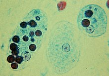
Trophozoites of the amoebic dysentery pathogen Entamoeba histolytica with ingested human red blood cells (dark circles)


The protozoan Ophryocystis elektroscirrha is a parasite of butterfly larvae, passed from female to caterpillar. Severely infected individuals are weak, unable to expand their wings, or unable to eclose, and have shortened lifespans, but parasite levels vary in populations. Infection creates a culling effect, whereby infected migrating animals are less likely to complete the migration. This results in populations with lower parasite loads at the end of the migration. This is not the case in laboratory or commercial rearing, where after a few generations, all individuals can be infected.

Crop tolerance to seawater

From Wikipedia, the free encyclopedia
Crop tolerance to seawater is the ability of an agricultural crop to withstand the high salinity induced by irrigation with seawater, or a mixture of fresh water and seawater. There are crops that can grow on seawater and demonstration farms have shown the feasibility. The government of the Netherlands reports a breakthrough in food security as specific varieties of potatoes, carrots, red onions, white cabbage and broccoli appear to thrive if they are irrigated with salt water.

Salt Farm Texel

The Salt Farm Texel, a farm on the island of Texel, The Netherlands, is testing the salt tolerance of crops under controlled field conditions. There are 56 experimental plots of 160 m2 each that are treated in eight replicas with seven different salt concentrations. These concentrations are obtained with intensive daily drip irrigations of 10 or more mm (i.e. more than 10 liter per m2 per day) with water having a salt concentration expressed in electric conductivity (EC) of 2, 4, 8, 12, 16, 20 and 35 dS/m. The range of EC values is obtained by mixing fresh water with the appropriate amount of seawater having a salinity corresponding to an EC value of about 50 dS/m. After planting, crops were allowed to germinate under fresh water conditions before the salt treatment started.

Soil salinity

The soil salinity is expressed in the electric conductivity of the extract of a saturated soil paste (ECe in dS/m).

Author Schleiff presented a classification of salt tolerance of crops based on ECe in dS/m [5] that may be summarized as follows:

Salt tolerance
ECe (dS/m) ^)
Tolerance classification
    < 2 very sensitive
    2 – 4 sensitive
    4 – 6 slightly sensitive
    6 – 8 moderately tolerant
    8 – 10 tolerant
    > 10 very tolerant
^) The crop performs well (no yield reduction) up to the soil salinity level listed in the table. Beyond that level, the yield goes down.

The main difference with the classification published by Richards in the USDA Agriculture Handbook No. 60, 1954  is that the classes are narrower with steps of 2 dS/m instead of 4. 

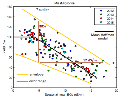
Maas–Hoffman model fitted to a data set.
In this example the crop has a salt tolerance (threshold) of ECe=7 dS/m beyond which the yield declines.
 
Data from Salt Farm Brochure. Boundaries (yellow) and error ranges (brown) have been added. The scatter is quite high. It is not known whether the yield percentages were computed year by year (A), or for all years combined (B). In case B the error ranges are still larger due to annual yield differences. No analysis of variance (Anova) was done to prove that the Maas-Hoffman model really is a statistically significant improvement over a simple, straightforward, downward sloping linear regression model.
 
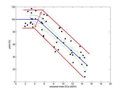
The Salt Farm Texel also published a graph of the yield-salinity relation of white cabbage. Boundary lines were added separately in red color. The boundaries suggest that the slope of the ellipse encompassing the confidence area of the breakpoint should be upward to the right instead of to the left. However the Texel document does not give an explanation of the construction of the ellipse.

Modeling

The Salt Farm Texel uses the Maas–Hoffman model for crop response to soil salinity. The model uses a response function starting with a horizontal line connected furtheron to a downward sloping line. The connection point is also called threshold or tolerance. Up to the threshold the crop is not affected by soil salinity while beyond it the yield starts declining. The model is fitted to the data by piecewise linear regression.

Results

Crop Variety   ^) Threshold *)
(ECe in dS/m)
Class
Potato   x) Mignonne      #) 4.1 slightly sensitive
Achilles 2.9 sensitive
Foc 2.1 sensitive
Met 1.9 very sensitive
"927" 3.4 sensitive
Carrot Cas 4.5 slightly sensitive
Ner 3.6 sensitive
Nat < 1 very sensitive
Ben < 1 very sensitive
"101" 3.0 sensitive
"102" 5.0 slightly sensitive
Pri 2.1 sensitive
Onion Alo 2.4 sensitive
Red 5.9 slightly sensitive
San 3.2 sensitive
Hyb 3.4 sensitive
Lettuce Batavia H < 1 very sensitive
Batavia S 2.3 sensitive
Butterhead L 1.8 very sensitive
Cabbage   White cabbage   #)   4.6 slightly sensitive
Broccoli 5.6 slightly sensitive
Barley Que 2014 3.3 sensitive           +)
Que 2015 1.7 very sensitive   +)
^) Many variety names are uncommon as they consist of 3 letters only
*) It is not known what the results would have been if the planting was not done under fresh water conditions but in saline conditions.
#) Graphs with scatter plots are shown in the report for these two varieties only. They show considerable variation both in Y (Yield) and X (ECe) direction.
x) For potato only one comparable value is known in literature, namely for the very sensitive variety white rose having a threshold of 1.7 dS/m
+) For barley, in contrast, the U.S. Salinity Laboratory mentions a threshold value of ECe = 8 dS/m, which makes it a tolerant crop 

Summary

The highest tolerance is found for the onion variety "Red" which classifies as slightly sensitive. All crops classify in the range from very sensitive over sensitive to slightly sensitive. There is no crop classified as tolerant, not even moderately tolerant.

S-curve model

In the Texel report, also the Van Genuchten-Gupta model (giving an S-curve) was used to find the soil salinity at the 90% yield point. The rationale for this was not given.

Lentils

Lentils
 
The Mediterranean Agronomic Institute, Valenzano, Bari, Italy South coast grew 5 cultivars of lentil irrigated with sea water of different salinity levels. Saline water was prepared by mixing fresh water (EC = 0.9 dS/m) with sea water (EC = 48 dS/m) to achieve salinity levels of 3.0, 6.0, 9.0 and 12.0 dS/m. Some of the results are shown in the following table:

Salinity
(dS/m)
Relative seedling length in % (control = 100%)
by cultivar
ILL4400 ILL5582 ILL5845 ILL5883 ILL8006
      3     98     83     82     98     96
      6     70     43     78     90     83
      9     57     48     63     52     62
    12     36     40     38     30     43

Halophytes

Turtleweed
 
Halophytes, or salt loving plants, can be irrigated with pure seawater with the aim to grow fodder crops. A trial was made by Glenn et al. to use halophytes for feeding of sheep and it was concluded that the animals thrived well.

Setting the yield of an alfalfa (lucerne) fodder crop irrigated with fresh water (2 kg/m2) at 100%, the following results were obtained for the yield of halophytic crops irrigated with seawater:

Crop Relative yield (%)
Atriplex lentiformis, Quailbush 90
Pickleweed, Turtleweed 89
Suaeda, Sea blite 88
Glasswort, Salicornia 87
Sesuvium, Sea purslane 85
Distichlis palmeri , Palmers grass 65
Atriplex cinerea , Coast salt bush 45
>
Barley (Hordeum vulgare)

Barley

After selecting the most salt tolerant strains, the University of California at Davis has grown barley irrigated with pure seawater and obtained half the normal yield per acre, i.e. half of the average yield per acre at national level. The experiment was conducted at Bodega Bay, North of San Francisco, in a laboratory on the Pacific Ocean.

Rice

A team led by Liu Shiping, a professor of agriculture at Yangzhou University, created rice varieties that can be grown in salt water, and achieve yields of 6.5 to 9.3 tons per hectare.[12][13]

Lettuce, Chard and Chicory

In a recent trial comparing three seawater and freshwater blends (i.e. 5%–10%–15% of seawater), some scientists found that lettuce productivity was negatively affected by 10% and 15% of seawater, whereas chard and chicory’s growth was not affected by any blend. Interestingly, water consumptions dropped and WUE significantly upturned in every tested crop accordingly with increased seawater concentrations. They concluded that certain amounts of seawater can be practically used in hydroponics, allowing freshwater saving and increasing certain mineral nutrients concentrations.

Halophile

From Wikipedia, the free encyclopedia
https://en.wikipedia.org/wiki/Halophile 

Halophiles are organisms that thrive in high salt concentrations. They are a type of extremophile organism. The name comes from the Greek word for "salt-loving". While most halophiles are classified into the Archaea domain, there are also bacterial halophiles and some eukaryota, such as the alga Dunaliella salina or fungus Wallemia ichthyophaga. Some well-known species give off a red color from carotenoid compounds, notably bacteriorhodopsin. Halophiles can be found anywhere with a concentration of salt five times greater than the salt concentration of the ocean, such as the Great Salt Lake in Utah, Owens Lake in California, the Dead Sea, and in evaporation ponds.

Classification

Halophiles are categorized as slight, moderate, or extreme, by the extent of their halotolerance. Slight halophiles prefer 0.3 to 0.8 M (1.7 to 4.8%—seawater is 0.6 M or 3.5%), moderate halophiles 0.8 to 3.4 M (4.7 to 20%), and extreme halophiles 3.4 to 5.1 M (20 to 30%) salt content. Halophiles require sodium chloride (salt) for growth, in contrast to halotolerant organisms, which do not require salt but can grow under saline conditions.

Lifestyle

High salinity represents an extreme environment to which relatively few organisms have been able to adapt and occupy. Most halophilic and all halotolerant organisms expend energy to exclude salt from their cytoplasm to avoid protein aggregation ('salting out'). To survive the high salinities, halophiles employ two differing strategies to prevent desiccation through osmotic movement of water out of their cytoplasm. Both strategies work by increasing the internal osmolarity of the cell. In the first (which is employed by the majority of halophilic bacteria, some archaea, yeasts, algae and fungi), organic compounds are accumulated in the cytoplasm—osmoprotectants which are known as compatible solutes. These can be either synthesised or accumulated from the environment. The most common compatible solutes are neutral or zwitterionic, and include amino acids, sugars, polyols, betaines, and ectoines, as well as derivatives of some of these compounds. 

The second, more radical adaptation involves the selective influx of potassium (K+) ions into the cytoplasm. This adaptation is restricted to the moderately halophilic bacterial order Halanaerobiales, the extremely halophilic archaeal family Halobacteriaceae, and the extremely halophilic bacterium Salinibacter ruber. The presence of this adaptation in three distinct evolutionary lineages suggests convergent evolution of this strategy, it being unlikely to be an ancient characteristic retained in only scattered groups or passed on through massive lateral gene transfer. The primary reason for this is the entire intracellular machinery (enzymes, structural proteins, etc.) must be adapted to high salt levels, whereas in the compatible solute adaptation, little or no adjustment is required to intracellular macromolecules; in fact, the compatible solutes often act as more general stress protectants, as well as just osmoprotectants.

Of particular note are the extreme halophiles or haloarchaea (often known as halobacteria), a group of archaea, which require at least a 2 M salt concentration and are usually found in saturated solutions (about 36% w/v salts). These are the primary inhabitants of salt lakes, inland seas, and evaporating ponds of seawater, such as the deep salterns, where they tint the water column and sediments bright colors. These species most likely perish if they are exposed to anything other than a very high-concentration, salt-conditioned environment. These prokaryotes require salt for growth. The high concentration of sodium chloride in their environment limits the availability of oxygen for respiration. Their cellular machinery is adapted to high salt concentrations by having charged amino acids on their surfaces, allowing the retention of water molecules around these components. They are heterotrophs that normally respire by aerobic means. Most halophiles are unable to survive outside their high-salt native environments. Indeed, many cells are so fragile that when placed in distilled water, they immediately lyse from the change in osmotic conditions. 

Halophiles may use a variety of energy sources. They can be aerobic or anaerobic. Anaerobic halophiles include phototrophic, fermentative, sulfate-reducing, homoacetogenic, and methanogenic species.

The Haloarchaea, and particularly the family Halobacteriaceae, are members of the domain Archaea, and comprise the majority of the prokaryotic population in hypersaline environments. Currently, 15 recognised genera are in the family. The domain Bacteria (mainly Salinibacter ruber) can comprise up to 25% of the prokaryotic community, but is more commonly a much lower percentage of the overall population. At times, the alga Dunaliella salina can also proliferate in this environment.

A comparatively wide range of taxa has been isolated from saltern crystalliser ponds, including members of these genera: Haloferax, Halogeometricum, Halococcus, Haloterrigena, Halorubrum, Haloarcula, and Halobacterium. However, the viable counts in these cultivation studies have been small when compared to total counts, and the numerical significance of these isolates has been unclear. Only recently has it become possible to determine the identities and relative abundances of organisms in natural populations, typically using PCR-based strategies that target 16 Svedberg small subunit ribosomal ribonucleic acid (16S rRNA) genes. While comparatively few studies of this type have been performed, results from these suggest that some of the most readily isolated and studied genera may not in fact be significant in the in situ community. This is seen in cases such as the genus Haloarcula, which is estimated to make up less than 0.1% of the in situ community, but commonly appears in isolation studies.

Genomic and proteomic signature

The comparative genomic and proteomic analysis showed distinct molecular signatures exist for environmental adaptation of halophiles. At the protein level, the halophilic species are characterized by low hydrophobicity, overrepresentation of acidic residues, underrepresentation of Cys, lower propensities for helix formation, and higher propensities for coil structure. The core of these proteins is less hydrophobic, such as DHFR, that was found to have narrower β-strands. At the DNA level, the halophiles exhibit distinct dinucleotide and codon usage.

Examples

Halobacterium is a genus of the Archaea that has a high tolerance for elevated levels of salinity. Some species of halobacteria have acidic proteins that resist the denaturing effects of salts. Halococcus is a specific genus of the family Halobacteriaceae.

Some hypersaline lakes are a habitat to numerous families of halophiles. For example, the Makgadikgadi Pans in Botswana form a vast, seasonal, high-salinity water body that manifests halophilic species within the diatom genus Nitzschia in the family Bacillariaceae, as well as species within the genus Lovenula in the family Diaptomidae. Owens Lake in California also contains a large population of the halophilic bacterium Halobacterium halobium.

Wallemia ichthyophaga is a basidiomycetous fungus, which requires at least 1.5 M sodium chloride for in vitro growth, and it thrives even in media saturated with salt. Obligate requirement for salt is an exception in fungi. Even species that can tolerate salt concentrations close to saturation (for example Hortaea werneckii) in almost all cases grow well in standard microbiological media without the addition of salt.

The fermentation of salty foods (such as soy sauce, Chinese fermented beans, salted cod, salted anchovies, sauerkraut, etc.) often involves halobacteria, as either essential ingredients or accidental contaminants. One example is Chromohalobacter beijerinckii, found in salted beans preserved in brine and in salted herring. Tetragenococcus halophilus is found in salted anchovies and soy sauce.

Artemia is a ubiquitous genus of small halophilic crustaceans living in salt lakes (such as Great Salt Lake) and solar salterns that can exist in water approaching the precipitation point of NaCl, 340 g L−1 and can withstand strong osmotic shocks thanks to its mitigating strategies for fluctuating salinity levels, such as its unique larval salt gland and osmoregulatory capacity.

North Ronaldsay sheep are a breed of sheep originating from Orkney, Scotland. They have limited access to fresh water sources on the island and to their only food source is seaweed. They have adapted to handle salt concentrations that would kill other breeds of sheep.

Friday, February 21, 2020

Psychrophile

From Wikipedia, the free encyclopedia
https://en.wikipedia.org/wiki/Psychrophile 

Psychrophiles or cryophiles (adj. psychrophilic or cryophilic) are extremophilic organisms that are capable of growth and reproduction in low temperatures, ranging from −20 °C to +10 °C. They are found in places that are permanently cold, such as the polar regions and the deep sea. They can be contrasted with thermophiles, which are organisms that thrive at unusually high temperatures. Psychrophile is Greek for 'cold-loving'.

Many such organisms are bacteria or archaea, but some eukaryotes such as lichens, snow algae, fungi, and wingless midges, are also classified as psychrophiles.

Biology

The lichen Xanthoria elegans can continue to photosynthesize at −24 °C.

Habitat

The cold environments that psychrophiles inhabit are ubiquitous on Earth, as a large fraction of our planetary surface experiences temperatures lower than 15 °C. They are present in permafrost, polar ice, glaciers, snowfields and deep ocean waters. These organisms can also be found in pockets of sea ice with high salinity content. Microbial activity has been measured in soils frozen below −39 °C. In addition to their temperature limit, psychrophiles must also adapt to other extreme environmental constraints that may arise as a result of their habitat. These constraints include high pressure in the deep sea, and high salt concentration on some sea ice.

Adaptations

Psychrophiles are protected from freezing and the expansion of ice by ice-induced desiccation and vitrification (glass transition), as long as they cool slowly. Free living cells desiccate and vitrify between −10 °C and −26 °C. Cells of multicellular organisms may vitrify at temperatures below −50 °C. The cells may continue to have some metabolic activity in the extracellular fluid down to these temperatures, and they remain viable once restored to normal temperatures.

They must also overcome the stiffening of their lipid cell membrane, as this is important for the survival and functionality of these organisms. To accomplish this, psychrophiles adapt lipid membrane structures that have a high content of short, unsaturated fatty acids. Compared to longer saturated fatty acids, incorporating this type of fatty acid allows for the lipid cell membrane to have a lower melting point, which increases the fluidity of the membranes. In addition, carotenoids are present in the membrane, which help modulate the fluidity of it.

Antifreeze proteins are also synthesized to keep psychrophiles' internal space liquid, and to protect their DNA when temperatures drop below water's freezing point. By doing so, the protein prevents any ice formation or recrystallization process from occurring.

The enzymes of these organisms have been hypothesized to engage in an activity-stability-flexibility relationship as a method for adapting to the cold; the flexibility of their enzyme structure will increase as a way to compensate for the freezing effect of their environment.

Certain cryophiles, such as Gram-negative bacteria Vibrio and Aeromonas spp., can transition into a viable but nonculturable (VBNC) state. During VBNC, a micro-organism can respirate and use substrates for metabolism – however, it cannot replicate. An advantage of this state is that it is highly reversible. It has been debated whether VBNC is an active survival strategy or if eventually the organism's cells will no longer be able to be revived. There is proof however it may be very effective – Gram positive bacteria Actinobacteria have been shown to have lived about 500,000 years in the permafrost conditions of Antarctica, Canada, and Siberia.

Taxonomic range

The wingless midge (Chironomidae) Belgica antarctica.

Psychrophiles include bacteria, lichens, fungi, and insects. 

Among the bacteria that can tolerate extreme cold are Arthrobacter sp., Psychrobacter sp. and members of the genera Halomonas, Pseudomonas, Hyphomonas, and Sphingomonas. Another example is Chryseobacterium greenlandensis, a psychrophile that was found in 120,000-year-old ice.
Umbilicaria antarctica and Xanthoria elegans are lichens that have been recorded photosynthesizing at temperatures ranging down to −24 °C, and they can grow down to around −10 °C. Some multicellular eukaryotes can also be metabolically active at sub-zero temperatures, such as some conifers; those in the Chironomidae family are still active at −16 °C.

Penicillium is a genus of fungi found in a wide range of environments including extreme cold.

Among the psychrophile insects, the Grylloblattidae or icebugs, found on mountaintops, have optimal temperatures between 1-4 °C. The wingless midge (Chironomidae) Belgica antarctica can tolerate salt, being frozen and strong ultraviolet, and has the smallest known genome of any insect. The small genome, of 99 million base pairs, is thought to be adaptive to extreme environments.

Psychrotrophic bacteria

Psychrotrophic microbes are able to grow at temperatures below 7 °C (44.6 °F), but have better growth rates at higher temperatures. Psychrotrophic bacteria and fungi are able to grow at refrigeration temperatures, and can be responsible for food spoilage. They provide an estimation of the product's shelf life, but also they can be found in soils, in surface and deep sea waters, in Antarctic ecosystems, and in foods.

Psychrotrophic bacteria are of particular concern to the dairy industry. Most are killed by pasteurization; however, they can be present in milk as post-pasteurization contaminants due to less than adequate sanitation practices. According to the Food Science Department at Cornell University, psychrotrophs are bacteria capable of growth at temperatures at or less than 7 °C (44.6 °F). At freezing temperatures, growth of psychrotrophic bacteria becomes negligible or virtually stops.

All three subunits of the RecBCD enzyme are essential for physiological activities of the enzyme in the Antarctic Pseudomonas syringae, namely, repairing of DNA damage and supporting the growth at low temperature. The RecBCD enzymes are exchangeable between the psychrophilic P. syringae and the mesophilic E. coli when provided with the entire protein complex from same species. However, the RecBC proteins (RecBCPs and RecBCEc) of the two bacteria are not equivalent; the RecBCEc is proficient in DNA recombination and repair, and supports the growth of P. syringae at low temperature, while RecBCPs is insufficient for these functions. Finally, both helicase and nuclease activity of the RecBCDPs are although important for DNA repair and growth of P. syringae at low temperature, the RecB-nuclease activity is not essential in vivo.

Versus psychrotroph

In 1940, ZoBell and Conn stated that they had never encountered "true psychrophiles" or organisms that grow best at relatively low temperatures. In 1958, J. L. Ingraham supported this by concluding that there are very few or possibly no bacteria that fit the textbook definitions of psychrophiles. Richard Y. Morita emphasizes this by using the term psychrotroph to describe organisms that do not meet the definition of psychrophiles. The confusion between the terms psychrotrophs and psychrophiles was started because investigators were unaware of the thermolability of psychrophilic organisms at the laboratory temperatures. Due to this, early investigators did not determine the cardinal temperatures for their isolates.

The similarity between these two is that they are both capable of growing at zero, but optimum and upper temperature limits for the growth are lower for psychrophiles compared to psychrotrophs. Psychrophiles are also more often isolated from permanently cold habitats compared to psychrotrophs. Although psychrophilic enzymes remain under-used because the cost of production and processing at low temperatures is higher than for the commercial enzymes that are presently in use, the attention and resurgence of research interest in psychrophiles and psychrotrophs will be a contributor to the betterment of the environment and the desire to conserve energy.

Entropy (information theory)

From Wikipedia, the free encyclopedia https://en.wikipedia.org/wiki/Entropy_(information_theory) In info...