Search This Blog

Tuesday, March 17, 2015

B vitamins


From Wikipedia, the free encyclopedia


B vitamins are a class of water-soluble vitamins that play important roles in cell metabolism. Though these vitamins share similar names, research shows that they are chemically distinct vitamins that often coexist in the same foods. In general, supplements containing all eight are referred to as a vitamin B complex. Individual B vitamin supplements are referred to by the specific name of each vitamin (e.g., B1, B2, B3 etc.).

List of B vitamins

B vitamin molecular functions

Vitamin Name Structure Molecular Function
Vitamin B1 Thiamine Thiamin.svg Thiamine plays a central role in the generation of energy from carbohydrates. It is involved in RNA and DNA production, as well as nerve function. Its active form is a coenzyme called thiamine pyrophosphate (TPP), which takes part in the conversion of pyruvate to acetyl coenzyme A (CoA) in metabolism.[1]
Vitamin B2 Riboflavin Riboflavin.svg Riboflavin is involved in the energy production for the electron transport chain, the citric acid cycle, as well as the catabolism of fatty acids (beta oxidation)[2]
Vitamin B3 Niacin
Niacin structure.svg
Niacin is composed of two structures: nicotinic acid and nicotinamide. There are two co-enzyme forms of niacin: nicotinamide adenine dinucleotide (NAD) and nicotinamide adenine dinucleotide phosphate (NADP). Both play an important role in energy transfer reactions in the metabolism of glucose, fat and alcohol.[3]
NAD carries hydrogens and their electrons during metabolic reactions, including the pathway from the citric acid cycle to the electron transport chain. NADP is a coenzyme in lipid and nucleic acid synthesis.[4]
Vitamin B5 Pantothenic acid Pantothenic acid structure.svg Pantothenic acid is involved in the oxidation of fatty acids and carbohydrates. Coenzyme A, which can be synthesised from pantothenic acid, is involved in the synthesis of amino acids, fatty acids, ketones, cholesterol,[5] phospholipids, steroid hormones, neurotransmitters (such as acetylcholine), and antibodies.[6]
Vitamin B6 pyridoxine, pyridoxal, pyridoxamine Pyridoxal-phosphate.svg The active form pyridoxal 5'-phosphate (PLP) (depicted) serves as a cofactor in many enzyme reactions mainly in amino acid metabolism including biosynthesis of neurotransmitters.
Vitamin B7 Biotin Biotin structure JA.png Biotin plays a key role in the metabolism of lipids, proteins and carbohydrates. It is a critical co-enzyme of four carboxylases: acetyl CoA carboxylase, which is involved in the synthesis of fatty acids from acetate; propionyl CoA carboxylase, involved in gluconeogenesis; β-methylcrotonyl CoA carboxylase, involved in the metabolism of leucin; and pyruvate CoA carboxylase, which is involved in the metabolism of energy, amino acids and cholesterol.[7]
Vitamin B9 Folic acid Folic acid.svg Folic acid acts as a co-enzyme in the form of tetrahydrofolate (THF), which is involved in the transfer of single-carbon units in the metabolism of nucleic acids and amino acids. THF is involved in pyrimidine nucleotide synthesis, so is needed for normal cell division, especially during pregnancy and infancy, which are times of rapid growth. Folate also aids in erythropoiesis, the production of red blood cells.[8]
Vitamin B12 Cobalamin Cobalamin.png Vitamin B12 is involved in the cellular metabolism of carbohydrates, proteins and lipids. It is essential in the production of blood cells in bone marrow, and for nerve sheaths and proteins.[9] Vitamin B12 functions as a co-enzyme in intermediary metabolism for the methionine synthase reaction with methylcobalamin, and the methylmalonyl CoA mutase reaction with adenosylcobalamin.[10]

B vitamin deficiency

Several named vitamin deficiency diseases may result from the lack of sufficient B vitamins. Deficiencies of other B vitamins result in symptoms that are not part of a named deficiency disease.

Vitamin Name Deficiency effects
Vitamin B1 thiamine Deficiency causes beriberi. Symptoms of this disease of the nervous system include weight loss, emotional disturbances, Wernicke's encephalopathy (impaired sensory perception), weakness and pain in the limbs, periods of irregular heartbeat, and edema (swelling of bodily tissues). Heart failure and death may occur in advanced cases. Chronic thiamin deficiency can also cause Korsakoff's syndrome, an irreversible dementia characterized by amnesia and compensatory confabulation.
Vitamin B2 riboflavin Deficiency causes ariboflavinosis. Symptoms may include cheilosis (cracks in the lips), high sensitivity to sunlight, angular cheilitis, glossitis (inflammation of the tongue), seborrheic dermatitis or pseudo-syphilis (particularly affecting the scrotum or labia majora and the mouth), pharyngitis (sore throat), hyperemia, and edema of the pharyngeal and oral mucosa.
Vitamin B3 niacin Deficiency, along with a deficiency of tryptophan causes pellagra. Symptoms include aggression, dermatitis, insomnia, weakness, mental confusion, and diarrhea. In advanced cases, pellagra may lead to dementia and death (the 3(+1) Ds: dermatitis, diarrhea, dementia, and death).
Vitamin B5 pantothenic acid Deficiency can result in acne and paresthesia, although it is uncommon.
Vitamin B6 pyridoxine, pyridoxal, pyridoxamine
Vitamin B7 biotin Deficiency does not typically cause symptoms in adults but may lead to impaired growth and neurological disorders in infants. Multiple carboxylase deficiency, an inborn error of metabolism, can lead to biotin deficiency even when dietary biotin intake is normal.
Vitamin B9 folic acid Deficiency results in a macrocytic anemia, and elevated levels of homocysteine. Deficiency in pregnant women can lead to birth defects. Supplementation is often recommended during pregnancy. Researchers have shown that folic acid might also slow the insidious effects of age on the brain.
Vitamin B12 cobalamin Deficiency results in a macrocytic anemia, elevated homocysteine, peripheral neuropathy, memory loss and other cognitive deficits. It is most likely to occur among elderly people, as absorption through the gut declines with age; the autoimmune disease pernicious anemia is another common cause. It can also cause symptoms of mania and psychosis. In rare extreme cases, paralysis can result.

B vitamin side effects


Because water-soluble B vitamins are eliminated in the urine, taking large doses of certain B vitamins usually only produces transient side-effects. General side effects may include restlessness, nausea and insomnia. These side-effects are almost always caused by dietary supplements and not foodstuffs.

Vitamin Name Tolerable Upper Intake Level Harmful effects
Vitamin B1 thiamine None[11] No known toxicity from oral intake. There are some reports of anaphylaxis caused by high-dose thiamin injections into the vein or muscle. However, the doses were greater than the quantity humans can physically absorb from oral intake.[11]
Vitamin B2 riboflavin None.[12] No evidence of toxicity based on limited human and animal studies. The only evidence of adverse effects associated with riboflavin comes from in vitro studies showing the production of reactive oxygen species (free radicals) when riboflavin was exposed to intense visible and UV light.[12]
Vitamin B3 niacin 35 mg/day from supplements, drugs or fortified food[13] Intake of 3000 mg/day of nicotinamide and 1500 mg/day of nicotinic acid are associated with nausea, vomiting, and signs and symptoms of liver toxicity. Other effects may include glucose intolerance, and (reversible) ocular effects. Additionally, the nicotinic acid form may cause vasodilatory effects, also known as flushing, including redness of the skin, often accompanied by an itching, tingling, or mild burning sensation, which is also often accompanied by pruritus, headaches, and increased intracranial blood flow, and occasionally accompanied by pain.[13] Medical practitioners prescribe recommended doses up to 2000 mg per day of niacin, usually in time release format, to combat arterial plaque development in cases of high lipid levels.[14]
Vitamin B5 pantothenic acid None No known toxicity
Vitamin B6 pyridoxine, pyridoxal, pyridoxamine
Main article: vitamin B6 § Toxicity
Vitamin B7 biotin None No known toxicity
Vitamin B9 folic acid 1 mg/day [15] Masks B12 deficiency, which can lead to permanent neurological damage[15]
Vitamin B12 cyanocobalamin None established.[16] Acne-like rash [causality is not conclusively established].[16][17]

B vitamin sources

B vitamins are found in whole unprocessed foods. Processed carbohydrates such as sugar and white flour tend to have lower B vitamin than their unprocessed counterparts. For this reason, it is required by law in many countries (including the United States) that the B vitamins thiamine, riboflavin, niacin, and folic acid be added back to white flour after processing. This is sometimes called "Enriched Flour" on food labels. B vitamins are particularly concentrated in meat such as turkey, tuna and liver.[18] Good sources for B vitamins include legumes (pulses or beans), whole grains, potatoes, bananas, chili peppers, tempeh, nutritional yeast, brewer's yeast, and molasses. Although the yeast used to make beer results in beers being a source of B vitamins,[19] their bioavailability ranges from poor to negative as drinking ethanol inhibits absorption of thiamine (B1),[20][21] riboflavin (B2),[22] niacin (B3),[23] biotin (B7),[24] and folic acid (B9).[25][26] In addition, each of the preceding studies further emphasizes that elevated consumption of beer and other ethanol-based drinks results in a net deficit of those B vitamins and the health risks associated with such deficiencies.

The B12 vitamin is of note because it is not available from plant products, making B12 deficiency a legitimate concern for vegans. Manufacturers of plant-based foods will sometimes report B12 content, leading to confusion about what sources yield B12. The confusion arises because the standard US Pharmacopeia (USP) method for measuring the B12 content does not measure the B12 directly. Instead, it measures a bacterial response to the food. Chemical variants of the B12 vitamin found in plant sources are active for bacteria, but cannot be used by the human body. This same phenomenon can cause significant over-reporting of B12 content in other types of foods as well.[27]

Another popular means of increasing one's vitamin B intake is through the use of dietary supplements. B vitamins are also commonly added to energy drinks, many of which have been marketed with large amounts of B vitamins[28] with claims that this will cause the consumer to "sail through your day without feeling jittery or tense."[28] Some nutritionists have been critical of these claims, pointing out for instance that while B vitamins do "help unlock the energy in foods," most Americans acquire the necessary amounts easily in their diets.[28]

Because they are soluble in water, excess B vitamins (such as may be ingested via supplements) are generally readily excreted, although individual absorption, use and metabolism may vary…"[28] The elderly and athletes may need to supplement their intake of B12 and other B vitamins due to problems in absorption and increased needs for energy production. In cases of severe deficiency B vitamins, especially B12, may also be delivered by injection to reverse deficiencies.[29] Both type 1 and type 2 diabetics may also be advised to supplement thiamine based on high prevalence of low plasma thiamine concentration and increased thiamine clearance associated with diabetes.[30] Also, Vitamin B9 (folic acid) deficiency in early embryo development has been linked to neural tube defects. Thus, women planning to become pregnant are usually encouraged to increase daily dietary folic acid intake and/or take a supplement.[31]

Related nutrients

Many of the following substances have been referred to as vitamins as they were once believed to be vitamins. They are no longer considered as such, and the numbers that were assigned to them now form the "gaps" in the true series of B-complex vitamins described above (e.g., there is no vitamin B4). Some of them, though not essential to humans, are essential in the diets of other organisms; others have no known nutritional value and may even be toxic under certain conditions.
  • Vitamin B4: can refer to the distinct chemicals choline, adenine, or carnitine.[32][33] Choline is synthesized by the human body, but not sufficiently to maintain good health, and is now considered an essential dietary nutrient.[34] Adenine is a nucleobase synthesized by the human body.[35] Carnitine is an essential dietary nutrient for certain worms, but not for humans.[36]
  • Vitamin B8: adenosine monophosphate (AMP), also known as adenylic acid.[37] Vitamin B8 may also refer to inositol.[38]
  • Vitamin B10: para-aminobenzoic acid (pABA or PABA), a chemical component of the folate molecule produced by plants and bacteria, and found in many foods.[39][40] It is best known as a UV-blocking sunscreen applied to the skin, and is sometimes taken orally for certain medical conditions.[39][41]
  • Vitamin B11: pteryl-hepta-glutamic acid—chick growth factor, which is a form of folic acid. Later found to be one of five folates necessary for humans. Vitamin Bc-conjugate was also found to be PHGA.
  • Vitamin B13: orotic acid.[42]
  • Vitamin B14: cell proliferant, anti-anemia, rat growth factor, and antitumor pterin phosphate named by Earl R. Norris. Isolated from human urine at 0.33ppm (later in blood), but later abandoned by him as further evidence did not confirm this. He also claimed this was not xanthopterin.
  • Vitamin B15: pangamic acid,[42] also known as pangamate. Promoted in various forms as a dietary supplement and drug; considered unsafe and subject to seizure by the US Food and Drug Administration.[43]
  • Vitamin B16: dimethylglycine (DMG)[44] is synthesized by the human body in the Citric acid (or Kreb) cycle.
  • Vitamin B17: nitrilosides, amygdalin or Laetrile. These substances are found in a number of seeds, sprouts, beans, tubers, and grains. While toxic in large quantities, proponents claim that it is effective in cancer treatment and prevention despite a lack of scientific evidence.[45]
  • Vitamin B20: L-carnitine.[44]
  • Vitamin Bf: carnitine.[37]
  • Vitamin Bm: myo-inositol, also called “mouse antialopaecia factor”.[46]
  • Vitamin Bp: “antiperosis factor”, which prevents perosis, a leg disorder, in chicks; can be replaced by choline and manganese salts.[36][37][47]
  • Vitamin BT: carnitine.[48][36]
  • Vitamin Bv: a type of B6 other than pyridoxine.
  • Vitamin BW: a type of biotin other than d-biotin.
  • Vitamin Bx: an alternative name for both pABA (see vitamin B10) and pantothenic acid.[36][41]

Cellular respiration


From Wikipedia, the free encyclopedia


Cellular respiration is the set of metabolic reactions and processes that take place in the cells of organisms to convert biochemical energy from nutrients into adenosine triphosphate (ATP), and then release waste products.[1] The reactions involved in respiration are catabolic reactions, which break large molecules into smaller ones, releasing energy in the process, as weak so-called "high-energy" bonds are replaced by stronger bonds in the products. Respiration is one of the key ways a cell gains useful energy to fuel cellular activity. Cellular respiration is considered an exothermic redox reaction which releases heat. The overall reaction occurs in a series of biochemical steps, most of which are redox reactions themselves. Although technically, cellular respiration is a combustion reaction, it clearly does not resemble one when it occurs in a living cell due to slow release of energy from the series of reactions.

Nutrients that are commonly used by animal and plant cells in respiration include sugar, amino acids and fatty acids, and the most common oxidizing agent (electron acceptor) is molecular oxygen (O2). The chemical energy stored in ATP (its third phosphate group is weakly bonded to the rest of the molecule and is cheaply broken allowing stronger bonds to form, thereby transferring energy for use by the cell) can then be used to drive processes requiring energy, including biosynthesis, locomotion or transportation of molecules across cell membranes.

Aerobic respiration


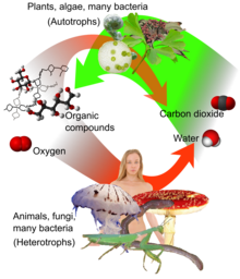
Aerobic respiration (red arrows) is the main means by which both fungi and animals utilize chemical energy in the form of organic compounds that were previously created through photosynthesis (green arrow).

Aerobic respiration requires oxygen in order to generate ATP. Although carbohydrates, fats, and proteins are consumed as reactants, it is the preferred method of pyruvate breakdown in glycolysis and requires that pyruvate enter the mitochondrion in order to be fully oxidized by the Krebs cycle. The products of this process are carbon dioxide and water, but the energy transferred is used to break strong bonds in ADP as the third phosphate group is added to form ATP (adenosine triphosphate), by substrate-level phosphorylation, NADH and FADH2

Simplified reaction: C6H12O6 (s) + 6 O2 (g) → 6 CO2 (g) + 6 H2O (l) + heat
ΔG = −2880 kJ per mol of C6H12O6

The negative ΔG indicates that the reaction can occur spontaneously.

The potential of NADH and FADH2 is converted to more ATP through an electron transport chain with oxygen as the "terminal electron acceptor". Most of the ATP produced by aerobic cellular respiration is made by oxidative phosphorylation. This works by the energy released in the consumption of pyruvate being used to create a chemiosmotic potential by pumping protons across a membrane. This potential is then used to drive ATP synthase and produce ATP from ADP and a phosphate group. Biology textbooks often state that 38 ATP molecules can be made per oxidised glucose molecule during cellular respiration (2 from glycolysis, 2 from the Krebs cycle, and about 34 from the electron transport system).[2] However, this maximum yield is never quite reached due to losses (leaky membranes) as well as the cost of moving pyruvate and ADP into the mitochondrial matrix, and current estimates range around 29 to 30 ATP per glucose.[2]

Aerobic metabolism is up to 15 times more efficient than anaerobic metabolism (which yields 2 molecules ATP per 1 molecule glucose). However some anaerobic organisms, such as methanogens are able to continue with anaerobic respiration, yielding more ATP by using other inorganic molecules (not oxygen) as final electron acceptors in the electron transport chain. They share the initial pathway of glycolysis but aerobic metabolism continues with the Krebs cycle and oxidative phosphorylation. The post-glycolytic reactions take place in the mitochondria in eukaryotic cells, and in the cytoplasm in prokaryotic cells.

Glycolysis


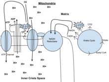
Out of the cytoplasm it goes into the Krebs cycle with the acetyl CoA. It then mixes with CO2 and makes 2 ATP, NADH, and FADH. From there the NADH and FADH go into the NADH reductase, which produces the enzyme. The NADH pulls the enzyme's electrons to send through the electron transport chain. The electron transport chain pulls H+ ions through the chain. From the electron transport chain, the released hydrogen ions make ADP for an end result of 32 ATP. 02 attracts itself to the left over electron to make water. Lastly, ATP leaves through the ATP channel and out of the mitochondria.

Glycolysis is a metabolic pathway that takes place in the cytosol of cells in all living organisms. This pathway can function with or without the presence of oxygen. In humans, aerobic conditions produce pyruvate and anaerobic conditions produce lactate. In aerobic conditions, the process converts one molecule of glucose into two molecules of pyruvate (pyruvic acid), generating energy in the form of two net molecules of ATP. Four molecules of ATP per glucose are actually produced, however, two are consumed as part of the preparatory phase. The initial phosphorylation of glucose is required to increase the reactivity (decrease its stability) in order for the molecule to be cleaved into two pyruvate molecules by the enzyme aldolase. During the pay-off phase of glycolysis, four phosphate groups are transferred to ADP by substrate-level phosphorylation to make four ATP, and two NADH are produced when the pyruvate are oxidized. The overall reaction can be expressed this way:
Glucose + 2 NAD+ + 2 Pi + 2 ADP → 2 pyruvate + 2 NADH + 2 ATP + 2 H+ + 2 H2O + heat
Starting with glucose, 1 ATP is used to donate a phosphate to glucose to produce glucose 6-phosphate. Glycogen can be converted into glucose 6-phosphate as well with the help of glycogen phosphorylase. During energy metabolism, glucose 6-phosphate becomes fructose 6-phosphate. An additional ATP is used to phosphorylate fructose 6-phosphate into fructose 1,6-disphosphate by the help of phosphofructokinase. Fructose 1,6-diphosphate then splits into two phosphorylated molecules with three carbon chains which later degrades into pyruvate.

Oxidative decarboxylation of pyruvate

Pyruvate is oxidized to acetyl-CoA and CO2 by the pyruvate dehydrogenase complex (PDC). The PDC contains multiple copies of three enzymes and is located in the mitochondria of eukaryotic cells and in the cytosol of prokaryotes. In the conversion of pyruvate to acetyl-CoA, one molecule of NADH and one molecule of CO2 is formed.

Citric acid cycle

This is also called the Krebs cycle or the tricarboxylic acid cycle. When oxygen is present, acetyl-CoA is produced from the pyruvate molecules created from glycolysis. When oxygen is present, the mitochondria will undergo aerobic respiration which leads to the Krebs cycle. However, if oxygen is not present, fermentation of the pyruvate molecule will occur. In the presence of oxygen, when acetyl-CoA is produced, the molecule then enters the citric acid cycle (Krebs cycle) inside the mitochondrial matrix, and gets oxidized to CO2 while at the same time reducing NAD to NADH. NADH can be used by the electron transport chain to create further ATP as part of oxidative phosphorylation. To fully oxidize the equivalent of one glucose molecule, two acetyl-CoA must be metabolized by the Krebs cycle. Two waste products, H2O and CO2, are created during this cycle.
The citric acid cycle is an 8-step process involving different enzymes and co-enzymes. During the cycle, acetyl-CoA (2 carbons) + oxaloacetate (4 carbons) yields citrate (6 carbons), which is rearranged to a more reactive form called isocitrate (6 carbons). Isocitrate is modified to become α-ketoglutarate (5 carbons), succinyl-CoA, succinate, fumarate, malate, and, finally, oxaloacetate. The net gain of high-energy compounds from one cycle is 3 NADH, 1 FADH2, and 1 GTP; the GTP may subsequently be used to produce ATP. Thus, the total yield from 1 glucose molecule (2 pyruvate molecules) is 6 NADH, 2 FADH2, and 2 ATP.

Oxidative phosphorylation

In eukaryotes, oxidative phosphorylation occurs in the mitochondrial cristae. It comprises the electron transport chain that establishes a proton gradient (chemiosmotic potential) across the boundary of inner membrane by oxidizing the NADH produced from the Krebs cycle. ATP is synthesized by the ATP synthase enzyme when the chemiosmotic gradient is used to drive the phosphorylation of ADP. The electrons are finally transferred to exogenous oxygen and, with the addition of two protons, water is formed.

Efficiency of ATP production

The table below describes the reactions involved when one glucose molecule is fully oxidized into carbon dioxide. It is assumed that all the reduced coenzymes are oxidized by the electron transport chain and used for oxidative phosphorylation.
Step coenzyme yield ATP yield Source of ATP
Glycolysis preparatory phase −2 Phosphorylation of glucose and fructose 6-phosphate uses two ATP from the cytoplasm.
Glycolysis pay-off phase 4 Substrate-level phosphorylation
2 NADH 3 or 5 Oxidative phosphorylation – Each NADH produces net 1.5 ATP (instead of usual 2.5) due to NADH transport over the mitochondrial membrane
Oxidative decarboxylation of pyruvate 2 NADH 5 Oxidative phosphorylation
Krebs cycle 2 Substrate-level phosphorylation
6 NADH 15 Oxidative phosphorylation
2 FADH2 3 Oxidative phosphorylation
Total yield 30  or 32 ATP From the complete oxidation of one glucose molecule to carbon dioxide and oxidation of all the reduced coenzymes.

Although there is a theoretical yield of 38 ATP molecules per glucose during cellular respiration, such conditions are generally not realized due to losses such as the cost of moving pyruvate (from glycolysis), phosphate, and ADP (substrates for ATP synthesis) into the mitochondria. All are actively transported using carriers that utilize the stored energy in the proton electrochemical gradient.
  • Pyruvate is taken up by a specific, low Km transporter to bring it into the mitochondrial matrix for oxidation by the pyruvate dehydrogenase complex.
  • The phosphate carrier (PiC) mediates the electroneutral exchange (antiport) of phosphate (H2PO4; Pi) for OH or symport of phosphate and protons (H+) across the inner membrane, and the driving force for moving phosphate ions into the mitochondria is the proton motive force.
  • The ATP-ADP translocase (also called adenine nucleotide translocase, ANT) is an antiporter and exchanges ADP and ATP across the inner membrane. The driving force is due to the ATP (−4) having a more negative charge than the ADP (−3), and thus it dissipates some of the electrical component of the proton electrochemical gradient.
The outcome of these transport processes using the proton electrochemical gradient is that more than 3 H+ are needed to make 1 ATP. Obviously this reduces the theoretical efficiency of the whole process and the likely maximum is closer to 28–30 ATP molecules.[2] In practice the efficiency may be even lower due to the inner membrane of the mitochondria being slightly leaky to protons.[3] Other factors may also dissipate the proton gradient creating an apparently leaky mitochondria. An uncoupling protein known as thermogenin is expressed in some cell types and is a channel that can transport protons. When this protein is active in the inner membrane it short circuits the coupling between the electron transport chain and ATP synthesis. The potential energy from the proton gradient is not used to make ATP but generates heat. This is particularly important in brown fat thermogenesis of newborn and hibernating mammals.

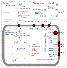
Stoichiometry of aerobic respiration and most known fermentation types in eucaryotic cell. [4] Numbers in circles indicate counts of carbon atoms in molecules, C6 is glucose C6H12O6, C1 carbon dioxide CO2. Mitochondrial outer membrane is omitted.

According to some of newer sources the ATP yield during aerobic respiration is not 36–38, but only about 30–32 ATP molecules / 1 molecule of glucose [4], because:
  • ATP : NADH+H+ and ATP : FADH2 ratios during the oxidative phosphorylation appear to be not 3 and 2, but 2.5 and 1.5 respectively. Unlike in the substrate-level phosphorylation, the stoichiometry here is difficult to establish.
    • ATP synthase produces 1 ATP / 3 H+. However the exchange of matrix ATP for cytosolic ADP and Pi (antiport with OH or symport with H+) mediated by ATP–ADP translocase and phosphate carrier consumes 1 H+ / 1 ATP due to regeneration of the transmembrane potential changed during this transfer, so the net ratio is 1 ATP : 4 H+.
    • The mitochondrial electron transport chain proton pump transfers across the inner membrane 10 H+ / 1 NADH+H+ (4 + 2 + 4) or 6 H+ / 1 FADH2 (2 + 4).
So the final stoichiometry is
1 NADH+H+ : 10 H+ : 10/4 ATP = 1 NADH+H+ : 2.5 ATP
1 FADH2 : 6 H+ : 6/4 ATP = 1 FADH2 : 1.5 ATP
  • ATP : NADH+H+ coming from glycolysis ratio during the oxidative phosphorylation is
    • 1.5, as for FADH2, if hydrogen atoms (2H++2e) are transferred from cytosolic NADH+H+ to mitochondrial FAD by the glycerol phosphate shuttle located in the inner mitochondrial membrane.
    • 2.5 in case of malate-aspartate shuttle transferring hydrogen atoms from cytosolic NADH+H+ to mitochondrial NAD+
So finally we have, per molecule of glucose
Altogether this gives 4 + 3 (or 5) + 20 + 3 = 30 (or 32) ATP per molecule of glucose

The total ATP yield in ethanol or lactic acid fermentation is only 2 molecules coming from glycolysis, because pyruvate is not transferred to the mitochondrion and finally oxidized to the carbon dioxide (CO2), but reduced to ethanol or lactic acid in the cytoplasm.[4]

Fermentation

Without oxygen, pyruvate (pyruvic acid) is not metabolized by cellular respiration but undergoes a process of fermentation. The pyruvate is not transported into the mitochondrion, but remains in the cytoplasm, where it is converted to waste products that may be removed from the cell. This serves the purpose of oxidizing the electron carriers so that they can perform glycolysis again and removing the excess pyruvate. Fermentation oxidizes NADH to NAD+ so it can be re-used in glycolysis. In the absence of oxygen, fermentation prevents the buildup of NADH in the cytoplasm and provides NAD+ for glycolysis. This waste product varies depending on the organism. In skeletal muscles, the waste product is lactic acid. This type of fermentation is called lactic acid fermentation. In strenuous exercise, when energy demands exceed energy supply, the respiratory chain cannot process all of the hydrogen atoms joined by NADH. During anaerobic glycolysis, NAD+ regenerates when pairs of hydrogen combine with pyruvate to form lactate. Lactate formation is catalyzed by lactate dehydrogenase in a reversible reaction. Lactate can also be used as an indirect precursor for liver glycogen. During recovery, when oxygen becomes available, NAD+ attaches to hydrogen from lactate to form ATP. In yeast, the waste products are ethanol and carbon dioxide. This type of fermentation is known as alcoholic or ethanol fermentation. The ATP generated in this process is made by substrate-level phosphorylation, which does not require oxygen.
Fermentation is less efficient at using the energy from glucose: only 2 ATP are produced per glucose, compared to the 38 ATP per glucose nominally produced by aerobic respiration. This is because the waste products of fermentation still contain chemical potential energy that can be released by oxidation. Ethanol, for example, can be burned in an internal combustion engine like gasoline. Glycolytic ATP, however, is created more quickly. For prokaryotes to continue a rapid growth rate when they are shifted from an aerobic environment to an anaerobic environment, they must increase the rate of the glycolytic reactions. For multicellular organisms, during short bursts of strenuous activity, muscle cells use fermentation to supplement the ATP production from the slower aerobic respiration, so fermentation may be used by a cell even before the oxygen levels are depleted, as is the case in sports that do not require athletes to pace themselves, such as sprinting.

Anaerobic respiration

Cellular respiration is the process by which biological fuels are oxidised in the presence of an inorganic electron acceptor (such as oxygen) to produce large amounts of energy, to drive the bulk production of ATP.
Anaerobic respiration is used by some microorganisms in which neither oxygen (aerobic respiration) nor pyruvate derivatives (fermentation) is the final electron acceptor. Rather, an inorganic acceptor such as sulfate or nitrate is used. Such organisms are typically found in unusual places such as underwater caves or near the lava shoots at the bottom of the ocean.

Many high-school biology textbooks[citation needed] incorrectly refer to fermentation (e.g., to lactate) as anaerobic respiration.

Monarch population rebounds as NRDC sues EPA for not stopping ‘Armageddon’–Are GMOs the problem?

| March 17, 2015 |
 
Original link:  http://geneticliteracyproject.org/2015/03/17/monarch-population-rebounds-as-nrdc-sues-epa-for-not-stopping-armageddon-are-gmos-the-problem/
 
chrysalys

The latest yearly count of the monarch butterfly shows a dramatic increase in their numbers, partly reversing at least temporarily recent sharp declines. Scientists with World Wildlife Fund Mexico estimate that some 56.5 million monarchs gathered for the winter after their trek across the United States—up more than 60 percent from last year, when 34 million were counted in Mexico’s Sierra Madre.

While the rebound is heartening, the overall picture is still not good, as the rebound came from very low levels, and remains well below the numbers  over the past 21 years. Butterfly migration counts in Mexico, their primary winter resting place, have only been kept since 1994.
overwintering-figure

The modestly better news came out shortly before the National Resources Defense Council turned its guns on the Environmental Protection Agency, claiming it has failed to heed the NRDC’s warnings, contained in a petition filed more than a year ago, about the dangers to monarchs posed by glyphosate. The NRDC filed the suit in U.S. District Court in New York. The advocacy group and numerous other organizations have pointed to the correlation of the butterfly population decline with the increased use of glyphosate, which is a very mild herbicide introduced decades ago (well before GM crops) to replace far more toxic chemicals. They often release charts such as this:

According to the NRDC news release:
“The longer EPA delays, the greater the risk we could lose the monarch migration,” Sylvia Fallon, an NRDC senior scientist and director of its Wildlife Conservation Project. … Experts say the primary cause for the population collapse is the skyrocketing use of the herbicide glyphosate (originally marketed as Roundup) on genetically modified corn and soybeans, which has wiped out much of the milkweed — a native wildflower — that monarchs need to survive. Since EPA last reviewed the safety of glyphosate in 1993, its use has increased ten-fold, yet the agency has never considered the herbicide’s impact on monarchs.
In effect, the NRDC is accusing the government of conspiring to suppress evidence that the popular herbicide, often paired with GMO crops, is responsible for killing off the monarch butterfly population. Other activist groups have used the same data to mount an offensive against all GMO crops.

“Monarch butterfly decline being caused by GMO agriculture,” blared a recent headline in NaturalNews.com, a junk science site that sells often bogus alternative health remedies, yet enjoys enormous popularity and even credibility among mainstream anti-GMO campaigners.

Does the science stand with the NRDC’s lawsuit and its claims?

Although more reliable statistics are only available for the past two decades, experts contest the simplistic narrative that the monarch butterfly population is mortally threatened.

“Monarchs are not in danger of extinction,” said Lincoln Brower, a monarch conservation scientist at Sweet Briar College. “What is endangered is their spectacular migration and overwintering behavior.”

What is not widely understood is that scientists measure the overwinter numbers in Mexico, not the numbers of monarchs. The situation, and the longterm trends, are not good, but even if the current declines continue, the monarch and other butterflies are not headed toward extinction.

Experts outside of the “activist circuit” believe the monarch butterfly population’s overwintering habitats have been in decline for decades, pre-dating both the widespread of glyphosate in agriculture in the 1980s and the use of GM crops in the mid-1990s.
image6
The decline in butterfly populations, while extremely concerning, is also not unique to the United States. It mirrors almost exactly what is occurring in other places around the world, most especially in Europe, where the growing of GMOs is almost totally nonexistent.

Scientists do know that certain herbicides kill the nuisance milkweed–the most common butterfly habitat in the United States. That’s certainly a contributing factor in altering migratory behavior. But how much, considering that glyphosate use has not accelerated in other places in the world where butterfly populations are in steep decline?

The largest factor in the decline is almost certainly the declining amount of productive agricultural land, in Europe and the United States in particular. Less milkweed—a bane of farmers—grows when there is less farm acreage. The reality is the land use for agriculture is going down while land not suitable for monarchs and other butterflies is going up. According to the USDA, land area designated as “Non-Agricultural”, mainly “Forest land” and “Special Uses”–rural parks, wilderness areas and rural transportation—has been increasing. The proportion of the land base in agricultural uses declined from 63 percent in 1949 to 51 percent in 2007, the latest year for which data are available.


2012 study in the journal Insect Conservation and Diversity estimated that between 1990 and 2010, milkweed prevalence declined 58 percent in Midwest agricultural areas where farmable acreage shrunk dramatically. Over the same time frame, the monarch population declined 81 percent. Additionally, according to Chip Taylor, director of Monarch Watch and a professor of ecology at the University of Kansas, a 2007 congressional ethanol mandate increased the price of corn and soybeans, which encouraged farmers to convert grassland–where milkweed grows–into cropland.

What role then does glyphosate play? 

The on the ground reality is certainly more complex than NRDC’s simplistic narrative. Corn and soybeans are the two biggest crops in the US, amounting to ~170 million acres.  Cotton is about 10 million acres. This graph shows the adoption of herbicide resistant (HT) crops and crops that are engineered to generate a pesticide found commonly in nature (BT). It’s important to note that Bt crops only affect pollinators that go after their pollen, but corn is wind pollinated (over a very short period of time) and soybeans are largely self pollinated.

image[12]

Now compare those trends to the population studies on monarch overwintering populations in Mexico: As you can see, the population crashed during the winter of 1997–when a fraction of the herbicide tolerant crops were being used relative to today. The overwintering numbers also increased dramatically at the turn of the century just as Ht crop usage and the use of glyphosate soared.

About caterpillars and butterflies

The issue with monarchs is complicated by the fact that as caterpillars they can eat only one food, the milkweed plant. Still monarchs thrived as our field crops increased, because the milkweed they rely on could survive tilling (the previous normal method of weed control), such that when its tap root was cut by tilling, often the large pieces healed themselves and the plant still came up in the spring. Milkweed was in fact fairly common in corn, cotton and soybean fields. So the larger population of monarchs we saw in the early 90s was largely because of the way we weeded our field crops.

Then GMO corn, cotton and soy were introduced that were resistant to glyphosate and many farmers quit tilling to control weeds and went to spraying glyphosate instead. Milkweed is not immune to glyphosate though and the amount of it in Midwest corn and soybean fields has gone down. But given all the land that is not farm land, that alone wouldn’t account for their decline. Indeed, Andrew Kniss, a weed expert at the University of Wyoming, wrote in a blog post that the use of herbicide-tolerant plants glyphosate has had only a limited impact on native plant diversity outside the border of the agricultural field.  So to understand the problem, one has to understand the insect itself.

The monarch spends the spring and summer in most of North America, and while in North America the monarchs will go through up to seven generations.  Each generation lasts from 6 to 12 weeks, depending on climate and food supply. The butterfly form itself normally lives from two to six weeks.

As monarchs migrate north, the female lays her eggs only on milkweed plants. The eggs take about a week to hatch into larvae (caterpillars). The larvae feed on the milkweed for about two weeks and then they attach themselves to a twig, shed their outer skin and change into a chrysalis. In about two weeks a full-grown monarch breaks free to start the next generation. Some of each new generation moves north with the warming spring weather. Note that while the caterpillars only live on milkweed, the adult form is free to sip nectar from any flowering plant they find.

As fall approaches a somewhat unique version of the monarch appears. Unlike their parents before them, they don’t immediately reproduce but instead falling temperatures make them avoid procreation and begin the migration back south. Depending on where they begin this trip, these last of the season monarchs will travel many hundreds or even thousands of miles to their winter grounds, mostly in Mexico (East of the Rockies, they will winter in Southern California, and there are a few populations of monarchs living year round in Southern Florida, California and Texas that don’t migrate at all).

On their way South, they store fat in their abdomens as it is needed for the long flights and to last through their winter hibernation, which lasts from November till about May. When they arrive in Mexico, the monarchs gather into dense clusters in the branches of the trees, and by late winter these clusters will contain hundreds to thousands of them. During their long hibernation they will remain perfectly still, clustered in these Mexican forests, surviving only off of their stored fat reserves.

As warm weather arrives in Mexico in February, they begin to move again and during the day the monarchs will once again begin to gather nectar. Ultimately they will mate and lay eggs as it is only their plump offspring which will make the return migration back to North America.

This complex life cycle shows why the key to their survival is multifaceted. Indeed, probably the most important need these insects have is the amount of nectar providing plants they can use to develop the reserves that are needed to make it through their long migration and hibernation period.

Finally there is also the pressure from the dwindling forest area in Mexico where much of their population over winters. The deforestation stems from illegal logging in Mexico. According to Politifact, which reviewed this controversy, it has reduced the areas where monarchs can migrate, affecting their lifecycle. A new study of the monarch butterflies’ winter nesting grounds in central Mexico showed that  small-scale logging is worse than previously thought. The reserve’s fairly small 33,482-acre core zone lost 41 acres of pine and fir trees so far in 2013, about half of that because of illegal logging while the rest of the loss was due to drought or disease-control removal of trees.

Mexico’s government had begun to protect the Monarch’s over-wintering grounds in recent years and in 2012 aerial photographs found very little deforestation due to logging over the previous year (at its peak in 2005, logging depleted as many as 1,140 acres each year).

Still this new study’s analysis of photos taken a decade apart showed that small-scale logging has never gone away and while it is not seen in year-to-year comparisons, the study found the losses by comparing 2001 photographs to the recent ones taken in 2011.

[See: “Trends in Deforestation and Forest Degradation after a Decade of Monitoring in the Monarch Butterfly Biosphere Reserve in Mexico“]

Given the multi-faceted nature of the problem, it seems clear that there are many factors causing the decline, and so the solution also has to address all of these factors. Indeed, many people are now coming to their aid by planting the flowering plants and milkweed they need to survive. Here is a chart of success stories–the creating of new monarch butterfly habitats.

While the total solution is not clear, words of caution, as many ecological conscious home gardeners have started planting milkweed in an attempt to help the monarchs. Caterpillars eat the weed because chemicals within the plant make them unpalatable to predators–but this same latex rich sap is also toxic to people and quite capable of causing severe eye injury, up to and including blindness if you get the sap in your eyes.

So if you do decide to plant it, don’t put it where kids can kids play in it and handle it carefully.

Arthur Doucette is a retired software developer, now writing about issues involving Genetic Engineering.

Criticism of democracy

From Wikipedia, the free encyclopedia https://en.wikipedia.org/wiki/Criticis...