Search This Blog

Thursday, October 4, 2018

Chromatin

From Wikipedia, the free encyclopedia
 
The major structures in DNA compaction: DNA, the nucleosome, the 10 nm "beads-on-a-string" fibre, the 30 nm chromatin fibre and the metaphase chromosome.

Chromatin is a complex of macromolecules found in cells, consisting of DNA, protein, and RNA. The primary functions of chromatin are 1) to package DNA into a more compact, denser shape, 2) to reinforce the DNA macromolecule to allow mitosis, 3) to prevent DNA damage, and 4) to control gene expression and DNA replication. The primary protein components of chromatin are histones that compact the DNA. Chromatin is only found in eukaryotic cells (cells with defined nuclei). Prokaryotic cells have a different organization of their DNA (the prokaryotic chromosome equivalent is called genophore and is localized within the nucleoid region).

Chromatin's structure is currently poorly understood despite being subjected to intense investigation. Its structure depends on several factors. The overall structure depends on the stage of the cell cycle. During interphase, the chromatin is structurally loose to allow access to RNA and DNA polymerases that transcribe and replicate the DNA. The local structure of chromatin during interphase depends on the genes present on the DNA. That DNA which codes genes that are actively transcribed ("turned on") is more loosely packaged and associated with RNA polymerases (referred to as euchromatin) while that DNA which codes inactive genes ("turned off") is more condensed and associated with structural proteins (heterochromatin). Epigenetic chemical modification of the structural proteins in chromatin also alters the local chromatin structure, in particular chemical modifications of histone proteins by methylation and acetylation. As the cell prepares to divide, i.e. enters mitosis or meiosis, the chromatin packages more tightly to facilitate segregation of the chromosomes during anaphase. During this stage of the cell cycle this makes the individual chromosomes in many cells visible by optical microscope.

In general terms, there are three levels of chromatin organization:
  1. DNA wraps around histone proteins forming nucleosomes; the "beads on a string" structure (euchromatin).
  2. Multiple histones wrap into a 30 nm fibre consisting of nucleosome arrays in their most compact form (heterochromatin). (Definitively established to exist in vitro, the 30-nanometer fibre was not seen in recent X-ray studies of human mitotic chromosomes.)
  3. Higher-level DNA packaging of the 30 nm fibre into the metaphase chromosome (during mitosis and meiosis).
There are, however, many cells that do not follow this organisation. For example, spermatozoa and avian red blood cells have more tightly packed chromatin than most eukaryotic cells, and trypanosomatid protozoa do not condense their chromatin into visible chromosomes for mitosis.

Dynamic chromatin structure and hierarchy

Chromatin undergoes various structural changes during a cell cycle. Histone proteins are the basic packer and arranger of chromatin and can be modified by various post-translational modifications to alter chromatin packing (Histone modification). Most of the modifications occur on the histone tail. The consequences in terms of chromatin accessibility and compaction depend both on the amino-acid that is modified and the type of modification. For example, Histone acetylation results in loosening and increased accessibility of chromatin for replication and transcription. Lysine tri-methylation can either be correlated with transcriptional activity (tri-methylation of histone H3 Lysine 4) or transcriptional repression and chromatin compaction (tri-methylation of histone H3 Lysine 9 or 27). Several studies suggested that different modifications could occur simultaneously. For example, it was proposed that a bivalent structure (with tri-methylation of both Lysine 4 and 27 on histone H3) was involved in mammalian early development.

Polycomb-group proteins play a role in regulating genes through modulation of chromatin structure.

DNA structure

The structures of A-, B-, and Z-DNA.

In nature, DNA can form three structures, A-, B-, and Z-DNA. A- and B-DNA are very similar, forming right-handed helices, whereas Z-DNA is a left-handed helix with a zig-zag phosphate backbone. Z-DNA is thought to play a specific role in chromatin structure and transcription because of the properties of the junction between B- and Z-DNA.

At the junction of B- and Z-DNA, one pair of bases is flipped out from normal bonding. These play a dual role of a site of recognition by many proteins and as a sink for torsional stress from RNA polymerase or nucleosome binding.

Nucleosomes and beads-on-a-string

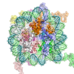
A cartoon representation of the nucleosome structure. From PDB: 1KX5​.

The basic repeat element of chromatin is the nucleosome, interconnected by sections of linker DNA, a far shorter arrangement than pure DNA in solution.

In addition to the core histones, there is the linker histone, H1, which contacts the exit/entry of the DNA strand on the nucleosome. The nucleosome core particle, together with histone H1, is known as a chromatosome. Nucleosomes, with about 20 to 60 base pairs of linker DNA, can form, under non-physiological conditions, an approximately 10 nm "beads-on-a-string" fibre. (Fig. 1-2).

The nucleosomes bind DNA non-specifically, as required by their function in general DNA packaging. There are, however, large DNA sequence preferences that govern nucleosome positioning. This is due primarily to the varying physical properties of different DNA sequences: For instance, adenine and thymine are more favorably compressed into the inner minor grooves. This means nucleosomes can bind preferentially at one position approximately every 10 base pairs (the helical repeat of DNA)- where the DNA is rotated to maximise the number of A and T bases that will lie in the inner minor groove.

30-nanometer chromatin fibre

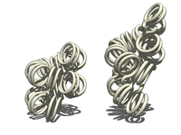
Two proposed structures of the 30 nm chromatin filament.
Left: 1 start helix "solenoid" structure.
Right: 2 start loose helix structure.
Note: the histones are omitted in this diagram - only the DNA is shown.

With addition of H1, the beads-on-a-string structure in turn coils into a 30 nm diameter helical structure known as the 30 nm fibre or filament. The precise structure of the chromatin fibre in the cell is not known in detail, and there is still some debate over this.

This level of chromatin structure is thought to be the form of heterochromatin, which contains mostly transcriptionally silent genes. EM studies have demonstrated that the 30 nm fibre is highly dynamic such that it unfolds into a 10 nm fiber ("beads-on-a-string") structure when transversed by an RNA polymerase engaged in transcription.

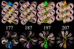
Four proposed structures of the 30 nm chromatin filament for DNA repeat length per nucleosomes ranging from 177 to 207 bp.
Linker DNA in yellow and nucleosomal DNA in pink.

The existing models commonly accept that the nucleosomes lie perpendicular to the axis of the fibre, with linker histones arranged internally. A stable 30 nm fibre relies on the regular positioning of nucleosomes along DNA. Linker DNA is relatively resistant to bending and rotation. This makes the length of linker DNA critical to the stability of the fibre, requiring nucleosomes to be separated by lengths that permit rotation and folding into the required orientation without excessive stress to the DNA. In this view, different lengths of the linker DNA should produce different folding topologies of the chromatin fiber. Recent theoretical work, based on electron-microscopy images of reconstituted fibers supports this view.

Spatial organization of chromatin in the cell nucleus

The spatial arrangement of the chromatin within the nucleus is not random - specific regions of the chromatin can be found in certain territories. Territories are, for example, the lamina-associated domains (LADs), and the topological association domains (TADs), which are bound together by protein complexes. Currently, polymer models such as the Strings & Binders Switch (SBS) model and the Dynamic Loop (DL) model are used to describe the folding of chromatin within the nucleus.

Cell-cycle dependent structural organization

  • Interphase: The structure of chromatin during interphase of mitosis is optimized to allow simple access of transcription and DNA repair factors to the DNA while compacting the DNA into the nucleus. The structure varies depending on the access required to the DNA. Genes that require regular access by RNA polymerase require the looser structure provided by euchromatin.

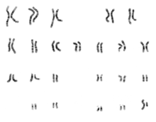
    1. Karyogram of human male using Giemsa staining, showing the classic metaphase chromatin structure.
    2. Metaphase: The metaphase structure of chromatin differs vastly to that of interphase. It is optimised for physical strength and manageability, forming the classic chromosome structure seen in karyotypes. The structure of the condensed chromatin is thought to be loops of 30 nm fibre to a central scaffold of proteins. It is, however, not well-characterised.The physical strength of chromatin is vital for this stage of division to prevent shear damage to the DNA as the daughter chromosomes are separated. To maximise strength the composition of the chromatin changes as it approaches the centromere, primarily through alternative histone H1 analogues.It should also be noted that, during mitosis, while most of the chromatin is tightly compacted, there are small regions that are not as tightly compacted. These regions often correspond to promoter regions of genes that were active in that cell type prior to entry into chromatosis. The lack of compaction of these regions is called bookmarking, which is an epigenetic mechanism believed to be important for transmitting to daughter cells the "memory" of which genes were active prior to entry into mitosis. This bookmarking mechanism is needed to help transmit this memory because transcription ceases during mitosis.

    Chromatin and bursts of transcription

    Chromatin and its interaction with enzymes has been researched, and a conclusion being made is that it is relevant and an important factor in gene expression. Vincent G. Allfrey, a professor at Rockefeller University, stated that RNA synthesis is related to histone acetylation. The lysine amino acid attached to the end of the histones is positively charged. The acetylation of these tails would make the chromatin ends neutral, allowing for DNA access.

    When the chromatin decondenses, the DNA is open to entry of molecular machinery. Fluctuations between open and closed chromatin may contribute to the discontinuity of transcription, or transcriptional bursting. Other factors are probably involved, such as the association and dissociation of transcription factor complexes with chromatin. The phenomenon, as opposed to simple probabilistic models of transcription, can account for the high variability in gene expression occurring between cells in isogenic populations.

    Alternative chromatin organizations

    During metazoan spermiogenesis, the spermatid's chromatin is remodeled into a more spaced-packaged, widened, almost crystal-like structure. This process is associated with the cessation of transcription and involves nuclear protein exchange. The histones are mostly displaced, and replaced by protamines (small, arginine-rich proteins). It is proposed that in yeast, regions devoid of histones become very fragile after transcription; HMO1 an HMGB protein helps in stabilizing nucleosomes-free chromatin.

    Chromatin and DNA repair

    The packaging of eukaryotic DNA into chromatin presents a barrier to all DNA-based processes that require recruitment of enzymes to their sites of action. To allow the critical cellular process of DNA repair, the chromatin must be remodeled. In eukaryotes, ATP dependent chromatin remodeling complexes and histone-modifying enzymes are two predominant factors employed to accomplish this remodeling process.

    Chromatin relaxation occurs rapidly at the site of a DNA damage. This process is initiated by PARP1 protein that starts to appear at DNA damage in less than a second, with half maximum accumulation within 1.6 seconds after the damage occurs. Next the chromatin remodeler Alc1 quickly attaches to the product of PARP1, and completes arrival at the DNA damage within 10 seconds of the damage. About half of the maximum chromatin relaxation, presumably due to action of Alc1, occurs by 10 seconds. This then allows recruitment of the DNA repair enzyme MRE11, to initiate DNA repair, within 13 seconds.

    γH2AX, the phosphorylated form of H2AX is also involved in the early steps leading to chromatin decondensation after DNA damage occurrence. The histone variant H2AX constitutes about 10% of the H2A histones in human chromatin. γH2AX (H2AX phosphorylated on serine 139) can be detected as soon as 20 seconds after irradiation of cells (with DNA double-strand break formation), and half maximum accumulation of γH2AX occurs in one minute. The extent of chromatin with phosphorylated γH2AX is about two million base pairs at the site of a DNA double-strand break. γH2AX does not, itself, cause chromatin decondensation, but within 30 seconds of irradiation, RNF8 protein can be detected in association with γH2AX. RNF8 mediates extensive chromatin decondensation, through its subsequent interaction with CHD4, a component of the nucleosome remodeling and deacetylase complex NuRD.

    After undergoing relaxation subsequent to DNA damage, followed by DNA repair, chromatin recovers to a compaction state close to its pre-damage level after about 20 min.

    Methods to investigate chromatin

    1. ChIP-seq (Chromatin immunoprecipitation sequencing), aimed against different histone modifications, can be used to identify chromatin states throughout the genome. Different modifications have been linked to various states of chromatin.
    2. DNase-seq (DNase I hypersensitive sites Sequencing) uses the sensitivity of accessible regions in the genome to the DNase I enzyme to map open or accessible regions in the genome.
    3. FAIRE-seq (Formaldehyde-Assisted Isolation of Regulatory Elements sequencing) uses the chemical properties of protein-bound DNA in a two-phase separation method to extract nucleosome depleted regions from the genome.
    4. ATAC-seq (Assay for Transposable Accessible Chromatin sequencing) uses the Tn5 transposase to integrate (synthetic) transposons into accessible regions of the genome consequentially highlighting the localisation of nucleosomes and transcription factors across the genome.
    5. DNA footprinting is a method aimed at identifying protein-bound DNA. It uses labeling and fragmentation coupled to gel electrophoresis to identify areas of the genome that have been bound by proteins.
    6. MNase-seq (Micrococcal Nuclease sequencing) uses the micrococcal nuclease enzyme to identify nucleosome positioning throughout the genome.
    7. Chromosome conformation capture determines the spatial organization of chromatin in the nucleus, by inferring genomic locations that physically interact.
    8. MACC profiling (Micrococcal nuclease ACCessibility profiling) uses titration series of chromatin digests with micrococcal nuclease to identify chromatin accessibility as well as to map nucleosomes and non-histone DNA-binding proteins in both open and closed regions of the genome.

    Chromatin and knots

    It has been a puzzle how decondensed interphase chromosomes remain essentially unknotted. The natural expectation is that in the presence of type II DNA topoisomerases that permit passages of double-stranded DNA regions through each other, all chromosomes should reach the state of topological equilibrium. The topological equilibrium in highly crowded interphase chromosomes forming chromosome territories would result in formation of highly knotted chromatin fibres. However, Chromosome Conformation Capture (3C) methods revealed that the decay of contacts with the genomic distance in interphase chromosomes is practically the same as in the crumpled globule state that is formed when long polymers condense without formation of any knots. To remove knots from highly crowded chromatin, one would need an active process that should not only provide the energy to move the system from the state of topological equilibrium but also guide topoisomerase-mediated passages in such a way that knots would be efficiently unknotted instead of making the knots even more complex. It has been shown that the process of chromatin-loop extrusion is ideally suited to actively unknot chromatin fibres in interphase chromosomes.

    Chromatin: alternative definitions

    The term, introduced by Walther Flemming, has multiple meanings:
    1. Simple and concise definition: Chromatin is a macromolecular complex of a DNA macromolecule and protein macromolecules (and RNA). The proteins package and arrange the DNA and control its functions within the cell nucleus.
    2. A biochemists’ operational definition: Chromatin is the DNA/protein/RNA complex extracted from eukaryotic lysed interphase nuclei. Just which of the multitudinous substances present in a nucleus will constitute a part of the extracted material partly depends on the technique each researcher uses. Furthermore, the composition and properties of chromatin vary from one cell type to the another, during development of a specific cell type, and at different stages in the cell cycle.
    3. The DNA + histone = chromatin definition: The DNA double helix in the cell nucleus is packaged by special proteins termed histones. The formed protein/DNA complex is called chromatin. The basic structural unit of chromatin is the nucleosome.
    Crops such as wheat and maize have undergone a breeding process lasting thousands of years, in the course of which mankind has gradually modified the properties of wild plants into highly cultivated variants. One motive was higher yields. A side effect of this breeding has been a reduction in genetic diversity and the loss of useful properties. This is demonstrated by an increased susceptibility to diseases, a lack of flavor or reduced vitamin and nutrient content in modern varieties. Now, for the first time, researchers from Brazil, the U.S. and Germany have created a new crop from a wild plant within a single generation using CRISPR-Cas9, a modern genome editing process. Starting with a wild tomato, they introduced a variety of crop features without losing the valuable genetic properties of the wild plant. The results have been published in the current issue of Nature Biotechnology.

    Read more at: https://phys.org/news/2018-10-biologists-crop-genome.html#jCp

    Histone

    From Wikipedia, the free encyclopedia
     
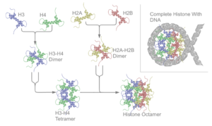
    Schematic representation of the assembly of the core histones into the nucleosome.

    In biology, histones are highly alkaline proteins found in eukaryotic cell nuclei that package and order the DNA into structural units called nucleosomes. They are the chief protein components of chromatin, acting as spools around which DNA winds, and playing a role in gene regulation. Without histones, the unwound DNA in chromosomes would be very long (a length to width ratio of more than 10 million to 1 in human DNA). For example, each human diploid cell (containing 23 pairs of chromosomes) has about 1.8 meters of DNA; wound on the histones, the diploid cell has about 90 micrometers (0.09 mm) of chromatin. When the diploid cells are duplicated and condensed during mitosis, the result is about 120 micrometers of chromosomes.

    Core histone H2A/H2B/H3/H4
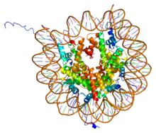
    Protein H2AFJ PDB 1aoi.png
    PDB rendering of Complex between nucleosome core particle (h3,h4,h2a,h2b) and 146 bp long DNA fragment based on 1aoi.
    Identifiers
    Symbol Histone
    Pfam PF00125
    Pfam clan CL0012
    InterPro IPR007125
    SCOP 1hio
    SUPERFAMILY 1hio

    linker histone H1 and H5 family
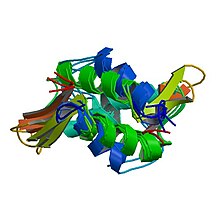
    PBB Protein HIST1H1B image.jpg
    PDB rendering of HIST1H1B based on 1ghc.
    Identifiers
    Symbol Linker_histone
    Pfam PF00538
    InterPro IPR005818
    SMART SM00526
    SCOP 1hst
    SUPERFAMILY 1hst

    Classes and histone variants

    Five major families of histones exist: H1/H5, H2A, H2B, H3, and H4. Histones H2A, H2B, H3 and H4 are known as the core histones, while histones H1/H5 are known as the linker histones.

    The core histones all exist as dimers, which are similar in that they all possess the histone fold domain: three alpha helices linked by two loops. It is this helical structure that allows for interaction between distinct dimers, particularly in a head-tail fashion (also called the handshake motif). The resulting four distinct dimers then come together to form one octameric nucleosome core, approximately 63 Angstroms in diameter (a solenoid (DNA)-like particle). Around 146 base pairs (bp) of DNA wrap around this core particle 1.65 times in a left-handed super-helical turn to give a particle of around 100 Angstroms across. The linker histone H1 binds the nucleosome at the entry and exit sites of the DNA, thus locking the DNA into place and allowing the formation of higher order structure. The most basic such formation is the 10 nm fiber or beads on a string conformation. This involves the wrapping of DNA around nucleosomes with approximately 50 base pairs of DNA separating each pair of nucleosomes (also referred to as linker DNA). Higher-order structures include the 30 nm fiber (forming an irregular zigzag) and 100 nm fiber, these being the structures found in normal cells. During mitosis and meiosis, the condensed chromosomes are assembled through interactions between nucleosomes and other regulatory proteins.

    Histones are subdivided into canonical replication-dependent histones that are expressed during the S-phase of cell cycle and replication-independent histone variants, expressed during the whole cell cycle. In animals, genes encoding canonical histones are typically clustered along the chromosome, lack introns and use a stem loop structure at the 3’ end instead of a polyA tail. Genes encoding histone variants are usually not clustered, have introns and their mRNAs are regulated with polyA tails. Complex multicellular organisms typically have a higher number of histone variants providing a variety of different functions. Recent data are accumulating about the roles of diverse histone variants highlighting the functional links between variants and the delicate regulation of organism development. Histone variants from different organisms, their classification and variant specific features can be found in "HistoneDB 2.0 - Variants" database.

    The following is a list of human histone proteins:

    Super family Family Subfamily Members
    Linker H1 H1F H1F0, H1FNT, H1FOO, H1FX
    H1H1 HIST1H1A, HIST1H1B, HIST1H1C, HIST1H1D, HIST1H1E, HIST1H1T
    Core H2A H2AF H2AFB1, H2AFB2, H2AFB3, H2AFJ, H2AFV, H2AFX, H2AFY, H2AFY2, H2AFZ
    H2A1 HIST1H2AA, HIST1H2AB, HIST1H2AC, HIST1H2AD, HIST1H2AE, HIST1H2AG, HIST1H2AI, HIST1H2AJ, HIST1H2AK, HIST1H2AL, HIST1H2AM
    H2A2 HIST2H2AA3, HIST2H2AC
    H2B H2BF H2BFM, H2BFS, H2BFWT
    H2B1 HIST1H2BA, HIST1H2BB, HIST1H2BC, HIST1H2BD, HIST1H2BE, HIST1H2BF, HIST1H2BG, HIST1H2BH, HIST1H2BI, HIST1H2BJ, HIST1H2BK, HIST1H2BL, HIST1H2BM, HIST1H2BN, HIST1H2BO
    H2B2 HIST2H2BE
    H3 H3A1 HIST1H3A, HIST1H3B, HIST1H3C, HIST1H3D, HIST1H3E, HIST1H3F, HIST1H3G, HIST1H3H, HIST1H3I, HIST1H3J
    H3A2 HIST2H3C
    H3A3 HIST3H3
    H4 H41 HIST1H4A, HIST1H4B, HIST1H4C, HIST1H4D, HIST1H4E, HIST1H4F, HIST1H4G, HIST1H4H, HIST1H4I, HIST1H4J, HIST1H4K, HIST1H4L
    H44 HIST4H4

    Structure

    The nucleosome core is formed of two H2A-H2B dimers and a H3-H4 tetramer, forming two nearly symmetrical halves by tertiary structure (C2 symmetry; one macromolecule is the mirror image of the other). The H2A-H2B dimers and H3-H4 tetramer also show pseudodyad symmetry. The 4 'core' histones (H2A, H2B, H3 and H4) are relatively similar in structure and are highly conserved through evolution, all featuring a 'helix turn helix turn helix' motif (DNA-binding protein motif that recognize specific DNA sequence). They also share the feature of long 'tails' on one end of the amino acid structure - this being the location of post-translational modification.

    It has been proposed that histone proteins are evolutionarily related to the helical part of the extended AAA+ ATPase domain, the C-domain, and to the N-terminal substrate recognition domain of Clp/Hsp100 proteins. Despite the differences in their topology, these three folds share a homologous helix-strand-helix (HSH) motif.

    Using an electron paramagnetic resonance spin-labeling technique, British researchers measured the distances between the spools around which eukaryotic cells wind their DNA. They determined the spacings range from 59 to 70 Å.

    In all, histones make five types of interactions with DNA:
    • Helix-dipoles form alpha-helixes in H2B, H3, and H4 cause a net positive charge to accumulate at the point of interaction with negatively charged phosphate groups on DNA
    • Hydrogen bonds between the DNA backbone and the amide group on the main chain of histone proteins
    • Nonpolar interactions between the histone and deoxyribose sugars on DNA
    • Salt bridges and hydrogen bonds between side chains of basic amino acids (especially lysine and arginine) and phosphate oxygens on DNA
    • Non-specific minor groove insertions of the H3 and H2B N-terminal tails into two minor grooves each on the DNA molecule
    The highly basic nature of histones, aside from facilitating DNA-histone interactions, contributes to their water solubility.

    Histones are subject to post translational modification by enzymes primarily on their N-terminal tails, but also in their globular domains. Such modifications include methylation, citrullination, acetylation, phosphorylation, SUMOylation, ubiquitination, and ADP-ribosylation. This affects their function of gene regulation.

    In general, genes that are active have less bound histone, while inactive genes are highly associated with histones during interphase[14]. It also appears that the structure of histones has been evolutionarily conserved, as any deleterious mutations would be severely maladaptive. All histones have a highly positively charged N-terminus with many lysine and arginine residues.

    History

    Histones were discovered in 1884 by Albrecht Kossel. The word "histone" dates from the late 19th century and is from the German word "Histon", a word itself of uncertain origin - perhaps from the Greek histanai or histos.

    In the early 1960s, before the types of histones were known and before histones were known to be highly conserved across taxonomically diverse organisms, James F. Bonner and his collaborators began a study of these proteins that were known to be tightly associated with the DNA in the nucleus of higher organisms. Bonner and his postdoctoral fellow Ru Chih C. Huang showed that isolated chromatin would not support RNA transcription in the test tube, but if the histones were extracted from the chromatin, RNA could be transcribed from the remaining DNA.  Their paper became a citation classic. Paul T'so and James Bonner had called together a World Congress on Histone Chemistry and Biology in 1964, in which it became clear that there was no consensus on the number of kinds of histone and that no one know how they would compare when isolated from different organisms. Bonner and his collaborators then developed methods to separate each type of histones, purified individual histones, compared amino acid compositions in the same histone from different organisms, and compared amino acid sequences  of the same histone from different organisms in collaboration with Emil Smith from UCLA. For example, they found Histone IV sequence to be highly conserved between peas and calf thymus. However, their work on the biochemical characteristics of individual histones did not reveal how the histones interacted with each other or with DNA to which they were tightly bound.

    Also in the 1960's, Allfrey and Mirsky had suggested, based on their analyses of histones, that acetylation and methylation of histones could provide a transcriptional control mechanism, but did not have available the kind of detailed analysis that later investigators were able to conduct to show how such regulation could be gene-specific. Until the early 1990s, histones were dismissed by most as inert packing material for eukaryotic nuclear DNA, a view based in part on the models of Mark Ptashne and others, who believed that transcription was activated by protein-DNA and protein-protein interactions on largely naked DNA templates, as is the case in bacteria.

    During the 1980s, Yahli Lorch and Roger Kornberg showed that a nucleosome on a core promoter prevents the initiation of transcription in vitro, and Michael Grunstein demonstrated that histones repress transcription in vivo, leading to the idea of the nucleosome as a general gene repressor. Relief from repression is believed to involve both histone modification and the action of chromatin-remodeling complexes. Vincent Allfrey and Alfred Mirsky earlier proposed a role of histone modification in transcriptional activation, regarded as a molecular manifestation of epigenetics. Michael Grunstein and David Allis found support for this proposal, in the importance of histone acetylation for transcription in yeast and the activity of the transcriptional activator Gcn5 as a histone acetyltransferase.

    The discovery of the H5 histone appears to date back to the 1970s, and it is now considered an isoform of Histone H1.

    Conservation across species

    Histones are found in the nuclei of eukaryotic cells, and in certain Archaea, namely Thermoproteales and Euryarchaea, but not in bacteria. The unicellular algae known as dinoflagellates were previously thought to be the only eukaryotes that completely lack histones, however, later studies showed that their DNA still encodes histone genes.

    Archaeal histones may well resemble the evolutionary precursors to eukaryotic histones. Histone proteins are among the most highly conserved proteins in eukaryotes, emphasizing their important role in the biology of the nucleus. In contrast mature sperm cells largely use protamines to package their genomic DNA, most likely because this allows them to achieve an even higher packaging ratio.

    There are some variant forms in some of the major classes. They share amino acid sequence homology and core structural similarity to a specific class of major histones but also have their own feature that is distinct from the major histones. These minor histones usually carry out specific functions of the chromatin metabolism. For example, histone H3-like CENPA is associated with only the centromere region of the chromosome. Histone H2A variant H2A.Z is associated with the promoters of actively transcribed genes and also involved in the prevention of the spread of silent heterochromatin. Furthermore, H2A.Z has roles in chromatin for genome stability. Another H2A variant H2A.X is phosphorylated at S139 in regions around double-strand breaks and marks the region undergoing DNA repair. Histone H3.3 is associated with the body of actively transcribed genes.

    Function

    Compacting DNA strands

    Histones act as spools around which DNA winds. This enables the compaction necessary to fit the large genomes of eukaryotes inside cell nuclei: the compacted molecule is 40,000 times shorter than an unpacked molecule.

    Chromatin regulation

    Histones undergo posttranslational modifications that alter their interaction with DNA and nuclear proteins. The H3 and H4 histones have long tails protruding from the nucleosome, which can be covalently modified at several places. Modifications of the tail include methylation, acetylation, phosphorylation, ubiquitination, SUMOylation, citrullination, and ADP-ribosylation. The core of the histones H2A and H2B can also be modified. Combinations of modifications are thought to constitute a code, the so-called "histone code". Histone modifications act in diverse biological processes such as gene regulation, DNA repair, chromosome condensation (mitosis) and spermatogenesis (meiosis).

    The common nomenclature of histone modifications is:
    • The name of the histone (e.g., H3)
    • The single-letter amino acid abbreviation (e.g., K for Lysine) and the amino acid position in the protein
    • The type of modification (Me: methyl, P: phosphate, Ac: acetyl, Ub: ubiquitin)
    • The number of modifications (only Me is known to occur in more than one copy per residue. 1, 2 or 3 is mono-, di- or tri-methylation)
    So H3K4me1 denotes the monomethylation of the 4th residue (a lysine) from the start (i.e., the N-terminal) of the H3 protein.

    Functions of histone modifications

    Schematic representation of histone modifications. Based on Rodriguez-Paredes and Esteller, Nature, 2011
    A huge catalogue of histone modifications have been described,
    but a functional understanding of most is still lacking.
    Collectively, it is thought that histone modifications may
    underlie a histone code, whereby combinations of histone
    modifications have specific meanings. However, most
    functional data concerns individual prominent histone
    modifications that are biochemically amenable to detailed study.

    Chemistry of histone modifications

    Lysine methylation

    Methyl lysine.svg

    The addition of one, two, or many methyl groups to lysine has little effect on the chemistry of the histone; methylation leaves the charge of the lysine intact and adds a minimal number of atoms so steric interactions are mostly unaffected. However, proteins containing Tudor, chromo or PHD domains, amongst others, can recognise lysine methylation with exquisite sensitivity and differentiate mono, di and tri-methyl lysine, to the extent that, for some lysines (e.g.: H4K20) mono, di and tri-methylation appear to have different meanings. Because of this, lysine methylation tends to be a very informative mark and dominates the known histone modification functions.

    Arginine methylation

    Methyl arginine.svg

    What was said above of the chemistry of lysine methylation also applies to arginine methylation, and some protein domains—e.g., Tudor domains—can be specific for methyl arginine instead of methyl lysine. Arginine is known to be mono- or di-methylated, and methylation can be symmetric or asymmetric, potentially with different meanings.

    Arginine citrullination

    Enzymes called peptidylarginine deiminases (PADs) hydrolyze the imine group of arginines and attach a keto group, so that there is one less positive charge on the amino acid residue. This process has been involved in the activation of gene expression by making the modified histones less tightly bound to DNA and thus making the chromatin more accessible. PADs can also produce the opposite effect by removing or inhibiting mono-methylation of arginine residues on histones and thus antagonizing the positive effect arginine methylation has on transcriptional activity.

    Lysine acetylation

    Acetyl lysine.tif

    Addition of an acetyl group has a major chemical effect on lysine as it neutralises the positive charge. This reduces electrostatic attraction between the histone and the negatively charged DNA backbone, loosening the chromatin structure; highly acetylated histones form more accessible chromatin and tend to be associated with active transcription. Lysine acetylation appears to be less precise in meaning than methylation, in that histone acetyltransferases tend to act on more than one lysine; presumably this reflects the need to alter multiple lysines to have a significant effect on chromatin structure. The modification includes H3K27ac.

    Serine/threonine/tyrosine phosphorylation

    Amino acid phosphorylations.tif

    Addition of a negatively charged phosphate group can lead to major changes in protein structure, leading to the well-characterised role of phosphorylation in controlling protein function. It is not clear what structural implications histone phosphorylation has, but histone phosphorylation has clear functions as a post-translational modification, and binding domains such as BRCT have been characterised.

    Functions in transcription

    Most well-studied histone modifications are involved in control of transcription.

    Actively transcribed genes

    Two histone modifications are particularly associated with active transcription:
    Trimethylation of H3 lysine 4 (H3K4me3)
    This trimethylation occurs at the promoter of active genes and is performed by the COMPASS complex. Despite the conservation of this complex and histone modification from yeast to mammals, it is not entirely clear what role this modification plays. However, it is an excellent mark of active promoters and the level of this histone modification at a gene’s promoter is broadly correlated with transcriptional activity of the gene. The formation of this mark is tied to transcription in a rather convoluted manner: early in transcription of a gene, RNA polymerase II undergoes a switch from initiating’ to ‘elongating’, marked by a change in the phosphorylation states of the RNA polymerase II C terminal domain (CTD). The same enzyme that phosphorylates the CTD also phosphorylates the Rad6 complex, which in turn adds a ubiquitin mark to H2B K123 (K120 in mammals). H2BK123Ub occurs throughout transcribed regions, but this mark is required for COMPASS to trimethylate H3K4 at promoters.
    Trimethylation of H3 lysine 36 (H3K36me3)
    This trimethylation occurs in the body of active genes and is deposited by the methyltransferase Set2. This protein associates with elongating RNA polymerase II, and H3K36Me3 is indicative of actively transcribed genes. H3K36Me3 is recognised by the Rpd3 histone deacetylase complex, which removes acetyl modifications from surrounding histones, increasing chromatin compaction and repressing spurious transcription. Increased chromatin compaction prevents transcription factors from accessing DNA, and reduces the likelihood of new transcription events being initiated within the body of the gene. This process therefore helps ensure that transcription is not interrupted.

    Repressed genes

    Three histone modifications are particularly associated with repressed genes:
    Trimethylation of H3 lysine 27 (H3K27me3)
    This histone modification is depositied by the polycomb complex PRC2. It is a clear marker of gene repression, and is likely bound by other proteins to exert a repressive function. Another polycomb complex, PRC1, can bind H3K27me3 and adds the histone modification H2AK119Ub which aids chromatin compaction. Based on this data it appears that PRC1 is recruited through the action of PRC2, however, recent studies show that PRC1 is recruited to the same sites in the absence of PRC2.
    Di and tri-methylation of H3 lysine 9 (H3K9me2/3)
    H3K9me2/3 is a well-characterised marker for heterochromatin, and is therefore strongly associated with gene repression. The formation of heterochromatin has been best studied in the yeast Schizosaccharomyces pombe, where it is initiated by recruitment of the RNA-induced transcriptional silencing (RITS) complex to double stranded RNAs produced from centromeric repeats. RITS recruits the Clr4 histone methyltransferase which deposits H3K9me2/3. This process is called histone methylation. H3K9Me2/3 serves as a binding site for the recruitment of Swi6 (heterochromatin protein 1 or HP1, another classic heterochromatin marker) which in turn recruits further repressive activities including histone modifiers such as histone deacetylases and histone methyltransferases.
    Trimethylation of H4 lysine 20 (H4K20me3)
    This modification is tightly associated with heterochromatin, although its functional importance remains unclear. This mark is placed by the Suv4-20h methyltransferase, which is at least in part recruited by heterochromatin protein 1.

    Bivalent promoters

    Analysis of histone modifications in embryonic stem cells (and other stem cells) revealed many gene promoters carrying both H3K4Me3 and H3K27Me3, in other words these promoters display both activating and repressing marks simultaneously. This peculiar combination of modifications marks genes that are poised for transcription; they are not required in stem cells, but are rapidly required after differentiation into some lineages. Once the cell starts to differentiate, these bivalent promoters are resolved to either active or repressive states depending on the chosen lineage.

    Other functions

    DNA damage

    Marking sites of DNA damage is an important function for histone modifications. It also protects DNA from getting destroyed by ultraviolet radiation of sun.
    Phosphorylation of H2AX at serine 139 (γH2AX)
    Phosphorylated H2AX (also known as gamma H2AX) is a marker for DNA double strand breaks, and forms part of the response to DNA damage. H2AX is phosphorylated early after detection of DNA double strand break, and forms a domain extending many kilobases either side of the damage. Gamma H2AX acts as a binding site for the protein MDC1, which in turn recruits key DNA repair proteins (this complex topic is well reviewed in) and as such, gamma H2AX forms a vital part of the machinery that ensures genome stability.
    Acetylation of H3 lysine 56 (H3K56Ac)
    H3K56Acx is required for genome stability. H3K56 is acetylated by the p300/Rtt109 complex, but is rapidly deacetylated around sites of DNA damage. H3K56 acetylation is also required to stabilise stalled replication forks, preventing dangerous replication fork collapses. Although in general mammals make far greater use of histone modifications than microorganisms, a major role of H3K56Ac in DNA replication exists only in fungi, and this has become a target for antibiotic development.

    DNA repair

    Trimethylation of H3 lysine 36 (H3K36me3)
    H3K36me3 has the ability to recruit the MSH2-MSH6 (hMutSα) complex of the DNA mismatch repair pathway. Consistently, regions of the human genome with high levels of H3K36me3 accumulate less somatic mutations due to mismatch repair activity.

    Chromosome condensation

    Phosphorylation of H3 at serine 10 (phospho-H3S10)
    The mitotic kinase aurora B phosphorylates histone H3 at serine 10, triggering a cascade of changes that mediate mitotic chromosome condensation. Condensed chromosomes therefore stain very strongly for this mark, but H3S10 phosphorylation is also present at certain chromosome sites outside mitosis, for example in pericentric heterochromatin of cells during G2. H3S10 phosphorylation has also been linked to DNA damage caused by R loop formation at highly transcribed sites.
    Phosphorylation H2B at serine 10/14 (phospho-H2BS10/14)
    Phosphorylation of H2B at serine 10 (yeast) or serine 14 (mammals) is also linked to chromatin condensation, but for the very different purpose of mediating chromosome condensation during apoptosis. This mark is not simply a late acting bystander in apoptosis as yeast carrying mutations of this residue are resistant to hydrogen peroxide-induced apoptotic cell death.

    Addiction

    Epigenetic modifications of histone tails in specific regions of the brain are of central importance in addictions. Once particular epigenetic alterations occur, they appear to be long lasting "molecular scars" that may account for the persistence of addictions.

    Cigarette smokers (about 15% of the US population) are usually addicted to nicotine. After 7 days of nicotine treatment of mice, acetylation of both histone H3 and histone H4 was increased at the FosB promoter in the nucleus accumbens of the brain, causing 61% increase in FosB expression. This would also increase expression of the splice variant Delta FosB. In the nucleus accumbens of the brain, Delta FosB functions as a "sustained molecular switch" and "master control protein" in the development of an addiction.

    About 7% of the US population is addicted to alcohol. In rats exposed to alcohol for up to 5 days, there was an increase in histone 3 lysine 9 acetylation in the pronociceptin promoter in the brain amygdala complex. This acetylation is an activating mark for pronociceptin. The nociceptin/nociceptin opioid receptor system is involved in the reinforcing or conditioning effects of alcohol.

    Methamphetamine addiction occurs in about 0.2% of the US population. Chronic methamphetamine use causes methylation of the lysine in position 4 of histone 3 located at the promoters of the c-fos and the C-C chemokine receptor 2 (ccr2) genes, activating those genes in the nucleus accumbens (NAc). c-fos is well known to be important in addiction. The ccr2 gene is also important in addiction, since mutational inactivation of this gene impairs addiction.

    Histone synthesis

    The first step of chromatin structure duplication is the synthesis of histone proteins: H1, H2A, H2B, H3, H4. These proteins are synthesized during S phase of the cell cycle. There are different mechanisms which contribute to the increase of histone synthesis.

    Yeast

    Yeast carry one or two copies of each histone gene, which are not clustered but rather scattered throughout chromosomes. Histone gene transcription is controlled by multiple gene regulatory proteins such as transcription factors which bind to histone promoter regions. In budding yeast, the candidate gene for activation of histone gene expression is SBF. SBF is a transcription factor that is activated in late G1 phase, when it dissociates from its repressor Whi5. This occurs when Whi5 is phosphorylated by Cdc8 which is a G1/S Cdk. Suppression of histone gene expression outside of S phases is dependent on Hir proteins which form inactive chromatin structure at the locus of histone genes, causing transcriptional activators to be blocked.

    Metazoans

    In metazoans the increase in the rate of histone synthesis is due to the increase in processing of pre-mRNA to its mature form as well as decrease in mRNA degradation; this results in an increase of active mRNA for translation of histone proteins. The mechanism for mRNA activation has been found to be the removal of a segment of the 3’ end of the mRNA strand, and is dependent on association with stem-loop binding protein (SLBP). SLBP also stabilizes histone mRNAs during S phase by blocking degradation by the 3’hExo nuclease. SLBP levels are controlled by cell-cycle proteins, causing SLBP to accumulate as cells enter S phase and degrade as cells leave S phase. SLBP are marked for degradation by phosphorylation at two threonine residues by cyclin dependent kinases, possibly cyclin A/ cdk2, at the end of S phase. Metazoans also have multiple copies of histone genes clustered on chromosomes which are localized in structures called Cajal bodies as determined by genome-wide chromosome conformation capture analysis (4C-Seq).

    Link between cell-cycle control machinery and histone synthesis

    Nuclear protein Ataxia-Telangiectasia (NPAT), also known as nuclear protein coactivator of histone transcription, is a transcription factor which activates histone gene transcription on chromosomes 1 and 6 of human cells. NPAT is also a substrate of cyclin E-Cdk2, which is required for the transition between G1 phase and S phase. NPAT activates histone gene expression only after it has been phosphorylated by the G1/S-Cdk cyclin E-Cdk2 in early S phase. This shows an important regulatory link between cell-cycle control and histone synthesis.

    Neurology

    From Wikipedia, the free encyclopedia

    Neurology
    PLoSBiol4.e126.Fig6fNeuron.jpg
    A network of dendrites from neurons in the hippocampus.
    System Nervous system
    Significant diseases Radiculopathy, neuropathy, visual snow, stroke, dementia, seizures and epilepsy, Alzheimer's disease, Attention deficit/hyperactivity disorder
    Significant tests Computed axial tomography, MRI scan, lumbar puncture
    Specialist Neurologist
    Glossary Glossary of medicine

    Neurology (from Greek: νεῦρον (neûron), "string, nerve" and the suffix -logia, "study of") is a branch of medicine dealing with disorders of the nervous system. Neurology deals with the diagnosis and treatment of all categories of conditions and disease involving the central and peripheral nervous systems (and their subdivisions, the autonomic and somatic nervous systems), including their coverings, blood vessels, and all effector tissue, such as muscle. Neurological practice relies heavily on the field of neuroscience, which is the scientific study of the nervous system.

    A neurologist is a physician specializing in neurology and trained to investigate, or diagnose and treat neurological disorders. Neurologists may also be involved in clinical research, clinical trials, and basic or translational research. While neurology is a nonsurgical specialty, its corresponding surgical specialty is neurosurgery.

    Significant overlap occurs between the fields of neurology and psychiatry, with the boundary between the two disciplines and the conditions they treat being somewhat nebulous.

    Scope

    A large number of neurological disorders have been described as listed. These can affect the central nervous system (brain and spinal cord), the peripheral nervous system, the autonomic nervous system, and the muscular system.

    History

    The academic discipline began between the 16th and 19th centuries with the work and research of many neurologists such as Thomas Willis, Robert Whytt, Matthew Baillie, Charles Bell, Moritz Heinrich Romberg, Duchenne de Boulogne, William A. Hammond, Jean-Martin Charcot, and John Hughlings Jackson.

    Training

    Neurologist
    Occupation
    Names Physician, Medical Practitioner
    Occupation type
    Profession
    Activity sectors
    Medicine
    Description
    Education required
    MD or DO (US), M.B.B.S. (UK), M.B. B.Ch. B.A.O. (Republic of Ireland)
    Fields of
    employment
    Hospitals, Clinics
    Polish neurologist Edward Flatau greatly influenced the developing field of neurology. He published a human brain atlas in 1894 and wrote a fundamental book on migraines in 1912.
     
    Jean-Martin Charcot is considered one of the fathers of neurology.
     
    Many neurologists also have additional training or interest in one area of neurology, such as stroke, epilepsy, neuromuscular, sleep medicine, pain management, or movement disorders.

    In the United States and Canada, neurologists are physicians having completed postgraduate training in neurology after graduation from medical school. Neurologists complete, on average, about 8 years of medical college education and clinical training,which includes obtaining a four-year undergraduate degree, a medical degree (DO or MD), which comprises an additional four years of study, then completing one year of basic clinical training and four years of residency. The four-year residency consists of one year of internal medicine internship training followed by three years of training in neurology.

    Some neurologists receive additional subspecialty training focusing on a particular area of the field. These training programs are called fellowships, and are one to two years in duration. Subspecialties include brain injury medicine, clinical neurophysiology, epilepsy, hospice and palliative medicine, neurodevelopmental disabilities, neuromuscular medicine, pain medicine, sleep medicine, neurocritical care, vascular neurology (stroke), behavioral neurology, child neurology, headache, multiple sclerosis, neuroimaging, neurorehabilitation.

    In Germany, a compulsory year of psychiatry must be done to complete a residency of neurology.
    In the United Kingdom and Ireland, neurology is a subspecialty of general (internal) medicine. After five to nine years of medical school and a year as a preregistration house officer (or two years on the Foundation Programme), a neurologist must pass the examination for Membership of the Royal College of Physicians (or the Irish equivalent) before completing two years of core medical training and then entering specialist training in neurology. A generation ago, some neurologists would have also spent a couple of years working in psychiatric units and obtain a diploma in psychological medicine. However, this requirement has become uncommon, and, now that a basic psychiatric qualification takes three years to obtain, the requirement is no longer practical. A period of research is essential, and obtaining a higher degree aids career progression. Many found it was eased after an attachment to the Institute of Neurology at Queen Square, London. Some neurologists enter the field of rehabilitation medicine (known as physiatry in the US) to specialise in neurological rehabilitation, which may include stroke medicine, as well as brain injuries.

    Physical examination

    During a neurological examination, the neurologist reviews the patient's health history with special attention to the current condition. The patient then takes a neurological exam. Typically, the exam tests mental status, function of the cranial nerves (including vision), strength, coordination, reflexes, and sensation. This information helps the neurologist determine whether the problem exists in the nervous system and the clinical localization. Localization of the pathology is the key process by which neurologists develop their differential diagnosis. Further tests may be needed to confirm a diagnosis and ultimately guide therapy and appropriate management.

    Clinical tasks

    Neurologists examine patients who are referred to them by other physicians in both the inpatient and outpatient settings. Neurologists begin their interactions with patients by taking a comprehensive medical history, and then performing a physical examination focusing on evaluating the nervous system. Components of the neurological examination include assessment of the patient's cognitive function, cranial nerves, motor strength, sensation, reflexes, coordination, and gait.

    In some instances, neurologists may order additional diagnostic tests as part of the evaluation. Commonly employed tests in neurology include imaging studies such as computed axial tomography (CAT) scans, magnetic resonance imaging (MRI), and ultrasound of major blood vessels of the head and neck. Neurophysiologic studies, including electroencephalography (EEG), needle electromyography (EMG), nerve conduction studies (NCSs) and evoked potentials are also commonly ordered. Neurologists frequently perform lumbar punctures to assess characteristics of a patient's cerebrospinal fluid. Advances in genetic testing have made genetic testing an important tool in the classification of inherited neuromuscular disease and diagnosis of many other neurogenetic diseases. The role of genetic influences on the development of acquired neurologic diseases is an active area of research.

    Some of the commonly encountered conditions treated by neurologists include headaches, radiculopathy, neuropathy, stroke, dementia, seizures and epilepsy, Alzheimer's disease, attention deficit/hyperactivity disorder, Parkinson's disease, Tourette's syndrome, multiple sclerosis, head trauma, sleep disorders, neuromuscular diseases, and various infections and tumors of the nervous system. Neurologists are also asked to evaluate unresponsive patients on life support to confirm brain death.

    Treatment options vary depending on the neurological problem. They can include referring the patient to a physiotherapist, prescribing medications, or recommending a surgical procedure.
    Some neurologists specialize in certain parts of the nervous system or in specific procedures. For example, clinical neurophysiologists specialize in the use of EEG and intraoperative monitoring to diagnose certain neurological disorders. Other neurologists specialize in the use of electrodiagnostic medicine studies – needle EMG and NCSs. In the US, physicians do not typically specialize in all the aspects of clinical neurophysiology – i.e. sleep, EEG, EMG, and NCSs. The American Board of Clinical Neurophysiology certifies US physicians in general clinical neurophysiology, epilepsy, and intraoperative monitoring. The American Board of Electrodiagnostic Medicine certifies US physicians in electrodiagnostic medicine and certifies technologists in nerve-conduction studies. Sleep medicine is a subspecialty field in the US under several medical specialties including anesthesiology, internal medicine, family medicine, and neurology. Neurosurgery is a distinct specialty that involves a different training path, and emphasizes the surgical treatment of neurological disorders.

    Also, many nonmedical doctors, those with doctoral degrees(usually PhDs) in subjects such as biology and chemistry, study and research the nervous system. Working in laboratories in universities, hospitals, and private companies, these neuroscientists perform clinical and laboratory experiments and tests to learn more about the nervous system and find cures or new treatments for diseases and disorders.

    A great deal of overlap occurs between neuroscience and neurology. Many neurologists work in academic training hospitals, where they conduct research as neuroscientists in addition to treating patients and teaching neurology to medical students.

    General caseload

    Neurologists are responsible for the diagnosis, treatment, and management of all the conditions mentioned above. When surgical or endovascular intervention is required, the neurologist may refer the patient to a neurosurgeon or an interventional neuroradiologist. In some countries, additional legal responsibilities of a neurologist may include making a finding of brain death when it is suspected that a patient has died. Neurologists frequently care for people with hereditary (genetic) diseases when the major manifestations are neurological, as is frequently the case. Lumbar punctures are frequently performed by neurologists. Some neurologists may develop an interest in particular subfields, such as stroke, dementia, movement disorders, neurointensive care, headaches, epilepsy, sleep disorders, chronic pain management, multiple sclerosis, or neuromuscular diseases.

    Overlapping areas

    Some overlap also occurs with other specialties, varying from country to country and even within a local geographic area. Acute head trauma is most often treated by neurosurgeons, whereas sequelae of head trauma may be treated by neurologists or specialists in rehabilitation medicine. Although stroke cases have been traditionally managed by internal medicine or hospitalists, the emergence of vascular neurology and interventional neuroradiology has created a demand for stroke specialists. The establishment of Joint Commission-certified stroke centers has increased the role of neurologists in stroke care in many primary, as well as tertiary, hospitals. Some cases of nervous system infectious diseases are treated by infectious disease specialists. Most cases of headache are diagnosed and treated primarily by general practitioners, at least the less severe cases. Likewise, most cases of sciatica are treated by general practitioners, though they may be referred to neurologists or surgeons (neurosurgeons or orthopedic surgeons). Sleep disorders are also treated by pulmonologists and psychiatrists. Cerebral palsy is initially treated by pediatricians, but care may be transferred to an adult neurologist after the patient reaches a certain age. Physical medicine and rehabilitation physicians also in the US diagnosis and treat patients with neuromuscular diseases through the use of electrodiagnostic studies (needle EMG and nerve-conduction studies) and other diagnostic tools. In the United Kingdom and other countries, many of the conditions encountered by older patients such as movement disorders, including Parkinson's disease, stroke, dementia, or gait disorders, are managed predominantly by specialists in geriatric medicine.

    Clinical neuropsychologists are often called upon to evaluate brain-behavior relationships for the purpose of assisting with differential diagnosis, planning rehabilitation strategies, documenting cognitive strengths and weaknesses, and measuring change over time (e.g., for identifying abnormal aging or tracking the progression of a dementia).

    Relationship to clinical neurophysiology

    In some countries, e.g. US and Germany, neurologists may subspecialize in clinical neurophysiology, the field responsible for EEG and intraoperative monitoring, or in electrodiagnostic medicine nerve conduction studies, EMG,and evoked potentials. In other countries, this is an autonomous specialty (e.g., United Kingdom, Sweden, Spain).

    Overlap with psychiatry

    Although mental illnesses are believed by many to be neurological disorders affecting the central nervous system, traditionally they are classified separately, and treated by psychiatrists. In a 2002 review article in the American Journal of Psychiatry, Professor Joseph B. Martin, Dean of Harvard Medical School and a neurologist by training, wrote, "the separation of the two categories is arbitrary, often influenced by beliefs rather than proven scientific observations. And the fact that the brain and mind are one makes the separation artificial anyway".

    Neurological disorders often have psychiatric manifestations, such as poststroke depression, depression and dementia associated with Parkinson's disease, mood and cognitive dysfunctions in Alzheimer's disease, and Huntington disease, to name a few. Hence, the sharp distinction between neurology and psychiatry is not always on a biological basis. The dominance of psychoanalytic theory in the first three-quarters of the 20th century has since then been largely replaced by a focus on pharmacology. Despite the shift to a medical model, brain science has not advanced to the point where scientists or clinicians can point to readily discernible pathologic lesions or genetic abnormalities that in and of themselves serve as reliable or predictive biomarkers of a given mental disorder.

    Neurological enhancement

    The emerging field of neurological enhancement highlights the potential of therapies to improve such things as workplace efficacy, attention in school, and overall happiness in personal lives. However, this field has also given rise to questions about neuroethics and the psychopharmacology of lifestyle drugs.

    Introduction to entropy

    From Wikipedia, the free encyclopedia https://en.wikipedia.org/wiki/Introduct...