Search This Blog

Wednesday, December 26, 2018

Locus coeruleus

From Wikipedia, the free encyclopedia

Locus coeruleus
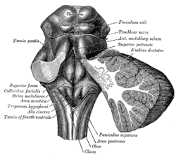
Gray709.png
Rhomboid fossa. (Locus coeruleus not labeled, but is very near [just lateral to] the facial colliculus, which is labeled at center left.)
Locus ceruleus - high mag.jpg
Micrograph showing the locus coeruleus. HE-LFB stain.
Details
Identifiers
Latinlocus caeruleus ("blue place")
MeSHD008125
NeuroNames583
NeuroLex IDbirnlex_905
TAA14.1.05.436 A14.1.05.706
FMA72478

The locus coeruleus (\-si-ˈrü-lē-əs\, also spelled locus caeruleus or locus ceruleus) is a nucleus in the pons of the brainstem involved with physiological responses to stress and panic. It is a part of the reticular activating system.

The locus coeruleus is the principal site for brain synthesis of norepinephrine (noradrenaline). The locus coeruleus and the areas of the body affected by the norepinephrine it produces are described collectively as the locus coeruleus-noradrenergic system or LC-NA system. Norepinephrine may also be released directly into the blood from the adrenal medulla.

Anatomy

Micrograph showing the locus coeruleus (upper-right of image) in an axial section of the pons. The fourth ventricle (quasi-triangular white area) is in the upper-left of the image. The midline is seen on the left. The large white area in the upper-left corner is where the cerebellum would be. HE-LFB stain.
 
Locus coeruleus highlighted in green.

The locus coeruleus (LC) is located in the posterior area of the rostral pons in the lateral floor of the fourth ventricle. It is composed of mostly medium-size neurons. Melanin granules inside the neurons of the LC contribute to its blue colour. Thus, it is also known as the nucleus pigmentosus pontis, meaning "heavily pigmented nucleus of the pons." The neuromelanin is formed by the polymerization of noradrenaline and is analogous to the black dopamine-based neuromelanin in the substantia nigra

In adult humans (19-78) the locus coeruleus has 22,000 to 51,000 total pigmented neurons that range in size between 31,000 and 60,000 μm3.

Connections

The projections of this nucleus reach far and wide. For example, they innervate the spinal cord, the brain stem, cerebellum, hypothalamus, the thalamic relay nuclei, the amygdala, the basal telencephalon, and the cortex. The norepinephrine from the LC has an excitatory effect on most of the brain, mediating arousal and priming the brain’s neurons to be activated by stimuli.

As an important homeostatic control center of the body, the locus coeruleus receives afferents from the hypothalamus. The cingulate gyrus and the amygdala also innervate the LC, allowing emotional pain and stressors to trigger noradrenergic responses. The cerebellum and afferents from the raphe nuclei also project to the LC, in particular the pontine raphe nucleus and dorsal raphe nucleus.

Inputs

The locus coeruleus receives inputs from a number of other brain regions, primarily:

Outputs

The projections from the locus coeruleus consist of neurons that utilize norepinephrine as their primary neurotransmitter. These projections include the following connections:

Function

It is related to many functions via its widespread projections. The LC-NA system modulates cortical, subcortical, cerebellar, brainstem, and spinal cord circuits. Some of the most important functions influenced by this system are:
The locus coeruleus is a part of the reticular activating system, and is almost completely inactivated in rapid eye movement sleep.

Pathophysiology


Research continues to reveal that norepinephrine (NE) is a critical regulator of numerous activities from stress response, the formation of memory to attention and arousal. Many neuropsychiatric disorders precipitate from alterations to NE modulated neurocircuitry: disorders of affect, anxiety disorders, PTSD, ADHD and Alzheimer’s disease. Alterations in the locus coeruleus (LC) accompany dysregulation of NE function and likely play a key role in the pathophysiology of these neuropsychiatric disorders.

In stress

The locus coeruleus is responsible for mediating many of the sympathetic effects during stress. The locus coeruleus is activated by stress, and will respond by increasing norepinephrine secretion, which in turn will alter cognitive function (through the prefrontal cortex), increase motivation (through nucleus accumbens), activate the hypothalamic-pituitary-adrenal axis, and increase the sympathetic discharge/inhibit parasympathetic tone (through the brainstem). Specific to the activation of the hypothalamo-pituitary adrenal axis, norepinephrine will stimulate the secretion of corticotropin-releasing factor from the hypothalamus, that induces adrenocorticotropic hormone release from the anterior pituitary and subsequent cortisol synthesis in the adrenal glands. Norepinephrine released from locus coeruleus will feedback to inhibit its production, and corticotropin-releasing hormone will feedback to inhibit its production, while positively feeding to the locus coeruleus to increase norepinephrine production.

The LC's role in cognitive function in relation to stress is complex and multi-modal. Norepinephrine released from the LC can act on α2 receptors to increase working memory, or an excess of NE may decrease working memory by binding to the lower-affinity α1 receptors.

Psychiatric research has documented that enhanced noradrenergic postsynaptic responsiveness in the neuronal pathway (brain circuit) that originates in the locus coeruleus and ends in the basolateral nucleus of the amygdala is a major factor in the pathophysiology of most stress-induced fear-circuitry disorders and especially in posttraumatic stress disorder (PTSD). The LC neurons are probably the origin of the first or second “leg” of the "PTSD circuit." An important 2005 study of deceased American army veterans from World War II has shown combat-related PTSD to be associated with a postmortem-diminished number of neurons in the locus coeruleus (LC) on the right side of the brain.

In opiate withdrawal

Opioids inhibit the firing of neurons in the locus coeruleus. When opioid consumption is stopped, the increased activity of the locus coeruleus contributes to the symptoms of opiate withdrawal. The alpha2 adrenoceptor agonist clonidine is used to counteract this withdrawal effect by decreasing adrenergic neurotransmission from the locus coeruleus.

Rett syndrome

The genetic defect of the transcriptional regulator MECP2 is responsible for Rett syndrome. A MECP2 deficiency has been associated to catecholaminergic dysfunctions related to autonomic and sympathoadrenergic system in mouse models of Rett Syndrome (RTT). The Locus Coeruleus is the major source of noradrenergic innervation in the brain and sends widespread connections to rostral (cerebral cortex, hippocampus, hypothalamus) and caudal (cerebellum, brainstem nuclei) brain areas. Indeed, an alteration of this structure could contribute to several symptoms observed in MECP2-deficient mice. Changes in the electrophysiological properties of cells in the locus ceruleus were shown. These Locus Coeruleus cell changes include hyperexcitability and decreased functioning of its noradrenergic innervation. A reduction of the tyrosine hydroxylase (TH) mRNA level, the rate-limiting enzyme in catecholamine synthesis, was detected in the whole pons of MECP2-null male as well as in adult heterozygous female mice. Using immunoquantification techniques, a decrease of TH protein staining level, number of locus coeruleus TH-expressing neurons and density of dendritic arborization surrounding the structure was shown in symptomatic MECP2-deficient mice. However, locus coeruleus cells are not dying but are more likely losing their fully mature phenotype, since no apoptotic neurons in the pons were detected. Researchers have concluded that, "Because these neurons are a pivotal source of norepinephrine throughout the brainstem and forebrain and are involved in the regulation of diverse functions disrupted in Rett Syndrome, such as respiration and cognition, we hypothesize that the locus ceruleus is a critical site at which loss of MECP2 results in CNS dysfunction. Restoration of normal locus ceruleus function may therefore be of potential therapeutic value in the treatment of Rett Syndrome." This could explain why a norepinephrine reuptake inhibitor (desipramine, DMI), which enhances the extracellular NE levels at all noradrenergic synapses, ameliorated some Rett syndrome symptoms in a mouse model of Rett syndrome.

Neurodegenerative diseases

The locus ceruleus is affected in many forms of neurodegenerative diseases: genetic and idiopathic Parkinson's disease, progressive supranuclear palsy, Pick's disease or Alzheimer's disease. It is also affected in Down syndrome. For example, there is up to 80% loss of locus ceruleus neurons in Alzheimer's disease. Mouse models of Alzheimer's disease show accelerated progression after chemical destruction of the locus ceruleus The norepinephrine from locus ceruleus cells in addition to its neurotransmitter role locally diffuses from "varicosities". As such it provides an endogenous anti-inflammatory agent in the microenvironment around the neurons, glial cells, and blood vessels in the neocortex and hippocampus. It has been shown that norepinephrine stimulates mouse microglia to suppress -induced production of cytokines and promotes phagocytosis of Aβ. This suggests that degeneration of the locus ceruleus might be responsible for increased Aβ deposition in AD brains. Degeneration of pigmented neurons in this region in Alzheimer's and Parkinson's disease can be visualized in vivo with Neuromelanin MRI.

History

It was discovered in 1784 by Félix Vicq-d'Azyr, redescribed later by Johann Christian Reil in 1809 and named by Joseph Wenzel and Karl Wenzel brothers in 1812. High monoamine oxidase activity in the rodent LC was found in 1959, monoamines were found in 1964 and noradrenergic ubiquitous projections in the 1970s.

Etymology

Coeruleus or caeruleus

The 'English' name locus coeruleus is actually a Latin expression consisting of the noun, locus, place or spot and the adjective coeruleus, dark blue or sky-blue. This was aptly translated into English as blue place in 1907 in the English translation of the official Latin anatomic nomenclature of 1895, Nomina Anatomica. The name of the locus coeruleus is derived from its azure appearance in unstained brain tissue. The color is due to light scattering from neuromelanin in noradrenergic (producing or activated by norepinephrine) nerve cell bodies. The phenomenon is magnified by the Falck-Hillarp technique°, which combines freeze-dried tissue and formaldehyde to fluoresce the catecholamines and serotonin contained in the tissue.

The spelling coeruleus is actually considered incorrect with dictionaries of classical Latin preferring caeruleus instead. Caeruleus is derived from caelum, hence the spelling with -ae, like caeluleus → caeruleus. Caelum in classical Latin could refer to the sky, the heaven or the vault of heaven.

In mediaeval Latin, orthographic variants like coelum for classical Latin caelum and cerulans for classical Latin caerulans can be spotted. 

In English, the color adjective cerulean is derived from Latin caeruleus. In addition, ceiling is ultimately derived from Latin caelum as well.

Official Latin nomenclature

The official Latin nomenclature, Nomina Anatomica as ratified in Basel in 1895 and in Jena in 1935 contained the orthographic correct form locus caeruleus. The Nomina Anatomica published in 1955 inadvertently introduced the incorrect spelling locus coeruleus, without any further explanation. The subsequent edition monophthongized the diphthong, resulting in locus ceruleus, as they proclaimed that: "All diphthongs should be eliminated". This form was retained in the subsequent edition. The following two editions from 1977 and 1983 reverted the orthography back to the incorrect spelling locus coeruleus, while the subsequent edition from 1989 eventually returned to the correct spelling locus caeruleus. The current edition of the Nomina Anatomica, rebaptized as Terminologia Anatomica, dictates locus caeruleus in its list of Latin expressions and correspondingly mentions locus caeruleus in its list of English equivalents. This is in line with the statement made by the chairman of the Terminologia Anatomica that "the committee decided that Latin terms when used in English should be in correct Latin".

Norepinephrine

From Wikipedia, the free encyclopedia

Norepinephrine
Norepinephrine.svg
Norepinephrine ball-and-stick model.png
Clinical data
Synonyms
  • NE, NA,
  • Noradrenaline,
  • (R)-(–)-Norepinephrine,
  • l-1-(3,4-Dihydroxyphenyl)-2-aminoethanol
Physiological data
Source tissueslocus coeruleus; sympathetic nervous system; adrenal medulla
Target tissuessystem-wide
Receptorsα1, α2, β1, β3
Agonistssympathomimetic drugs, clonidine, isoprenaline
AntagonistsTricyclic antidepressants, beta blockers, antipsychotics
Precursordopamine
Biosynthesisdopamine β-monooxygenase
MetabolismMAO-A; COMT
Identifiers
CAS Number
PubChem CID
IUPHAR/BPS
DrugBank
ChemSpider
KEGG
ECHA InfoCard100.000.088 Edit this at Wikidata

Noradrenaline (NA), also called norepinephrine (NE) or noradrenalin, is an organic chemical in the catecholamine family that functions in the brain and body as a hormone and neurotransmitter. The name "noradrenaline", derived from Latin roots meaning "at/alongside the kidneys", is more commonly used in the United Kingdom; in the United States, "norepinephrine," derived from Greek roots having that same meaning, is usually preferred. "Norepinephrine" is also the international nonproprietary name given to the drug. Regardless of which name is used for the substance itself, parts of the body that produce or are affected by it are referred to as noradrenergic.

The general function of norepinephrine is to mobilize the brain and body for action. Norepinephrine release is lowest during sleep, rises during wakefulness, and reaches much higher levels during situations of stress or danger, in the so-called fight-or-flight response. In the brain, norepinephrine increases arousal and alertness, promotes vigilance, enhances formation and retrieval of memory, and focuses attention; it also increases restlessness and anxiety. In the rest of the body, norepinephrine increases heart rate and blood pressure, triggers the release of glucose from energy stores, increases blood flow to skeletal muscle, reduces blood flow to the gastrointestinal system, and inhibits voiding of the bladder and gastrointestinal motility.

In the brain, noradrenaline is produced in nuclei that are small yet exert powerful effects on other brain areas. The most important of these nuclei is the locus coeruleus, located in the pons. Outside the brain, norepinephrine is used as a neurotransmitter by sympathetic ganglia located near the spinal cord or in the abdomen, and it is also released directly into the bloodstream by the adrenal glands. Regardless of how and where it is released, norepinephrine acts on target cells by binding to and activating noradrenergic receptors located on the cell surface.

A variety of medically important drugs work by altering the actions of noradrenaline systems. Noradrenaline itself is widely used as an injectable drug for the treatment of critically low blood pressure. Beta blockers, which counter some of the effects of noradrenaline, are frequently used to treat glaucoma, migraine, and a range of cardiovascular problems. Alpha blockers, which counter a different set of noradrenaline effects, are used to treat several cardiovascular and psychiatric conditions. Alpha-2 agonists often have a sedating effect, and are commonly used as anesthesia-enhancers in surgery, as well as in treatment of drug or alcohol dependence. Many important psychiatric drugs exert strong effects on noradrenaline systems in the brain, resulting in side-effects that may be helpful or harmful.

Structure

Norepinephrine is a catecholamine and a phenethylamine. Its structure differs from that of epinephrine only in that epinephrine has a methyl group attached to its nitrogen, whereas the methyl group is replaced by a hydrogen atom in norepinephrine. The prefix nor- is derived as an abbreviation of the word "normal", used to indicate a demethylated compound.

Chemical diagram of the structure of a norepinephrine molecule.
Norepinephrine structure
 
Chemical diagram of the structure of an epinephrine molecule.
Epinephrine structure
 
Chemical diagram of a catechol structure.
Catechol structure

Biochemical mechanisms

Biosynthesis

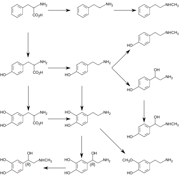
Biosynthetic pathways for catecholamines and trace amines in the human brain
 
Graphic of catecholamine and trace amine biosynthesis


Norepinephrine is synthesized from the amino acid tyrosine by a series of enzymatic steps in the adrenal medulla and postganglionic neurons of the sympathetic nervous system. While the conversion of tyrosine to dopamine occurs predominantly in the cytoplasm, the conversion of dopamine to norepinephrine by dopamine β-monooxygenase occurs predominantly inside neurotransmitter vesicles. The metabolic pathway is:
Phenylalanine → Tyrosine → L-DOPA → Dopamine → Norepinephrine
Thus the direct precursor of norepinephrine is dopamine, which is synthesized indirectly from the essential amino acid phenylalanine or the non-essential amino acid tyrosine. These amino acids are found in nearly every protein and, as such, are provided by ingestion of protein-containing food, with tyrosine being the most common. 

Phenylalanine is converted into tyrosine by the enzyme phenylalanine hydroxylase, with molecular oxygen (O2) and tetrahydrobiopterin as cofactors. Tyrosine is converted into L-DOPA by the enzyme tyrosine hydroxylase, with tetrahydrobiopterin, O2, and probably ferrous iron (Fe2+) as cofactors. L-DOPA is converted into dopamine by the enzyme aromatic L-amino acid decarboxylase (also known as DOPA decarboxylase), with pyridoxal phosphate as a cofactor. Dopamine is then converted into norepinephrine by the enzyme dopamine β-monooxygenase (formerly known as dopamine β-hydroxylase), with O2 and ascorbic acid as cofactors.

Norepinephrine itself can further be converted into epinephrine by the enzyme phenylethanolamine N-methyltransferase with S-adenosyl-L-methionine as cofactor.

Degradation

In mammals, norepinephrine is rapidly degraded to various metabolites. The initial step in the breakdown can be catalyzed by either of the enzymes monoamine oxidase (mainly monoamine oxidase A) or COMT. From there the breakdown can proceed by a variety of pathways. The principal end products are either Vanillylmandelic acid or a conjugated form of MHPG, both of which are thought to be biologically inactive and are excreted in the urine.

Norepinephrine degradation. Metabolizing enzymes are shown in boxes.

Functions

Cellular effects

Adrenergic receptors in the mammal brain and body
Family Receptor Type Mechanism
Alpha α1 Gq-coupled. Increase IP3 and calcium by
activating phospholipase C.
α2 Gi/Go-coupled. Decrease cAMP by
inhibiting adenylate cyclase.
Beta β1 Gs-coupled. Increase cAMP by
activating adenylate cyclase.
β2
β3

Like many other biologically active substances, norepinephrine exerts its effects by binding to and activating receptors located on the surface of cells. Two broad families of norepinephrine receptors have been identified, known as alpha and beta adrenergic receptors. Alpha receptors are divided into subtypes α1 and α2; beta receptors into subtypes β1, β2, and β3. All of these function as G protein-coupled receptors, meaning that they exert their effects via a complex second messenger system. Alpha-2 receptors usually have inhibitory effects, but many are located pre-synaptically (i.e., on the surface of the cells that release norepinephrine), so the net effect of alpha-2 activation is often a decrease in the amount of norepinephrine released. Alpha-1 receptors and all three types of beta receptors usually have excitatory effects.

Storage, release, and reuptake

Cartoon diagram of a noradrenergic synapse, showing the synthetic and metabolic mechanisms as well as the things that can happen after release.
Norepinephrine (labeled "noradrenaline" in this drawing) processing in a synapse. After release norepinephrine can either be taken up again by the presynaptic terminal, or broken down by enzymes.
 
Inside the brain norepinephrine functions as a neurotransmitter, and is controlled by a set of mechanisms common to all monoamine neurotransmitters. After synthesis, norepinephrine is transported from the cytosol into synaptic vesicles by the vesicular monoamine transporter (VMAT). Norepinephrine is stored in these vesicles until it is ejected into the synaptic cleft, typically after an action potential causes the vesicles to release their contents directly into the synaptic cleft through a process called exocytosis.

Once in the synapse, norepinephrine binds to and activates receptors. After an action potential, the norepinephrine molecules quickly become unbound from their receptors. They are then absorbed back into the presynaptic cell, via reuptake mediated primarily by the norepinephrine transporter (NET). Once back in the cytosol, norepinephrine can either be broken down by monoamine oxidase or repackaged into vesicles by VMAT, making it available for future release.

Sympathetic nervous system

Schema of the sympathetic nervous system, showing the sympathetic ganglia and the parts of the body to which they connect.
 
Norepinephrine is the main neurotransmitter used by the sympathetic nervous system, which consists of about two dozen sympathetic chain ganglia located next to the spinal cord, plus a set of prevertebral ganglia located in the chest and abdomen. These sympathetic ganglia are connected to numerous organs, including the eyes, salivary glands, heart, lungs, liver, gallbladder, stomach, intestines, kidneys, urinary bladder, reproductive organs, muscles, skin, and adrenal glands. Sympathetic activation of the adrenal glands causes the part called the adrenal medulla to release norepinephrine (as well as epinephrine) into the bloodstream, from which, functioning as a hormone, it gains further access to a wide variety of tissues.

Broadly speaking, the effect of norepinephrine on each target organ is to modify its state in a way that makes it more conducive to active body movement, often at a cost of increased energy use and increased wear and tear. This can be contrasted with the acetylcholine-mediated effects of the parasympathetic nervous system, which modifies most of the same organs into a state more conducive to rest, recovery, and digestion of food, and usually less costly in terms of energy expenditure.

The sympathetic effects of norepinephrine include:
  • In the eyes, an increase in production of tears, making the eyes more moist, and pupil dilation through contraction of the iris dilator.
  • In the heart, an increase in the amount of blood pumped.
  • In brown adipose tissue, an increase in calories burned to generate body heat.
  • Multiple effects on the immune system. The sympathetic nervous system is the primary path of interaction between the immune system and the brain, and several components receive sympathetic inputs, including the thymus, spleen, and lymph nodes. However the effects are complex, with some immune processes activated while others are inhibited.
  • In the arteries, constriction of blood vessels, causing an increase in blood pressure.
  • In the kidneys, release of renin and retention of sodium in the bloodstream.
  • In the liver, an increase in production of glucose, either by glycogenolysis after a meal or by gluconeogenesis when food has not recently been consumed. Glucose is the body's main energy source in most conditions.
  • In the pancreas, increased release of glucagon, a hormone whose main effect is to increase the production of glucose by the liver.
  • In skeletal muscles, an increase in glucose uptake.
  • In adipose tissue (i.e., fat cells), an increase in lipolysis, that is, conversion of fat to substances that can be used directly as energy sources by muscles and other tissues.
  • In the stomach and intestines, a reduction in digestive activity. This results from a generally inhibitory effect of norepinephrine on the enteric nervous system, causing decreases in gastrointestinal mobility, blood flow, and secretion of digestive substances.

Central nervous system

Brain areas containing noradrenergic neurons.

The noradrenergic neurons in the brain form a neurotransmitter system, that, when activated, exerts effects on large areas of the brain. The effects are manifested in alertness, arousal, and readiness for action.

Noradrenergic neurons (i.e., neurons whose primary neurotransmitter is norepinephrine) are comparatively few in number, and their cell bodies are confined to a few relatively small brain areas, but they send projections to many other brain areas and exert powerful effects on their targets. These noradrenergic cell groups were first mapped in 1964 by Annica Dahlström and Kjell Fuxe, who assigned them labels starting with the letter "A" (for "aminergic"). In their scheme, areas A1 through A7 contain the neurotransmitter norepinephrine (A8 through A14 contain dopamine). Noradrenergic cell group A1 is located in the caudal ventrolateral part of the medulla, and plays a role in the control of body fluid metabolism. Noradrenergic cell group A2 is located in a brainstem area called the solitary nucleus; these cells have been implicated in a variety of responses, including control of food intake and responses to stress. Cell groups A5 and A7 project mainly to the spinal cord.

The most important source of norepinephrine in the brain is the locus coeruleus, which contains noradrenergic cell group A6 and adjoins cell group A4. The locus coeruleus is quite small in absolute terms—in primates it is estimated to contain around 15,000 neurons, less than one millionth of the neurons in the brain—but it sends projections to every major part of the brain and also to the spinal cord.

The level of activity in the locus coeruleus correlates broadly with vigilance and speed of reaction. LC activity is low during sleep and drops to virtually nothing during the REM (dreaming) state. It runs at a baseline level during wakefulness, but increases temporarily when a person is presented with any sort of stimulus that draws attention. Unpleasant stimuli such as pain, difficulty breathing, bladder distension, heat or cold generate larger increases. Extremely unpleasant states such as intense fear or intense pain are associated with very high levels of LC activity.

Norepinephrine released by the locus coeruleus affects brain function in a number of ways. It enhances processing of sensory inputs, enhances attention, enhances formation and retrieval of both long term and working memory, and enhances the ability of the brain to respond to inputs by changing the activity pattern in the prefrontal cortex and other areas. The control of arousal level is strong enough that drug-induced suppression of the LC has a powerful sedating effect.

There is great similarity between situations that activate the locus coeruleus in the brain and situations that activate the sympathetic nervous system in the periphery: the LC essentially mobilizes the brain for action while the sympathetic system mobilizes the body. It has been argued that this similarity arises because both are to a large degree controlled by the same brain structures, particularly a part of the brainstem called the nucleus gigantocellularis.

Pharmacology

A large number of important drugs exert their effects by interacting with norepinephrine systems in the brain or body. Their uses include treatment of cardiovascular problems, shock, and a variety of psychiatric conditions. These drugs are divided into: sympathomimetic drugs which mimic or enhance at least some of the effects of norepinephrine released by the sympathetic nervous system; sympatholytic drugs, in contrast, block at least some of the effects. Both of these are large groups with diverse uses, depending on exactly which effects are enhanced or blocked.

Norepinephrine itself is classified as a sympathomimetic drug: its effects when given by intravenous injection of increasing heart rate and force and constricting blood vessels make it very useful for treating medical emergencies that involve critically low blood pressure. Surviving Sepsis Campaign recommended norepinephrine as first line agent in treating septic shock which is unresponsive to fluid resuscitation, supplemented by vasopressin and epinephrine. Dopamine usage is restricted only to highly selected patients.

Beta blockers

These are sympatholytic drugs that block the effects of beta adrenergic receptors while having little or no effect on alpha receptors. They are sometimes used to treat high blood pressure, atrial fibrillation and congestive heart failure, but recent reviews have concluded that other types of drugs are usually superior for those purposes. Beta blockers may be a viable choice for other cardiovascular conditions, though, including angina and Marfan syndrome. They are also widely used to treat glaucoma, most commonly in the form of eyedrops. Because of their effects in reducing anxiety symptoms and tremor, they have sometimes been used by entertainers, public speakers and athletes to reduce performance anxiety, although they are not medically approved for that purpose and are banned by the International Olympic Committee.

However, the usefulness of beta blockers is limited by a range of serious side effects, including slowing of heart rate, a drop in blood pressure, asthma, and reactive hypoglycemia. The negative effects can be particularly severe in people who suffer from diabetes.

Alpha blockers

These are sympatholytic drugs that block the effects of adrenergic alpha receptors while having little or no effect on beta receptors. Drugs belonging to this group can have very different effects, however, depending on whether they primarily block alpha-1 receptors, alpha-2 receptors, or both. Alpha-2 receptors, as described elsewhere in this article, are frequently located on norepinephrine-releasing neurons themselves and have inhibitory effects on them; consequently blockage of alpha-2 receptors usually results in an increase in norepinephrine release. Alpha-1 receptors are usually located on target cells and have excitatory effects on them; consequently blockage of alpha-1 receptors usually results in blocking some of the effects of norepinephrine. Drugs such as phentolamine that act on both types of receptors can produce a complex combination of both effects. In most cases when the term "alpha blocker" is used without qualification, it refers to a selective alpha-1 antagonist.

Selective alpha-1 blockers have a variety of uses. Because one of their effects is to relax the muscles in the neck of the bladder, they are often used to treat benign prostatic hyperplasia, and to help with the expulsion of bladder stones. Alpha-blockers also likely help people pass their kidney stones. Their effects on the central nervous system make them useful for treating generalized anxiety disorder, panic disorder, and posttraumatic stress disorder. They may, however, have significant side-effects, including a drop in blood pressure.

Some antidepressants function partly as selective alpha-2 blockers, but the best-known drug in that class is yohimbine, which is extracted from the bark of the African yohimbe tree. Yohimbine acts as a male potency enhancer, but its usefulness for that purpose is limited by serious side-effects including anxiety and insomnia. Overdoses can cause a dangerous increase in blood pressure. Yohimbine is banned in many countries, but in the United States, because it is extracted from a plant rather than chemically synthesized, it is sold over the counter as a nutritional supplement.

Alpha-2 agonists

These are sympathomimetic drugs that activate alpha-2 receptors or enhance their effects. Because alpha-2 receptors are inhibitory and many are located presynaptically on norepinephrine-releasing cells, the net effect of these drugs is usually to reduce the amount of norepinephrine released. Drugs in this group that are capable of entering the brain often have strong sedating effects, due to their inhibitory effects on the locus coeruleus. Clonidine, for example, is used for the treatment of anxiety disorders and insomnia, and also as a sedative premedication for patients about to undergo surgery. Xylazine, another drug in this group, is also a powerful sedative and is often used in combination with ketamine as a general anaesthetic for veterinary surgery—in the United States it has not been approved for use in humans.

Stimulants and antidepressants

These are drugs whose primary effects are thought to be mediated by different neurotransmitter systems (dopamine for stimulants, serotonin for antidepressants), but many also increase levels of norepinephrine in the brain. Amphetamine, for example, is a stimulant that increases release of norepinephrine as well as dopamine. Monoamine oxidase inhibitors are antidepressants that inhibit the metabolic degradation of norepinephrine as well as serotonin. In some cases it is difficult to distinguish the norepinephrine-mediated effects from the effects related to other neurotransmitters.

Diseases and disorders

A number of important medical problems involve dysfunction of the norepinephrine system in the brain or body.

Sympathetic hyperactivation

Hyperactivation of the sympathetic nervous system is not a recognized condition in itself, but it is a component of a number of conditions, as well as a possible consequence of taking sympathomimetic drugs. It causes a distinctive set of symptoms including aches and pains, rapid heartbeat, elevated blood pressure, sweating, palpitations, anxiety, headache, paleness, and a drop in blood glucose. If sympathetic activity is elevated for an extended time, it can cause weight loss and other stress-related body changes. 

The list of conditions that can cause sympathetic hyperactivation includes severe brain injury, spinal cord damage, heart failure, high blood pressure, kidney disease, and various types of stress.

Pheochromocytoma

A pheochromocytoma is a rarely occurring tumor of the adrenal medulla, caused either by genetic factors or certain types of cancer. The consequence is a massive increase in the amount of norepinephrine and epinephrine released into the bloodstream. The most obvious symptoms are those of sympathetic hyperactivation, including particularly a rise in blood pressure that can reach fatal levels. The most effective treatment is surgical removal of the tumor.

Stress

Stress, to a physiologist, means any situation that threatens the continued stability of the body and its functions. Stress affects a wide variety of body systems: the two most consistently activated are the hypothalamic-pituitary-adrenal axis and the norepinephrine system, including both the sympathetic nervous system and the locus coeruleus-centered system in the brain. Stressors of many types evoke increases in noradrenergic activity, which mobilizes the brain and body to meet the threat. Chronic stress, if continued for a long time, can damage many parts of the body. A significant part of the damage is due to the effects of sustained norepinephrine release, because of norepinephrine's general function of directing resources away from maintenance, regeneration, and reproduction, and toward systems that are required for active movement. The consequences can include slowing of growth (in children), sleeplessness, loss of libido, gastrointestinal problems, impaired disease resistance, slower rates of injury healing, depression, and increased vulnerability to addiction.

ADHD

Attention deficit hyperactivity disorder is a psychiatric condition involving problems with attention, hyperactivity, and impulsiveness. It is most commonly treated using stimulant drugs such as methylphenidate (Ritalin), whose primary effect is to increase dopamine levels in the brain, but drugs in this group also generally increase brain levels of norepinephrine, and it has been difficult to determine whether these actions are involved in their clinical value. Also there is substantial evidence that many people with ADHD show "biomarkers" involving altered norepinephrine processing. Several drugs whose primary effects are on norepinephrine, including guanfacine, clonidine, and atomoxetine, have been tried as treatments for ADHD, and found to have effects comparable to those of stimulants.

Autonomic failure

Several conditions, including Parkinson's disease, diabetes and so-called pure autonomic failure, can cause a loss of norepinephrine-secreting neurons in the sympathetic nervous system. The symptoms are widespread, the most serious being a reduction in heart rate and an extreme drop in resting blood pressure, making it impossible for severely affected people to stand for more than a few seconds without fainting. Treatment can involve dietary changes or drugs.

Comparative biology and evolution

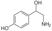
Chemical structure of octopamine, which serves as the homologue of norepinephrine in many invertebrate species
 
Norepinephrine has been reported to exist in a wide variety of animal species, including protozoa, placozoa and cnidaria (jellyfish and related species), but not in ctenophores (comb jellies), whose nervous systems differ greatly from those of other animals. It is generally present in deuterostomes (vertebrates, etc.), but in protostomes (arthropods, molluscs, flatworms, nematodes, annelids, etc.) it is replaced by octopamine, a closely related chemical with a closely related synthesis pathway. In insects, octopamine has alerting and activating functions that correspond (at least roughly) with the functions of norepinephrine in vertebrates. It has been argued that octopamine evolved to replace norepinephrine rather than vice versa; however, the nervous system of amphioxus (a primitive chordate) has been reported to contain octopamine but not norepinephrine, which presents difficulties for that hypothesis.

History

Early in the twentieth century Walter Cannon, who had popularized the idea of a sympathoadrenal system preparing the body for fight and flight, and his colleague Arturo Rosenblueth developed a theory of two sympathins, sympathin E (excitatory) and sympathin I (inhibitory), responsible for these actions. The Belgian pharmacologist Zénon Bacq as well as Canadian and US-American pharmacologists between 1934 and 1938 suggested that noradrenaline might be a sympathetic transmitter. In 1939, Hermann Blaschko and Peter Holtz independently identified the biosynthetic mechanism for norepinephrine in the vertebrate body. In 1945 Ulf von Euler published the first of a series of papers that established the role of norepinephrine as a neurotransmitter. He demonstrated the presence of norepinephrine in sympathetically innervated tissues and brain, and adduced evidence that it is the sympathin of Cannon and Rosenblueth.

Introduction to entropy

From Wikipedia, the free encyclopedia https://en.wikipedia.org/wiki/Introduct...