Search This Blog

Wednesday, December 4, 2019

Karyotype

From Wikipedia, the free encyclopedia
https://en.wikipedia.org/wiki/Karyotype
 
Karyotyping is the process by which cytogeneticists take photographs of chromosomes in order to determine the chromosome complement of an individual, including the number of chromosomes and any abnormalities. The term is also used for the complete set of chromosomes in a species or in an individual organism and for a test that detects this complement or measures the number.

Karyotypes describe the chromosome count of an organism and what these chromosomes look like under a light microscope. Attention is paid to their length, the position of the centromeres, banding pattern, any differences between the sex chromosomes, and any other physical characteristics. The preparation and study of karyotypes is part of cytogenetics.

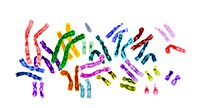
Karyogram of human male using Giemsa staining

The study of whole sets of chromosomes is sometimes known as karyology. The chromosomes are depicted (by rearranging a photomicrograph) in a standard format known as a karyogram or idiogram: in pairs, ordered by size and position of centromere for chromosomes of the same size. 

The basic number of chromosomes in the somatic cells of an individual or a species is called the somatic number and is designated 2n. In the germ-line (the sex cells) the chromosome number is n (humans: n = 23). Thus, in humans 2n = 46. 

So, in normal diploid organisms, autosomal chromosomes are present in two copies. There may, or may not, be sex chromosomes. Polyploid cells have multiple copies of chromosomes and haploid cells have single copies. 

The study of karyotypes is important for cell biology and genetics, and the results may be used in evolutionary biology (karyosystematics) and medicine. Karyotypes can be used for many purposes; such as to study chromosomal aberrations, cellular function, taxonomic relationships, and to gather information about past evolutionary events.

History of karyotype studies

Chromosomes were first observed in plant cells by Carl Wilhelm von Nägeli in 1842. Their behavior in animal (salamander) cells was described by Walther Flemming, the discoverer of mitosis, in 1882. The name was coined by another German anatomist, Heinrich von Waldeyer in 1888. It is New Latin from Ancient Greek κάρυον karyon, "kernel", "seed", or "nucleus", and τύπος typos, "general form")
The next stage took place after the development of genetics in the early 20th century, when it was appreciated that chromosomes (that can be observed by karyotype) were the carrier of genes. Lev Delaunay in 1922 seems to have been the first person to define the karyotype as the phenotypic appearance of the somatic chromosomes, in contrast to their genic contents. The subsequent history of the concept can be followed in the works of C. D. Darlington and Michael JD White.

Investigation into the human karyotype took many years to settle the most basic question: how many chromosomes does a normal diploid human cell contain? In 1912, Hans von Winiwarter reported 47 chromosomes in spermatogonia and 48 in oogonia, concluding an XX/XO sex determination mechanism. Painter in 1922 was not certain whether the diploid of humans was 46 or 48, at first favoring 46, but revised his opinion from 46 to 48, and he correctly insisted on humans having an XX/XY system. Considering the techniques of the time, these results were remarkable. 

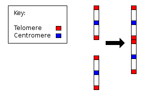
Fusion of ancestral chromosomes left distinctive remnants of telomeres, and a vestigial centromere
 
In textbooks, the number of human chromosomes remained at 48 for over thirty years. New techniques were needed to correct this error. Joe Hin Tjio working in Albert Levan's lab was responsible for finding the approach:
  1. Using cells in tissue culture
  2. Pretreating cells in a hypotonic solution, which swells them and spreads the chromosomes
  3. Arresting mitosis in metaphase by a solution of colchicine
  4. Squashing the preparation on the slide forcing the chromosomes into a single plane
  5. Cutting up a photomicrograph and arranging the result into an indisputable karyogram.
The work took place in 1955, and was published in 1956. The karyotype of humans includes only 46 chromosomes. The great apes have 48 chromosomes. Human chromosome 2 is now known to be a result of an end-to-end fusion of two ancestral ape chromosomes.

Observations on karyotypes

Staining

The study of karyotypes is made possible by staining. Usually, a suitable dye, such as Giemsa, is applied after cells have been arrested during cell division by a solution of colchicine usually in metaphase or prometaphase when most condensed. In order for the Giemsa stain to adhere correctly, all chromosomal proteins must be digested and removed. For humans, white blood cells are used most frequently because they are easily induced to divide and grow in tissue culture. Sometimes observations may be made on non-dividing (interphase) cells. The sex of an unborn fetus can be determined by observation of interphase cells (see amniotic centesis and Barr body).

Observations

Six different characteristics of karyotypes are usually observed and compared:
  1. Differences in absolute sizes of chromosomes. Chromosomes can vary in absolute size by as much as twenty-fold between genera of the same family. For example, the legumes Lotus tenuis and Vicia faba each have six pairs of chromosomes, yet V. faba chromosomes are many times larger. These differences probably reflect different amounts of DNA duplication.
  2. Differences in the position of centromeres. These differences probably came about through translocations.
  3. Differences in relative size of chromosomes. These differences probably arose from segmental interchange of unequal lengths.
  4. Differences in basic number of chromosomes. These differences could have resulted from successive unequal translocations which removed all the essential genetic material from a chromosome, permitting its loss without penalty to the organism (the dislocation hypothesis) or through fusion. Humans have one pair fewer chromosomes than the great apes. Human chromosome 2 appears to have resulted from the fusion of two ancestral chromosomes, and many of the genes of those two original chromosomes have been translocated to other chromosomes.
  5. Differences in number and position of satellites. Satellites are small bodies attached to a chromosome by a thin thread.
  6. Differences in degree and distribution of heterochromatic regions. Heterochromatin stains darker than euchromatin. Heterochromatin is packed tighter. Heterochromatin consists mainly of genetically inactive and repetitive DNA sequences as well as containing a larger amount of Adenine-Thymine pairs. Euchromatin is usually under active transcription and stains much lighter as it has less affinity for the giemsa stain. Euchromatin regions contain larger amounts of Guanine-Cytosine pairs. The staining technique using giemsa staining is called G banding and therefore produces the typical "G-Bands".
A full account of a karyotype may therefore include the number, type, shape and banding of the chromosomes, as well as other cytogenetic information.
Variation is often found:
  1. between the sexes,
  2. between the germ-line and soma (between gametes and the rest of the body),
  3. between members of a population (chromosome polymorphism),
  4. in geographic specialization, and
  5. in mosaics or otherwise abnormal individuals.

Human karyotype

human karyotype (male)
 
The normal human karyotypes contain 22 pairs of autosomal chromosomes and one pair of sex chromosomes (allosomes). Normal karyotypes for females contain two X chromosomes and are denoted 46,XX; males have both an X and a Y chromosome denoted 46,XY. Any variation from the standard karyotype may lead to developmental abnormalities.

Diversity and evolution of karyotypes

Although the replication and transcription of DNA is highly standardized in eukaryotes, the same cannot be said for their karyotypes, which are highly variable. There is variation between species in chromosome number, and in detailed organization, despite their construction from the same macromolecules. This variation provides the basis for a range of studies in evolutionary cytology

In some cases there is even significant variation within species. In a review, Godfrey and Masters conclude:
In our view, it is unlikely that one process or the other can independently account for the wide range of karyotype structures that are observed ... But, used in conjunction with other phylogenetic data, karyotypic fissioning may help to explain dramatic differences in diploid numbers between closely related species, which were previously inexplicable.
Although much is known about karyotypes at the descriptive level, and it is clear that changes in karyotype organization has had effects on the evolutionary course of many species, it is quite unclear what the general significance might be.
We have a very poor understanding of the causes of karyotype evolution, despite many careful investigations ... the general significance of karyotype evolution is obscure.
— Maynard Smith

Changes during development

Instead of the usual gene repression, some organisms go in for large-scale elimination of heterochromatin, or other kinds of visible adjustment to the karyotype.
  • Chromosome elimination. In some species, as in many sciarid flies, entire chromosomes are eliminated during development.
  • Chromatin diminution (founding father: Theodor Boveri). In this process, found in some copepods and roundworms such as Ascaris suum, portions of the chromosomes are cast away in particular cells. This process is a carefully organised genome rearrangement where new telomeres are constructed and certain heterochromatin regions are lost. In A. suum, all the somatic cell precursors undergo chromatin diminution.
  • X-inactivation. The inactivation of one X chromosome takes place during the early development of mammals. In placental mammals, the inactivation is random as between the two Xs; thus the mammalian female is a mosaic in respect of her X chromosomes. In marsupials it is always the paternal X which is inactivated. In human females some 15% of somatic cells escape inactivation, and the number of genes affected on the inactivated X chromosome varies between cells: in fibroblast cells up about 25% of genes on the Barr body escape inactivation.

Number of chromosomes in a set

A spectacular example of variability between closely related species is the muntjac, which was investigated by Kurt Benirschke and his colleague Doris Wurster. The diploid number of the Chinese muntjac, Muntiacus reevesi, was found to be 46, all telocentric. When they looked at the karyotype of the closely related Indian muntjac, Muntiacus muntjak, they were astonished to find it had female = 6, male = 7 chromosomes.
They simply could not believe what they saw ... They kept quiet for two or three years because they thought something was wrong with their tissue culture ... But when they obtained a couple more specimens they confirmed [their findings].
— Hsu p. 73-4
The number of chromosomes in the karyotype between (relatively) unrelated species is hugely variable. The low record is held by the nematode Parascaris univalens, where the haploid n = 1; and an ant: Myrmecia pilosula. The high record would be somewhere amongst the ferns, with the adder's tongue fern Ophioglossum ahead with an average of 1262 chromosomes. Top score for animals might be the shortnose sturgeon Acipenser brevirostrum at 372 chromosomes. The existence of supernumerary or B chromosomes means that chromosome number can vary even within one interbreeding population; and aneuploids are another example, though in this case they would not be regarded as normal members of the population.

Fundamental number

The fundamental number, FN, of a karyotype is the number of visible major chromosomal arms per set of chromosomes. Thus, FN ≤ 2 x 2n, the difference depending on the number of chromosomes considered single-armed (acrocentric or telocentric) present. Humans have FN = 82, due to the presence of five acrocentric chromosome pairs: 13, 14, 15, 21, and 22 (the human Y chromosome is also acrocentric). The fundamental autosomal number or autosomal fundamental number, FNa or AN, of a karyotype is the number of visible major chromosomal arms per set of autosomes (non-sex-linked chromosomes).

Ploidy

Ploidy is the number of complete sets of chromosomes in a cell.
  • Polyploidy, where there are more than two sets of homologous chromosomes in the cells, occurs mainly in plants. It has been of major significance in plant evolution according to Stebbins. The proportion of flowering plants which are polyploid was estimated by Stebbins to be 30–35%, but in grasses the average is much higher, about 70%. Polyploidy in lower plants (ferns, horsetails and psilotales) is also common, and some species of ferns have reached levels of polyploidy far in excess of the highest levels known in flowering plants.Polyploidy in animals is much less common, but it has been significant in some groups.
    Polyploid series in related species which consist entirely of multiples of a single basic number are known as euploid.
  • Haplo-diploidy, where one sex is diploid, and the other haploid. It is a common arrangement in the Hymenoptera, and in some other groups.
  • Endopolyploidy occurs when in adult differentiated tissues the cells have ceased to divide by mitosis, but the nuclei contain more than the original somatic number of chromosomes. In the endocycle (endomitosis or endoreduplication) chromosomes in a 'resting' nucleus undergo reduplication, the daughter chromosomes separating from each other inside an intact nuclear membrane.
    In many instances, endopolyploid nuclei contain tens of thousands of chromosomes (which cannot be exactly counted). The cells do not always contain exact multiples (powers of two), which is why the simple definition 'an increase in the number of chromosome sets caused by replication without cell division' is not quite accurate.
    This process (especially studied in insects and some higher plants such as maize) may be a developmental strategy for increasing the productivity of tissues which are highly active in biosynthesis.
    The phenomenon occurs sporadically throughout the eukaryote kingdom from protozoa to humans; it is diverse and complex, and serves differentiation and morphogenesis in many ways.
  • See palaeopolyploidy for the investigation of ancient karyotype duplications.

Aneuploidy

Aneuploidy is the condition in which the chromosome number in the cells is not the typical number for the species. This would give rise to a chromosome abnormality such as an extra chromosome or one or more chromosomes lost. Abnormalities in chromosome number usually cause a defect in development. Down syndrome and Turner syndrome are examples of this.

Aneuploidy may also occur within a group of closely related species. Classic examples in plants are the genus Crepis, where the gametic (= haploid) numbers form the series x = 3, 4, 5, 6, and 7; and Crocus, where every number from x = 3 to x = 15 is represented by at least one species. Evidence of various kinds shows that trends of evolution have gone in different directions in different groups. In primates, the great apes have 24x2 chromosomes whereas humans have 23x2. Human chromosome 2 was formed by a merger of ancestral chromosomes, reducing the number.

Chromosomal polymorphism

Some species are polymorphic for different chromosome structural forms. The structural variation may be associated with different numbers of chromosomes in different individuals, which occurs in the ladybird beetle Chilocorus stigma, some mantids of the genus Ameles, the European shrew Sorex araneus. There is some evidence from the case of the mollusc Thais lapillus (the dog whelk) on the Brittany coast, that the two chromosome morphs are adapted to different habitats.

Species trees

The detailed study of chromosome banding in insects with polytene chromosomes can reveal relationships between closely related species: the classic example is the study of chromosome banding in Hawaiian drosophilids by Hampton L. Carson

In about 6,500 sq mi (17,000 km2), the Hawaiian Islands have the most diverse collection of drosophilid flies in the world, living from rainforests to subalpine meadows. These roughly 800 Hawaiian drosophilid species are usually assigned to two genera, Drosophila and Scaptomyza, in the family Drosophilidae.

The polytene banding of the 'picture wing' group, the best-studied group of Hawaiian drosophilids, enabled Carson to work out the evolutionary tree long before genome analysis was practicable. In a sense, gene arrangements are visible in the banding patterns of each chromosome. Chromosome rearrangements, especially inversions, make it possible to see which species are closely related.

The results are clear. The inversions, when plotted in tree form (and independent of all other information), show a clear "flow" of species from older to newer islands. There are also cases of colonization back to older islands, and skipping of islands, but these are much less frequent. Using K-Ar dating, the present islands date from 0.4 million years ago (mya) (Mauna Kea) to 10mya (Necker). The oldest member of the Hawaiian archipelago still above the sea is Kure Atoll, which can be dated to 30 mya. The archipelago itself (produced by the Pacific plate moving over a hot spot) has existed for far longer, at least into the Cretaceous. Previous islands now beneath the sea (guyots) form the Emperor Seamount Chain.

All of the native Drosophila and Scaptomyza species in Hawaiʻi have apparently descended from a single ancestral species that colonized the islands, probably 20 million years ago. The subsequent adaptive radiation was spurred by a lack of competition and a wide variety of niches. Although it would be possible for a single gravid female to colonise an island, it is more likely to have been a group from the same species.

There are other animals and plants on the Hawaiian archipelago which have undergone similar, if less spectacular, adaptive radiations.

Chromosome banding

Chromosomes display a banded pattern when treated with some stains. Bands are alternating light and dark stripes that appear along the lengths of chromosomes. Unique banding patterns are used to identify chromosomes and to diagnose chromosomal aberrations, including chromosome breakage, loss, duplication, translocation or inverted segments. A range of different chromosome treatments produce a range of banding patterns: G-bands, R-bands, C-bands, Q-bands, T-bands and NOR-bands.

Depiction of karyotypes

Types of banding

Cytogenetics employs several techniques to visualize different aspects of chromosomes:
  • G-banding is obtained with Giemsa stain following digestion of chromosomes with trypsin. It yields a series of lightly and darkly stained bands — the dark regions tend to be heterochromatic, late-replicating and AT rich. The light regions tend to be euchromatic, early-replicating and GC rich. This method will normally produce 300–400 bands in a normal, human genome.
  • R-banding is the reverse of G-banding (the R stands for "reverse"). The dark regions are euchromatic (guanine-cytosine rich regions) and the bright regions are heterochromatic (thymine-adenine rich regions).
  • C-banding: Giemsa binds to constitutive heterochromatin, so it stains centromeres.The name is derived from centromeric or constitutive heterochromatin. The preparations undergo alkaline denaturation prior to staining leading to an almost complete depurination of the DNA. After washing the probe the remaining DNA is renatured again and stained with Giemsa solution consisting of methylene azure, methylene violet, methylene blue, and eosin. Heterochromatin binds a lot of the dye, while the rest of the chromosomes absorb only little of it. The C-bonding proved to be especially well-suited for the characterization of plant chromosomes.
  • Q-banding is a fluorescent pattern obtained using quinacrine for staining. The pattern of bands is very similar to that seen in G-banding.They can be recognized by a yellow fluorescence of differing intensity. Most part of the stained DNA is heterochromatin. Quinacrin (atebrin) binds both regions rich in AT and in GC, but only the AT-quinacrin-complex fluoresces. Since regions rich in AT are more common in heterochromatin than in euchromatin, these regions are labelled preferentially. The different intensities of the single bands mirror the different contents of AT. Other fluorochromes like DAPI or Hoechst 33258 lead also to characteristic, reproducible patterns. Each of them produces its specific pattern. In other words: the properties of the bonds and the specificity of the fluorochromes are not exclusively based on their affinity to regions rich in AT. Rather, the distribution of AT and the association of AT with other molecules like histones, for example, influences the binding properties of the fluorochromes.
  • T-banding: visualize telomeres.
  • Silver staining: Silver nitrate stains the nucleolar organization region-associated protein. This yields a dark region where the silver is deposited, denoting the activity of rRNA genes within the NOR.

Classic karyotype cytogenetics

Karyogram from a human female lymphocyte probed for the Alu sequence using FISH.
 
In the "classic" (depicted) karyotype, a dye, often Giemsa (G-banding), less frequently mepacrine (quinacrine), is used to stain bands on the chromosomes. Giemsa is specific for the phosphate groups of DNA. Quinacrine binds to the adenine-thymine-rich regions. Each chromosome has a characteristic banding pattern that helps to identify them; both chromosomes in a pair will have the same banding pattern.

Karyotypes are arranged with the short arm of the chromosome on top, and the long arm on the bottom. Some karyotypes call the short and long arms p and q, respectively. In addition, the differently stained regions and sub-regions are given numerical designations from proximal to distal on the chromosome arms. For example, Cri du chat syndrome involves a deletion on the short arm of chromosome 5. It is written as 46,XX,5p-. The critical region for this syndrome is deletion of p15.2 (the locus on the chromosome), which is written as 46,XX,del(5)(p15.2).

Multicolor FISH (mFISH) and spectral karyotype (SKY technique)

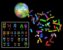
Spectral karyogram of a human female
 
Multicolor FISH and the older spectral karyotyping are molecular cytogenetic techniques used to simultaneously visualize all the pairs of chromosomes in an organism in different colors. Fluorescently labeled probes for each chromosome are made by labeling chromosome-specific DNA with different fluorophores. Because there are a limited number of spectrally distinct fluorophores, a combinatorial labeling method is used to generate many different colors. Fluorophore combinations are captured and analyzed by a fluorescence microscope using up to 7 narrow-banded fluorescence filters or, in the case of spectral karyotyping, by using an interferometer attached to a fluorescence microscope. In the case of an mFISH image, every combination of fluorochromes from the resulting original images is replaced by a pseudo color in a dedicated image analysis software. Thus, chromosomes or chromosome sections can be visualized and identified, allowing for the analysis of chromosomal rearrangements. In the case of spectral karyotyping, image processing software assigns a pseudo color to each spectrally different combination, allowing the visualization of the individually colored chromosomes.

Spectral human karyotype

Multicolor FISH is used to identify structural chromosome aberrations in cancer cells and other disease conditions when Giemsa banding or other techniques are not accurate enough.

Digital karyotyping

Digital karyotyping is a technique used to quantify the DNA copy number on a genomic scale. Short sequences of DNA from specific loci all over the genome are isolated and enumerated. This method is also known as virtual karyotyping.

Chromosome abnormalities

Chromosome abnormalities can be numerical, as in the presence of extra or missing chromosomes, or structural, as in derivative chromosome, translocations, inversions, large-scale deletions or duplications. Numerical abnormalities, also known as aneuploidy, often occur as a result of nondisjunction during meiosis in the formation of a gamete; trisomies, in which three copies of a chromosome are present instead of the usual two, are common numerical abnormalities. Structural abnormalities often arise from errors in homologous recombination. Both types of abnormalities can occur in gametes and therefore will be present in all cells of an affected person's body, or they can occur during mitosis and give rise to a genetic mosaic individual who has some normal and some abnormal cells. 

Chromosomal abnormalities that lead to disease in humans include
  • Turner syndrome results from a single X chromosome (45,X or 45,X0).
  • Klinefelter syndrome, the most common male chromosomal disease, otherwise known as 47,XXY, is caused by an extra X chromosome.
  • Edwards syndrome is caused by trisomy (three copies) of chromosome 18.
  • Down syndrome, a common chromosomal disease, is caused by trisomy of chromosome 21.
  • Patau syndrome is caused by trisomy of chromosome 13.
  • Trisomy 9, believed to be the 4th most common trisomy, has many long lived affected individuals but only in a form other than a full trisomy, such as trisomy 9p syndrome or mosaic trisomy 9. They often function quite well, but tend to have trouble with speech.
  • Also documented are trisomy 8 and trisomy 16, although they generally do not survive to birth.
Some disorders arise from loss of just a piece of one chromosome, including
  • Cri du chat (cry of the cat), from a truncated short arm on chromosome 5. The name comes from the babies' distinctive cry, caused by abnormal formation of the larynx.
  • 1p36 Deletion syndrome, from the loss of part of the short arm of chromosome 1.
  • Angelman syndrome – 50% of cases have a segment of the long arm of chromosome 15 missing; a deletion of the maternal genes, example of imprinting disorder.
  • Prader-Willi syndrome – 50% of cases have a segment of the long arm of chromosome 15 missing; a deletion of the paternal genes, example of imprinting disorder.

Task allocation and partitioning of social insects

 
Task allocation and partitioning is the way that tasks are chosen, assigned, subdivided, and coordinated (here, within a single colony of social insects). Closely associated are issues of communication that enable these actions to occur. This entry focuses exclusively on social insects. For information on human task allocation and partitioning, see division of labour, task analysis, and workflow.

Definitions

  • Task allocation "... is the process that results in specific workers being engaged in specific tasks, in numbers appropriate to the current situation. [It] operates without any central or hierarchical control..." The concept of task allocation is individual-centric. It focuses on decisions by individuals about what task to perform. However, different biomathematical models give different weights to inter-individual interactions vs. environmental stimuli.
  • Task partitioning is the division of one task into sequential actions done by more than one individual. The focus here is on the task, and its division, rather than on the individuals performing it. For example, "hygienic behavior" is a task in which worker bees uncap and remove diseased brood cells that may be affected by American foulbrood (Paenibacillus larvae) or the parasitic mite Varroa destructor. In this case, individual bees often focus on either uncapping or removing diseased brood. Therefore, the task is partitioned, and performed by multiple individuals.

Introduction

Social living provides a multitude of advantages to its practitioners, including predation risk reduction, environmental buffering, food procurement, and possible mating advantages. The most advanced form of sociality is eusociality, characterized by overlapping generations, cooperative care of the young, and reproductive division of labor, which includes sterility or near-sterility of the overwhelming majority of colony members. With few exceptions, all the practitioners of eusociality are insects of the orders Hymenoptera (ants, bees, and wasps), Isoptera (termites), Thysanoptera (thrips), and Hemiptera (aphids). Social insects have been extraordinarily successful ecologically and evolutionarily. This success has at its most pronounced produced colonies 1) having a persistence many times the lifespan of most individuals of the colony, and 2) numbering thousands or even millions of individuals. Social insects can exhibit division of labor with respect to non-reproductive tasks, in addition to the aforementioned reproductive one. In some cases this takes the form of markedly different, alternative morphological development (polymorphism), as in the case of soldier castes in ants, termites, thrips, and aphids, while in other cases it is age-based (temporal polyethism), as with honey bee foragers, who are the oldest members of the colony (with the exception of the queen). Evolutionary biologists are still debating the fitness-advantage gained by social insects due to their advanced division of labor and task allocation, but hypotheses include: increased resilience against a fluctuating environment, reduced energy costs of continuously switching tasks, increased longevity of the colony as a whole, or reduced rate of pathogen transmission. Division of labor, large colony sizes, temporally-changing colony needs, and the value of adaptability and efficiency under Darwinian competition, all form a theoretical basis favoring the existence of evolved communication in social insects. Beyond the rationale, there is well-documented empirical evidence of communication related to tasks; examples include the waggle dance of honey bee foragers, trail marking by ant foragers such as the red harvester ants, and the propagation via pheromones of an alarm state in Africanized honey bees.

Worker Polymorphism

One of the most well known mechanisms of task allocation is worker polymorphism, where workers within a colony have morphological differences. This difference in size is determined by the amount of food workers are fed as larvae, and is set once workers emerge from their pupae. Workers may vary just in size (monomorphism) or size and bodily proportions (allometry). An excellent example of the monomorphism is in bumblebees (Bombus spp.). Bumblebee workers display a large amount of body size variation which is normally distributed. The largest workers may be ten times the mass of the smallest workers. Worker size is correlated with several tasks: larger workers tend to forage, while smaller workers tend to perform brood care and nest thermoregulation. Size also affects task efficiency. Larger workers are better at learning, have better vision, carry more weight, and fly at a greater range of temperatures. However, smaller workers are more resistant to starvation. In other eusocial insects as well, worker size can determine what polymorphic role they become. For instance, larger workers in Myrmecocystus mexicanus (a North America species of honeypot ant) tend to become repletes, or workers so engorged with food that they become immobile and act a living food storage for the rest of the colonies.

In many ants and termites, on the other hand, workers vary in both size and bodily proportions, which have a bimodal distribution. This is present in approximately one in six ant genera. In most of these there are two developmentally distinct pathways, or castes, into which workers can develop. Typically members of the smaller caste are called minors and members of the larger caste are called majors or soldiers. There is often variation in size within each caste. The term soldiers may be apt, as in Cephalotes, but in many species members of the larger caste act primarily as foragers or food processors. In a few ant species, such as certain Pheidole species, there is a third caste, called supersoldiers.

Temporal polyethism

Temporal polyethism is a mechanism of task allocation, and is ubiquitous among eusocial insect colonies. Tasks in a colony are allocated among workers based on their age. Newly emerged workers perform tasks within the nest, such as brood care and nest maintenance, and progress to tasks outside the nest, such as foraging, nest defense, and corpse removal as they age. In honeybees, the youngest workers exclusively clean cells, which is then followed by tasks related to brood care and nest maintenance from about 2–11 days of age. From 11– 20 days, they transition to receiving and storing food from foragers, and at about 20 days workers begin to forage. Similar temporal polyethism patterns can be seen in primitive species of wasps, such as Ropalidia marginata as well as the eusocial wasp Vespula germanica. Young workers feed larvae, and then transition to nest building tasks, followed by foraging. Many species of ants also display this pattern. This pattern is not rigid, though. Workers of certain ages have strong tendencies to perform certain tasks, but may perform other tasks if there is enough need. For instance, removing young workers from the nest will cause foragers, especially younger foragers, to revert to tasks such as caring for brood. These changes in task preference are caused by epigenetic changes over the life of the individual. Honeybee workers of different ages show substantial differences in DNA methylation, which causes differences in gene expression. Reverting foragers to nurses by removing younger workers causes changes in DNA methylation similar to younger workers. Temporal polyethism is not adaptive because of maximized efficiency; indeed older workers are actually more efficient at brood care than younger workers in some ant species. Rather it allows workers with the lowest remaining life expectancy to perform the most dangerous tasks. Older workers tend to perform riskier tasks, such as foraging, which has high risks of predation and parasitism, while younger workers perform less dangerous tasks, such as brood care. If workers experience injuries, which shortens their life expectancies, they will start foraging sooner than healthy workers of the same age.

Response-Threshold Model

A dominant theory of explaining the self-organized division of labor in social insect societies such as honey bee colonies is the Response-Threshold Model. It predicts that individual worker bees have inherent thresholds to stimuli associated with different tasks. Individuals with the lowest thresholds will preferentially perform that task. Stimuli could include the “search time” that elapses while a foraging bee waits to unload her nectar and pollen to a receiver bee at the hive, the smell of diseased brood cells, or any other combination of environmental inputs that an individual worker bee encounters. The Response-Threshold Model only provides for effective task allocation in the honey bee colony if thresholds are varied among individual workers. This variation originates from the considerable genetic diversity among worker daughters of a colony due to the queen’s multiple matings.

Network representation of information flow and task allocation

To explain how colony-level complexity arises from the interactions of several autonomous individuals, a network-based approach has emerged as a promising area of social insect research. Social insect colonies can be viewed as a self-organized network, in which interacting elements (i.e. nodes) communicate with each other. As decentralized networks, colonies are capable of distributing information rapidly which facilitates robust responsiveness to their dynamic environments. The efficiency of information flow is critical for colony-level flexibility because worker behavior is not controlled by a centralized leader but rather is based on local information. 

Social insect networks are often non-randomly distributed, wherein a few individuals act as ‘hubs,’ having disproportionately more connections to other nestmates than other workers in the colony. In harvester ants, the total interactions per ant during recruitment for outside work is right-skewed, meaning that some ants are more highly connected than others. Computer simulations of this particular interaction network demonstrated that inter-individual variation in connectivity patterns expedites information flow among nestmates. 

Task allocation within a social insect colony can be modeled using a network-based approach, in which workers are represented by nodes, which are connected by edges that signify inter-node interactions. Workers performing a common task form highly connected clusters, with weaker links across tasks. These weaker, cross-task connections are important for allowing task-switching to occur between clusters. This approach is potentially problematic because connections between workers are not permanent, and some information is broadcast globally, e.g. through pheromones, and therefore does not rely on interaction networks. One alternative approach to avoid this pitfall is to treat tasks as nodes and workers as fluid connections. 

To demonstrate how time and space constraints of individual-level interactions affect colony function, social insect network approaches can also incorporate spatiotemporal dynamics. These effects can impose upper bounds to information flow rate in the network. For example, the rate of information flow through Temnothorax rugatulus ant colonies is slower than would be predicted if time spent traveling and location within the nest were not considered. In Formica fusca L. ant colonies, a network analysis of spatial effects on feeding and the regulation of food storage revealed that food is distributed heterogeneously within colony, wherein heavily loaded workers are located centrally within the nest and those storing less food were located at the periphery.

Studies of inter-nest pheromone trail networks maintained by super-colonies of Argentine ants (Linepithema humile) have shown that different colonies establish networks with very similar topologies. Insights from these analyses revealed that these networks – which are used to guide workers transporting brood, workers and food between nests – are formed through a pruning process, in which individual ants initially create a complex network of trails, which are then refined to eliminate extraneous edges, resulting in a shorter, more efficient inter-nest network. 

Long-term stability of interaction networks has been demonstrated in Odontomachus hastatus ants, in which initially highly connected ants remain highly connected over an extended time period. Conversely, Temnothorax rugatulus ant workers are not persistent in their interactive role, which might suggest that social organization is regulated differently among different eusocial species.

A network is pictorially represented as a graph, but can equivalently be represented as an adjacency list or adjacency matrix. Traditionally, workers are the nodes of the graph, but Fewell prefers to make the tasks the nodes, with workers as the links. O'Donnell has coined the term "worker connectivity" to stand for "communicative interactions that link a colony's workers in a social network and affect task performance". He has pointed out that connectivity provides three adaptive advantages compared to individual direct perception of needs:
  1. It increases both the physical and temporal reach of information. With connectivity, information can travel farther and faster, and additionally can persist longer, including both direct persistence (i.e. through pheromones), memory effects, and by initiating a sequence of events.
  2. It can help overcome task inertia and burnout, and push workers into performing hazardous tasks. For reasons of indirect fitness, this latter stimulus should not be necessary if all workers in the colony are highly related genetically, but that is not always the case.
  3. Key individuals may possess superior knowledge, or have catalytic roles. Examples, respectively, are a sentry who has detected an intruder, or the colony queen.
O'Donnell provides a comprehensive survey, with examples, of factors that have a large bearing on worker connectivity. They include:
  • graph degree
  • size of the interacting group, especially if the network has a modular structure
  • sender distribution (i.e. a small number of controllers vs. numerous senders)
  • strength of the interaction effect, which includes strength of the signal sent, recipient sensitivity, and signal persistence (i.e. pheromone signal vs. sound waves)
  • recipient memory, and its decay function
  • socially-transmitted inhibitory signals, as not all interactions provide positive stimulus
  • specificity of both the signal and recipient response
  • signal and sensory modalities, and activity and interaction rates

Task taxonomy and complexity

Anderson, Franks, and McShea have broken down insect tasks (and subtasks) into a hierarchical taxonomy; their focus is on task partitioning and its complexity implications. They classify tasks as individual, group, team, or partitioned; classification of a task depends on whether there are multiple vs. individual workers, whether there is division of labor, and whether subtasks are done concurrently or sequentially. Note that in their classification, in order for an action to be considered a task, it must contribute positively to inclusive fitness; if it must be combined with other actions to achieve that goal, it is considered to be a subtask. In their simple model, they award 1, 2, or 3 points to the different tasks and subtasks, depending on its above classification. Summing all tasks and subtasks point values down through all levels of nesting allows any task to be given a score that roughly ranks relative complexity of actions.

Note: model-building

All models are simplified abstractions of the real-life situation. There exists a basic tradeoff between model precision and parameter precision. A fixed amount of information collected, will, if split amongst the many parameters of an overly precise model, result in at least some of the parameters being represented by inadequate sample sizes. Because of the often limited quantities and limited precision of data from which to calculate parameters values in non-human behavior studies, such models should generally be kept simple. Therefore, we generally should not expect models for social insect task allocation or task partitioning to be as elaborate as human workflow ones, for example.

Metrics for division of labor

With increased data, more elaborate metrics for division of labor within the colony become possible. Gorelick and Bertram survey the applicability of metrics taken from a wide range of other fields. They argue that a single output statistic is desirable, to permit comparisons across different population sizes and different numbers of tasks. But they also argue that the input to the function should be a matrix representation (of time spent by each individual on each task), in order to provide the function with better data. They conclude that "... normalized matrix-input generalizations of Shannon's and Simpson's index ... should be the indices of choice when one wants to simultaneously examine division of labor amongst all individuals in a population". Note that these indexes, used as metrics of biodiversity, now find a place measuring division of labor.

Genetics of aggression

From Wikipedia, the free encyclopedia
 
The field of psychology has been greatly influenced by the study of genetics. Decades of research have demonstrated that both genetic and environmental factors play a role in a variety of behaviors in humans and animals (e.g. Grigorenko & Sternberg, 2003). The genetic basis of aggression, however, remains poorly understood. Aggression is a multi-dimensional concept, but it can be generally defined as behavior that inflicts pain or harm on another.

Genetic-developmental theory states that individual differences in a continuous phenotype result from the action of a large number of genes, each exerting an effect that works with environmental factors to produce the trait. This type of trait is influenced by multiple factors making it more complex and difficult to study than a simple Mendelian trait (one gene for one phenotype).

History

Past thought on genetic factors influencing aggression tended to seek answers from chromosomal abnormalities. Specifically, four decades ago, the XYY genotype was (erroneously) believed by many to be correlated with aggression. In 1965 and 1966, researchers at the MRC Clinical & Population Cytogenetics Research Unit led by Dr. Court Brown at Western General Hospital in Edinburgh reported finding a much higher than expected nine XYY men (2.9%) averaging almost 6 ft. tall in a survey of 314 patients at the State Hospital for Scotland; seven of the nine XYY patients were mentally retarded. In their initial reports published before examining the XYY patients, the researchers suggested they might have been hospitalized because of aggressive behavior. When the XYY patients were examined, the researchers found their assumptions of aggressive behavior were incorrect. Unfortunately, many science and medicine textbooks quickly and uncritically incorporated the initial, incorrect assumptions about XYY and aggression—including psychology textbooks on aggression.

The XYY genotype first gained wide notoriety in 1968 when it was raised as a part of a defense in two murder trials in Australia and France. In the United States, five attempts to use the XYY genotype as a defense were unsuccessful—in only one case in 1969 was it allowed to go to a jury—which rejected it.

Results from several decades of long-term follow-up of scores of unselected XYY males identified in eight international newborn chromosome screening studies in the 1960s and 1970s have replaced pioneering but biased studies from the 1960s (that used only institutionalized XYY men), as the basis for current understanding of the XYY genotype and established that XYY males are characterized by increased height but are not characterized by aggressive behavior. Though the link currently between genetics and aggression has turned to an aspect of genetics different from chromosomal abnormalities, it is important to understand where the research started and the direction it is moving towards today.

Heritability

Aggression, as well as other behavioral traits, is studied genetically based on its heritability through generations. Heritability models of aggression are mainly based on animals due to the ethical concern in using humans for genetic study. Animals are first selectively bred and then placed in a variety of environmental conditions, allowing researchers to examine the differences of selection in the aggression of animals.

Research methods

As with other topics in behavioral genetics, aggression is studied in three main experimental ways to help identify what role genetics plays in the behavior:
  • Heritability studies – studies focused to determine whether a trait, such as aggression, is heritable and how it is inherited from parent to offspring. These studies make use of genetic linkage maps to identify genes associated with certain behaviors such as aggression.
  • Mechanism experiments – studies to determine the biological mechanisms that lead certain genes to influence types of behavior like aggression.
  • Genetic behavior correlation studies – studies that use scientific data and attempt to correlate it with actual human behavior. Examples include twin studies and adoption studies.
These three main experimental types are used in animal studies, studies testing heritability and molecular genetics, and gene/environment interaction studies. Recently, important links between aggression and genetics have been studied and the results are allowing scientists to better understand the connections.

Selective breeding

The heritability of aggression has been observed in many animal strains after noting that some strains of birds, dogs, fish, and mice seem to be more aggressive than other strains. Selective breeding has demonstrated that it is possible to select for genes that lead to more aggressive behavior in animals. Selective breeding examples also allow researchers to understand the importance of developmental timing for genetic influences on aggressive behavior. A study done in 1983 (Cairns) produced both highly aggressive male and female strains of mice dependent on certain developmental periods to have this more aggressive behavior expressed. These mice were not observed to be more aggressive during the early and later stages of their lives, but during certain periods of time (in their middle-age period) were more violent and aggressive in their attacks on other mice. Selective breeding is a quick way to select for specific traits and see those selected traits within a few generations of breeding. These characteristics make selective breeding an important tool in the study of genetics and aggressive behavior.

Mouse studies

Mice are often used as a model for human genetic behavior since mice and humans have homologous genes coding for homologous proteins that are used for similar functions at some biological levels. Mice aggression studies have led to some interesting insight in human aggression. Using reverse genetics, the DNA of genes for the receptors of many neurotransmitters have been cloned and sequenced, and the role of neurotransmitters in rodent aggression has been investigated using pharmacological manipulations. Serotonin has been identified in the offensive attack by male mice against intruder male mice. Mutants were made by manipulating a receptor for serotonin by deleting a gene for the serotonin receptor. These mutant male mice with the knockout alleles exhibited normal behavior in everyday activities such as eating and exploration, but when prompted, attacked intruders with twice the intensity of normal male mice. In offense aggression in mice, males with the same or similar genotypes were more likely to fight than males that encountered males of other genotypes. Another interesting finding in mice dealt with mice reared alone. These mice showed a strong tendency to attack other male mice upon their first exposure to the other animals. The mice reared alone were not taught to be more aggressive; they simply exhibited the behavior. This implicates the natural tendency related to biological aggression in mice since the mice reared alone lacked a parent to model aggressive behavior.

Oxidative stress arises as a result of excess production of reactive oxygen species in relation to defense mechanisms, including the action of antioxidants such as superoxide dismutase 1 (SOD1). Knockout of the Sod1 gene was experimentally introduced in male mice leading to impaired antioxidant defense. These mice were designated (Sod1-/-). The Sod1-/- male mice proved to be more aggressive than both heterozygous knockout males (Sod1+/-) that were 50% deficient in SOD1, and wild-type males (Sod1+/+). The basis for the association of oxidative stress with increased aggression has not yet been determined.

Biological mechanisms

Experiments designed to study biological mechanisms are utilized when exploring how aggression is influenced by genetics. Molecular genetics studies allow many different types of behavioral traits to be examined by manipulating genes and studying the effect(s) of the manipulation.

Molecular genetics

A number of molecular genetics studies have focused on manipulating candidate aggression genes in mice and other animals to induce effects that can be possibly applied to humans. Most studies have focused on polymorphisms of serotonin receptors, dopamine receptors, and neurotransmitter metabolizing enzymes. Results of these studies have led to linkage analysis to map the serotonin-related genes and impulsive aggression, as well as dopamin-related genes and proactive aggression. In particular, the serotonin 5-HT seems to be an influence in inter-male aggression either directly or through other molecules that use the 5-HT pathway. 5-HT normally dampens aggression in animals and humans. Mice missing specific genes for 5-HT were observed to be more aggressive than normal mice and were more rapid and violent in their attacks. Other studies have been focused on neurotransmitters. Studies of a mutation in the neurotransmitter metabolizing enzyme monoamine oxidase A (MAO-A) have been shown to cause a syndrome that includes violence and impulsivity in humans. Studies of the molecular genetics pathways are leading to the production of pharmaceuticals to fix the pathway problems and hopefully show an observed change in aggressive behavior.

Human behavior genetics

In determining if a trait is related to genetic factors or environmental factors, twin studies and adoption studies are used. These studies examine correlations based on similarity of a trait and a person's genetic or environmental factors that could influence the trait. Aggression has been examined via both twin studies and adoption studies.

Twin studies

Twin studies manipulate the environmental factors of behavior by examining if identical twins raised apart are different from twins raised together. Before the advancement of molecular genetics, twin studies were almost the only mode of investigation of genetic influences on personality. Heritability was estimated as twice the difference between the correlation for identical, or monozygotic, twins and that for fraternal, or dizygotic, twins. Early studies indicated that personality was fifty percent genetic. Current thinking holds that each individual picks and chooses from a range of stimuli and events largely on the basis of his genotype creating a unique set of experiences; basically meaning that people create their own environments.

Operator (computer programming)

From Wikipedia, the free encyclopedia https://en.wikipedia.org/wiki/Operator_(computer_programmin...