Search This Blog

Friday, February 21, 2020

Psychrophile

From Wikipedia, the free encyclopedia
https://en.wikipedia.org/wiki/Psychrophile 

Psychrophiles or cryophiles (adj. psychrophilic or cryophilic) are extremophilic organisms that are capable of growth and reproduction in low temperatures, ranging from −20 °C to +10 °C. They are found in places that are permanently cold, such as the polar regions and the deep sea. They can be contrasted with thermophiles, which are organisms that thrive at unusually high temperatures. Psychrophile is Greek for 'cold-loving'.

Many such organisms are bacteria or archaea, but some eukaryotes such as lichens, snow algae, fungi, and wingless midges, are also classified as psychrophiles.

Biology

The lichen Xanthoria elegans can continue to photosynthesize at −24 °C.

Habitat

The cold environments that psychrophiles inhabit are ubiquitous on Earth, as a large fraction of our planetary surface experiences temperatures lower than 15 °C. They are present in permafrost, polar ice, glaciers, snowfields and deep ocean waters. These organisms can also be found in pockets of sea ice with high salinity content. Microbial activity has been measured in soils frozen below −39 °C. In addition to their temperature limit, psychrophiles must also adapt to other extreme environmental constraints that may arise as a result of their habitat. These constraints include high pressure in the deep sea, and high salt concentration on some sea ice.

Adaptations

Psychrophiles are protected from freezing and the expansion of ice by ice-induced desiccation and vitrification (glass transition), as long as they cool slowly. Free living cells desiccate and vitrify between −10 °C and −26 °C. Cells of multicellular organisms may vitrify at temperatures below −50 °C. The cells may continue to have some metabolic activity in the extracellular fluid down to these temperatures, and they remain viable once restored to normal temperatures.

They must also overcome the stiffening of their lipid cell membrane, as this is important for the survival and functionality of these organisms. To accomplish this, psychrophiles adapt lipid membrane structures that have a high content of short, unsaturated fatty acids. Compared to longer saturated fatty acids, incorporating this type of fatty acid allows for the lipid cell membrane to have a lower melting point, which increases the fluidity of the membranes. In addition, carotenoids are present in the membrane, which help modulate the fluidity of it.

Antifreeze proteins are also synthesized to keep psychrophiles' internal space liquid, and to protect their DNA when temperatures drop below water's freezing point. By doing so, the protein prevents any ice formation or recrystallization process from occurring.

The enzymes of these organisms have been hypothesized to engage in an activity-stability-flexibility relationship as a method for adapting to the cold; the flexibility of their enzyme structure will increase as a way to compensate for the freezing effect of their environment.

Certain cryophiles, such as Gram-negative bacteria Vibrio and Aeromonas spp., can transition into a viable but nonculturable (VBNC) state. During VBNC, a micro-organism can respirate and use substrates for metabolism – however, it cannot replicate. An advantage of this state is that it is highly reversible. It has been debated whether VBNC is an active survival strategy or if eventually the organism's cells will no longer be able to be revived. There is proof however it may be very effective – Gram positive bacteria Actinobacteria have been shown to have lived about 500,000 years in the permafrost conditions of Antarctica, Canada, and Siberia.

Taxonomic range

The wingless midge (Chironomidae) Belgica antarctica.

Psychrophiles include bacteria, lichens, fungi, and insects. 

Among the bacteria that can tolerate extreme cold are Arthrobacter sp., Psychrobacter sp. and members of the genera Halomonas, Pseudomonas, Hyphomonas, and Sphingomonas. Another example is Chryseobacterium greenlandensis, a psychrophile that was found in 120,000-year-old ice.
Umbilicaria antarctica and Xanthoria elegans are lichens that have been recorded photosynthesizing at temperatures ranging down to −24 °C, and they can grow down to around −10 °C. Some multicellular eukaryotes can also be metabolically active at sub-zero temperatures, such as some conifers; those in the Chironomidae family are still active at −16 °C.

Penicillium is a genus of fungi found in a wide range of environments including extreme cold.

Among the psychrophile insects, the Grylloblattidae or icebugs, found on mountaintops, have optimal temperatures between 1-4 °C. The wingless midge (Chironomidae) Belgica antarctica can tolerate salt, being frozen and strong ultraviolet, and has the smallest known genome of any insect. The small genome, of 99 million base pairs, is thought to be adaptive to extreme environments.

Psychrotrophic bacteria

Psychrotrophic microbes are able to grow at temperatures below 7 °C (44.6 °F), but have better growth rates at higher temperatures. Psychrotrophic bacteria and fungi are able to grow at refrigeration temperatures, and can be responsible for food spoilage. They provide an estimation of the product's shelf life, but also they can be found in soils, in surface and deep sea waters, in Antarctic ecosystems, and in foods.

Psychrotrophic bacteria are of particular concern to the dairy industry. Most are killed by pasteurization; however, they can be present in milk as post-pasteurization contaminants due to less than adequate sanitation practices. According to the Food Science Department at Cornell University, psychrotrophs are bacteria capable of growth at temperatures at or less than 7 °C (44.6 °F). At freezing temperatures, growth of psychrotrophic bacteria becomes negligible or virtually stops.

All three subunits of the RecBCD enzyme are essential for physiological activities of the enzyme in the Antarctic Pseudomonas syringae, namely, repairing of DNA damage and supporting the growth at low temperature. The RecBCD enzymes are exchangeable between the psychrophilic P. syringae and the mesophilic E. coli when provided with the entire protein complex from same species. However, the RecBC proteins (RecBCPs and RecBCEc) of the two bacteria are not equivalent; the RecBCEc is proficient in DNA recombination and repair, and supports the growth of P. syringae at low temperature, while RecBCPs is insufficient for these functions. Finally, both helicase and nuclease activity of the RecBCDPs are although important for DNA repair and growth of P. syringae at low temperature, the RecB-nuclease activity is not essential in vivo.

Versus psychrotroph

In 1940, ZoBell and Conn stated that they had never encountered "true psychrophiles" or organisms that grow best at relatively low temperatures. In 1958, J. L. Ingraham supported this by concluding that there are very few or possibly no bacteria that fit the textbook definitions of psychrophiles. Richard Y. Morita emphasizes this by using the term psychrotroph to describe organisms that do not meet the definition of psychrophiles. The confusion between the terms psychrotrophs and psychrophiles was started because investigators were unaware of the thermolability of psychrophilic organisms at the laboratory temperatures. Due to this, early investigators did not determine the cardinal temperatures for their isolates.

The similarity between these two is that they are both capable of growing at zero, but optimum and upper temperature limits for the growth are lower for psychrophiles compared to psychrotrophs. Psychrophiles are also more often isolated from permanently cold habitats compared to psychrotrophs. Although psychrophilic enzymes remain under-used because the cost of production and processing at low temperatures is higher than for the commercial enzymes that are presently in use, the attention and resurgence of research interest in psychrophiles and psychrotrophs will be a contributor to the betterment of the environment and the desire to conserve energy.

Medical microbiology

From Wikipedia, the free encyclopedia
 
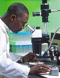
A microbiologist examining cultures under a dissecting microscope.

Medical microbiology, the large subset of microbiology that is applied to medicine, is a branch of medical science concerned with the prevention, diagnosis and treatment of infectious diseases. In addition, this field of science studies various clinical applications of microbes for the improvement of health. There are four kinds of microorganisms that cause infectious disease: bacteria, fungi, parasites and viruses, and one type of infectious protein called prion.

A medical microbiologist studies the characteristics of pathogens, their modes of transmission, mechanisms of infection and growth. Using this information, a treatment can be devised. Medical microbiologists often serve as consultants for physicians, providing identification of pathogens and suggesting treatment options. Other tasks may include the identification of potential health risks to the community or monitoring the evolution of potentially virulent or resistant strains of microbes, educating the community and assisting in the design of health practices. They may also assist in preventing or controlling epidemics and outbreaks of disease. Not all medical microbiologists study microbial pathology; some study common, non-pathogenic species to determine whether their properties can be used to develop antibiotics or other treatment methods. 

Epidemiology, the study of the patterns, causes, and effects of health and disease conditions in populations, is an important part of medical microbiology, although the clinical aspect of the field primarily focuses on the presence and growth of microbial infections in individuals, their effects on the human body, and the methods of treating those infections. In this respect the entire field, as an applied science, can be conceptually subdivided into academic and clinical sub-specialties, although in reality there is a fluid continuum between public health microbiology and clinical microbiology, just as the state of the art in clinical laboratories depends on continual improvements in academic medicine and research laboratories.

History

Anton van Leeuwenhoek was the first to observe microorganisms using a microscope.

In 1676, Anton van Leeuwenhoek observed bacteria and other microorganisms, using a single-lens microscope of his own design.

In 1796, Edward Jenner developed a method using cowpox to successfully immunize a child against smallpox. The same principles are used for developing vaccines today.

Following on from this, in 1857 Louis Pasteur also designed vaccines against several diseases such as anthrax, fowl cholera and rabies as well as pasteurization for food preservation.

In 1867 Joseph Lister is considered to be the father of antiseptic surgery. By sterilizing the instruments with diluted carbolic acid and using it to clean wounds, post-operative infections were reduced, making surgery safer for patients.

In the years between 1876 and 1884 Robert Koch provided much insight into infectious diseases. He was one of the first scientists to focus on the isolation of bacteria in pure culture. This gave rise to the germ theory, a certain microorganism being responsible for a certain disease. He developed a series of criteria around this that have become known as the Koch's postulates.

A major milestone in medical microbiology is the Gram stain. In 1884 Hans Christian Gram developed the method of staining bacteria to make them more visible and differentiated under a microscope. This technique is widely used today.

In 1910 Paul Ehrlich tested multiple combinations of arsenic based chemicals on infected rabbits with syphilis. Ehrlich then found that arsphenamine was found effective against syphilis spirochetes. The arsphenamines was then made available in 1910, known as Salvarsan.

In 1929 Alexander Fleming developed the most commonly used antibiotic substance both at the time and now: penicillin

In 1939 Gerhard Domagk found Prontosil red protected mice from pathogenic streptococci and staphylococci without toxicity. Domagk received the Nobel Prize in physiology, or medicine, for the discovery of the sulfa drug

DNA sequencing, a method developed by Walter Gilbert and Frederick Sanger in 1977, caused a rapid change the development of vaccines, medical treatments and diagnostic methods. Some of these include synthetic insulin which was produced in 1979 using recombinant DNA and the first genetically engineered vaccine was created in 1986 for hepatitis B.

In 1995 a team at The Institute for Genomic Research sequenced the first bacterial genome; Haemophilus influenzae. A few months later, the first eukaryotic genome was completed. This would prove invaluable for diagnostic techniques.

Commonly treated infectious diseases

Bacterial

Viral

Parasitic

Fungal

Causes and transmission of infectious diseases

Infections may be caused by bacteria, viruses, fungi, and parasites. The pathogen that causes the disease may be exogenous (acquired from an external source; environmental, animal or other people, e.g. Influenza) or endogenous (from normal flora e.g. Candidiasis).

The site at which a microbe enters the body is referred to as the portal of entry. These include the respiratory tract, gastrointestinal tract, genitourinary tract, skin, and mucous membranes. The portal of entry for a specific microbe is normally dependent on how it travels from its natural habitat to the host.

There are various ways in which disease can be transmitted between individuals. These include:
  • Direct contact - Touching an infected host, including sexual contact
  • Indirect contact - Touching a contaminated surface
  • Droplet contact - Coughing or sneezing
  • Fecal–oral route - Ingesting contaminated food or water sources
  • Airborne transmission - Pathogen carrying spores
  • Vector transmission - An organism that does not cause disease itself but transmits infection by conveying pathogens from one host to another
  • Fomite transmission - An inanimate object or substance capable of carrying infectious germs or parasites
  • Environmental - Hospital-acquired infection (Nosocomial infections)
Like other pathogens, viruses use these methods of transmission to enter the body, but viruses differ in that they must also enter into the host's actual cells. Once the virus has gained access to the host's cells, the virus' genetic material (RNA or DNA) must be introduced to the cell. Replication between viruses is greatly varied and depends on the type of genes involved in them. Most DNA viruses assemble in the nucleus while most RNA viruses develop solely in cytoplasm.

The mechanisms for infection, proliferation, and persistence of a virus in cells of the host are crucial for its survival. For example, some diseases such as measles employ a strategy whereby it must spread to a series of hosts. In these forms of viral infection, the illness is often treated by the body's own immune response, and therefore the virus is required to disperse to new hosts before it is destroyed by immunological resistance or host death. In contrast, some infectious agents such as the Feline leukemia virus, are able to withstand immune responses and are capable of achieving long-term residence within an individual host, whilst also retaining the ability to spread into successive hosts.

Diagnostic tests

Identification of an infectious agent for a minor illness can be as simple as clinical presentation; such as gastrointestinal disease and skin infections. In order to make an educated estimate as to which microbe could be causing the disease, epidemiological factors need to be considered; such as the patient's likelihood of exposure to the suspected organism and the presence and prevalence of a microbial strain in a community.

Diagnosis of infectious disease is nearly always initiated by consulting the patient's medical history and conducting a physical examination. More detailed identification techniques involve microbial culture, microscopy, biochemical tests and genotyping. Other less common techniques (such as X-rays, CAT scans, PET scans or NMR) are used to produce images of internal abnormalities resulting from the growth of an infectious agent.

Microbial culture

Four nutrient agar plates growing colonies of common Gram negative bacteria.

Microbiological culture is the primary method used for isolating infectious disease for study in the laboratory. Tissue or fluid samples are tested for the presence of a specific pathogen, which is determined by growth in a selective or differential medium.

The 3 main types of media used for testing are:
  • Solid culture: A solid surface is created using a mixture of nutrients, salts and agar. A single microbe on an agar plate can then grow into colonies (clones where cells are identical to each other) containing thousands of cells. These are primarily used to culture bacteria and fungi.
  • Liquid culture: Cells are grown inside a liquid media. Microbial growth is determined by the time taken for the liquid to form a colloidal suspension. This technique is used for diagnosing parasites and detecting mycobacteria.
  • Cell culture: Human or animal cell cultures are infected with the microbe of interest. These cultures are then observed to determine the effect the microbe has on the cells. This technique is used for identifying viruses.

Microscopy

Culture techniques will often use a microscopic examination to help in the identification of the microbe. Instruments such as compound light microscopes can be used to assess critical aspects of the organism. This can be performed immediately after the sample is taken from the patient and is used in conjunction with biochemical staining techniques, allowing for resolution of cellular features. Electron microscopes and fluorescence microscopes are also used for observing microbes in greater detail for research.

Biochemical tests

Fast and relatively simple biochemical tests can be used to identify infectious agents. For bacterial identification, the use of metabolic or enzymatic characteristics are common due to their ability to ferment carbohydrates in patterns characteristic of their genus and species. Acids, alcohols and gases are usually detected in these tests when bacteria are grown in selective liquid or solid media, as mentioned above. In order to perform these tests en masse, automated machines are used. These machines perform multiple biochemical tests simultaneously, using cards with several wells containing different dehydrated chemicals. The microbe of interest will react with each chemical in a specific way, aiding in its identification.

Serological methods are highly sensitive, specific and often extremely rapid laboratory tests used to identify different types of microorganisms. The tests are based upon the ability of an antibody to bind specifically to an antigen. The antigen (usually a protein or carbohydrate made by an infectious agent) is bound by the antibody, allowing this type of test to be used for organisms other than bacteria. This binding then sets off a chain of events that can be easily and definitively observed, depending on the test. More complex serological techniques are known as immunoassays. Using a similar basis as described above, immunoassays can detect or measure antigens from either infectious agents or the proteins generated by an infected host in response to the infection.

Polymerase chain reaction

Polymerase chain reaction (PCR) assays are the most commonly used molecular technique to detect and study microbes. As compared to other methods, sequencing and analysis is definitive, reliable, accurate, and fast. Today, quantitative PCR is the primary technique used, as this method provides faster data compared to a standard PCR assay. For instance, traditional PCR techniques require the use of gel electrophoresis to visualize amplified DNA molecules after the reaction has finished. quantitative PCR does not require this, as the detection system uses fluorescence and probes to detect the DNA molecules as they are being amplified. In addition to this, quantitative PCR also removes the risk of contamination that can occur during standard PCR procedures (carrying over PCR product into subsequent PCRs). Another advantage of using PCR to detect and study microbes is that the DNA sequences of newly discovered infectious microbes or strains can be compared to those already listed in databases, which in turn helps to increase understanding of which organism is causing the infectious disease and thus what possible methods of treatment could be used. This technique is the current standard for detecting viral infections such as AIDS and hepatitis.

Treatments

Once an infection has been diagnosed and identified, suitable treatment options must be assessed by the physician and consulting medical microbiologists. Some infections can be dealt with by the body's own immune system, but more serious infections are treated with antimicrobial drugs. Bacterial infections are treated with antibacterials (often called antibiotics) whereas fungal and viral infections are treated with antifungals and antivirals respectively. A broad class of drugs known as antiparasitics are used to treat parasitic diseases

Medical microbiologists often make treatment recommendations to the patient's physician based on the strain of microbe and its antibiotic resistances, the site of infection, the potential toxicity of antimicrobial drugs and any drug allergies the patient has.

Antibiotic resistance tests: bacteria in the culture on the left are sensitive to the antibiotics contained in the white, paper discs. Bacteria in the culture on the right are resistant to most of the antibiotics.

In addition to drugs being specific to a certain kind of organism (bacteria, fungi, etc.), some drugs are specific to a certain genus or species of organism, and will not work on other organisms. Because of this specificity, medical microbiologists must consider the effectiveness of certain antimicrobial drugs when making recommendations. Additionally, strains of an organism may be resistant to a certain drug or class of drug, even when it is typically effective against the species. These strains, termed resistant strains, present a serious public health concern of growing importance to the medical industry as the spread of antibiotic resistance worsens. Antimicrobial resistance is an increasingly problematic issue that leads to millions of deaths every year.

Whilst drug resistance typically involves microbes chemically inactivating an antimicrobial drug or a cell mechanically stopping the uptake of a drug, another form of drug resistance can arise from the formation of biofilms. Some bacteria are able to form biofilms by adhering to surfaces on implanted devices such as catheters and prostheses and creating an extracellular matrix for other cells to adhere to. This provides them with a stable environment from which the bacteria can disperse and infect other parts of the host. Additionally, the extracellular matrix and dense outer layer of bacterial cells can protect the inner bacteria cells from antimicrobial drugs.

Medical microbiology is not only about diagnosing and treating disease, it also involves the study of beneficial microbes. Microbes have been shown to be helpful in combating infectious disease and promoting health. Treatments can be developed from microbes, as demonstrated by Alexander Fleming's discovery of penicillin as well as the development of new antibiotics from the bacterial genus Streptomyces among many others. Not only are microorganisms a source of antibiotics but some may also act as probiotics to provide health benefits to the host, such as providing better gastrointestinal health or inhibiting pathogens.

Virology

From Wikipedia, the free encyclopedia
Gamma phage, an example of a virus

Virology is the study of viral – submicroscopic, parasitic particles of genetic material contained in a protein coat – and virus-like agents. It focuses on the following aspects of viruses: their structure, classification and evolution, their ways to infect and exploit host cells for reproduction, their interaction with host organism physiology and immunity, the diseases they cause, the techniques to isolate and culture them, and their use in research and therapy. Virology is considered to be a subfield of microbiology or of medicine.

Virus structure and classification

A major branch of virology is virus classification. Viruses can be classified according to the host cell they infect: animal viruses, plant viruses, fungal viruses, and bacteriophages (viruses infecting bacterium, which include the most complex viruses). Another classification uses the geometrical shape of their capsid (often a helix or an icosahedron) or the virus's structure (e.g. presence or absence of a lipid envelope). Viruses range in size from about 30 nm to about 450 nm, which means that most of them cannot be seen with light microscopes. The shape and structure of viruses has been studied by electron microscopy, NMR spectroscopy, and X-ray crystallography

The most useful and most widely used classification system distinguishes viruses according to the type of nucleic acid they use as genetic material and the viral replication method they employ to coax host cells into producing more viruses:
The latest report by the International Committee on Taxonomy of Viruses (2005) lists 5450 viruses, organized in over 2,000 species, 287 genera, 73 families and 3 orders.

Virologists also study subviral particles, infectious entities notably smaller and simpler than viruses:
  • viroids (naked circular RNA molecules infecting plants),
  • satellites (nucleic acid molecules with or without a capsid that require a helper virus for infection and reproduction), and
  • prions (proteins that can exist in a pathological conformation that induces other prion molecules to assume that same conformation).
Taxa in virology are not necessarily monophyletic, as the evolutionary relationships of the various virus groups remain unclear. Three hypotheses regarding their origin exist:
  1. Viruses arose from non-living matter, separately from yet in parallel to cells, perhaps in the form of self-replicating RNA ribozymes similar to viroids.
  2. Viruses arose by genome reduction from earlier, more competent cellular life forms that became parasites to host cells and subsequently lost most of their functionality; examples of such tiny parasitic prokaryotes are Mycoplasma and Nanoarchaea.
  3. Viruses arose from mobile genetic elements of cells (such as transposons, retrotransposons or plasmids) that became encapsulated in protein capsids, acquired the ability to "break free" from the host cell and infect other cells.
Of particular interest here is mimivirus, a giant virus that infects amoebae and encodes much of the molecular machinery traditionally associated with bacteria. Two possibilities are that it is a simplified version of a parasitic prokaryote or it originated as a simpler virus that acquired genes from its host.
The evolution of viruses, which often occurs in concert with the evolution of their hosts, is studied in the field of viral evolution

While viruses reproduce and evolve, they do not engage in metabolism, do not move, and depend on a host cell for reproduction. The often-debated question of whether they are alive or not is a matter of definition that does not affect the biological reality of viruses.

Viral diseases and host defenses

One main motivation for the study of viruses is the fact that they cause many important infectious diseases, among them the common cold, influenza, rabies, measles, many forms of diarrhea, hepatitis, Dengue fever, yellow fever, polio, smallpox and AIDS. Herpes simplex causes cold sores and genital herpes and is under investigation as a possible factor in Alzheimer's.

Some viruses, known as oncoviruses, contribute to the development of certain forms of cancer. The best studied example is the association between Human papillomavirus and cervical cancer: almost all cases of cervical cancer are caused by certain strains of this sexually transmitted virus. Another example is the association of infection with hepatitis B and hepatitis C viruses and liver cancer.

Some subviral particles also cause disease: the transmissible spongiform encephalopathies, which include Kuru, Creutzfeldt–Jakob disease and bovine spongiform encephalopathy ("mad cow disease"), are caused by prions, hepatitis D is due to a satellite virus.

The study of the manner in which viruses cause disease is viral pathogenesis. The degree to which a virus causes disease is its virulence.

When the immune system of a vertebrate encounters a virus, it may produce specific antibodies which bind to the virus and neutralize its infectivity or mark it for destruction. Antibody presence in blood serum is often used to determine whether a person has been exposed to a given virus in the past, with tests such as ELISA. Vaccinations protect against viral diseases, in part, by eliciting the production of antibodies. Monoclonal antibodies, specific to the virus, are also used for detection, as in fluorescence microscopy.

A second defense of vertebrates against viruses, cell-mediated immunity, involves immune cells known as T cells: the body's cells constantly display short fragments of their proteins on the cell's surface, and if a T cell recognizes a suspicious viral fragment there, the host cell is destroyed and the virus-specific T-cells proliferate. This mechanism is jump-started by certain vaccinations. 

RNA interference, an important cellular mechanism found in plants, animals and many other eukaryotes, most likely evolved as a defense against viruses. An elaborate machinery of interacting enzymes detects double-stranded RNA molecules (which occur as part of the life cycle of many viruses) and then proceeds to destroy all single-stranded versions of those detected RNA molecules.

Every lethal viral disease presents a paradox: killing its host is obviously of no benefit to the virus, so how and why did it evolve to do so? Today it is believed that most viruses are relatively benign in their natural hosts; some viral infection might even be beneficial to the host. The lethal viral diseases are believed to have resulted from an "accidental" jump of the virus from a species in which it is benign to a new one that is not accustomed to it (see zoonosis). For example, viruses that cause serious influenza in humans probably have pigs or birds as their natural host, and HIV is thought to derive from the benign non-human primate virus SIV.

While it has been possible to prevent (certain) viral diseases by vaccination for a long time, the development of antiviral drugs to treat viral diseases is a comparatively recent development. The first such drug was interferon, a substance that is naturally produced when an infection is detected and stimulates other parts of the immune system.

Molecular biology research and viral therapy

Bacteriophages, the viruses which infect bacteria, can be relatively easily grown as viral plaques on bacterial cultures. Bacteriophages occasionally move genetic material from one bacterial cell to another in a process known as transduction, and this horizontal gene transfer is one reason why they served as a major research tool in the early development of molecular biology. The genetic code, the function of ribozymes, the first recombinant DNA and early genetic libraries were all arrived at using bacteriophages. Certain genetic elements derived from viruses, such as highly effective promoters, are commonly used in molecular biology research today. 

Growing animal viruses outside of the living host animal is more difficult. Classically, fertilized chicken eggs have often been used, but cell cultures are increasingly employed for this purpose today.
Since some viruses that infect eukaryotes need to transport their genetic material into the host cell's nucleus, they are attractive tools for introducing new genes into the host (known as transformation or transfection). Modified retroviruses are often used for this purpose, as they integrate their genes into the host's chromosomes.

This approach of using viruses as gene vectors is being pursued in the gene therapy of genetic diseases. An obvious problem to be overcome in viral gene therapy is the rejection of the transforming virus by the immune system.

Phage therapy, the use of bacteriophages to combat bacterial diseases, was a popular research topic before the advent of antibiotics and has recently seen renewed interest.

Oncolytic viruses are viruses that preferably infect cancer cells. While early efforts to employ these viruses in the therapy of cancer failed, there have been reports in 2005 and 2006 of encouraging preliminary results.

Sequencing of viruses

As most viruses are too small to be seen by a light microscope, sequencing is one of the main tools in virology to identify and study the virus. Traditional Sanger sequencing and next-generation sequencing (NGS) are used to sequence viruses in basic and clinical research, as wells as for the diagnosis of emerging viral infections, molecular epidemiology of viral pathogens, and drug-resistance testing. There are more than 2.3 million unique viral sequences in GenBank . Recently, NGS has surpassed traditional Sanger as the most popular approach for generating viral genomes .

Other uses of viruses

A new application of genetically engineered viruses in nanotechnology was recently described; see the uses of viruses in material science and nanotechnology. For a use in mapping neurons see the applications of pseudorabies in neuroscience.

History of virology

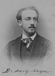
Adolf Mayer in 1875
 
Dmitri Ivanovsky, ca. 1915
 
An old, bespectacled man wearing a suit and sitting at a bench by a large window. The bench is covered with small bottles and test tubes. On the wall behind him is a large old-fashioned clock below frick u which are four small enclosed shelves on which sit many neatly labelled bottles.
Martinus Beijerinck in his laboratory in 1921.
 
The word virus appeared in 1599 and originally meant "venom".

A very early form of vaccination known as variolation was developed several thousand years ago in China. It involved the application of materials from smallpox sufferers in order to immunize others. In 1717 Lady Mary Wortley Montagu observed the practice in Istanbul and attempted to popularize it in Britain, but encountered considerable resistance. In 1796 Edward Jenner developed a much safer method, using cowpox to successfully immunize a young boy against smallpox, and this practice was widely adopted. Vaccinations against other viral diseases followed, including the successful rabies vaccination by Louis Pasteur in 1886. The nature of viruses however was not clear to these researchers.

In 1892, the Russian biologist Dmitry Ivanovsky used a Chamberland filter to try to isolate the bacteria that caused tobacco mosaic disease. His experiments showed that crushed leaf extracts from infected tobacco plants remained infectious after filtration. Ivanovsky reported a minuscule infectious agent or toxin, capable of passing the filter, may be being produced by a bacterium.

In 1898 Martinus Beijerinck repeated Ivanovski's work but went further and passed the "filterable agent" from plant to plant, found the action undiminished, and concluded it infectious—replicating in the host—and thus not a mere toxin. He called it contagium vivum fluidum. The question of whether the agent was a "living fluid" or a particle was however still open. 

In 1903 it was suggested for the first time that transduction by viruses might cause cancer. In 1908 Bang and Ellerman showed that a filterable virus could transmit chicken leukemia, data largely ignored till the 1930s when leukemia became regarded as cancerous. In 1911 Peyton Rous reported the transmission of chicken sarcoma, a solid tumor, with a virus, and thus Rous became "father of tumor virology". The virus was later called Rous sarcoma virus 1 and understood to be a retrovirus. Several other cancer-causing retroviruses have since been described. 

The existence of viruses that infect bacteria (bacteriophages) was first recognized by Frederick Twort in 1911, and, independently, by Félix d'Herelle in 1917. As bacteria could be grown easily in culture, this led to an explosion of virology research.

The cause of the devastating Spanish flu pandemic of 1918 was initially unclear. In late 1918, French scientists showed that a "filter-passing virus" could transmit the disease to people and animals, fulfilling Koch's postulates.

In 1926 it was shown that scarlet fever is caused by a bacterium that is infected by a certain bacteriophage.

While plant viruses and bacteriophages can be grown comparatively easily, animal viruses normally require a living host animal, which complicates their study immensely. In 1931 it was shown that influenza virus could be grown in fertilized chicken eggs, a method that is still used today to produce vaccines. In 1937, Max Theiler managed to grow the yellow fever virus in chicken eggs and produced a vaccine from an attenuated virus strain; this vaccine saved millions of lives and is still being used today.

Max Delbrück, an important investigator in the area of bacteriophages, described the basic "life cycle" of a virus in 1937: rather than "growing", a virus particle is assembled from its constituent pieces in one step; eventually it leaves the host cell to infect other cells. The Hershey–Chase experiment in 1952 showed that only DNA and not protein enters a bacterial cell upon infection with bacteriophage T2. Transduction of bacteria by bacteriophages was first described in the same year.

In 1949 John F. Enders, Thomas Weller and Frederick Robbins reported growth of poliovirus in cultured human embryonal cells, the first significant example of an animal virus grown outside of animals or chicken eggs. This work aided Jonas Salk in deriving a polio vaccine from deactivated polio viruses; this vaccine was shown to be effective in 1955.

The first virus that could be crystalized and whose structure could therefore be elucidated in detail was tobacco mosaic virus (TMV), the virus that had been studied earlier by Ivanovski and Beijerink. In 1935, Wendell Stanley achieved its crystallization for electron microscopy and showed that it remains active even after crystallization. Clear X-ray diffraction pictures of the crystallized virus were obtained by Bernal and Fankuchen in 1941. Based on such pictures, Rosalind Franklin proposed the full structure of the tobacco mosaic virus in 1955. Also in 1955, Heinz Fraenkel-Conrat and Robley Williams showed that purified TMV RNA and its capsid (coat) protein can self-assemble into functional virions, suggesting that this assembly mechanism is also used within the host cell, as Delbrück had proposed earlier.

In 1963, the Hepatitis B virus was discovered by Baruch Blumberg who went on to develop a hepatitis B vaccine. 

In 1965, Howard Temin described the first retrovirus: a virus whose RNA genome was reverse transcribed into complementary DNA (cDNA), then integrated into the host's genome and expressed from that template. The viral enzyme reverse transcriptase, which along with integrase is a distinguishing trait of retroviruses, was first described in 1970, independently by Howard Temin and David Baltimore. The first retrovirus infecting humans was identified by Robert Gallo in 1974. Later it was found that reverse transcriptase is not specific to retroviruses; retrotransposons which code for reverse transcriptase are abundant in the genomes of all eukaryotes. About 10-40% of the human genome derives from such retrotransposons.

In 1975 the functioning of oncoviruses was clarified considerably. Until that time, it was thought that these viruses carried certain genes called oncogenes which, when inserted into the host's genome, would cause cancer. Michael Bishop and Harold Varmus showed that the oncogene of Rous sarcoma virus is in fact not specific to the virus but is contained in the genome of healthy animals of many species. The oncovirus can switch this pre-existing benign proto-oncogene on, turning it into a true oncogene that causes cancer.

1976 saw the first recorded outbreak of Ebola virus disease, a highly lethal virally transmitted disease.

In 1977, Frederick Sanger achieved the first complete sequencing of the genome of any organism, the bacteriophage Phi X 174. In the same year, Richard Roberts and Phillip Sharp independently showed that the genes of adenovirus contain introns and therefore require gene splicing. It was later realized that almost all genes of eukaryotes have introns as well.

A worldwide vaccination campaign led by the UN World Health Organization resulted in the eradication of smallpox in 1979.

In 1982, Stanley Prusiner discovered prions and showed that they cause scrapie.

The first cases of AIDS were reported in 1981, and HIV, the retrovirus causing it, was identified in 1983 by Luc Montagnier, Françoise Barré-Sinoussi and Robert Gallo. Tests detecting HIV infection by detecting the presence of HIV antibody were developed. Subsequent tremendous research efforts turned HIV into the best studied virus. Human Herpes Virus 8, the cause of Kaposi's sarcoma which is often seen in AIDS patients, was identified in 1994. Several antiretroviral drugs were developed in the late 1990s, decreasing AIDS mortality dramatically in developed countries. Treatment that exists for HIV includes a multitude of different drugs collectively termed Highly Active Antiretroviral Therapy (HAART). HAART attacks many different aspects of the HIV virus, effectively reducing its effects below the limit of detection. However, when the administration of HAART is discontinued, HIV will bounce back. This is because HAART does not attack latently infected HIV cells, which can reactivate.

The Hepatitis C virus was identified using novel molecular cloning techniques in 1987, leading to screening tests that dramatically reduced the incidence of post-transfusion hepatitis.

The first attempts at gene therapy involving viral vectors began in the early 1980s, when retroviruses were developed that could insert a foreign gene into the host's genome. They contained the foreign gene but did not contain the viral genome and therefore could not reproduce. Tests in mice were followed by tests in humans, beginning in 1989. The first human studies attempted to correct the genetic disease severe combined immunodeficiency (SCID), but clinical success was limited. In the period from 1990 to 1995, gene therapy was tried on several other diseases and with different viral vectors, but it became clear that the initially high expectations were overstated. In 1999 a further setback occurred when 18-year-old Jesse Gelsinger died in a gene therapy trial. He suffered a severe immune response after having received an adenovirus vector. Success in the gene therapy of two cases of X-linked SCID was reported in 2000.

In 2002 it was reported that poliovirus had been synthetically assembled in the laboratory, representing the first synthetic organism. Assembling the 7741-base genome from scratch, starting with the virus's published RNA sequence, took about two years. In 2003 a faster method was shown to assemble the 5386-base genome of the bacteriophage Phi X 174 in 2 weeks.

The giant mimivirus, in some sense an intermediate between tiny prokaryotes and ordinary viruses, was described in 2003 and sequenced in 2004.

The strain of Influenza A virus subtype H1N1 that killed up to 50 million people during the Spanish flu pandemic in 1918 was reconstructed in 2005. Sequence information was pieced together from preserved tissue samples of flu victims; viable virus was then synthesized from this sequence. The 2009 flu pandemic involved another strain of Influenza A H1N1, commonly known as "swine flu".

By 1985, Harald zur Hausen had shown that two strains of Human papillomavirus (HPV) cause most cases of cervical cancer. Two vaccines protecting against these strains were released in 2006.

In 2006 and 2007 it was reported that introducing a small number of specific transcription factor genes into normal skin cells of mice or humans can turn these cells into pluripotent stem cells, known as induced pluripotent stem cells. The technique uses modified retroviruses to transform the cells; this is a potential problem for human therapy since these viruses integrate their genes at a random location in the host's genome, which can interrupt other genes and potentially causes cancer.

In 2008, Sputnik virophage was described, the first known virophage: it uses the machinery of a helper virus to reproduce and inhibits reproduction of that helper virus. Sputnik reproduces in amoeba infected by mamavirus, a relative of the mimivirus mentioned above and the largest known virus to date.

An endogenous retrovirus (ERV) is a retrovirus whose genome has been permanently incorporated into the germ-line genome of some organism and that is therefore copied with each reproduction of that organism. It is estimated that about 9 percent of the human genome have their origin in ERVs. In 2015 it was shown that proteins from an ERV are actively expressed in 3-day-old human embryos and appear to play a role in embryonal development and protect embryos from infection by other viruses.

Equality (mathematics)

From Wikipedia, the free encyclopedia https://en.wikipedia.org/wiki/Equality_...