Search This Blog

Tuesday, September 14, 2021

Sleep medicine

From Wikipedia, the free encyclopedia
 
Sleep medicine
SystemRespiratory system, Cardiovascular system, Nervous system
Significant diseasesInsomnia, Sleep apnoea, Narcolepsy
Significant testsSleep study
SpecialistSleep medicine physician

Sleep Medicine Physician
Occupation
NamesPhysician
Activity sectors
Medicine, Psychiatry
Description
Education required
Sleep diary layout example

Sleep medicine is a medical specialty or subspecialty devoted to the diagnosis and therapy of sleep disturbances and disorders. From the middle of the 20th century, research has provided increasing knowledge and answered many questions about sleep-wake functioning. The rapidly evolving field has become a recognized medical subspecialty in some countries. Dental sleep medicine also qualifies for board certification in some countries. Properly organized, minimum 12-month, postgraduate training programs are still being defined in the United States. In some countries, the sleep researchers and the physicians who treat patients may be the same people.

The first sleep clinics in the United States were established in the 1970s by interested physicians and technicians; the study, diagnosis and treatment of obstructive sleep apnea were their first tasks. As late as 1999, virtually any American physician, with no specific training in sleep medicine, could open a sleep laboratory.

Disorders and disturbances of sleep are widespread and can have significant consequences for affected individuals as well as economic and other consequences for society. The US National Transportation Safety Board has, according to Dr. Charles Czeisler, member of the Institute of Medicine and Director of the Harvard University Medical School Division of Sleep Medicine at Brigham and Women's Hospital, discovered that the leading cause (31%) of fatal-to-the-driver heavy truck crashes is fatigue related (though rarely associated directly with sleep disorders, such as sleep apnea), with drugs and alcohol as the number two cause (29%). Sleep deprivation has also been a significant factor in dramatic accidents, such as the Exxon Valdez oil spill, the nuclear incidents at Chernobyl and Three Mile Island and the explosion of the space shuttle Challenger.

Scope and classification

Competence in sleep medicine requires an understanding of a plethora of very diverse disorders, many of which present with similar symptoms such as excessive daytime sleepiness, which, in the absence of volitional sleep deprivation, "is almost inevitably caused by an identifiable and treatable sleep disorder," such as sleep apnea, narcolepsy, idiopathic hypersomnia, Kleine-Levin syndrome, menstrual-related hypersomnia, idiopathic recurrent stupor, or circadian rhythm disturbances. Another common complaint is insomnia, a set of symptoms that can have many causes, physical and mental. Management in the varying situations differs greatly and cannot be undertaken without a correct diagnosis.

ICSD, The International Classification of Sleep Disorders, was restructured in 1990, in relation to its predecessor, to include only one code for each diagnostic entry and to classify disorders by pathophysiologic mechanism, as far as possible, rather than by primary complaint. Training in sleep medicine is multidisciplinary, and the present structure was chosen to encourage a multidisciplinary approach to diagnosis. Sleep disorders often do not fit neatly into traditional classification; differential diagnoses cross medical systems. Minor revisions and updates to the ICSD were made in 1997 and in following years. The present classification system in fact follows the groupings suggested by Nathaniel Kleitman, the "father of sleep research", in his seminal 1939 book Sleep and Wakefulness.

The revised ICSD, ICSD-R, placed the primary sleep disorders in the subgroups (1) dyssomnias, which include those that produce complaints of insomnia or excessive sleepiness, and (2) the parasomnias, which do not produce those primary complaints but intrude into or occur during sleep. A further subdivision of the dyssomnias preserves the integrity of circadian rhythm sleep disorders, as was mandated by about 200 doctors and researchers from all over the world who participated in the process between 1985–1990. The last two subgroups were (3) the medical or psychiatric sleep disorder section and (4) the proposed new disorders section. The authors found the heading "medical or psychiatric" less than ideal but better than the alternative "organic or non-organic", which seemed more likely to change in the future. Detailed reporting schemes aimed to provide data for further research. A second edition, called ICSD-2, was published in 2005.

MeSH, Medical Subject Headings, a service of the US National Library of Medicine and the National Institutes of Health, uses similar broad categories: (1) dyssomnias, including narcolepsy, apnea, and the circadian rhythm sleep disorders, (2) parasomnias, which include, among others, bruxism (tooth-grinding), sleepwalking and bedwetting, and (3) sleep disorders caused by medical or psychiatric conditions. The system used produces "trees," approaching each diagnosis from up to several angles such that each disorder may be known by several codes.

DSM-IV-TR, the Diagnostic and Statistical Manual of Mental Disorders, Fourth Edition, Text Revision, using the same diagnostic codes as the International Statistical Classification of Diseases and Related Health Problems (ICD), divides sleep disorders into three groups: (1) primary sleep disorders, both the dyssomnias and the parasomnias, presumed to result from an endogenous disturbance in sleep-wake generating or timing mechanisms, (2) those secondary to mental disorders and (3) those related to a general medical condition or substance abuse.

Recent thinking opens for a common cause for mood and sleep disorders occurring in the same patient; a 2010 review states that, in humans, "single nucleotide polymorphisms in Clock and other clock genes have been associated with depression" and that the "evidence that mood disorders are associated with disrupted or at least inappropriately timed circadian rhythms suggests that treatment strategies or drugs aimed at restoring 'normal' circadian rhythmicity may be clinically useful."

History

A 16th-century physician wrote that many laborers dozed off exhausted at the start of each night; sexual intercourse with their wives typically occurring in the watching period, after a recuperative first sleep. Anthropologists find that isolated societies without electric light sleep in a variety of patterns; seldom do they resemble our modern habit of sleeping in one single eight-hour bout. Much has been written about dream interpretation, from biblical times to Freud, but sleep itself was historically seen as a passive state of not-awake.

The concept of sleep medicine belongs to the second half of the 20th century. Due to the rapidly increasing knowledge about sleep, including the growth of the research field chronobiology from about 1960 and the discoveries of REM sleep (1952–53) and sleep apnea (first described in the medical literature in 1965), the medical importance of sleep was recognized. The medical community began paying more attention than previously to primary sleep disorders, such as sleep apnea, as well as the role and quality of sleep in other conditions. By the 1970s in the US, and in many western nations within the two following decades, clinics and laboratories devoted to the study of sleep and the treatment of its disorders had been founded. Most sleep doctors were primarily concerned with apnea; some were experts in narcolepsy. There was as yet nothing to restrict the use of the title "sleep doctor", and a need for standards arose.

Basic medical training has paid little attention to sleep problems; according to Benca in her review Diagnosis and Treatment of Chronic Insomnia (2005), most doctors are "not well trained with respect to sleep and sleep disorders," and a survey in 1990–91 of 37 American medical schools showed that sleep and sleep disorders were "covered" in less than two hours of total teaching time, on average. Benca's review cites a 2002 survey by Papp et al. of more than 500 primary care physicians who self-reported their knowledge of sleep disorders as follows: Excellent – 0%; Good – 10%, Fair – 60%; and Poor – 30%. The review of more than 50 studies indicates that both doctors and patients appear reluctant to discuss sleep complaints, in part because of perceptions that treatments for insomnia are ineffective or associated with risks, and:

Physicians may avoid exploring problems such as sleep difficulties in order to avoid having to deal with issues that could take up more than the normal allotted time for a patient.

Also, an editorial in the American College of Chest Physicians' (pulmonologists') journal CHEST in 1999 was quite concerned about the Conundrums in Sleep Medicine. The author, then chair of her organization's Sleep Section, asked "What is required to set up a sleep laboratory? Money and a building! Anyone can open a sleep laboratory, and it seems that just about everyone is." On the accreditation process for sleep laboratories, she continues: "This accreditation, however, is currently not required by most states, or more importantly, by most insurance carriers for reimbursements... There is also an American Board of Sleep Medicine (ABSM) that certifies individuals as sleep specialists. This certification presumably makes those individuals more qualified to run a sleep laboratory; however, the certification is not required to run a laboratory or to read sleep studies." Her concern at the turn of the century was:

Not all patients with hypersomnia have sleep apnea, and other diagnoses may be missed if the physician is only trained to diagnose and treat sleep apnea. Also, when a physician runs a sleep laboratory, they are "assumed" to be a sleep expert and are asked to evaluate and treat all types of sleep disorders when they are not adequately trained to do so.

In the UK, knowledge of sleep medicine and possibilities for diagnosis and treatment seem to lag. Guardian.co.uk quotes the director of the Imperial College Healthcare Sleep Centre: "One problem is that there has been relatively little training in sleep medicine in this country – certainly there is no structured training for sleep physicians." The Imperial College Healthcare site shows attention to obstructive sleep apnea syndrome (OSA) and very few other disorders, specifically not including insomnia.

Training and certification

Worldwide

The World Federation of Sleep Research & Sleep Medicine Societies (WFSRSMS) was founded in 1987. As its name implies, members are concerned with basic and clinical research as well as medicine. Member societies in the Americas are the American Academy of Sleep Medicine (AASM), publisher of the Journal of Clinical Sleep Medicine; the Sleep Research Society (SRS), publisher of SLEEP; the Canadian Sleep Society (CSS) and the Federation of Latin American Sleep Societies (FLASS). WFSRSMS promotes both sleep research and physician training and education.

Africa

The Colleges of Medicine of South Africa (CMSA) provide the well-defined specialty Diploma in Sleep Medicine of the College of Neurologists of South Africa: DSM(SA), which was first promulgated by the Health Professions Council in 2007. The newly formed South African Society of Sleep Medicine (SASSM) was launched at its inaugural congress in February 2010. The society's membership is diverse; it includes general practitioners, ENT surgeons, pulmonologists, cardiologists, endocrinologists and psychiatrists.

Asia

WFSRSMS members in Asia include the Australasian Sleep Association (ASA) of New Zealand and Australia and the Asian Sleep Research Society (ASRS), an umbrella organization for the societies of several Asian nations.

Europe

The European Sleep Research Society (ESRS) is a member of the WFSRSMS. The Assembly of National Sleep Societies (ANSS), which includes both medical and scientific organizations from 26 countries as of 2007, is a formal body of the ESRS. The ESRS has published European Accreditation Guidelines for SMCs (Sleep Medicine Centres), the first of several proposed guidelines to coordinate and promote sleep science and medicine in Europe.

United States

Polysomnography (PSG) is a multi-parametric test used as a diagnostic tool in sleep medicine.

The American Academy of Sleep Medicine (AASM), founded in 1978, administered the certification process and sleep medicine examination for doctors until 1990. Its independent daughter entity the American Board of Sleep Medicine (ABSM) was incorporated in 1991 and took over the aforementioned responsibilities. As of 2007, the ABSM ceased administering its examination, as it conceded that an examination process recognized by the American Board of Medical Specialties (ABMS) was advantageous to the field. Candidates who passed the ABSM exam in 1978–2006 retain lifetime certification as Diplomates of that organization.

The American Board of Psychiatry and Neurology (ABPN), and the corresponding boards of Internal Medicine, of Pediatrics, and of Otolaryngology (ear, nose and throat, ENT) now administer collectively the Sleep Medicine Certification exam for their members. Each board supervises the required 12 months of formal training for its candidates, while the exam is administered to all of them at the same time in the same place. For the first five years, 2007–2011, during "grandfathering", there was a "practice pathway" for ABSM certified specialists while additional, coordinated requirements were to be added after 2011. The ABPN provides information about the pathways, requirements and the exam on its website. Additionally, there are currently four boards of the American Osteopathic Association Bureau of Osteopathic Specialists that administer Sleep Medicine Certification exams. The American Osteopathic boards of Family Medicine, Internal Medicine, Neurology & Psychiatry, and Ophthalmology & Otolaryngology grant certificates of added qualification to qualified candidate physicians.

Sleep medicine is now a recognized subspecialty within anesthesiology, internal medicine, family medicine, pediatrics, otolaryngology, psychiatry and neurology in the US. Certification in Sleep Medicine by the several "Member Boards" of the ABMS shows that the specialist:

has demonstrated expertise in the diagnosis and management of clinical conditions that occur during sleep, that disturb sleep, or that are affected by disturbances in the wake–sleep cycle. This specialist is skilled in the analysis and interpretation of comprehensive polysomnography, and well-versed in emerging research and management of a sleep laboratory.

Pulmonologists, already subspecialists within internal medicine, may be accepted to sit for the board and be certified in Sleep Medicine after just a six-month fellowship, building on their knowledge of sleep-related breathing problems, rather than the usual twelve-month fellowship required of other specialists.

Sleep dentistry (bruxism, snoring and sleep apnea), while not recognized as one of the nine dental specialties, qualifies for board-certification by the American Board of Dental Sleep Medicine (ABDSM). The resulting Diplomate status is recognized by the AASM, and these dentists are organized in the Academy of Dental Sleep Medicine (USA). The qualified dentists collaborate with sleep doctors at accredited sleep centers and can provide several types of oral appliances or upper airway surgery to treat or manage sleep-related breathing disorders as well as tooth-grinding and clenching.

Laboratories for sleep-related breathing disorders are accredited by the AASM and are required to follow the Code of Medical Ethics of the American Medical Association. The new and very detailed Standards for Accreditation are available online. Sleep disorder centers, or clinics, are accredited by the same body, whether hospital-based, university-based or "freestanding"; they are required to provide testing and treatment for all sleep disorders and to have on staff a sleep specialist who has been certified by the American Board of Sleep Medicine and otherwise meet similar standards.

Diagnostic methods

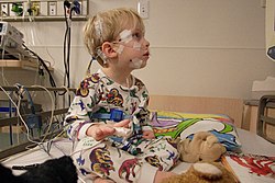
Pediatric polysomnography

The taking of a thorough medical history while keeping in mind alternative diagnoses and the possibility of more than one ailment in the same patient is the first step. Symptoms for very different sleep disorders may be similar and it must be determined whether any psychiatric problems are primary or secondary.

The patient history includes previous attempts at treatment and coping and a careful medication review. Differentiation of transient from chronic disorders and primary from secondary ones influences the direction of evaluation and treatment plans.

The Epworth Sleepiness Scale (ESS), designed to give an indication of sleepiness and correlated with sleep apnea, or other questionnaires designed to measure excessive daytime sleepiness, are diagnostic tools that can be used repeatedly to measure results of treatment.

A sleep diary, also called sleep log or sleep journal, kept by a patient at home for at least two weeks, while subjective, may help determine the extent and nature of sleep disturbance and the level of alertness in the normal environment. A parallel journal kept by a parent or bed partner, if any, can also be helpful. Sleep logs can also be used for self-monitoring and in connection with behavioral and other treatment. The image at the top of this page, with nighttime in the middle and the weekend in the middle, shows a layout that can aid in noticing trends

An actigraph unit is a motion-sensing device worn on the wrist, generally for one or two weeks. It gives a gross picture of sleep-wake cycles and is often used to verify the sleep diary. It is cost-efficient when full polysomnography is not required.

Polysomnography is performed in a sleep laboratory while the patient sleeps, preferably at his or her usual sleeping time. The polysomnogram (PSG) objectively records sleep stages and respiratory events. It shows multiple channels of electroencephalogram (EEG), electrooculogram (EOG), electrocardiogram (ECG), nasal and oral airflow, abdominal, chest and leg movements and blood oxygen levels. A single part of a polysomnogram is sometimes measured at home with portable equipment, for example oximetry, which records blood oxygen levels throughout the night. Polysomnography is not routinely used in the evaluation of patients with insomnia or circadian rhythm disorders, except as needed to rule out other disorders. It will usually be a definitive test for sleep apnea.

Home Sleep Tests (HST)or Home Sleep Apnea Tests (HSAT) are types of sleep studies that can be performed in a patient's home to identify obstructive sleep apnea. These devices are increasing in utilization due to their convenience and cost effectiveness.

A Multiple Sleep Latency Test (MSLT) is often performed during the entire day after polysomnography while the electrodes and other equipment are still in place. The patient is given nap opportunities every second hour; the test measures the number of minutes it takes from the start of a daytime nap period to the first signs of sleep. It is a measure of daytime sleepiness; it also shows whether REM sleep is achieved in a short nap, a typical indication of narcolepsy.

Imaging studies may be performed if a patient is to be evaluated for neurodegenerative disease or to determine the obstruction in obstructive sleep apnea.

Sleep Questionnaires: There are some validated questionnaires in sleep medicine such as:

Treatments

 
Normison (temazepam) is a benzodiazepine commonly prescribed for insomnia and other sleep disorders.

When sleep complaints are secondary to pain, other medical or psychiatric diagnoses, or substance abuse, it may be necessary to treat both the underlying cause and the sleep problems.

When the underlying cause of sleep problems is not immediately obvious, behavioral treatments are usually the first suggested. These range from patient education about sleep hygiene to cognitive behavioral therapy (CBT). Studies of both younger and older adults have compared CBT to medication and found that CBT should be considered a first-line and cost-effective intervention for chronic insomnia, not least because gains may be maintained at long-term follow-up. Sleep physicians and psychologists, at least in the US, are not in agreement about who should perform CBT nor whether sleep centers should be required to have psychologists on staff. In the UK the number of CBT-trained therapists is limited so CBT is not widely available on the NHS.

Behavioral therapies include progressive relaxation, stimulus control (to reassociate the bed with sleepiness), limiting time-in-bed to increase sleep efficiency and debunking misconceptions about sleep.

Pharmacotherapy is necessary for some conditions. Medication may be useful for acute insomnia and for some of the parasomnias. It is almost always needed, along with scheduled short naps and close follow-up, in the treatment of narcolepsy and idiopathic hypersomnia.

Chronic circadian rhythm disorders, the most common of which is delayed sleep phase disorder, may be managed by specifically-timed bright light therapy, usually in the morning, darkness therapy in the hours before bedtime, and timed oral administration of the hormone melatonin. Chronotherapy has also been prescribed for circadian rhythm disorders, though results are generally short-lived. Stimulants may also be prescribed. When these therapies are unsuccessful, counseling may be indicated to help a person adapt to and live with the condition. People with these disorders who have chosen a lifestyle in conformity with their sleeping schedules have no need of treatment, though they may need the diagnosis in order to avoid having to meet for appointments or meetings during their sleep time.

Continuous positive airway pressure (CPAP), Bilevel Continuous Positive Airway Pressure (BiPAP), or similar machines can be used nightly at home to effectively manage sleep-related breathing disorders such as apnea. In milder cases, oral appliances may be effective alternate treatments. For mild cases in obese people, weight reduction may be sufficient, but it is usually recommended as an adjunct to CPAP treatment since sustaining weight loss is difficult. In some cases, upper airway surgery, generally performed by an otolaryngologist/head & neck surgeon or occasionally an oral and maxillofacial surgeon, is indicated. The treatments prevent airway collapse, which interrupts breathing during sleep. A 2001 study published by Hans-Werner Gessmann in the Journal of Sleep Medicine and Sleep Psychology found that patients who practiced a series of electrical stimulations of suprahyoidal tongue muscles for 20 minutes a day showed a marked decline in sleep apnea symptoms after two months. Patients experienced an average of 36% fewer apnea episodes after successfully completing the treatments.

According to the National Cancer Institute (NCI), about 50% of cancer patients have trouble sleeping. Difficulty sleeping can include Restless Leg Syndrome (RLS), sleeping that is fragmented, or insomnia. Some reports show that up to 80% of patients who are undergoing cancer treatments experience some form of insomnia. One of the significant reasons for sleeping problems is stress, uncertainty, and fear. Other patients have difficulty sleeping directly due to their treatments while others experience pain that affects sleep quality. Other factors include diet and less than optimum sleeping conditions. Cancer has also been shown to be a cause of increased sleep apnea, which adds to the potential issues.

See also


Prion

From Wikipedia, the free encyclopedia

Prion diseases
Histology bse.jpg
Microscopic "holes" are characteristic in prion-affected tissue sections, causing the tissue to develop a "spongy" architecture. This causes deterioration of the sponge-like tissue in the brain.
Pronunciation
SpecialtyInfectious disease

Prions are misfolded proteins with the ability to transmit their misfolded shape onto normal variants of the same protein. They characterize several fatal and transmissible neurodegenerative diseases in humans and many other animals. It is not known what causes the normal protein to misfold, but the abnormal three-dimensional structure is suspected of conferring infectious properties, collapsing nearby protein molecules into the same shape. The word prion derives from "proteinaceous infectious particle". The hypothesized role of a protein as an infectious agent stands in contrast to all other known infectious agents such as viroids, viruses, bacteria, fungi, and parasites, all of which contain nucleic acids (DNA, RNA, or both).

Prion isoforms of the prion protein (PrP), whose specific function is uncertain, are hypothesized as the cause of transmissible spongiform encephalopathies (TSEs), including scrapie in sheep, chronic wasting disease (CWD) in deer, bovine spongiform encephalopathy (BSE) in cattle (commonly known as "mad cow disease") and Creutzfeldt–Jakob disease (CJD) in humans. All known prion diseases in mammals affect the structure of the brain or other neural tissue; all are progressive, have no known effective treatment, and are always fatal. Until 2015, all known mammalian prion diseases were considered to be caused by the prion protein (PrP); however in 2015 multiple system atrophy (MSA) was hypothesized to be caused by a prion form of alpha-synuclein.

Prions form abnormal aggregates of proteins called amyloids, which accumulate in infected tissue and are associated with tissue damage and cell death. Amyloids are also responsible for several other neurodegenerative diseases such as Alzheimer's disease and Parkinson's disease. Prion aggregates are stable, and this structural stability means that prions are resistant to denaturation by chemical and physical agents: they cannot be destroyed by ordinary disinfection or cooking. This makes disposal and containment of these particles difficult.

A prion disease is a type of proteopathy, or disease of structurally abnormal proteins. In humans, prions are believed to be the cause of Creutzfeldt–Jakob disease (CJD), its variant (vCJD), Gerstmann–Sträussler–Scheinker syndrome (GSS), fatal familial insomnia (FFI), and kuru. There is also evidence suggesting prions may play a part in the process of Alzheimer's disease, Parkinson's disease, and amyotrophic lateral sclerosis (ALS); these have been termed prion-like diseases. Several yeast proteins have also been identified as having prionogenic properties. Prion replication is subject to epimutation and natural selection just as for other forms of replication, and their structure varies slightly between species.

Etymology and pronunciation

The word prion, coined in 1982 by Stanley B. Prusiner, is derived from protein and infection, hence prion, and is short for "proteinaceous infectious particle", in reference to its ability to self-propagate and transmit its conformation to other proteins. Its main pronunciation is /ˈprɒn/ (About this soundlisten), although /ˈprɒn/, as the homographic name of the bird (prions or whalebirds) is pronounced, is also heard. In his 1982 paper introducing the term, Prusiner specified that it is "pronounced pree-on."

Prion protein

Structure

The protein that prions are made of (PrP) is found throughout the body, even in healthy people and animals. However, PrP found in infectious material has a different structure and is resistant to proteases, the enzymes in the body that can normally break down proteins. The normal form of the protein is called PrPC, while the infectious form is called PrPSc – the C refers to 'cellular' PrP, while the Sc refers to 'scrapie', the prototypic prion disease, occurring in sheep. While PrPC is structurally well-defined, PrPSc is certainly polydisperse and defined at a relatively poor level. PrP can be induced to fold into other more-or-less well-defined isoforms in vitro, and their relationship to the form(s) that are pathogenic in vivo is not yet clear.

PrPC

PrPC is a normal protein found on the membranes of cells, "including several blood components of which platelets constitute the largest reservoir in humans." It has 209 amino acids (in humans), one disulfide bond, a molecular mass of 35–36 kDa and a mainly alpha-helical structure. Several topological forms exist; one cell surface form anchored via glycolipid and two transmembrane forms. The normal protein is not sedimentable; meaning that it cannot be separated by centrifuging techniques. Its function is a complex issue that continues to be investigated. PrPC binds copper (II) ions with high affinity. The significance of this finding is not clear, but it is presumed to relate to PrP structure or function. PrPC is readily digested by proteinase K and can be liberated from the cell surface in vitro by the enzyme phosphoinositide phospholipase C (PI-PLC), which cleaves the glycophosphatidylinositol (GPI) glycolipid anchor. PrP has been reported to play important roles in cell-cell adhesion and intracellular signaling in vivo, and may therefore be involved in cell-cell communication in the brain.

PrPres

Protease-resistant PrPSc-like protein (PrPres) is the name given to any isoform of PrPc which is structurally altered and converted into a misfolded proteinase K-resistant form in vitro. To model conversion of PrPC to PrPSc in vitro, Saborio et al. rapidly converted PrPC into a PrPres by a procedure involving cyclic amplification of protein misfolding. The term "PrPres" has been used to distinguish between PrPSc, which is isolated from infectious tissue and associated with the transmissible spongiform encephalopathy agent. For example, unlike PrPSc, PrPres may not necessarily be infectious.

PrPSc

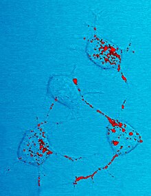
Photomicrograph of mouse neurons showing red stained inclusions identified as scrapies prion protein.
Prion protein (stained in red) revealed in a photomicrograph of neural tissue from a scrapie-infected mouse.

The infectious isoform of PrP, known as PrPSc, or simply the prion, is able to convert normal PrPC proteins into the infectious isoform by changing their conformation, or shape; this, in turn, alters the way the proteins interconnect. PrPSc always causes prion disease. Although the exact 3D structure of PrPSc is not known, it has a higher proportion of β-sheet structure in place of the normal α-helix structure. Aggregations of these abnormal isoforms form highly structured amyloid fibers, which accumulate to form plaques. The end of each fiber acts as a template onto which free protein molecules may attach, allowing the fiber to grow. Under most circumstances, only PrP molecules with an identical amino acid sequence to the infectious PrPSc are incorporated into the growing fiber. However, rare cross-species transmission is also possible.

Normal function of PrP

The physiological function of the prion protein remains poorly understood. While data from in vitro experiments suggest many dissimilar roles, studies on PrP knockout mice have provided only limited information because these animals exhibit only minor abnormalities. In research done in mice, it was found that the cleavage of PrP proteins in peripheral nerves causes the activation of myelin repair in Schwann cells and that the lack of PrP proteins caused demyelination in those cells.

PrP and regulated cell death

MAVS, RIP1, and RIP3 are prion-like proteins found in other parts of the body. They also polymerise into filamentous amyloid fibers which initiate regulated cell death in the case of a viral infection to prevent the spread of virions to other, surrounding cells.

PrP and long-term memory

A review of evidence in 2005 suggested that PrP may have a normal function in maintenance of long-term memory. As well, a 2004 study found that mice lacking genes for normal cellular PrP protein show altered hippocampal long-term potentiation. A recent study that might explain why this is found that neuronal protein CPEB has a similar genetic sequence to yeast prion proteins. The prion-like formation of CPEB is essential for maintaining long-term synaptic changes associated with long term memory formation.

PrP and stem cell renewal

A 2006 article from the Whitehead Institute for Biomedical Research indicates that PrP expression on stem cells is necessary for an organism's self-renewal of bone marrow. The study showed that all long-term hematopoietic stem cells express PrP on their cell membrane and that hematopoietic tissues with PrP-null stem cells exhibit increased sensitivity to cell depletion.

PrP and innate immunity

There is some evidence that PrP may play a role in innate immunity, as the expression of PRNP, the PrP gene, is upregulated in many viral infections and PrP has antiviral properties against many viruses, including HIV.

Prion replication

Heterodimer model of prion propagation
 
Fibril model of prion propagation.

The first hypothesis that tried to explain how prions replicate in a protein-only manner was the heterodimer model. This model assumed that a single PrPSc molecule binds to a single PrPC molecule and catalyzes its conversion into PrPSc. The two PrPSc molecules then come apart and can go on to convert more PrPC. However, a model of prion replication must explain both how prions propagate, and why their spontaneous appearance is so rare. Manfred Eigen showed that the heterodimer model requires PrPSc to be an extraordinarily effective catalyst, increasing the rate of the conversion reaction by a factor of around 1015. This problem does not arise if PrPSc exists only in aggregated forms such as amyloid, where cooperativity may act as a barrier to spontaneous conversion. What is more, despite considerable effort, infectious monomeric PrPSc has never been isolated.

An alternative model assumes that PrPSc exists only as fibrils, and that fibril ends bind PrPC and convert it into PrPSc. If this were all, then the quantity of prions would increase linearly, forming ever longer fibrils. But exponential growth of both PrPSc and of the quantity of infectious particles is observed during prion disease. This can be explained by taking into account fibril breakage. A mathematical solution for the exponential growth rate resulting from the combination of fibril growth and fibril breakage has been found. The exponential growth rate depends largely on the square root of the PrPC concentration. The incubation period is determined by the exponential growth rate, and in vivo data on prion diseases in transgenic mice match this prediction. The same square root dependence is also seen in vitro in experiments with a variety of different amyloid proteins.

The mechanism of prion replication has implications for designing drugs. Since the incubation period of prion diseases is so long, an effective drug does not need to eliminate all prions, but simply needs to slow down the rate of exponential growth. Models predict that the most effective way to achieve this, using a drug with the lowest possible dose, is to find a drug that binds to fibril ends and blocks them from growing any further.

Researchers at Dartmouth College discovered that endogenous host cofactor molecules such as the phospholipid molecule (e.g phosphaditylethanolamine) and polyanions (e.g. single stranded RNA molecules) are necessary to form PrPSc molecules with high levels of specific infectivity in vitro, whereas protein-only PrPSc molecules appear to lack significant levels of biological infectivity.

Transmissible spongiform encephalopathies

Diseases caused by prions
Affected animal(s) Disease
Sheep, Goat Scrapie[59]
Cattle Mad cow disease[59]
Camel[60] Camel spongiform encephalopathy (CSE)
Mink[59] Transmissible mink encephalopathy (TME)
White-tailed deer, elk, mule deer, moose[59] Chronic wasting disease (CWD)
Cat[59] Feline spongiform encephalopathy (FSE)
Nyala, Oryx, Greater Kudu[59] Exotic ungulate encephalopathy (EUE)
Ostrich[61] Spongiform encephalopathy
(unknown if transmissible)
Human Creutzfeldt–Jakob disease (CJD)[59]
Iatrogenic Creutzfeldt–Jakob disease (iCJD)
Variant Creutzfeldt–Jakob disease (vCJD)
Familial Creutzfeldt–Jakob disease (fCJD)
Sporadic Creutzfeldt–Jakob disease (sCJD)
Gerstmann–Sträussler–Scheinker syndrome (GSS)[59]
Fatal familial insomnia (FFI)[62]
Kuru[59]
Familial spongiform encephalopathy[63]
Variably protease-sensitive prionopathy (VPSPr)

Prions cause neurodegenerative disease by aggregating extracellularly within the central nervous system to form plaques known as amyloids, which disrupt the normal tissue structure. This disruption is characterized by "holes" in the tissue with resultant spongy architecture due to the vacuole formation in the neurons. Other histological changes include astrogliosis and the absence of an inflammatory reaction. While the incubation period for prion diseases is relatively long (5 to 20 years), once symptoms appear the disease progresses rapidly, leading to brain damage and death. Neurodegenerative symptoms can include convulsions, dementia, ataxia (balance and coordination dysfunction), and behavioural or personality changes.

Many different mammalian species can be affected by prion diseases, as the prion protein (PrP) is very similar in all mammals. Due to small differences in PrP between different species it is unusual for a prion disease to transmit from one species to another. The human prion disease variant Creutzfeldt–Jakob disease, however, is thought to be caused by a prion that typically infects cattle, causing bovine spongiform encephalopathy and is transmitted through infected meat.

All known prion diseases are untreatable and fatal. However, a vaccine developed in mice may provide insight into providing a vaccine to resist prion infections in humans. Additionally, in 2006 scientists announced that they had genetically engineered cattle lacking a necessary gene for prion production – thus theoretically making them immune to BSE, building on research indicating that mice lacking normally occurring prion protein are resistant to infection by scrapie prion protein. In 2013, a study revealed that 1 in 2,000 people in the United Kingdom might harbour the infectious prion protein that causes vCJD.

Until 2015 all known mammalian prion diseases were considered to be caused by the prion protein, PrP; in 2015 multiple system atrophy was found to be transmissible and was hypothesized to be caused by a new prion, the misfolded form of a protein called alpha-synuclein. The endogenous, properly folded form of the prion protein is denoted PrPC (for Common or Cellular), whereas the disease-linked, misfolded form is denoted PrPSc (for Scrapie), after one of the diseases first linked to prions and neurodegeneration. The precise structure of the prion is not known, though they can be formed spontaneously by combining PrPC, homopolymeric polyadenylic acid, and lipids in a protein misfolding cyclic amplification (PMCA) reaction even in the absence of pre-existing infectious prions. This result is further evidence that prion replication does not require genetic information.

Transmission

It has been recognized that prion diseases can arise in three different ways: acquired, familial, or sporadic. It is often assumed that the diseased form directly interacts with the normal form to make it rearrange its structure. One idea, the "Protein X" hypothesis, is that an as-yet unidentified cellular protein (Protein X) enables the conversion of PrPC to PrPSc by bringing a molecule of each of the two together into a complex.

The primary method of infection in animals is through ingestion. It is thought that prions may be deposited in the environment through the remains of dead animals and via urine, saliva, and other body fluids. They may then linger in the soil by binding to clay and other minerals.

A University of California research team has provided evidence for the theory that infection can occur from prions in manure. And, since manure is present in many areas surrounding water reservoirs, as well as used on many crop fields, it raises the possibility of widespread transmission. It was reported in January 2011 that researchers had discovered prions spreading through airborne transmission on aerosol particles, in an animal testing experiment focusing on scrapie infection in laboratory mice. Preliminary evidence supporting the notion that prions can be transmitted through use of urine-derived human menopausal gonadotropin, administered for the treatment of infertility, was published in 2011.

Prions in plants

In 2015, researchers at The University of Texas Health Science Center at Houston found that plants can be a vector for prions. When researchers fed hamsters grass that grew on ground where a deer that died with chronic wasting disease (CWD) was buried, the hamsters became ill with CWD, suggesting that prions can bind to plants, which then take them up into the leaf and stem structure, where they can be eaten by herbivores, thus completing the cycle. It is thus possible that there is a progressively accumulating number of prions in the environment.

Sterilization

Infectious particles possessing nucleic acid are dependent upon it to direct their continued replication. Prions, however, are infectious by their effect on normal versions of the protein. Sterilizing prions, therefore, requires the denaturation of the protein to a state in which the molecule is no longer able to induce the abnormal folding of normal proteins. In general, prions are quite resistant to proteases, heat, ionizing radiation, and formaldehyde treatments, although their infectivity can be reduced by such treatments. Effective prion decontamination relies upon protein hydrolysis or reduction or destruction of protein tertiary structure. Examples include sodium hypochlorite, sodium hydroxide, and strongly acidic detergents such as LpH. 134 °C (273 °F) for 18 minutes in a pressurized steam autoclave has been found to be somewhat effective in deactivating the agent of disease. Ozone sterilization is currently being studied as a potential method for prion denaturation and deactivation. Renaturation of a completely denatured prion to infectious status has not yet been achieved; however, partially denatured prions can be renatured to an infective status under certain artificial conditions.

The World Health Organization recommends any of the following three procedures for the sterilization of all heat-resistant surgical instruments to ensure that they are not contaminated with prions:

  1. Immerse in 1N sodium hydroxide and place in a gravity-displacement autoclave at 121 °C for 30 minutes; clean; rinse in water; and then perform routine sterilization processes.
  2. Immerse in 1N sodium hypochlorite (20,000 parts per million available chlorine) for 1 hour; transfer instruments to water; heat in a gravity-displacement autoclave at 121 °C for 1 hour; clean; and then perform routine sterilization processes.
  3. Immerse in 1N sodium hydroxide or sodium hypochlorite (20,000 parts per million available chlorine) for 1 hour; remove and rinse in water, then transfer to an open pan and heat in a gravity-displacement (121 °C) or in a porous-load (134 °C) autoclave for 1 hour; clean; and then perform routine sterilization processes.

Degradation resistance in nature

Overwhelming evidence shows that prions resist degradation and persist in the environment for years, and proteases do not degrade them. Experimental evidence shows that unbound prions degrade over time, while soil-bound prions remain at stable or increasing levels, suggesting that prions likely accumulate in the environment. One 2015 study by US scientists found that repeated drying and wetting may render soil bound prions less infectious, although this was dependent on the soil type they were bound to.

Fungi

Proteins showing prion-type behavior are also found in some fungi, which has been useful in helping to understand mammalian prions. Fungal prions do not appear to cause disease in their hosts. In yeast, protein refolding to the prion configuration is assisted by chaperone proteins such as Hsp104. All known prions induce the formation of an amyloid fold, in which the protein polymerises into an aggregate consisting of tightly packed beta sheets. Amyloid aggregates are fibrils, growing at their ends, and replicate when breakage causes two growing ends to become four growing ends. The incubation period of prion diseases is determined by the exponential growth rate associated with prion replication, which is a balance between the linear growth and the breakage of aggregates.

Fungal proteins exhibiting templated conformational change were discovered in the yeast Saccharomyces cerevisiae by Reed Wickner in the early 1990s. For their mechanistic similarity to mammalian prions, they were termed yeast prions. Subsequent to this, a prion has also been found in the fungus Podospora anserina. These prions behave similarly to PrP, but, in general, are nontoxic to their hosts. Susan Lindquist's group at the Whitehead Institute has argued some of the fungal prions are not associated with any disease state, but may have a useful role; however, researchers at the NIH have also provided arguments suggesting that fungal prions could be considered a diseased state. There is evidence that fungal proteins have evolved specific functions that are beneficial to the microorganism that enhance their ability to adapt to their diverse environments.

Research into fungal prions has given strong support to the protein-only concept, since purified protein extracted from cells with a prion state has been demonstrated to convert the normal form of the protein into a misfolded form in vitro, and in the process, preserve the information corresponding to different strains of the prion state. It has also shed some light on prion domains, which are regions in a protein that promote the conversion into a prion. Fungal prions have helped to suggest mechanisms of conversion that may apply to all prions, though fungal prions appear distinct from infectious mammalian prions in the lack of cofactor required for propagation. The characteristic prion domains may vary between species – e.g., characteristic fungal prion domains are not found in mammalian prions.

Fungal prions
Protein Natural host Normal function Prion state Prion phenotype Year identified
Ure2p Saccharomyces cerevisiae Nitrogen catabolite repressor [URE3] Growth on poor nitrogen sources 1994
Sup35p S. cerevisiae Translation termination factor [PSI+] Increased levels of nonsense suppression 1994
HET-S Podospora anserina Regulates heterokaryon incompatibility [Het-s] Heterokaryon formation between incompatible strains
Rnq1p S. cerevisiae Protein template factor [RNQ+], [PIN+] Promotes aggregation of other prions
Swi1 S. cerevisiae Chromatin remodeling [SWI+] Poor growth on some carbon sources 2008
Cyc8 S. cerevisiae Transcriptional repressor [OCT+] Transcriptional derepression of multiple genes 2009
Mot3 S. cerevisiae Nuclear transcription factor [MOT3+] Transcriptional derepression of anaerobic genes 2009
Sfp1 S. cerevisiae Putative transcription factor [ISP+] Antisuppression 2010

Treatments

There are no effective treatments for prion diseases. Clinical trials in humans have not met with success and have been hampered by the rarity of prion diseases. Although some potential treatments have shown promise in the laboratory, none have been effective once the disease has commenced.

In other diseases

Prion-like domains have been found in a variety of other mammalian proteins. Some of these proteins have been implicated in the ontogeny of age-related neurodegenerative disorders such as amyotrophic lateral sclerosis (ALS), frontotemporal lobar degeneration with ubiquitin-positive inclusions (FTLD-U), Alzheimer's disease, Parkinson's disease, and Huntington's disease. They are also implicated in some forms of systemic amyloidosis including AA amyloidosis that develops in humans and animals with inflammatory and infectious diseases such as tuberculosis, Crohn's disease, rheumatoid arthritis, and HIV AIDS. AA amyloidosis, like prion disease, may be transmissible. This has given rise to the 'prion paradigm', where otherwise harmless proteins can be converted to a pathogenic form by a small number of misfolded, nucleating proteins.

The definition of a prion-like domain arises from the study of fungal prions. In yeast, prionogenic proteins have a portable prion domain that is both necessary and sufficient for self-templating and protein aggregation. This has been shown by attaching the prion domain to a reporter protein, which then aggregates like a known prion. Similarly, removing the prion domain from a fungal prion protein inhibits prionogenesis. This modular view of prion behaviour has led to the hypothesis that similar prion domains are present in animal proteins, in addition to PrP. These fungal prion domains have several characteristic sequence features. They are typically enriched in asparagine, glutamine, tyrosine and glycine residues, with an asparagine bias being particularly conducive to the aggregative property of prions. Historically, prionogenesis has been seen as independent of sequence and only dependent on relative residue content. However, this has been shown to be false, with the spacing of prolines and charged residues having been shown to be critical in amyloid formation.

Bioinformatic screens have predicted that over 250 human proteins contain prion-like domains (PrLD). These domains are hypothesized to have the same transmissible, amyloidogenic properties of PrP and known fungal proteins. As in yeast, proteins involved in gene expression and RNA binding seem to be particularly enriched in PrLD's, compared to other classes of protein. In particular, 29 of the known 210 proteins with an RNA recognition motif also have a putative prion domain. Meanwhile, several of these RNA-binding proteins have been independently identified as pathogenic in cases of ALS, FTLD-U, Alzheimer's disease, and Huntington's disease.

Role in neurodegenerative disease

The pathogenicity of prions and proteins with prion-like domains is hypothesized to arise from their self-templating ability and the resulting exponential growth of amyloid fibrils. The presence of amyloid fibrils in patients with degenerative diseases has been well documented. These amyloid fibrils are seen as the result of pathogenic proteins that self-propagate and form highly stable, non-functional aggregates. While this does not necessarily imply a causal relationship between amyloid and degenerative diseases, the toxicity of certain amyloid forms and the overproduction of amyloid in familial cases of degenerative disorders supports the idea that amyloid formation is generally toxic.

Specifically, aggregation of TDP-43, an RNA-binding protein, has been found in ALS/MND patients, and mutations in the genes coding for these proteins have been identified in familial cases of ALS/MND. These mutations promote the misfolding of the proteins into a prion-like conformation. The misfolded form of TDP-43 forms cytoplasmic inclusions in afflicted neurons, and is found depleted in the nucleus. In addition to ALS/MND and FTLD-U, TDP-43 pathology is a feature of many cases of Alzheimer's disease, Parkinson's disease and Huntington's disease. The misfolding of TDP-43 is largely directed by its prion-like domain. This domain is inherently prone to misfolding, while pathological mutations in TDP-43 have been found to increase this propensity to misfold, explaining the presence of these mutations in familial cases of ALS/MND. As in yeast, the prion-like domain of TDP-43 has been shown to be both necessary and sufficient for protein misfolding and aggregation.

Similarly, pathogenic mutations have been identified in the prion-like domains of heterogeneous nuclear riboproteins hnRNPA2B1 and hnRNPA1 in familial cases of muscle, brain, bone and motor neuron degeneration. The wild-type form of all of these proteins show a tendency to self-assemble into amyloid fibrils, while the pathogenic mutations exacerbate this behaviour and lead to excess accumulation.

Weaponization

Prions can be employed as a weaponized agent. With potential fatality rates of 100%, it makes prions a very effective bio-weapon choice. However, one unfavorable aspect is that prions have very long incubation periods. On the other hand, persistent and heavy exposure of prions to intestine, might shorten the overall onset. Also, one large benefit for using prions in warfare, is that detecting prions and decontaminating them is rather difficult.

History

In the 1950s, Carleton Gajdusek began research which eventually showed that kuru could be transmitted to chimpanzees by what was possibly a new infectious agent, work for which he eventually won the 1976 Nobel prize. During the 1960s, two London-based researchers, radiation biologist Tikvah Alper and biophysicist John Stanley Griffith, developed the hypothesis that the transmissible spongiform encephalopathies are caused by an infectious agent consisting solely of proteins. Earlier investigations by E.J. Field into scrapie and kuru had found evidence for the transfer of pathologically inert polysaccharides that only become infectious post-transfer, in the new host. Alper and Griffith wanted to account for the discovery that the mysterious infectious agent causing the diseases scrapie and Creutzfeldt–Jakob disease resisted ionizing radiation. Griffith proposed three ways in which a protein could be a pathogen.

In the first hypothesis, he suggested that if the protein is the product of a normally suppressed gene, and introducing the protein could induce the gene's expression, that is, wake the dormant gene up, then the result would be a process indistinguishable from replication, as the gene's expression would produce the protein, which would then go wake the gene up in other cells.

His second hypothesis forms the basis of the modern prion theory, and proposed that an abnormal form of a cellular protein can convert normal proteins of the same type into its abnormal form, thus leading to replication. His third hypothesis proposed that the agent could be an antibody if the antibody was its own target antigen, as such an antibody would result in more and more antibody being produced against itself. However, Griffith acknowledged that this third hypothesis was unlikely to be true due to the lack of a detectable immune response.

Francis Crick recognized the potential significance of the Griffith protein-only hypothesis for scrapie propagation in the second edition of his "Central dogma of molecular biology" (1970): While asserting that the flow of sequence information from protein to protein, or from protein to RNA and DNA was "precluded", he noted that Griffith's hypothesis was a potential contradiction (although it was not so promoted by Griffith). The revised hypothesis was later formulated, in part, to accommodate reverse transcription (which both Howard Temin and David Baltimore discovered in 1970).

In 1982, Stanley B. Prusiner of the University of California, San Francisco, announced that his team had purified the hypothetical infectious protein, which did not appear to be present in healthy hosts, though they did not manage to isolate the protein until two years after Prusiner's announcement. The protein was named a prion, for "proteinacious infectious particle", derived from the words protein and infection. When the prion was discovered, Griffith's first hypothesis, that the protein was the product of a normally silent gene was favored by many. It was subsequently discovered, however, that the same protein exists in normal hosts but in different form.

Following the discovery of the same protein in different form in uninfected individuals, the specific protein that the prion was composed of was named the prion protein (PrP), and Griffith's second hypothesis that an abnormal form of a host protein can convert other proteins of the same type into its abnormal form, became the dominant theory. Prusiner won the Nobel Prize in Physiology or Medicine in 1997 for his research into prions.

See also

Monday, September 13, 2021

Fatal insomnia

From Wikipedia, the free encyclopedia
 
Fatal insomnia
Cranial imaging of a FFI patient.jpg
Cranial imaging of an FFI patient. In the MRI, there are abnormal signals in the bilateral frontoparietal subcortical area. MRA showed smaller distal branches of cerebral arteries.

SpecialtyPsychiatry, Sleep medicine, Neuropathology
SymptomsProgressive insomnia leading to dementia and death.
ComplicationsPermanent state of hypnagogia later in the illness
Usual onsetMiddle age
TypesFatal familial insomnia, sporadic fatal insomnia
CausesGenetic mutation, sporadic form very rare
Risk factorsFamily history
Diagnostic methodSuspected based on symptoms, Supported by Sleep study, PET scan and genetic testing (If Familial form is suspected)
Differential diagnosisAlzheimer’s disease, frontotemporal dementia, other transmissible spongiform encephalopathies
PreventionNone
TreatmentSupportive care
MedicationNone
PrognosisAlways fatal
FrequencyFewer than 40 families worldwide are known to carry the gene associated with the disease, 24 sporadic cases diagnosed (as of 2016) 

Fatal insomnia is an extremely rare genetic disorder that results in trouble sleeping as its hallmark symptom. The problems with sleeping typically start out gradually and worsen over time. Other symptoms may include speech problems, coordination problems, and dementia. It results in death within a few months to a few years.

It is a prion disease of the brain. It is usually caused by a mutation to the gene encoding protein PrPC. It has two forms: fatal familial insomnia (FFI), which is autosomal dominant and sporadic fatal insomnia (sFI) which is due to a noninherited mutation. Diagnosis is suspected based on symptoms and can be supported by a sleep study, a PET scan, and genetic testing if the patient's family has a history of the disease. Similar to other prion diseases, the diagnosis can only be confirmed by a brain autopsy at post-mortem.

Fatal insomnia has no known cure and involves progressively worsening insomnia, which leads to hallucinations, delirium, confusional states like that of dementia, and eventually death. The average survival time from onset of symptoms is 18 months. The first recorded case was an Italian man, who died in Venice in 1765.

Signs and symptoms

The disease has four stages:

  1. Characterized by worsening insomnia, resulting in panic attacks, paranoia, and phobias. This stage lasts for about four months.
  2. Hallucinations and panic attacks become noticeable, continuing for about five months.
  3. Complete inability to sleep is followed by rapid loss of weight. This lasts for about three months.
  4. Dementia, during which the person becomes unresponsive or mute over the course of six months, is the final stage of the disease, after which death follows.

Other symptoms include: profuse sweating, miosis (pinpoint pupils), sudden entrance into menopause for women, or impotence for men, neck stiffness, and elevation of blood pressure and heart rate. The sporadic form of the disease often presents with double vision. Constipation is common as well. As the disease progresses, the person becomes stuck in a state of pre-sleep limbo, or hypnagogia, which is the state just before sleep in healthy individuals. During these stages, people commonly and repeatedly move their limbs as if dreaming.

The age of onset is variable, ranging from 18 to 60 years, with an average of 50. The disease can be detected prior to onset by genetic testing. Death usually occurs between 6–36 months from onset. The presentation of the disease varies considerably from person to person, even among people within the same family, in the sporadic form for example, sleep problems are not commonly reported and early symptoms are ataxia, cognitive impairment, and double vision.

Cause

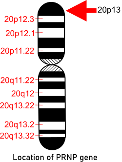
Idiogram of chromosome 20 showing gene PRP location

The gene PRNP that provides instructions for making the prion protein PrPC is located on the short (p) arm of chromosome 20 at position p13. Both people with FFI and those with familial Creutzfeldt–Jakob disease (fCJD) carry a mutation at codon 178 of the prion protein gene. FFI is also invariably linked to the presence of the methionine codon at position 129 of the mutant allele, whereas fCJD is linked to the presence of the valine codon at that position. "The disease is where there is a change of amino acid at position 178 when an asparagine (N) is found instead of the normal aspartic acid (D). This has to be accompanied with a methionine at position 129."

Pathophysiology

In itself, the presence of prions causes reduced glucose use by the thalamus and a mild hypo-metabolism of the cingulate cortex. The extent of this symptom varies between two variations of the disease, these being those presenting methionine homozygotes at codon 129 and methionine/valine heterozygotes being the most severe in the later one. Given the relationship between the involvement of the thalamus in regulating sleep and alertness, a causal relationship can be drawn, and is often mentioned as the cause.

Diagnosis

Diagnosis is suspected based on symptoms. Further work up often include a sleep study and PET scan. Confirmation of the familial form is by genetic testing.

Differential diagnosis

Other diseases involving the mammalian prion protein are known. Some are transmissible (TSEs, including FFI) such as kuru, bovine spongiform encephalopathy (BSE, also known as "mad cow disease") in cattle, and chronic wasting disease in American deer and American elk in some areas of the United States and Canada, as well as Creutzfeldt–Jakob disease (CJD). Until recently, prion diseases were only thought to be transmissible by direct contact with infected tissue, such as from eating infected tissue, transfusion, or transplantation; research suggests that prions can be transmitted by aerosols, but that the general public is not at risk of airborne infection.

Treatments

Treatment involves palliative care. Sleeping pills, including barbiturates, have not been found to be helpful; contrarily, they have been suggested to worsen the symptoms.

Prognosis

Similar to other prion diseases, the disease is invariably fatal. Life expectancy ranges from seven months to six years, with an average of 18 months.

Epidemiology

Hypnogram comparing the sleep pattern of a healthy control with five FFI patients, who display decreased sleep efficiency and disrupted sleep cycles. [W: wake; R: REM; N1-3: NREM sleep stages].

In 1998, 40 families were known to carry the gene for FFI globally: eight German, five Italian, four American, two French, two Australian, two British, one Japanese, and one Austrian. In the Basque Country, Spain, 16 family cases of the 178N mutation were seen between 1993 and 2005 related to two families with a common ancestor in the 18th century. In 2011, another family was added to the list when researchers found the first man in the Netherlands with FFI. While he had lived in the Netherlands for 19 years, he was of Egyptian descent. Other prion diseases are similar to FFI and could be related, but are missing the D178N gene mutation.

As of 2016, 24 cases of sporadic fatal insomnia have been diagnosed. Unlike in FFI, sFI sufferers do not have the D178N mutation in the PRNP-prion gene; they all have a different mutation in the same gene causing methionine homozygosity at codon 129. Nonetheless the methionine presence in lieu of the valine (Val129) is what causes the sporadic form of disease. The targeting of this mutation is another strategy that has been suggested as possible for treatment, or hopefully as cure for the disease.

Silvano, 1983, Bologna, Italy

In late 1983, Italian neurologist/sleep expert Dr. Ignazio Roiter received a patient at the University of Bologna hospital's sleep institute. The man, known only as Silvano, decided in a rare moment of consciousness to be recorded for future studies and to donate his brain for research in hopes of finding a cure for future victims.

Unnamed patient of Schenkein and Montagna, 2001

One person was able to exceed the average survival time by nearly one year with various strategies, including vitamin therapy and meditation, using different stimulants and hypnotics, and even complete sensory deprivation in an attempt to induce sleep at night and increase alertness during the day. He managed to write a book and drive hundreds of miles in this time, but nonetheless, over the course of his trials, the person succumbed to the classic four-stage progression of the illness.

Egyptian man, 2011, Netherlands

Timeline of an FFI patient (same as the one above this one)

In 2011, the first reported case in the Netherlands was of a 57-year-old man of Egyptian descent. The man came in with symptoms of double vision and progressive memory loss, and his family also noted he had recently become disoriented, paranoid, and confused. While he tended to fall asleep during random daily activities, he experienced vivid dreams and random muscular jerks during normal slow-wave sleep. After four months of these symptoms, he began to have convulsions in his hands, trunk, and lower limbs while awake. The person died at age 58, seven months after the onset of symptoms. An autopsy revealed mild atrophy of the frontal cortex and moderate atrophy of the thalamus. The latter is one of the most common signs of FFI.

Research

Still with unclear benefit in humans, a number of treatments have had tentative success in slowing disease progression in animal models, including pentosan polysulfate, mepacrine, and amphotericin B. As of 2016, a study investigating doxycycline is being carried out.

In 2009, a mouse model was made for FFI. These mice expressed a humanized version of the PrP protein that also contains the D178N FFI mutation. These mice appear to have progressively fewer and shorter periods of uninterrupted sleep, damage in the thalamus, and early deaths, similar to humans with FFI.

The Prion Alliance was established by husband and wife duo Eric Minikel and Sonia Vallabh after Vallabh's mother was diagnosed with the fatal disease. They conduct research at the Broad Institute to develop therapeutics for human prion diseases. Other research interests involve identifying biomarkers to track the progression of prion disease in living people.

Popular culture

  • In the 2018 novel and online short story series "Tales from the Gas Station" the main character, Jack, believes that he suffers from fatal familial insomnia.
  • Charlie Huston's 2010 novel Sleepless concerns an epidemic of a fatal insomnia that is frequently compared to FFI by characters in the story.
  • In Something's Killing Me with BD Wong, November 2017 (season one, episode five), "Family Curse", FFI is the topic.
  • Nancy Kress's novelette Pathways concerns research into FFI.
  • The 2019 movie, A Score to Settle, starring Nicolas Cage, uses FFI as a plot element.
  • The 2019 movie, Awoken, uses FFI as a major plot element.
  • FFI is a major plot element and is described in detail in the Lewis episode "Falling Darkness".
  • In the first episode of the 2020 TV series Next, one of the main characters confesses to have the disease and says "It's a real thing, look it up." It also features this Wikipedia article about the disease depicted in a book encyclopedia (Episode 6, 9:4
  • In the novel Siphonophore by Jaimie Batchan (2021), the fictional author of the book is diagnosed with the disease midway through writing the novel, under the name of Prionic Fatal Insomnia.
  • In Destiny 2: Beyond Light, the Bray family all suffer from fatal familial insomnia due to their patriarch's experiments with genetic engineering.
  • Law and Order: Special Victims Unit, season 12, episode 19 – Dr. Huang diagnoses a witness who suffers from fatal familial insomnia. The witness is disoriented; unable to sleep; sweats severely and presents altered pupils. Dr. Huang explains to Detectives Olivia and Elliot that the referred disease mainly affects people from a small town in Italy, where the witness came from.

Eradication of suffering

From Wikipedia, the free encyclopedia https://en.wikipedia.org/wiki/Eradication_of_suffering ...