Search This Blog

Thursday, March 9, 2023

Modern valence bond theory

From Wikipedia, the free encyclopedia
 

Modern valence bond theory is the application of valence bond theory [ VBT ] with computer programs that are competitive in accuracy and economy with programs for the Hartree–Fock or post-Hartree-Fock methods. The latter methods dominated quantum chemistry from the advent of digital computers because they were easier to program. The early popularity of valence bond methods thus declined. It is only recently that the programming of valence bond methods has improved. These developments are due to and described by Gerratt, Cooper, Karadakov and Raimondi (1997); Li and McWeeny (2002); Joop H. van Lenthe and co-workers (2002); Song, Mo, Zhang and Wu (2005); and Shaik and Hiberty (2004)

While Molecular Orbital Theory [MOT] describes the electronic wavefunction as a linear combination of basis functions that are centered on the various atoms in a species (Linear combination of atomic orbitals), VBT describes the electronic wavefunction as a linear combination of several valence bond structures. Each of these valence bond structures can be described using linear combinations of either atomic orbitals, delocalized atomic orbitals (Coulson-Fischer theory), or even molecular orbital fragments. Although this is often overlooked, MOT and VBT are equally valid ways of describing the electronic wavefunction, and are actually related by a unitary transformation. Assuming MOT and VBT are applied at the same level of theory, this relationship ensures that they will describe the same wavefunction, but will do so in different forms.

Theory

Bonding in H2

Heitler and London's original work on VBT attempts to approximate the electronic wavefunction as a covalent combination of localized basis functions on the bonding atoms. In VBT, wavefunctions are described as the sums and differences of VB determinants, which enforce the antisymmetric properties required by the Pauli exclusion principle. Taking H2 as an example, the VB determinant is

In this expression, N is a normalization constant, and a and b are basis functions that are localized on the two hydrogen atoms, often considered simply to be 1s atomic orbitals. The numbers are an index to describe the electron (i.e. a(1) represents the concept of ‘electron 1’ residing in orbital a). ɑ and β describe the spin of the electron. The bar over b in indicates that the electron associated with orbital b has β spin (in the first term, electron 2 is in orbital b, and thus electron 2 has β spin). By itself, a single VB determinant is not a proper spin-eigenfunction, and thus cannot describe the true wavefunction. However, by taking the sum and difference (linear combinations) of VB determinants, two approximate wavefunctions can be obtained:

These are four valence bond structures that can contribute to the VBT description of bonding in a hydrogen molecule. The Heitler-London (covalent) structure is the largest contributor, while the ionic structures are minor contributors. The triplet structure is a negligible contributor.

ΦHL is the wavefunction as described by Heiter and London originally, and describes the covalent bonding between orbitals a and b in which the spins are paired, as expected for a chemical bond. ΦT is a representation of the bond that where the electron spins are parallel, resulting in a triplet state. This is a highly repulsive interaction, so this description of the bonding will not play a major role in determining the wave function.

Other ways of describing the wavefunction can also be constructed. Specifically, instead of considering a covalent interaction, the ionic interactions can be considered, resulting in the wavefunction

This wavefunction describes the bonding in H2 as the ionic interaction between an H+ and an H-.

Since none of these wavefunctions, ΦHL (covalent bonding) or ΦI (ionic bonding) perfectly approximates the wavefunction, a combination of these two can be used to describe the total wavefunction

where λ and μ are coefficients that can vary from 0 to 1. In determining the lowest energy wavefunction, these coefficients can be varied until a minimum energy is reached. λ will be larger in bonds that have more covalency, while μ will be larger in bonds that are more ionic. In the specific case of H2, λ ≈ 0.75, and μ ≈ 0.25.

The orbitals that were used as the basis (a and b) do not necessarily have to be localized on the atoms involved in bonding. Orbitals that are partially delocalized onto the other atom involved in bonding can also be used, as in the Coulson-Fischer theory. Even the molecular orbitals associated with a portion of a molecule can be used as a basis set, a processes referred to as using fragment orbitals.

For more complicated molecules, ΦVBT could consider several possible structures that all contribute in various degrees (there would be several coefficients, not just λ and μ). An example of this is the Kekule and Dewar structures used in describing benzene.

Note that all normalization constants were ignored in the discussion above for simplicity.

Relationship to Molecular Orbital Theory

History

The application of VBT and MOT to computations that attempt to approximate the Schrödinger equation began near the middle of the 20th century, but MOT quickly became the preferred approach between the two. The relative computational ease of doing calculations with non-overlapping orbitals in MOT is said to have contributed to its popularity. In addition, the successful explanation of π-systems, pericyclic reactions, and extended solids further cemented MOT as the preeminent approach. Despite this, the two theories are just two different ways of representing the same wavefunction. As shown below, at the same level of theory, the two methods lead to the same results.

H2 - Molecular Orbital vs Valence Bond Theory

The relationship between MOT and VBT can be made more clear by directly comparing the results of the two theories for the hydrogen molecule, H2. Using MOT, the same basis orbitals (a and b) can be used to describe the bonding. Combining them in a constructive and destructive manner gives two spin-orbitals

The ground state wavefunction of H2 would be that where the σ orbital is doubly occupied, which is expressed as the following Slater determinant (as required by MOT)

This expression for the wavefunction can be shown to be equivalent to the following wavefunction

which is now expressed in terms of VB determinants. This transformation does not alter the wavefunction in any way, only the way that the wavefunction is represented. This process of going from an MO description to a VB description can be referred to as ‘mapping MO wavefunctions onto VB wavefunctions’, and is fundamentally the same process as that used to generate localized molecular orbitals.

Rewriting the VB wavefunction derived above, we can clearly see the relationship between MOT and VBT

Thus, at its simplest level, MOT is just VBT, where the covalent and ionic contributions (the first and second terms, respectively) are equal. This is the basis of the claim that MOT does not correctly predict the dissociation of molecules. When MOT includes configuration interaction (MO-CI), this allows the relative contributions of the covalent and ionic contributions to be altered. This leads to the same description of bonding for both VBT and MO-CI. In conclusion, the two theories, when brought to a high enough level of theory, will converge. Their distinction is in the way they are built up to that description.

Note that in all of the aforementioned discussions, as with the derivation of H2 for VBT, normalization constants were ignored for simplicity.

'Failures' of Valence Bond Theory

When describing the relationship between MOT and VBT, there are a few examples that are commonly cited as ‘failures’ of VBT. However, these often arise from an incomplete or inaccurate use of VBT.

Triplet Ground State of Oxygen

It is known that O2 has a triplet ground state, but a classic Lewis structure depiction of oxygen would not indicate that any unpaired electrons exist. Perhaps because Lewis structures and VBT often depict the same structure as the most stable state, this misinterpretation has persisted. However, as has been consistently demonstrated with VBT calculations, the lowest energy state is that with two, three electron π-bonds, which is the triplet state.

Ionization Energy of Methane

The photoelectron spectrum (PES) of methane is commonly used as an argument as to why MO theory is superior to VBT. From an MO calculation (or even just a qualitative MOT diagram), it can be seen that the HOMO is a triply degenerate state, while the HOMO-1 is a single degenerate state. By invoking Koopman's Theorem, one can predict that there would be two distinct peaks in the ionization spectrum of methane. Those would be by exciting an electron from the t2 orbitals or the a1 orbital, which would result in a 3:1 ratio in intensity. This is corroborated by experiment. However, when one examines the VB description of CH4, it is clear that there are 4 equivalent bonds between C and H. If one were to invoke Koopman's Theorem (which is implicitly done when claiming that VBT is inadequate to describe PES), a single ionization energy peak would be predicted. However, Koopman's Theorem cannot be applied to orbitals that are not the canonical molecular orbitals, and thus a different approach is required to understand the ionization potentials of methane from VBT. To do this, the ionized product, CH4+ must be analyzed. The VB wavefunction of CH4+ would be an equal combination of 4 structures, each having 3 two-electron bonds, and 1 one-electron bond. Based on group theory arguments, these states must give rise to a triply degenerate T2 state and a single degenerate A1 state. A diagram showing the relative energies of the states is shown below, and it can be seen that there exist two distinct transitions from the CH4 state with 4 equivalent bonds to the two CH4+ states.

Two distinct states for CH4+ exist (A1 and T2), both of which result from the ionization of CH4. This gives rise to the two unique peaks on the photoelectron spectrum of methane.

Valence Bond Theory Methods

Listed below are a few notable VBT methods that are applied in modern computational software packages.

Generalized VBT (GVB)

This was one of the first ab initio computational methods developed that utilized VBT. Using Coulson-Fischer type basis orbitals, this method uses singly-occupied, instead of doubly-occupied orbitals, as the basis set. This allows from the distance between paired electrons to increase during variational optimization, lowering the resultant energy.  The total wavefunction is described by a single set of orbitals, rather than a linear combination of multiple VB structures. GVB is considered to be a user-friendly method for new practitioners. 

Spin-Coupled Generalized Valence Bond Theory (SCGVB, or sometimes SCVB/full GVB)

SCGVB is an extension of GVB that still uses delocalized orbitals, whose delocalization can adjust with molecular structure. In addition, the electronic wavefunction is still a single product of orbitals. The difference is that the spin functions are allowed to adjust simultaneously with the orbitals during energy minimization procedures. This is considered to be one of the best VB descriptions of the wavefunction that relies on only a single configuration. 

Complete Active Space Valence Bond Method (CASVB)

This is a method that often gets confused as a traditional VB method. Instead, this is a localization procedure that maps the full configuration interaction Hartree-Fock wavefunction (CASSCF) onto valence bond structures. 

Spin-coupled theory

There are a large number of different valence bond methods. Most use n valence bond orbitals for n electrons. If a single set of these orbitals is combined with all linear independent combinations of the spin functions, we have spin-coupled valence bond theory. The total wave function is optimized using the variational method by varying the coefficients of the basis functions in the valence bond orbitals and the coefficients of the different spin functions. In other cases only a sub-set of all possible spin functions is used. Many valence bond methods use several sets of the valence bond orbitals. Be warned that different authors use different names for these different valence bond methods.

Valence bond programs

Several groups have produced computer programs for modern valence bond calculations that are freely available.

Molecular geometry

From Wikipedia, the free encyclopedia
 
Geometry of the water molecule with values for O-H bond length and for H-O-H bond angle between two bonds

Molecular geometry is the three-dimensional arrangement of the atoms that constitute a molecule. It includes the general shape of the molecule as well as bond lengths, bond angles, torsional angles and any other geometrical parameters that determine the position of each atom.

Molecular geometry influences several properties of a substance including its reactivity, polarity, phase of matter, color, magnetism and biological activity. The angles between bonds that an atom forms depend only weakly on the rest of molecule, i.e. they can be understood as approximately local and hence transferable properties.

Determination

The molecular geometry can be determined by various spectroscopic methods and diffraction methods. IR, microwave and Raman spectroscopy can give information about the molecule geometry from the details of the vibrational and rotational absorbance detected by these techniques. X-ray crystallography, neutron diffraction and electron diffraction can give molecular structure for crystalline solids based on the distance between nuclei and concentration of electron density. Gas electron diffraction can be used for small molecules in the gas phase. NMR and FRET methods can be used to determine complementary information including relative distances, dihedral angles, angles, and connectivity. Molecular geometries are best determined at low temperature because at higher temperatures the molecular structure is averaged over more accessible geometries (see next section). Larger molecules often exist in multiple stable geometries (conformational isomerism) that are close in energy on the potential energy surface. Geometries can also be computed by ab initio quantum chemistry methods to high accuracy. The molecular geometry can be different as a solid, in solution, and as a gas.

The position of each atom is determined by the nature of the chemical bonds by which it is connected to its neighboring atoms. The molecular geometry can be described by the positions of these atoms in space, evoking bond lengths of two joined atoms, bond angles of three connected atoms, and torsion angles (dihedral angles) of three consecutive bonds.

The influence of thermal excitation

Since the motions of the atoms in a molecule are determined by quantum mechanics, "motion" must be defined in a quantum mechanical way. The overall (external) quantum mechanical motions translation and rotation hardly change the geometry of the molecule. (To some extent rotation influences the geometry via Coriolis forces and centrifugal distortion, but this is negligible for the present discussion.) In addition to translation and rotation, a third type of motion is molecular vibration, which corresponds to internal motions of the atoms such as bond stretching and bond angle variation. The molecular vibrations are harmonic (at least to good approximation), and the atoms oscillate about their equilibrium positions, even at the absolute zero of temperature. At absolute zero all atoms are in their vibrational ground state and show zero point quantum mechanical motion, so that the wavefunction of a single vibrational mode is not a sharp peak, but an exponential of finite width (the wavefunction for n = 0 depicted in the article on the quantum harmonic oscillator). At higher temperatures the vibrational modes may be thermally excited (in a classical interpretation one expresses this by stating that "the molecules will vibrate faster"), but they oscillate still around the recognizable geometry of the molecule.

To get a feeling for the probability that the vibration of molecule may be thermally excited, we inspect the Boltzmann factor β ≡ exp(−ΔE/kT), where ΔE is the excitation energy of the vibrational mode, k the Boltzmann constant and T the absolute temperature. At 298 K (25 °C), typical values for the Boltzmann factor β are:

  • β = 0.089 for ΔE = 500 cm−1
  • β = 0.008 for ΔE = 1000 cm−1
  • β = 0.0007 for ΔE = 1500 cm−1.

(The reciprocal centimeter is an energy unit that is commonly used in infrared spectroscopy; 1 cm−1 corresponds to 1.23984×10⁻⁴ eV). When an excitation energy is 500 cm−1, then about 8.9 percent of the molecules are thermally excited at room temperature. To put this in perspective: the lowest excitation vibrational energy in water is the bending mode (about 1600 cm−1). Thus, at room temperature less than 0.07 percent of all the molecules of a given amount of water will vibrate faster than at absolute zero.

As stated above, rotation hardly influences the molecular geometry. But, as a quantum mechanical motion, it is thermally excited at relatively (as compared to vibration) low temperatures. From a classical point of view it can be stated that at higher temperatures more molecules will rotate faster, which implies that they have higher angular velocity and angular momentum. In quantum mechanical language: more eigenstates of higher angular momentum become thermally populated with rising temperatures. Typical rotational excitation energies are on the order of a few cm−1. The results of many spectroscopic experiments are broadened because they involve an averaging over rotational states. It is often difficult to extract geometries from spectra at high temperatures, because the number of rotational states probed in the experimental averaging increases with increasing temperature. Thus, many spectroscopic observations can only be expected to yield reliable molecular geometries at temperatures close to absolute zero, because at higher temperatures too many higher rotational states are thermally populated.

Bonding

Molecules, by definition, are most often held together with covalent bonds involving single, double, and/or triple bonds, where a "bond" is a shared pair of electrons (the other method of bonding between atoms is called ionic bonding and involves a positive cation and a negative anion).

Molecular geometries can be specified in terms of 'bond lengths', 'bond angles' and 'torsional angles'. The bond length is defined to be the average distance between the nuclei of two atoms bonded together in any given molecule. A bond angle is the angle formed between three atoms across at least two bonds. For four atoms bonded together in a chain, the torsional angle is the angle between the plane formed by the first three atoms and the plane formed by the last three atoms.

There exists a mathematical relationship among the bond angles for one central atom and four peripheral atoms (labeled 1 through 4) expressed by the following determinant. This constraint removes one degree of freedom from the choices of (originally) six free bond angles to leave only five choices of bond angles. (Note that the angles θ11, θ22, θ33, and θ44 are always zero and that this relationship can be modified for a different number of peripheral atoms by expanding/contracting the square matrix.)

Molecular geometry is determined by the quantum mechanical behavior of the electrons. Using the valence bond approximation this can be understood by the type of bonds between the atoms that make up the molecule. When atoms interact to form a chemical bond, the atomic orbitals of each atom are said to combine in a process called orbital hybridisation. The two most common types of bonds are sigma bonds (usually formed by hybrid orbitals) and pi bonds (formed by unhybridized p orbitals for atoms of main group elements). The geometry can also be understood by molecular orbital theory where the electrons are delocalised.

An understanding of the wavelike behavior of electrons in atoms and molecules is the subject of quantum chemistry.

Isomers

Isomers are types of molecules that share a chemical formula but have difference geometries, resulting in different properties:

  • A pure substance is composed of only one type of isomer of a molecule (all have the same geometrical structure).
  • Structural isomers have the same chemical formula but different physical arrangements, often forming alternate molecular geometries with very different properties. The atoms are not bonded (connected) together in the same orders.
    • Functional isomers are special kinds of structural isomers, where certain groups of atoms exhibit a special kind of behavior, such as an ether or an alcohol.
  • Stereoisomers may have many similar physicochemical properties (melting point, boiling point) and at the same time very different biochemical activities. This is because they exhibit a handedness that is commonly found in living systems. One manifestation of this chirality or handedness is that they have the ability to rotate polarized light in different directions.
  • Protein folding concerns the complex geometries and different isomers that proteins can take.

Types of molecular structure

A bond angle is the geometric angle between two adjacent bonds. Some common shapes of simple molecules include:

  • Linear: In a linear model, atoms are connected in a straight line. The bond angles are set at 180°. For example, carbon dioxide and nitric oxide have a linear molecular shape.
  • Trigonal planar: Molecules with the trigonal planar shape are somewhat triangular and in one plane (flat). Consequently, the bond angles are set at 120°. For example, boron trifluoride.
  • Angular: Angular molecules (also called bent or V-shaped) have a non-linear shape. For example, water (H2O), which has an angle of about 105°. A water molecule has two pairs of bonded electrons and two unshared lone pairs.
  • Tetrahedral: Tetra- signifies four, and -hedral relates to a face of a solid, so "tetrahedral" literally means "having four faces". This shape is found when there are four bonds all on one central atom, with no extra unshared electron pairs. In accordance with the VSEPR (valence-shell electron pair repulsion theory), the bond angles between the electron bonds are arccos(−1/3) = 109.47°. For example, methane (CH4) is a tetrahedral molecule.
  • Octahedral: Octa- signifies eight, and -hedral relates to a face of a solid, so "octahedral" means "having eight faces". The bond angle is 90 degrees. For example, sulfur hexafluoride (SF6) is an octahedral molecule.
  • Trigonal pyramidal: A trigonal pyramidal molecule has a pyramid-like shape with a triangular base. Unlike the linear and trigonal planar shapes but similar to the tetrahedral orientation, pyramidal shapes require three dimensions in order to fully separate the electrons. Here, there are only three pairs of bonded electrons, leaving one unshared lone pair. Lone pair – bond pair repulsions change the bond angle from the tetrahedral angle to a slightly lower value. For example, ammonia (NH3).

VSEPR table

The bond angles in the table below are ideal angles from the simple VSEPR theory (pronounced "Vesper Theory"), followed by the actual angle for the example given in the following column where this differs. For many cases, such as trigonal pyramidal and bent, the actual angle for the example differs from the ideal angle, and examples differ by different amounts. For example, the angle in H2S (92°) differs from the tetrahedral angle by much more than the angle for H2O (104.48°) does.

Atoms bonded to
central atom
Lone pairs Electron domains
(Steric number)
Shape Ideal bond angle
(example's bond angle)
Example Image
2 0 2 linear 180° CO2 Linear-3D-balls.png
3 0 3 trigonal planar 120° BF3 Trigonal-3D-balls.png
2 1 3 bent 120° (119°) SO2 Bent-3D-balls.png
4 0 4 tetrahedral 109.5° CH4 AX4E0-3D-balls.png
3 1 4 trigonal pyramidal 109.5° (106.8°) NH3 Pyramidal-3D-balls.png
2 2 4 bent 109.5° (104.48°) H2O Bent-3D-balls.png
5 0 5 trigonal bipyramidal 90°, 120° PCl5 Trigonal-bipyramidal-3D-balls.png
4 1 5 seesaw ax–ax 180° (173.1°),
eq–eq 120° (101.6°),
ax–eq 90°
SF4 Seesaw-3D-balls.png
3 2 5 T-shaped 90° (87.5°), 180° (175°) ClF3 T-shaped-3D-balls.png
2 3 5 linear 180° XeF2 Linear-3D-balls.png
6 0 6 octahedral 90°, 180° SF6 AX6E0-3D-balls.png
5 1 6 square pyramidal 90° (84.8°) BrF5 Square-pyramidal-3D-balls.png
4 2 6 square planar 90°, 180° XeF4 Square-planar-3D-balls.png
7 0 7 pentagonal bipyramidal 90°, 72°, 180° IF7 Pentagonal-bipyramidal-3D-balls.png
6 1 7 pentagonal pyramidal 72°, 90°, 144° XeOF5 Pentagonal-pyramidal-3D-balls.png
5 2 7 pentagonal planar 72°, 144° XeF5 Pentagonal-planar-3D-balls.png
8 0 8 square antiprismatic
XeF2−8 Square-antiprismatic-3D-balls.png
9 0 9 tricapped trigonal prismatic
ReH2−9 AX9E0-3D-balls.png

3D representations

  • Line or stick – atomic nuclei are not represented, just the bonds as sticks or lines. As in 2D molecular structures of this type, atoms are implied at each vertex.
Formic-acid-3D-stick.png
L-aspartic-acid-3D-sticks.png
ATP-xtal-3D-sticks.png
Endohedral fullerene.png
NorbornylCation ElectronDensity.jpg
WinsteinYellow.jpg
  • Ball and stick – atomic nuclei are represented by spheres (balls) and the bonds as sticks.
Methanol-3D-balls.png
Methanol struktur.png
PropyleneGlycol-stickAndBall.png
3LRI SolutionStructureAndBackboneDynamicsOfHumanLong arg3 insulin-Like Growth Factor 1 02.png
Methanol.pdb.png
Ubiquitin spheres.png
P-cresol-spaceFilling.png
3GF1 Insulin-Like Growth Factor Nmr 10 01.png
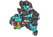
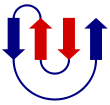
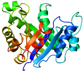
  • Cartoon – a representation used for proteins where loops, beta sheets, and alpha helices are represented diagrammatically and no atoms or bonds are explicitly represented (e.g. the protein backbone is represented as a smooth pipe).
Beta-meander1.png
MreB.png
Anthrax toxin protein key motif.svg
8tim TIM barrel.png

The greater the amount of lone pairs contained in a molecule, the smaller the angles between the atoms of that molecule. The VSEPR theory predicts that lone pairs repel each other, thus pushing the different atoms away from them.

Lewis structure

From Wikipedia, the free encyclopedia
 
A lewis structure of a water molecule, composed of two hydrogen atoms and one oxygen atom sharing valence electrons
Lewis structure of a water molecule.

Lewis structures, also known as Lewis dot formulas, Lewis dot structures, electron dot structures, or Lewis electron dot structures (LEDS), are diagrams that show the bonding between atoms of a molecule, as well as the lone pairs of electrons that may exist in the molecule. A Lewis structure can be drawn for any covalently bonded molecule, as well as coordination compounds. The Lewis structure was named after Gilbert N. Lewis, who introduced it in his 1916 article The Atom and the Molecule. Lewis structures extend the concept of the electron dot diagram by adding lines between atoms to represent shared pairs in a chemical bond.

Lewis structures show each atom and its position in the structure of the molecule using its chemical symbol. Lines are drawn between atoms that are bonded to one another (pairs of dots can be used instead of lines). Excess electrons that form lone pairs are represented as pairs of dots, and are placed next to the atoms.

Although main group elements of the second period and beyond usually react by gaining, losing, or sharing electrons until they have achieved a valence shell electron configuration with a full octet of (8) electrons, hydrogen (H) can only form bonds which share just two electrons.

Construction and electron counting

The total number of electrons represented in a Lewis structure is equal to the sum of the numbers of valence electrons on each individual atom. Non-valence electrons are not represented in Lewis structures.

Once the total number of available electrons has been determined, electrons must be placed into the structure according to these steps:

  1. The atoms are first connected by single bonds.
  2. If t is the total number of electrons and n the number of single bonds, t-2n electrons remain to be placed. These should be placed as lone pairs: one pair of dots for each pair of electrons available. Lone pairs should initially be placed on outer atoms (other than hydrogen) until each outer atom has eight electrons in bonding pairs and lone pairs; extra lone pairs may then be placed on the central atom. When in doubt, lone pairs should be placed on more electronegative atoms first.
  3. Once all lone pairs are placed, atoms (especially the central atoms) may not have an octet of electrons. In this case, the atoms must form a double bond; a lone pair of electrons is moved to form a second bond between the two atoms. As the bonding pair is shared between the two atoms, the atom that originally had the lone pair still has an octet; the other atom now has two more electrons in its valence shell.

Lewis structures for polyatomic ions may be drawn by the same method. When counting electrons, negative ions should have extra electrons placed in their Lewis structures; positive ions should have fewer electrons than an uncharged molecule. When the Lewis structure of an ion is written, the entire structure is placed in brackets, and the charge is written as a superscript on the upper right, outside the brackets.

A simpler method has been proposed for constructing Lewis structures, eliminating the need for electron counting: the atoms are drawn showing the valence electrons; bonds are then formed by pairing up valence electrons of the atoms involved in the bond-making process, and anions and cations are formed by adding or removing electrons to/from the appropriate atoms.

A trick is to count up valence electrons, then count up the number of electrons needed to complete the octet rule (or with hydrogen just 2 electrons), then take the difference of these two numbers. The answer is the number of electrons that make up the bonds. The rest of the electrons just go to fill all the other atoms' octets.

Another simple and general procedure to write Lewis structures and resonance forms has been proposed.

Formal charge

In terms of Lewis structures, formal charge is used in the description, comparison, and assessment of likely topological and resonance structures by determining the apparent electronic charge of each atom within, based upon its electron dot structure, assuming exclusive covalency or non-polar bonding. It has uses in determining possible electron re-configuration when referring to reaction mechanisms, and often results in the same sign as the partial charge of the atom, with exceptions. In general, the formal charge of an atom can be calculated using the following formula, assuming non-standard definitions for the markup used:

where:

  • is the formal charge.
  • represents the number of valence electrons in a free atom of the element.
  • represents the number of unshared electrons on the atom.
  • represents the total number of electrons in bonds the atom has with another.

The formal charge of an atom is computed as the difference between the number of valence electrons that a neutral atom would have and the number of electrons that belong to it in the Lewis structure. Electrons in covalent bonds are split equally between the atoms involved in the bond. The total of the formal charges on an ion should be equal to the charge on the ion, and the total of the formal charges on a neutral molecule should be equal to zero.

Resonance

For some molecules and ions, it is difficult to determine which lone pairs should be moved to form double or triple bonds, and two or more different resonance structures may be written for the same molecule or ion. In such cases it is usual to write all of them with two-way arrows in between (see Example below). This is sometimes the case when multiple atoms of the same type surround the central atom, and is especially common for polyatomic ions.

When this situation occurs, the molecule's Lewis structure is said to be a resonance structure, and the molecule exists as a resonance hybrid. Each of the different possibilities is superimposed on the others, and the molecule is considered to have a Lewis structure equivalent to some combination of these states.

The nitrate ion (NO3), for instance, must form a double bond between nitrogen and one of the oxygens to satisfy the octet rule for nitrogen. However, because the molecule is symmetrical, it does not matter which of the oxygens forms the double bond. In this case, there are three possible resonance structures. Expressing resonance when drawing Lewis structures may be done either by drawing each of the possible resonance forms and placing double-headed arrows between them or by using dashed lines to represent the partial bonds (although the latter is a good representation of the resonance hybrid which is not, formally speaking, a Lewis structure).

When comparing resonance structures for the same molecule, usually those with the fewest formal charges contribute more to the overall resonance hybrid. When formal charges are necessary, resonance structures that have negative charges on the more electronegative elements and positive charges on the less electronegative elements are favored.

Single bonds can also be moved in the same way to create resonance structures for hypervalent molecules such as sulfur hexafluoride, which is the correct description according to quantum chemical calculations instead of the common expanded octet model.

The resonance structure should not be interpreted to indicate that the molecule switches between forms, but that the molecule acts as the average of multiple forms.

Example

The formula of the nitrite ion is NO2.

  1. Nitrogen is the least electronegative atom of the two, so it is the central atom by multiple criteria.
  2. Count valence electrons. Nitrogen has 5 valence electrons; each oxygen has 6, for a total of (6 × 2) + 5 = 17. The ion has a charge of −1, which indicates an extra electron, so the total number of electrons is 18.
  3. Connect the atoms by single bonds. Each oxygen must be bonded to the nitrogen, which uses four electrons—two in each bond.
  4. Place lone pairs. The 14 remaining electrons should initially be placed as 7 lone pairs. Each oxygen may take a maximum of 3 lone pairs, giving each oxygen 8 electrons including the bonding pair. The seventh lone pair must be placed on the nitrogen atom.
  5. Satisfy the octet rule. Both oxygen atoms currently have 8 electrons assigned to them. The nitrogen atom has only 6 electrons assigned to it. One of the lone pairs on an oxygen atom must form a double bond, but either atom will work equally well. Therefore, there is a resonance structure.
  6. Tie up loose ends. Two Lewis structures must be drawn: Each structure has one of the two oxygen atoms double-bonded to the nitrogen atom. The second oxygen atom in each structure will be single-bonded to the nitrogen atom. Place brackets around each structure, and add the charge (−) to the upper right outside the brackets. Draw a double-headed arrow between the two resonance forms.
Nitrite-ion-lewis-canonical.svg

Alternative formations

Two varieties of condensed structural formula, both showing butane
A skeletal diagram of butane

Chemical structures may be written in more compact forms, particularly when showing organic molecules. In condensed structural formulas, many or even all of the covalent bonds may be left out, with subscripts indicating the number of identical groups attached to a particular atom. Another shorthand structural diagram is the skeletal formula (also known as a bond-line formula or carbon skeleton diagram). In a skeletal formula, carbon atoms are not signified by the symbol C but by the vertices of the lines. Hydrogen atoms bonded to carbon are not shown—they can be inferred by counting the number of bonds to a particular carbon atom—each carbon is assumed to have four bonds in total, so any bonds not shown are, by implication, to hydrogen atoms.

Other diagrams may be more complex than Lewis structures, showing bonds in 3D using various forms such as space-filling diagrams.

Usage and limitations

Despite their simplicity and development in the early twentieth century, when understanding of chemical bonding was still rudimentary, Lewis structures capture many of the key features of the electronic structure of a range of molecular systems, including those of relevance to chemical reactivity. Thus, they continue to enjoy widespread use by chemists and chemistry educators. This is especially true in the field of organic chemistry, where the traditional valence-bond model of bonding still dominates, and mechanisms are often understood in terms of curve-arrow notation superimposed upon skeletal formulae, which are shorthand versions of Lewis structures. Due to the greater variety of bonding schemes encountered in inorganic and organometallic chemistry, many of the molecules encountered require the use of fully delocalized molecular orbitals to adequately describe their bonding, making Lewis structures comparatively less important (although they are still common).

It is important to note that there are simple and archetypal molecular systems for which a Lewis description, at least in unmodified form, is misleading or inaccurate. Notably, the naive drawing of Lewis structures for molecules known experimentally to contain unpaired electrons (e.g., O2, NO, and ClO2) leads to incorrect inferences of bond orders, bond lengths, and/or magnetic properties. A simple Lewis model also does not account for the phenomenon of aromaticity. For instance, Lewis structures do not offer an explanation for why cyclic C6H6 (benzene) experiences special stabilization beyond normal delocalization effects, while C4H4 (cyclobutadiene) actually experiences a special destabilization. Molecular orbital theory provides the most straightforward explanation for these phenomena.

Bayesian inference

From Wikipedia, the free encyclopedia https://en.wikipedia.org/wiki/Bayesian_inference Bayesian inference ( / ...