Search This Blog

Monday, June 19, 2023

Combat medic

From Wikipedia, the free encyclopedia
 
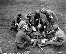
A U.S. Army Medical Corps team at work during the Battle of Normandy
 
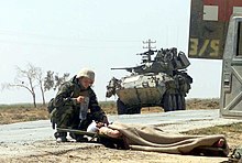
U.S. Navy Hospital Corpsman providing treatment to a wounded Iraqi soldier, 2003.

A combat medic is responsible for providing emergency medical treatment at a point of wounding in a combat or training environment, as well as primary care and health protection and evacuation from a point of injury or illness. Additionally, medics may also be responsible for the creation, oversight, and execution of long-term patient care plans in consultation with or in the absence of a readily available doctor or advanced practice provider. Combat medics may be used in hospitals and clinics, where they have the opportunity to work in additional roles, such as operating medical and laboratory equipment and performing and assisting with procedures.

Israel Defense Forces medical personnel coordinating relief efforts after the 2010 Haiti earthquake

Canada

Training

All Regular Force Medical Technicians begin training with the Canadian Forces Leadership and Recruit School in Saint-Jean-sur-Richelieu, Quebec. Then they begin Occupational training at The Canadian Forces Health Services Training Center in Borden, Ontario, where they are taught how to maintain medical supplies and equipment, screen patients, implement patient care plans, treat medical conditions, administer diagnostic procedures, medications, and continuous infusions. They also learn how to manage airways and assist with minor surgical procedures setting up deployed medical facilities and treating casualties in an operational and CBRNE environment over the course of 47-48 weeks. They also earn the Primary Care Paramedic Level 1 certification. Advanced training can include, Preventive Medicine, Biomedical electronics, and Aviation Physiology, among others.

Assignments

Medical Technicians may be posted to any Canadian ship, or base as part of the base emergency response or as part of a clinic or hospital. They can also be deployed as the medical detachment of an infantry platoon, as part of a MEDEVAC helicopter team, or on a naval vessel. It is customary for soldiers to refer to their platoon medic as "Doc", similar to the US tradition.

United States

Training and certifications

All military medical training in the United States takes place at Joint Base San Antonio, Fort Sam Houston, Texas. Here, among other medical jobs, Army Combat Medics, Air Force Medical Technicians, and Navy Hospital Corpsman complete their respective medical training programs. While there are similarities in the training and skills, each branch also incorporates training specific to their services’ needs and mission.

Although Combat Medics are certified at the EMT-B (Emergency Medical Technician, Basic) level upon graduation, their scope of practice often parallels and sometimes surpasses that of a paramedic. Their scope is expanded upon by the medical provider(s) assigned to the unit, who oversees the protocols and training of assigned medical personnel. Army medics follow a career progression sequence, wherein each rank above Specialist/Corporal (E4) comes with additional required knowledge, skills, and competencies.

Medics remain very versatile and may even diagnose illnesses and perform procedures usually performed by Advanced Practice Providers (Physician Assistants/Nurse Practitioners), and Physicians. Combat Paramedic Course, Prolonged Field Care Course, Flight Medic/Critical Care Course, and advanced Tactical Combat Casualty Care (TCCC) such as cadaver labs are some of the educational opportunities available to medics as they progress, which include the use of goats as training aids due to their similarity to human physiology.

Although most of the training translates into civilian certifications/licenses, medics often train and practice on skills and with medications outside of their civilian counterparts' scope of practice. Many programs are dedicated to and who provide preference for military medics transitioning into Paramedic, Registered Nurse (RN), and Physician Assistant (PA) educations.

Assignments

Hospitals and clinics

Medics may also be assigned to combat support hospital units, forward surgical teams, and military treatment facilities and clinics where they can fulfill almost any role, from administrative duties to laboratory and medical equipment operations.

Field units

When assigned to non-medical field units such as infantry, armored cavalry, artillery, combat engineers and military police, the personnel of the medical platoon are organic to the Headquarters and Headquarters Company/Detachment. The platoon is usually composed of three sections: Ambulance/Evac Section, Combat Medic/Line Medic Section, and the BAS/Treatment section. Each section is led by team leader, and the scope of practice all falls under the medical providers.

Ambulance/Evac medics function just like a civilian ambulance would. They are responsible for responding to and transporting patients from a point of injury to, as well as between medical care facilities.

The Battalion Aid Station (BAS)/Treatment Medics function similar to an urgent care/ small emergency department depending on size and resources. Under the direction of the medical provider and team leaders or sergeants, they intake, triage, and stabilize or treat patients for transfer to another tier or discharge. Battalion Aid Stations are more mobile than Combat Support Hospitals, but have less resources available. They are intended to move as the unit advances, whereas a Combat Support Hospital would have a more long-term fixed position.

Line medics are the most independent of the 3. Although they belong to Headquarters, they are attached to other platoons within a company and oversee the medical care of the soldiers assigned to them which can be anywhere from 30 to 60 Soldiers. They become a part of the group they are assigned to an with the exception of a few tasks, they do everything that their assigned soldiers do from training to missions. They are the ones who would be the first to treat an injured person and direct their immediate medical care, and are often the first to recognize when something is wrong with one of their soldiers because they spend so much time with them. They may also be the subject matter expert in advising leaders of medical planning for missions. Their co-location with the troops they are assigned allows them to easily monitor ongoing health.

Being a line medic requires them to carry whatever a regular soldier carries, in addition to an aid bag and other medical supplies. These medics must be very physically fit, and able to function well in highly stressful tactical situations. They are expected to be very independent and function on their own as extensions of the provider. They make field diagnosis and manage the conditions appropriately, deferring to the provider when necessary.

In the U.S. armed forces, service members in line units often refer to their assigned combat medic or hospital corpsman as "Doc."

Geneva Convention protection

Israel Defense Forces field doctors training in Israel

In 1864, sixteen European states adopted the first-ever Geneva Convention to save lives and alleviate the suffering of wounded and sick persons in the battlefield, as well as to protect trained medical personnel as non-combatants, in the act of rendering aid.

Chapter IV, Article 25 of the Geneva Convention states that: "Members of the armed forces specially trained for employment, should the need arise, as hospital orderlies, nurses or auxiliary stretcher-bearers, in the search for or the collection, transport or treatment of the wounded and sick shall likewise be respected and protected if they are carrying out these duties at the time when they come into contact with the enemy or fall into his hands." Article 29 reads: "Members of the personnel designated in Article 25 who have fallen into the hands of the enemy, shall be prisoners of war, but shall be employed on their medical duties insofar as the need arises."

According to the Geneva Convention, knowingly firing at a medic wearing clear insignia is a war crime.

In modern times, most combat medics carry a personal weapon, to be used to protect themselves and the wounded or sick in their care. By convention this is limited to small arms (including rifles). During World War II, for example, Allied medics serving the European and Mediterranean areas usually carried the M1911A1 pistol while those serving the Pacific theater carried pistols or M1 carbines. The German medics (Sanitätssoldaten) in medical units were issued with standard Kar98K, while the infantry level stretcher-bearers (Krankenträgeren) and medical NCOs (Sanitätsunteroffiziere) were issued Luger or Walther pistols. When and if they use their arms offensively, they then sacrifice their protection under the Geneva Conventions. In today's combat environment, many times non-conventional forces do not follow the Geneva Conventions, and actually deliberately target medical personnel identified by their equipment or insignia. Consequently, based on the tactical environment medics in some armies carry an M4 in addition to their pistol.

History

Capsarii depicted tending to injured soldiers on Trajan's Column

The Roman Army used combat medics which were referred to as Capsarii after the box (capsa) of bandages which they carried. Forts could also have hospitals integrated into their designs.

Surgeon Dominique Jean Larrey directed the Grande Armée of Napoleon to develop mobile field hospitals, or ambulances volantes ('flying ambulances'), in addition to a corps of trained and equipped soldiers (infirmiers tenues de service) to aid those on the battlefield. Before Larrey's initiative in the 1790s, wounded soldiers were either left amid the fighting until the combat ended or their comrades would carry them to the rear line.

During the American Civil War, musicians had the double duty of acting as stretcher-bearers to move the wounded to field hospitals and assisting surgeons operating on patients. However, the results of using musicians as medical assistants were uneven, and while some became adept in the role others were more hindrance than help. Surgeon (Major) Jonathan Letterman, Medical Director of the Army of the Potomac, realized a need for an integrated medical treatment and evacuation system, equipped with dedicated vehicles, organizations, facilities, and personnel. The Letterman plan for a dedicated ambulance corps was first implemented in September 1862 at the Battle of Antietam, Maryland, where it proved its worth. Soon the U.S. Ambulance Corps became an integral part of the Union Army. The Confederate States Army also implemented an ambulance corps, but it was plagued with shortages of men and materiel, making its job more difficult.

The United States Army's need for medical and scientific specialty officers to support combat operations resulted in the creation of two temporary components: the U.S. Army Ambulance Service, established on June 23, 1917, and the Sanitary Corps, established on June 30, 1917. Officers of the Sanitary Corps served in medical logistics, hospital administration, patient administration, resource management, x-ray, laboratory engineering, physical reconstruction, gas defense, and venereal disease control. They were dedicated members of the medical team that enabled American generals to concentrate on enemy threats rather than epidemic threats. On August 4, 1947, Congress created the Navy Medical Service Corps.

In the United States, a report entitled "Accidental Death and Disability: The Neglected Disease of Modern Society (1966)", was published by National Academy of Sciences and the National Research Council. Better known as "The White Paper" to emergency providers, it revealed that soldiers who were seriously wounded on the battlefields of Vietnam had a better survival rate than those individuals who were seriously injured in motor vehicle accidents on California freeways. Early research attributed these differences in outcome to a number of factors, including comprehensive trauma care, rapid transport to designated trauma facilities, and a new type of medical corpsman, one who was trained to perform certain critical advanced medical procedures such as fluid replacement and airway management, which allowed the victim to survive the journey to definitive care.

Red Cross, Red Crescent, and Red Star of David

Norwegian Army medics wearing red cross armbands during an exercise in 2007
 
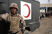
An Iraqi Ground Forces medic next to his military ambulance featuring a red crescent emblem

The International Committee of the Red Cross, a private humanitarian institution based in Switzerland, provided the first official symbol for medical personnel. The first Geneva Convention, originally called for "Amelioration of the Condition of the Wounded and Sick in Armed Forces in the Field", officially adopted the red cross on a field of white as the identifying emblem. This symbol was meant to signify to enemy combatants that the medic qualifies as a non-combatant, at least while providing medical care. Islamic countries use a Red Crescent instead, originating from the Russo-Turkish War, when the Ottoman Empire declared that it would use a red crescent instead of a red cross as its emblem, although it agreed to respect the red cross used by the opposing Russian Empire.

Although these symbols were officially sponsored by the International Federation of Red Cross and Red Crescent Societies, the Magen David Adom, Israel's emergency relief service, use the "Magen David", a red star of David on a white background. To enable the MDA to become a fully recognized and participating member of the International Red Cross and Red Crescent Movement, Protocol III was adopted, authorizing the use of the Red Crystal. For indicative use on foreign territory, any national society can incorporate its unique symbol into the Red Crystal. Under Protocol III, the MDA continues to employ the red Magen David for domestic use, and employs the Red Crystal on international relief missions.

Modern day

A U.S. Army combat medic examining a young child during the War in Afghanistan in 2009. Note that the medic lacks distinguishing features or medic insignia, to prevent targeting by insurgents.

Medical personnel from most Western nations carry weapons for protection of themselves and their patients but remain designated non-combatants, wearing the red cross, crescent or crystal. In the United States Armed Forces, MEDEVAC vehicles display a large Red Cross on a white background. However, ground forces do not display this due to increased targeting of medical personnel by insurgents.

Traditionally, most United States medical personnel also wore a distinguishing red cross, to denote their protection as non-combatants under the Geneva Convention. This practice continued into World War II. However, the enemies faced by professional armies in more recent conflicts are often insurgents who either do not recognize the Geneva Convention or choose not to adhere to it, and thus readily engage all personnel, irrespective of non-combatant status. As their non-combatant status is not respected, many US medics no longer wear non-combatant markings. This can enable medics to be used as medically trained soldiers, fighting aggressively rather than just in self-defence. Combat Medics in the United States Army and United States Navy Hospital Corpsmen are virtually indistinguishable from regular combat troops, except for the extra medical equipment they carry.

The modern-day interpretation of the U.S. Army doctrine requires medics to carry one primary weapon and, if possible, a secondary weapon. It is also common to find American combat medics who are no longer wearing the red or white cross because it is considered unethical to do so when the combat medic is carrying a weapon and could engage in actual combat.

In the U.S. Navy, enlisted medical personnel are known as corpsmen, not medics. The colloquial form of address for a Hospital Corpsman and Army Medics is "Doc". In the Army and U.S. Marine Corps, this term is generally used as a sign of respect. The U.S. Navy deploys FMF Hospital Corpsman attached to U.S. Marine Corps units as part of the Fleet Marine Force. Since the U.S. Marine Corps is part of the Department of the Navy, it relies on Navy corpsmen and other Naval medical personnel for medical care.

U.S. Air Force aerospace medical services technicians have frequently served attached to U.S. Army units in recent conflicts. Though all combat medical personnel are universally referred to as "medic", within different branches of the U.S. military, the skill level, quality of training and scope of work performed by medics varies from branch to branch and unit to unit.

As a result of the 2005 BRAC, the U.S. Department of Defense has moved most medical training for all branches of the armed forces to Fort Sam Houston of Joint Base San Antonio. A new Medical Education and Training Campus was constructed and the Air Force's 937th Training Group and Naval Hospital Corps School were relocated to Fort Sam Houston, joining the Army's existing Army Medical Department Center & School. Although each service has some training particular to its branch, the bulk of the course material and instruction is shared between medical personnel of the different services.
Battlefield medicine

From Wikipedia, the free encyclopedia
 
An illustration of the Wound Man, showing a variety of wounds from the Feldbuch der Wundarznei (Field manual for the treatment of wounds) by Hans von Gersdorff, (1517); illustration by Hans Wechtlin.

Battlefield medicine, also called field surgery and later combat casualty care, is the treatment of wounded combatants and non-combatants in or near an area of combat. Civilian medicine has been greatly advanced by procedures that were first developed to treat the wounds inflicted during combat. With the advent of advanced procedures and medical technology, even polytrauma can be survivable in modern wars. Battlefield medicine is a category of military medicine.

Chronology of battlefield medical advances

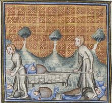
A wounded knight is carried on a medieval stretcher.
  • During Alexander the Great’s military campaigns in the fourth century BC, tourniquets were used to stanch the bleeding of wounded soldiers. Romans used them to control bleeding, especially during amputations. These tourniquets were narrow straps made of bronze, using leather only for comfort.
  • An early stretcher, likely made of wicker over a frame, appears in a manuscript from c.1380. Simple stretchers were common with militaries right through the middle of the 20th century.
  • During the Battle of Shrewsbury in 1403, Prince Henry had an arrow removed from his face using a specially designed surgical instrument.
  • Ambulances or dedicated vehicles for the purpose of carrying injured persons were first used by Spanish soldiers during the Siege of Málaga (1487).
  • French military surgeon Ambroise Paré (1510–90) pioneered modern battlefield wound treatment. His two main contributions to battlefield medicine are the use of dressing to treat wounds and the use of ligature to stop bleeding during amputation.
  • The practice of triage, pioneered by Dominique Jean Larrey during the Napoleonic Wars (1803–1815). He also pioneered the use of ambulances in the midst of combat ('ambulances volantes', or flying ambulances). Prior to this, military ambulances had waited for combat to cease before collecting the wounded by which time many casualties would have succumbed to their injuries.
  • Russian surgeon Nikolay Ivanovich Pirogov was one of the first surgeons to use ether as an anaesthetic in 1847, as well as the very first surgeon to use anaesthesia in a field operation during the Crimean War.
  • American Civil War surgeon Jonathan Letterman (1824–72) originated modern methods of medical organization within armies.
  • The Relief Society for Wounded Soldiers, forerunner of the International Committee of the Red Cross (ICRC) was founded in 1863 in Geneva. The ICRC advocated for the establishment of national aid societies for battlefield medical relief, and stood behind the First Geneva Convention of 1864 which provided neutrality for medics, ambulances, and hospitals.
  • In the late 19th century, the influence of notable medical practitioners like Friedrich von Esmarch and members of the Venerable Order of Saint John pushing for every adult man and woman to be taught the basics of first aid eventually led to institutionalised first-aid courses amongst the military and standard first-aid kits for every soldier.
  • Advances in surgery - especially amputation - during the Napoleonic Wars and First World War on the battlefield of the Somme.
  • Medical advances also provided kinder methods for treatment of battlefield injuries, such as antiseptic ointments, which replaced boiling oil for cauterizing amputations.
  • During the Spanish Civil War there were two major advances. The first one was the invention of a practical method for transporting blood. Developed in Barcelona by Duran i Jordà, the technique mixed the blood of the donors with the same blood type and then, using Grífols glass tubes and a refrigerator truck, transported the blood to the frontline. A few weeks later Norman Bethune developed a similar service. The second advance was the invention of the mobile operating room by the Catalan Moisès Broggi, who worked for the International Brigades.
A US Army soldier, wounded by a Japanese sniper, undergoes surgery during the Bougainville Campaign in World War II.
  • The establishment of fully equipped and mobile field hospitals such as the Mobile Army Surgical Hospital was first practiced by the United States in World War II. It was succeeded in 2006 by the Combat Support Hospital.
  • The use of helicopters as ambulances, or MEDEVACs, was first practiced in Burma in 1944. The first MEDEVAC under fire was done in Manila in 1945 where over 70 troops were extracted in five helicopters, one and two at a time.
  • The extension of emergency medicine to pre-hospital settings through the use of emergency medical technicians.
  • The use of remote physiological monitoring devices on soldiers to show vital signs and biomechanical data to the medic and MEDEVAC crew before and during trauma. This allows medicine and treatment to be administered as soon as possible in the field and during extraction. Similar telemetry units are used in crewed spaceflight, where a flight surgeon at the Command Center can monitor vital signs. This can help to see issues before larger problems occur, such as elevated carbon dioxide levels, or a rise in body temperature indicating a possible infection.

History of Tactical Combat Casualty Care (TCCC)

In 1989, the Commander of the Naval Special Warfare Command (NAVSPECWARCOM) established a research program to conduct studies on medical and physiologic issues. The research concluded that extremity hemorrhage was a leading cause of preventable death in the battlefield. At that time, proper care and treatment was not provided immediately which often resulted in death. This insight prompted a systematic reevaluation of all aspects of battlefield trauma care that was conducted from 1993 to 1996 as a joint effort by special operations medical personnel and the Uniformed Services University of the Health Sciences. Through this 3-year research, the first version of the TCCC guidelines were created to train soldiers to provide effective intervention on the battlefield. The TCCC aims to combine good medicine with good small-unit tactics. One very important aspect that the TCCC outlined was the use of tourniquets, initially there was a belief that the use of tourniquets led to the preventable loss of an extremity due to ischemia but after careful literature search the committee arrived at the conclusion that there was not enough information out there to confirm this claim. The TCCC therefore outline the appropriate usage of tourniquets to provide effective first aid on the battlefield.

After the TCCC article was published in 1996, the program undertook 4 parallel efforts during the next 5-year period. These efforts are as follows:

  1. Presenting TCCC concepts to senior Department of Defense (DoD) line and medical leaders and advocating for their use.
  2. Identifying and developing responses to representative types of TCCC casualty scenarios.
  3. Initiating TCCC’s first strategic partnership with civilian trauma organizations—the Prehospital Trauma Life Support (PHTLS) Committee, the National Association of Emergency Medical Technicians (NAEMT), and the American College of Surgeons Committee on Trauma (ACS-COT).
  4. Expanding TCCC training beyond medical personnel to include SEAL and 75th Ranger Regiment combat leaders and nonmedical unit members.

Current applications of battlefield medicine

Over the past decade combat medicine has improved drastically. Everything has been given a complete overhaul from the training to the gear. In 2011, all enlisted military medical training for the U.S. Navy, Air Force, and Army were located under one command, the Medical Education and Training Campus (METC). After attending a basic medical course there (which is similar to a civilian EMT course), the students go on to advanced training in Tactical Combat Casualty Care.

Tactical combat casualty care (TCCC)

Tactical combat casualty care is becoming the standard of care for the tactical management of combat casualties within the Department of Defense and is the sole standard of care endorsed by both the American College of Surgeons and the National Association of EMT's for casualty management in tactical environments.

Tactical combat casualty care is built around three definitive phases of casualty care:

  1. Care Under Fire: Care rendered at the scene of the injury while both the medic and the casualty are under hostile fire. Available medical equipment is limited to that carried by each operator and the medic. This stage focuses on a quick assessment, and placing a tourniquet on any major bleed.
  2. Tactical Field Care: Rendered once the casualty is no longer under hostile fire. Medical equipment is still limited to that carried into the field by mission personnel. Time prior to evacuation may range from a few minutes to many hours. Care here may include advanced airway treatment, IV therapy, etc. The treatment rendered varies depending on the skill level of the provider as well as the supplies available. This is when a corpsman/medic will make a triage and evacuation decision.
  3. Tactical Evacuation Care (TACEVAC): Rendered while the casualty is evacuated to a higher echelon of care. Any additional personnel and medical equipment pre-staged in these assets will be available during this phase.

Since "90% of combat deaths occur on the battlefield before the casualty ever reaches a medical treatment facility" (Col. Ron Bellamy) TCCC focuses training on major hemorrhaging and airway complications such as a tension-pneumothorax. This has driven the casualty fatality rate down to less than 9%.

Interventions used

Listed below are interventions that a TCCC provider may be expected to perform depending on the phase of TCCC they are at and their level of training. This list is not comprehensive and may be subject to change with future revisions in TCCC guidelines.

Hemorrhage control interventions include the use of extremity tourniquets, junctional tourniquets, trauma dressings, wound packing with compressed gauze and hemostatic dressings, and direct pressure. Newer devices approved for use by the CoTCCC for hemorrhage control include the iTClamp and XStat. Pharmacological options also include tranexamic acid, and hemostatic agents such as zeolite and chitosan.

In managing a casualty’s airway, a TCCC provider may position the casualty in the recovery position or utilize airway adjuncts such as nasopharyngeal airways, oropharyngeal airways, and supraglottic airways. They may also utilize the jaw thrust and head-tilt/ chin-lift maneuver to open a casualty's airway. Advanced TCCC providers may also perform endotracheal intubation and cricothyroidotomy.

Respiratory management largely revolves around the use of chest seals, vented and unvented, and needle decompressions to manage tension pneumothoraxes.

In circulation management a TCCC provider may obtain intravenous/ intraosseous access for the administration of fluids such as normal saline, lactated Ringer’s solution, whole blood, and colloids and plasma substitutes for fluid resuscitation. This also provides a route for the administration of other drugs in accordance with the provider’s scope of practice.

Head injuries would indicate for cervical spine immobilization to the best of the provider’s abilities if deemed appropriate in a given setting, or the use of devices such as a cervical collar.

As trauma-induced hypothermia is a leading cause of battlefield deaths, a provider may also perform hypothermia prevention can be accomplished through the use of a Hypothermia Prevention and Management Kit or emergency blanket, the placement of a casualty on an insulated surface, and the removal of wet clothing from a casualty’s body.

Care under fire

Care under fire is care provided at the point of injury immediately upon wounding while the casualty and care provider remain under effective hostile fire. The casualty should be encouraged to provide self-aid and continue remain engaged in the firefight if possible. If unable to do so, the casualty should be encouraged to move behind cover or "play dead". Due to the high risk of injury to the care-provider and limited resources at this phase, care provided to the casualty should be limited to controlling life-threatening hemorrhage with tourniquets and preventing airway obstruction by placing casualty in the recovery position. The primary focus during care under fire should be winning the firefight to prevent further casualties and further wounding of existing casualties.

Tactical field care

Tactical field care phase begins when the casualty and care-provider are no longer under imminent threat of injury by hostile actions. Though the level of danger is lessened, care-providers should exercise caution and maintain good situational awareness as the tactical situation may be fluid and subject to change. The tactical field care phase enables the provision of more comprehensive care according to care providers' levels of training, tactical considerations, and available resources. Major tasks that are to be completed in the tactical field care phase include the rapid trauma survey, the triage of all casualties, and the transport decision.

Tactical evacuation care

Tactical evacuation care refers to care provided when a casualty is being evacuated and en-route to higher levels of medical care. Care providers at this phase are at even less risk of imminent harm as result of hostile actions. Due to improved access to resources and the tactical situation, more advanced interventions can be provided to casualties such as endotracheal intubation. Patient re-assessments and the addressing of issues that were not or were inadequately addressed previously are also major components of this phase.

In tactical evacuation (TACEVAC), casualties are moved from a hostile environment to a safer and more secure location to receive advanced medical care. Tactical evacuation techniques use a combination of air, ground and water units to conduct the mission depending on the location of the incident and medical centres. Ground vehicle evacuations are more prevalent in urban locations that are in close proximity to medical facilities. Requests for evacuation of casualties and pertinent information are typically communicated through 9-Line MEDEVAC and MIST reports.

Tactical evaluation is an umbrella term that encompasses both medical evacuation (MEDEVAC) and casualty evacuation (CASEVAC). Medical evacuation platforms are typically not engaged in combat except in self-defence and defence of patients. MEDEVAC takes place using special dedicated medical assets marked with a red cross. Casualty evacuation is through non-medical platforms and may include a Quick-Reaction force aided by air support.

For aircraft involved TACEVAC situations there are many considerations that need to be accounted for. Firstly, the flying rules vary widely depending on the aircraft and units in play. The list of determinants to create the TACEVAC strategy include the distances and altitudes involved, time of day, passenger capacity, hostile threat, availability of medical equipment/personnel, and icing conditions. As mentioned TACEVAC is more advanced than TCCC, it also includes training to/for:

  • improve breathing 
  • provide supplemental oxygen 
  • administer Tranexamic acid (TXA) 
  • deal with traumatic brain injuries 
  • fluid resuscitation
  • blood product administration
  • blood transfusion
  • preventing and treating hypothermia

Canadian armed forces

There are three levels of tactical combat casualty care providers in the Canadian Armed Forces.

Combat first aid

Every soldier receives a two-day combat first aid training course. The course focuses on treating hemorrhages, using tourniquets and applying dressings, and basic training for casualty management.

Tactical combat casualty care

A select number of soldiers are chosen to participate in an intense 2-week tactical combat casualty care course where soldiers are provided with additional training. Overall, they are trained to work as medic extenders since they work under the direction of medics.

Tactical medicine

The tactical medicine (TACMED) course is offered exclusively to medics. The tactical medicine program provides training for advanced tactical combat casualty care and is the highest level of care provided by the Canadian Armed Forces in a battlefield setting. Medics are trained to treat and manage patients using the MARCHE protocol. The MARCHE protocol prioritizes potential preventable causes of death in warfare as follows:

  1. Massive hemorrhage control
  2. Airway management
  3. Respiratory management
  4. Circulation
    1. Bleeding control
    2. Intravenous (IV)/ intraosseous (IO) access
    3. Fluid resuscitation
    4. Tourniquet reassessment
  5. Hypothermia prevention
  6. Head injuries
  7. Eye injuries
  8. Everything else
    1. Monitor patient
    2. Pain management
    3. Head-to-toe assessment
    4. Address all wounds found
    5. Antibiotics
    6. Tactical evacuation preparation
    7. Documentation of care and findings

United States

Care under fire

Care under fire happens at the point of injury. According to tactical combat casualty care guidelines, the most effective way to reduce further morbidity and mortality is to return fire at enemy combatants by all personnel. The priority is to continue the combat mission, gain fire superiority, and then treat casualties. The only medical treatment rendered in care under fire is the application of direct pressure on massive bleeding. Tactical combat casualty care recommends a tourniquet as the single most important treatment at the point of injury. It is recommended during care under fire to quickly place tourniquets over clothing, high, and tight; the tourniquet should be reassessed when out of danger in the tactical field care phase.

Tactical field care

Tactical field care is considered to be the backbone of Tactical Combat Casualty Care and consists of care rendered by first responders or prehospital medical personnel while still in the tactical environment. The acronyms MARCH and PAWS help personnel remember crucial treatment steps while under duress.

MARCH

The MARCH acronym is used by personnel to remember the proper order of treatment for casualties.

Massive hemorrhage. The most potentially survivable cause of death is hemorrhage from extremity bleeds, however more than 90% of 4596 combat mortalities post September 11, 2001 died of hemorrhage associated injuries. It is recommended to apply a Committee on Tactical Combat Casualty Care (CoTCCC) approved tourniquet for any life-threatening extremity hemorrhages. Tourniquets during tactical field care should be placed under clothing 2 to 3 inches above the wound, with application time written on the tourniquet.

Airway. Non-patent or closed airway is another survivable cause of death. Airway injuries typically occur due to inhalation burns or maxillofacial trauma. If a person is conscious and speaking they have a patent open airway, while nasopharyngeal airway could benefit those who are unconscious and breathing. However, unconscious casualties who are not breathing could require surgical cricothyroidotomy, as endotracheal intubation is highly difficult in tactical settings.

Respirations. Tension pneumothorax (PTX) develops when air trapped in the chest cavity displaces functional lung tissue and puts pressure on the heart causing cardiac arrest. Thus, open chest wounds must be sealed using a vented chest seal. Tension pneumothorax should be decompressed using a needle chest decompression (NCD) with a 14 gauge, 3.25 inch needle with a catheter. Ventilation and/or oxygenation should be supported as required.

Circulation. It is more important to stem the flow of bleeding than to infuse fluids, and only casualties in shock or those who need intravenous (IV) medications should have IV access. Signs of shock include unconsciousness or altered mental status, and/or abnormal radial pulse. IV should be applied using an 18 gauge catheter and saline lock in tactical field care, secured by transparent would-dressing film. Tranexamic acid (TXA) should be given as soon as possible to casualties in or at risk of hemorrhagic shock. An intraosseous (IO) device could also be used for administering fluids if IV access is not feasible.

Head injury/hypothermia. Secondary brain injury is worsened by hypotension (systolic blood pressure under 90 mmHg), hypoxia (peripheral capillary oxygen saturation under 90%), and hypothermia (whole body temperature below 95 Fahrenheit or 35 Celsius). Medical personnel can use the Military Acute Concussion Evaluation (MACE), while non-medical personnel can use the alert, verbal, pain, unresponsive (AVPU) scale to identify traumatic brain injury. The "lethal triad" is a combination of hypothermia, acidosis, and coagulopathy in trauma patients. Since hypothermia can occur regardless of ambient temperature due to blood loss, the Hypothermia Prevention and Management Kit (HPMK) is recommended for all casualties.

PAWS

The PAWS acronym is used by personnel to remember additional casualty care items that should be addressed.

Pain. Proper management of pain reduces stress on a casualty's mind and body, and have reduced incidents of post-traumatic stress disorder (PTSD). Pain management is shown to reduce harmful patient movement, improves compliance and cooperation, and allows for easier transport as well as improved health outcomes.

Antibiotics. All battlefield wounds are considered contaminated, and thus any penetrating injury should receive antibiotics at the point of injury as well as in tactical field care. The recommended parenteral antibiotics are 1g ertapenem or 2g cefotetan, which can treat multi drug-resistant bacteria. If the casualty can tolerate oral fluids, 400mg moxifloxacin can be administered orally instead of ertapenem or cefotetan.

Wounds. Assessing the casualty for additional wounds improves morbidity and mortality. First responders must address burns, open fractures, facial trauma, amputation dressings, and security of tourniquets. Prior to movement, reassessment of wounds and interventions is very important. Casualties with penetrating trauma to the chest or abdomen should receive priority evacuation due to the possibility of internal hemorrhage.

Splinting. Explosions (such as from improvised explosive device or land mines) that cause lower extremity traumatic amputation cause forces to move upward through the body, which may cause further bone disruption, hollow organ collapse, or internal bleeding. Thus, first responders should use the Combat Ready Clamp (CRoC), the Junctional Emergency Treatment Tool (JETT), or the SAM Junctional Tourniquet to control junctional hemorrhage and stabilize the pelvis. In cases of penetrative eye trauma, responders should first perform a rapid field test of visual acuity, then tape a rigid shield over the eye to prevent further damage, and also give 400mg oral moxifloxacin as soon as possible. Pressure must never by applied to an eye suspected of penetrative injury.

Evaluating effectiveness

In order to evaluate the effectiveness of Tactical Combat Casualty Care, a study was conducted which analyzed US military casualties who died from an injury that occurred while they were deployed to Afghanistan or Iraq from October 2001 to June 2011. Of the 4,596 casualties, 87% died in the pre-medical treatment facility, prior to receiving surgical care. Of the casualties in the pre-medical treatment facility, 75.7% of the prehospital deaths were non-survivable, while 24.3% of deaths were potentially survivable. Instantaneous non-survivable mortalities included physical dismemberment, catastrophic brain injury, and destructive cardiovascular injury. Non-instantaneous non-survivable mortalities included severe traumatic brain injury, thoracic vascular injury, high spinal cord injury, and destructive abdominal pelvic injury. These injuries are very difficult to treat given currently fielded medical therapies such as Tactical Combat Casualty Care.

In terms of potentially survivable mortalities, 8.0% of mortalities were associated with airway obstruction. Majority of mortalities (90.9%) which were classified as potentially survivable mortalities were attributed to hemorrhage, with 67.3% of the hemorrhage being truncal, 19.2% junctional, and 13.5% extremity. During the study period, there were no effective protocols put in place to control junctional or truncal sources of hemorrhage in the battlefield, which suggests a gap in medical treatment capability.

This study shows the majority of battlefield casualties which occur prior to receiving surgical care are non-survivable. However, of the casualties which are survivable, the majority of deaths can be attributed to hemorrhages. Developing protocol which can control and temporize hemorrhage in the battlefield would improve the effectiveness of Tactical Combat Casualty Care, and decreases the number of casualties in the battlefield.

Another study analyzed the effectiveness of tourniquets for hemorrhage control, which are used in Tactical Combat Casualty Care. A four-year retrospective analysis showed that out of 91 soldiers who were treated with tourniquets, 78% of tourniquets were applied effectively. The success rate for tourniquets applied to upper limbs was 94% while the success rate for tourniquets applied to lower limbs was 71%. The difference between the success rates can be attributed to the tourniquets themselves, as in another study, tourniquets applied on healthy volunteers resulted in a much lower success rate for lower limbs in comparison to upper limbs. Therefore, the tourniquets themselves can be redesigned to increase its effectiveness and improve Tactical Combat Casualty Care.

A prospective study of all trauma patients treated at the Canadian-led Role 3 multinational medical unit (Role 3 MMU) established at Kandahar Airfield Base between February 7, 2006, to May 20, 2006, was conducted to examine how Tactical Combat Casualty Care interventions are delivered. The study concluded that tourniquets are effective, but must be used appropriately. The distinction between venous and arterial tourniquets must be reinforced in Tactical Combat Casualty Care training. Tactical Combat Casualty Care courses must also train soldiers to remove tourniquets for the purposes of reassessing trauma after the patient and caregiver is no longer under enemy fire. This is because the risks of iatrogenic ischemic injury of prolonged use of tourniquets outweigh the risks of increased blood loss.

The study also identified technical errors in performing needle decompressions. All needle decompressions were performed at least 2 cm medial to the mid-clavicular line and well within the cardiac box. This may result in injury to the heart and surrounding vasculature. Tactical Combat Casualty Care training must reinforce using landmarks when performing needle decompressions. This is especially useful since soldiers may have to perform this procedure in poor lighting conditions.

Dopamine agonist

From Wikipedia, the free encyclopedia
 
Dopamine agonist
Drug class
Skeletal structure diagram of dopamine
The skeletal structure of dopamine
Class identifiers
UseParkinson's disease, hyperprolactinemia, restless legs syndrome
ATC codeN04BC
Biological targetDopamine receptors
External links
MeSHD010300

A dopamine agonist (DA) is a compound that activates dopamine receptors. There are two families of dopamine receptors, D2-like and D1-like, and they are all G protein-coupled receptors. D1- and D5-receptors belong to the D1-like family and the D2-like family includes D2, D3 and D4 receptors. Dopamine agonists are primarily used in the treatment of Parkinson's disease, and to a lesser extent, in hyperprolactinemia and restless legs syndrome. They are also used off-label in the treatment of clinical depression. The use of dopamine agonists is associated with impulse control disorders and dopamine agonist withdrawal syndrome (DAWS).

Medical uses

Parkinson's disease

Dopamine agonists are mainly used in the treatment of Parkinson's disease.  The cause of Parkinson's is not fully known but genetic factors, for example specific genetic mutations, and environmental triggers have been linked to the disease. In Parkinson's disease dopaminergic neurons that produce the neurotransmitter dopamine in the brain slowly break down and can eventually die. With decreasing levels of dopamine the brain can't function properly and causes abnormal brain activity, which ultimately leads to the symptoms of Parkinson's disease.

There are two fundamental ways of treating Parkinson's disease, either by replacing dopamine or mimicking its effect.

Dopamine agonists act directly on the dopamine receptors and mimic dopamine's effect. Dopamine agonists have two subclasses: ergoline and non-ergoline agonists. Both subclasses target dopamine D2-type receptors. Types of ergoline agonists are cabergoline and bromocriptine and examples of non-ergoline agonists are pramipexole, ropinirole and rotigotine. Ergoline agonists are much less used nowadays because of the risk of cartilage formation in heart valves.

Treatment of depression in Parkinson's patients

Depressive symptoms and disorders are common in patients with Parkinson's disease and can affect their quality of life. Increased anxiety can accentuate the symptoms of Parkinson's and is therefore essential to treat. Instead of conventional antidepressant medication in treating depression, treatment with dopamine agonists has been suggested. It is mainly thought that dopamine agonists help with treating depressive symptoms and disorders by alleviating motor complications, which is one of the main symptoms of Parkinson's disease.  Although preliminary evidence of clinical trials has shown interesting results, further research is crucial to establish the anti-depressive effects of dopamine agonists in treating depressive symptoms and disorders in those with Parkinson's.

Hyperprolactinemia

Dopamine is a prolactin-inhibiting factor (PIFs) since it lowers the prolactin-releasing factors (PRFs) synthesis and secretion through D2-like receptors. That is why dopamine agonists are the first-line treatment in hyperprolactinemia. Ergoline-derived agents, bromocriptine and cabergoline are mostly used in treatment. Research shows that these agents reduce the size of prolactinomas by suppressing the hypersecretion of prolactin resulting in normal gonadal function.

Restless leg syndrome

Numerous clinical trials have been performed to assess the use of dopamine agonists for the treatment of restless leg syndrome (RLS). RLS is identified by the strong urge to move and is a dopamine-dependent disorder. RLS symptoms decrease with the use of drugs that stimulate dopamine receptors and increase dopamine levels, such as dopamine agonists.

Adverse effects

Side effects

Dopamine agonists are mainly used to treat Parkinson’s disease but are also used to treat hyperprolactinemia and restless legs syndrome. The side effects are mainly recorded in treatment for Parkinson’s disease where dopamine agonists are commonly used, especially as first-line treatment with levodopa.

Dopamine agonists are divided into two subgroups or drug classes, first-generation and newer agents. Ergoline derived agonists are the first generation and are not used as much as the newer generation the non-ergoline derived agonists. Ergoline derived agonists are said to be "dirtier" drugs because of their interaction with other receptors than dopamine receptors, therefore they cause more side effects. Ergoline derived agonists are for example bromocriptine, cabergoline, pergolide and lisuride. Non-ergoline agonists are pramipexole, ropinirole, rotigotine, piribedil and apomorphine.

The most common adverse effects are constipation, nausea and headaches. Other serious side effects are hallucinations, peripheral edema, gastrointestinal ulcers, pulmonary fibrosis and psychosis.

Dopamine agonists have been linked to cardiac problems. Side effects such as hypotension, myocardial infarction, congestive heart failure, cardiac fibrosis, pericardial effusion and tachycardia. A high risk for valvular heart disease has been established in association with ergot-derived agonists especially in elderly patients with hypertension.

Somnolence and sleep attacks have been reported as an adverse effect that happen to almost 30% of patients using dopamine agonists. Daytime sleepiness, insomnia and other sleep disturbances have been reported as well.

Impulse control disorder that is described as gambling, hypersexuality, compulsive shopping and binge eating is one serious adverse effect of dopamine agonists.

After long-term use of dopamine agonist a withdrawal syndrome may occur when discontinuing or during dose reduction. The following side effects are possible: anxiety, panic attacks, dysphoria, depression, agitation, irritability, suicidal ideation, fatigue, orthostatic hypotension, nausea, vomiting, diaphoresis, generalised pain, and drug cravings. For some individuals, these withdrawal symptoms are short-lived and make a full recovery, for others a protracted withdrawal syndrome may occur with withdrawal symptoms persisting for months or years.

Interactions

Dopamine agonists interact with a number of drugs but there is little evidence that they interact with other Parkinson’s drugs. In most cases there is no reason not to co-administer Parkinson's drugs. Although there has been an indication that the use of dopamine agonists with L-DOPA can cause psychosis therefore it is recommended that either the use of dopamine agonists be discontinued or the dose of L-DOPA reduced. Since ergot-dopamine agonist have antihypertensive qualities it is wise to monitor blood pressure when using dopamine agonists with antihypertensive drugs to ensure that the patient does not get hypotension. That includes the drug sildenafil which is commonly used to treat erectile dysfunction but also used for pulmonary hypertension.

There is evidence that suggests that since ergot dopamine agonists are metabolized by CYP3A4 enzyme concentration rises with the use of CYP3A4 inhibitors. For example, in one study bromocriptine was given with a CYP3A4 inhibitor and the AUC (e. Area under the curve) increased 268%. Ropinirole is a non-ergot derived dopamine agonist and concomitant use with a CYP1A2 inhibitor can result in a higher concentration of ropinirole. When discontinuing the CYP1A2 inhibitor, if using both drugs, there is a change that a dose adjustment for ropinirole is needed. There is also evidence the dopamine agonists inhibit various CYP enzymes and therefore they may inhibit the metabolism of certain drugs.

Pharmacology

Ergoline class

Pharmacokinetics of Bromocriptine

The absorption of the oral dose is approximately 28% however, only 6% reaches the systemic circulation unchanged, due to a substantial first-pass effect. Bromocriptine reaches mean peak plasma levels in about 1–1.5 hours after a single oral dose. The drug has high protein binding, ranging from 90-96% bound to serum albumin. Bromocriptine is metabolized by CYP3A4 and excreted primarily in the feces via biliary secretion. Metabolites and parent drugs are mostly excreted via the liver, but also 6% via the kidney. It has a half-life of 2–8 hours.

Pharmacokinetics of Pergolide

Pergolide has a long half-life of about 27 hours and reaches a mean peak plasma level in about 2–3 hours after a single oral dose. The protein binding is 90% and the drug is mainly metabolized in the liver by CYP3A4 and CYP2D6. The major route of excretion is through the kidneys.

Drug

Maintenance

Half-life

Protein binding Peak plasma Metabolism Excretion
Bromocriptine

Oral, 2.5–40 mg/day

2–8 hours 90-96% 1-1,5 hours

Hepatic, via CYP3A4, 93% first-pass metabolism

Bile, 94-98%

Renal, 2-6%

Pergolide

Oral, 0.05 mg/day Usual response up to 0.1 mg per day

27 hours 90% 2–3 hours Extensively hepatic Renal, 50%

Fecal 50%

Non-Ergoline class

Pharmacokinetics of Pramipexole

Pramipexole reaches maximum plasma concentration 1–3 hours post-dose. It is about 15% bound to plasma proteins and the metabolism is minimal. Pramipexole has a long half-life, around 27 hours. The drug is mostly excreted in the urine, around 90%, but also in feces.

Pharmacokinetics of Ropinirole

Ropinirole is rapidly absorbed after a single oral dose, reaching plasma concentration in approximately 1–2 hours. The half-life is around 5–6 hours. Ropinirole is heavily metabolized by the liver and in vitro studies show that the enzyme involved in the metabolism of ropinirole is CYP1A2.

Pharmacokinetics of Rotigotine

Since rotigotine is a transdermal patch it provides continuous drug delivery over 24 hours. It has a half-life of 3 hours and the protein binding is around 92% in vitro and 89.5% in vivo. Rotigotine is extensively and rapidly metabolized in the liver and by the CYP enzymes. The drug is mostly excreted in urine (71%), but also in feces (23%).

Drug

Maintenance

Half-life

Protein binding Peak plasma Metabolism Excretion
Pramipexole

Oral, 0.125 mg 3x/day (IR) Oral, 0.375 mg/day (ER)

8–12 hours 15% 1–3 hours Minimal < 10% Urine 90%

Fecal 2%

Ropinirole

Oral, 0.25 mg 3x/day (IR) Oral, 2 mg/day (ER)

5–6 hours 10-40% 1–2 hours Hepatic, via P450 CYP1A2 — can increase ↑ INR Renal > 88%
Rotigotine

Transdermal, 2 – 4 mg/day

3 hours

92%

24 hours Hepatic (CYP-mediated). Urine 71%

Fecal 23%

Mechanism of action

The dopamine receptors are 7-transmembrane domains and are members of the G protein-coupled receptors (GPCR) superfamily. Dopamine receptors have five subtypes, D1 through D5, the subtypes can be divided into two subclasses due to their mechanism of action on adenylate cyclase enzyme, D1-like receptors (D1 and D5) and D2-like receptors (D2, D3 and D4). D1-like receptors are primarily coupled to Gαs/olf proteins and activates adenylate cyclase which increases intracellular levels of cAMP, they also activate the Gβγ complex and the N-type Ca2+ channel. D2-like receptors decrease intracellular levels of the second messenger cAMP by inhibiting adenylate cyclase.

Bromocriptine

Bromocriptine is an ergot derivative, semi-synthetic. Bromocriptine is a D2 receptor agonist and D1 receptor antagonist with a binding affinity to D2 receptors of anterior pituitary cells, exclusively on lactotrophs. Bromocriptine stimulates Na+, K+-ATPase activity and/or cytosolic Ca2+ elevation and therefore reduction of prolactin which leads to no production of cAMP.

Pramipexole

Pramipexole is a highly active non-ergot D2-like receptor agonist with a higher binding affinity to D3 receptors rather than D2 or D4 receptors. The mechanism of action of pramipexole is mostly unknown, it is thought to be involved in the activation of dopamine receptors in the area of the brain where the striatum and the substantia nigra is located. This stimulation of dopamine receptors in the striatum may lead to the better movement performance.

Structure–activity relationship

When dealing with agonists it can be extremely complex to confirm relationships between structure and biological activity. Agonists generate responses from living tissues. Therefore, their activity depends both on their efficacy to activate receptors and their affinity to bind to receptors.

Crossing the blood brain barrier

Many molecules are unable to cross the blood brain barrier (BBB). Molecules must be small, non-polar and lipophilic to cross over. If compounds do not possess these qualities they must have a specific transporter that can transport them over the BBB. Dopamine cannot diffuse across the BBB because of the catechol group, it is too polar and therefore unable to enter the brain. The catechol group is a dihydroxy benzene ring.

The synthesis of dopamine consists of three stages. The synthesis process starts with an amino acid, called L-Tyrosine. In the second stage Levodopa (L-dopa) is formed by adding a phenol group to the benzene ring of L-Tyrosine. The formation of L-dopa from L-tyrosine is catalyzed by the enzyme tyrosine hydroxylase. The third stage is the formation of dopamine by removing the carboxylic acid group from L-dopa, catalysed by the enzyme dopa decarboxylase.

Levodopa is also too polar to cross the blood brain barrier but it happens to be an amino acid so it has a specialized transporter called L-type amino acid transporter or LAT-1 that helps it diffuse through the barrier.

Dopamine

When dopamine interacts with ATP, which is a component of some dopamine receptors, it has a significant preference for a trans-conformation of the dopamine molecule. The dopamine-ATP complex is stabilised by hydrogen bonding between catechol hydroxyls and purine nitrogens and by electrostatic interactions between the protonated ammonium group of dopamine and a negative phosphate group. Two conformers of dopamine have been identified as alpha- and beta-conformers in which the catechol ring is coplanar with the plane of the ethylamine side chain. They are substantial in agonist-receptor interactions.

Ergoline derivatives

Central dopaminergic agonist properties of semisynthetic ergoline derivatives lergotrile, pergolide, bromocriptine and lisuride have been established. Some studies suggest that ergot alkaloids have the properties of mixed agonist-antagonist with regards to certain presynaptic and postsynaptic receptors. N-n-Propyl groups (chemical formula: –CH2CH2CH3) frequently enhance dopamine agonist effects in the ergoline derivatives.

The (+)-enantiomer displays notably diminished activity whereas the (-)-enantiomer possess potent dopamine agonist properties.

Bromocriptine

Bromocriptine has an ergot alkaloid structure. Ergot alkaloids are divided into 2 groups; amino acid ergot alkaloids and amine ergot alkaloids, bromocriptine is part of the former group. It contains a bromine halogen on the ergot structure which increases the affinity for the D2-receptor but often reduces the efficacy. The similarity between the dopamine structure and the ergoline ring in bromocriptine is likely the cause for its action on the dopamine receptors. It has shown to have equal affinity for D2- and D3-receptor and much lower affinity for D1-receptor.

Non-ergoline derivatives

Non-ergoline dopamine receptor agonists have higher binding affinity to dopamine D3-receptors than dopamine D2-receptors. This binding affinity is related to D2 and D3 receptor homology, the homology between them has a high degree of sequence and is closest in their transmembrane domains, were they share around 75% of the amino acid.

Apomorphine

Apomorphine has a catechol element and belongs to a class called β-phenylethylamines and its main components are similar to the dopamine structure. The effect that apomorphine has on the dopamine receptors can also be linked to the similarities between its structure and dopamine. It is a chiral molecule and thus can be acquired in both the R and S form, the R form is the one that is used in therapy. When apomorphine interacts with the dopamine receptor, or the ATP on the receptor, the catechol and nitrogen are important to stabilize the structure with hydrogen bonding. The position of the hydroxyl groups is also important and monohydroxy derivatives have been found to be less potent than the dihydroxy groups. There are a number of stability concerns with apomorphine such as oxidation and racemization.

Rotigotine

Rotigotine is a phenolic amine and thus has poor oral bioavailability and fast clearance from the body. Therefore, it has been formulated as a transdermal patch, first and foremost to prevent first pass metabolism in the liver.

Members

Examples of dopamine agonists include:

Partial agonist

Agonists of full/unknown efficacy

Some, such as fenoldopam, are selective for dopamine receptor D1.

Related class of drugs: Indirect agonists

There are two classes of drugs that act as indirect agonists of dopamine receptors: dopamine reuptake inhibitors and dopamine releasing agents. These are not considered dopamine agonists, since they have no specific agonist activity at dopamine receptors, but they are nonetheless related. Indirect agonists are prescribed for a wider range of conditions than standard dopamine agonists.

The most commonly prescribed indirect agonists of dopamine receptors include:

Other examples include:

History

Since the late 1960 Levodopa (L-DOPA) has been used to treat Parkinson’s disease but there has always been a debate whether the treatment is worth the side effects. Around 1970 clinicians started using the dopamine agonist apomorphine alongside L-DOPA to minimize the side effects caused by L-DOPA, the dopamine agonists bind to the dopamine receptor in the absence of dopamine. Apomorphine had limited use since it had considerable side effects and difficulty with administration. In 1974 bromocriptine was use widely after clinicians discovered its benefits in treating Parkinsons. When using the two drug classes together there is a possibility to reduce the amount of L-DOPA by 20-30% and thus keeping the fluctuating motor responses to a minimum. Dopamine agonists are often used in younger people as monotherapy and as initial therapy instead of L-DOPA. Although it is important to know that there is a correlation between the two drugs, if l-DOPA doesn't work dopamine agonists are also ineffective.

The early dopamine agonists, such as bromocriptine, were ergot derived and activated the D2-receptor. They induced major side effects such as fibrosis of cardiac valves. It is considered that the reason they induced such side effects is that they activate many types of receptors.

Because of the major adverse effects of ergot derived dopamine agonists they are generally not used anymore and were mostly abandoned in favor of non-ergot agonists such as pramipexole, ropinirole and rotigotine. They do not induce as serious side effects although common side effects are nausea, edema and hypotension. Patients have also shown impaired impulse control such as overspending, hypersexuality and gambling.

Memory and trauma

From Wikipedia, the free encyclopedia https://en.wikipedia.org/wiki/Memory_and_trauma ...