Search This Blog

Tuesday, June 20, 2023

Syncope (medicine)

From Wikipedia, the free encyclopedia

Syncope
Other namesFainting, blacking out, passing out, swooning
Pietro Longhi 027.jpg
A 1744 oil painting by Pietro Longhi called Fainting
Pronunciation
SpecialtyNeurology, cardiology
SymptomsLoss of consciousness and muscle strength
ComplicationsInjury
Usual onsetFast onset
DurationShort duration
TypesCardiac, reflex, orthostatic hypotension
CausesDecrease in blood flow to brain
Diagnostic methodMedical history, physical examination, electrocardiogram
TreatmentBased on underlying cause
PrognosisDepends on underlying cause
Frequency~5 per 1,000 per year

Syncope, commonly known as fainting, or passing out, is a loss of consciousness and muscle strength characterized by a fast onset, short duration, and spontaneous recovery. It is caused by a decrease in blood flow to the brain, typically from low blood pressure. There are sometimes symptoms before the loss of consciousness such as lightheadedness, sweating, pale skin, blurred vision, nausea, vomiting, or feeling warm. Syncope may also be associated with a short episode of muscle twitching. Psychiatric causes can also be determined when a patient experiences fear, anxiety, or panic; particularly before a stressful event, usually medical in nature. When consciousness and muscle strength are not completely lost, it is called presyncope. It is recommended that presyncope be treated the same as syncope.

Causes range from non-serious to potentially fatal. There are three broad categories of causes: heart or blood vessel related; reflex, also known as neurally mediated; and orthostatic hypotension. Issues with the heart and blood vessels are the cause in about 10% and typically the most serious while neurally mediated is the most common. Heart related causes may include an abnormal heart rhythm, problems with the heart valves or heart muscle and blockages of blood vessels from a pulmonary embolism or aortic dissection among others. Neurally mediated syncope occurs when blood vessels expand and heart rate decreases inappropriately. This may occur from either a triggering event such as exposure to blood, pain, strong feelings or a specific activity such as urination, vomiting, or coughing. Neurally mediated syncope may also occur when an area in the neck known as the carotid sinus is pressed. The third type of syncope is due to a drop in blood pressure when changing position such as when standing up. This is often due to medications that a person is taking but may also be related to dehydration, significant bleeding or infection. There also seems to be a genetic component to syncope.

A medical history, physical examination, and electrocardiogram (ECG) are the most effective ways to determine the underlying cause. The ECG is useful to detect an abnormal heart rhythm, poor blood flow to the heart muscle and other electrical issues, such as long QT syndrome and Brugada syndrome. Heart related causes also often have little history of a prodrome. Low blood pressure and a fast heart rate after the event may indicate blood loss or dehydration, while low blood oxygen levels may be seen following the event in those with pulmonary embolism. More specific tests such as implantable loop recorders, tilt table testing or carotid sinus massage may be useful in uncertain cases. Computed tomography (CT) is generally not required unless specific concerns are present. Other causes of similar symptoms that should be considered include seizure, stroke, concussion, low blood oxygen, low blood sugar, drug intoxication and some psychiatric disorders among others. Treatment depends on the underlying cause. Those who are considered at high risk following investigation may be admitted to hospital for further monitoring of the heart.

Syncope affects about three to six out of every thousand people each year. It is more common in older people and females. It is the reason for one to three percent of visits to emergency departments and admissions to hospital. Up to half of women over the age of 80 and a third of medical students describe at least one event at some point in their lives. Of those presenting with syncope to an emergency department, about 4% died in the next 30 days. The risk of a poor outcome, however, depends very much on the underlying cause.

Causes

Causes range from non-serious to potentially fatal. There are three broad categories of causes: heart or blood vessel related; reflex, also known as neurally mediated; and orthostatic hypotension. Issues with the heart and blood vessels are the cause in about 10% and typically the most serious while neurally mediated is the most common. There also seems to be a genetic component to syncope. A recent genetic study has identified first risk locus for syncope and collapse. The lead genetic variant, residing at chromosome 2q31.1, is an intergenic variant approximately 250 kb downstream of the ZNF804A gene. The variant effected the expression of ZNF804A, making this gene the strongest driver of the association.

Neurally mediated syncope

Reflex syncope or neurally mediated syncope occurs when blood vessels expand and heart rate decreases inappropriately leading to poor blood flow to the brain. This may occur from either a triggering event such as exposure to blood, pain, strong feelings, or a specific activity such as urination, vomiting, or coughing. Vasovagal syncope

Vasovagal (situational) syncope is one of the most common types which may occur in response to any of a variety of triggers, such as scary, embarrassing or uneasy situations, during blood drawing, or moments of sudden unusually high stress. There are many different syncope syndromes which all fall under the umbrella of vasovagal syncope related by the same central mechanism. First, the person is usually predisposed to decreased blood pressure by various environmental factors. A lower than expected blood volume, for instance, from taking a low-salt diet in the absence of any salt-retaining tendency. Or heat causing vaso-dilation and worsening the effect of the relatively insufficient blood volume. The next stage is the adrenergic response. If there is underlying fear or anxiety (e.g., social circumstances), or acute fear (e.g., acute threat, needle phobia), the vaso-motor centre demands an increased pumping action by the heart (flight or fight response). This is set in motion via the adrenergic (sympathetic) outflow from the brain, but the heart is unable to meet requirements because of the low blood volume, or decreased return. A feedback response to the medulla is triggered via the afferent vagus nerve. The high (ineffective) sympathetic activity is thereby modulated by vagal (parasympathetic) outflow leading to excessive slowing of heart rate. The abnormality lies in this excessive vagal response causing loss of blood flow to the brain. The tilt-table test typically evokes the attack. Avoiding what brings on the syncope and possibly greater salt intake is often all that is needed.

Associated symptoms may be felt in the minutes leading up to a vasovagal episode and are referred to as the prodrome. These consist of light-headedness, confusion, pallor, nausea, salivation, sweating, tachycardia, blurred vision, and sudden urge to defecate among other symptoms.

Vasovagal syncope can be considered in two forms:

  • Isolated episodes of loss of consciousness, unheralded by any warning symptoms for more than a few moments. These tend to occur in the adolescent age group and may be associated with fasting, exercise, abdominal straining, or circumstances promoting vaso-dilation (e.g., heat, alcohol). The subject is invariably upright. The tilt-table test, if performed, is generally negative.
  • Recurrent syncope with complex associated symptoms. This is neurally mediated syncope (NMS). It is associated with any of the following: preceding or succeeding sleepiness, preceding visual disturbance ("spots before the eyes"), sweating, lightheadedness. The subject is usually but not always upright. The tilt-table test, if performed, is generally positive. It is relatively uncommon.

Syncope has been linked with psychological triggers. This includes fainting in response to the sight or thought of blood, needles, pain, and other emotionally stressful situations. One theory in evolutionary psychology is that fainting at the sight of blood might have evolved as a form of playing dead which increased survival from attackers and might have slowed blood loss in a primitive environment. "Blood-injury phobia", as this is called, is experienced by about 15% of people. It is often possible to manage these symptoms with specific behavioral techniques.

Another evolutionary psychology view is that some forms of fainting are non-verbal signals that developed in response to increased inter-group aggression during the paleolithic. A non-combatant who has fainted signals that she or he is not a threat. This would explain the association between fainting and stimuli such as bloodletting and injuries seen in blood-injection-injury type phobias such as needle phobia as well as the gender differences.

Much of this pathway was discovered in animal experiments by Bezold (Vienna) in the 1860s. In animals, it may represent a defence mechanism when confronted by danger ("playing possum").

Situational syncope

Syncope may be caused by specific behaviors including coughing, urination, defecation, vomiting, swallowing (deglutition), and following exercise. Manisty et al. note: "Deglutition syncope is characterised by loss of consciousness on swallowing; it has been associated not only with ingestion of solid food, but also with carbonated and ice-cold beverages, and even belching." Fainting can occur in "cough syncope" following severe fits of coughing, such as that associated with pertussis or "whooping cough". Neurally mediated syncope may also occur when an area in the neck known as the carotid sinus is pressed. A normal response to carotid sinus massage is reduction in blood pressure and slowing of the heart rate. Especially in people with hypersensitive carotid sinus syndrome this response can cause syncope or presyncope.

Cardiac

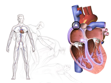
Heart-related causes may include an abnormal heart rhythm, problems with the heart valves or heart muscle, or blockages of blood vessels from a pulmonary embolism or aortic dissection, among others.

Syncope from bradycardia

Cardiac arrhythmias

The most common cause of cardiac syncope is cardiac arrhythmia (abnormal heart rhythm) wherein the heart beats too slowly, too rapidly, or too irregularly to pump enough blood to the brain. Some arrhythmias can be life-threatening.

Two major groups of arrhythmias are bradycardia and tachycardia. Bradycardia can be caused by heart blocks. Tachycardias include SVT (supraventricular tachycardia) and VT (ventricular tachycardia). SVT does not cause syncope except in Wolff-Parkinson-White syndrome. Ventricular tachycardia originate in the ventricles. VT causes syncope and can result in sudden death. Ventricular tachycardia, which describes a heart rate of over 100 beats per minute with at least three irregular heartbeats as a sequence of consecutive premature beats, can degenerate into ventricular fibrillation, which is rapidly fatal without cardiopulmonary resuscitation (CPR) and defibrillation.

Long QT syndrome can cause syncope when it sets off ventricular tachycardia or torsades de pointes. The degree of QT prolongation determines the risk of syncope. Brugada syndrome also commonly presents with syncope secondary to arrhythmia.

Typically, tachycardic-generated syncope is caused by a cessation of beats following a tachycardic episode. This condition, called tachycardia-bradycardia syndrome, is usually caused by sinoatrial node dysfunction or block or atrioventricular block.

Obstructive cardiac lesion

Blockages in major vessels or within the heart can also impede blood flow to the brain. Aortic stenosis and mitral stenosis are the most common examples. Major valves of the heart become stiffened and reduce the efficiency of the hearts pumping action. This may not cause symptoms at rest but with exertion, the heart is unable to keep up with increased demands leading to syncope. Aortic stenosis presents with repeated episodes of syncope. Rarely, cardiac tumors such as atrial myxomas can also lead to syncope.

Structural cardiopulmonary disease

Diseases involving the shape and strength of the heart can be a cause of reduced blood flow to the brain, which increases risk for syncope. The most common cause in this category is fainting associated with an acute myocardial infarction or ischemic event. The faint in this case is primarily caused by an abnormal nervous system reaction similar to the reflex faints. Women are significantly more likely to experience syncope as a presenting symptom of a myocardial infarction. In general, faints caused by structural disease of the heart or blood vessels are particularly important to recognize, as they are warning of potentially life-threatening conditions.

Among other conditions prone to trigger syncope (by either hemodynamic compromise or by a neural reflex mechanism, or both), some of the most important are hypertrophic cardiomyopathy, acute aortic dissection, pericardial tamponade, pulmonary embolism, aortic stenosis, and pulmonary hypertension.

Other cardiac causes

Sick sinus syndrome, a sinus node dysfunction, causing alternating bradycardia and tachycardia. Often there is a long pause (asystole) between heartbeats.

Adams-Stokes syndrome is a cardiac syncope that occurs with seizures caused by complete or incomplete heart block. Symptoms include deep and fast respiration, weak and slow pulse, and respiratory pauses that may last for 60 seconds.

Subclavian steal syndrome arises from retrograde (reversed) flow of blood in the vertebral artery or the internal thoracic artery, due to a proximal stenosis (narrowing) and/or occlusion of the subclavian artery. Symptoms such as syncope, lightheadedness, and paresthesias occur while exercising the arm on the affected side (most commonly the left).

Aortic dissection (a tear in the aorta) and cardiomyopathy can also result in syncope.

Various medications, such as beta blockers, may cause bradycardia induced syncope.

A pulmonary embolism can cause obstructed blood vessels and is the cause of syncope in less than 1% of people who present to the emergency department.

Blood pressure

Orthostatic (postural) hypotensive syncope is caused primarily by an excessive drop in blood pressure when standing up from a previous position of lying or sitting down. When the head is elevated above the feet the pull of gravity causes blood pressure in the head to drop. This is sensed by stretch receptors in the walls of vessels in the carotid sinus and aortic arch. These receptors then trigger a sympathetic nervous response to compensate and redistribute blood back into the brain. The sympathetic response causes peripheral vasoconstriction and increased heart rate. These together act to raise blood pressure back to baseline. Apparently healthy individuals may experience minor symptoms ("lightheadedness", "greying-out") as they stand up if blood pressure is slow to respond to the stress of upright posture. If the blood pressure is not adequately maintained during standing, faints may develop. However, the resulting "transient orthostatic hypotension" does not necessarily signal any serious underlying disease. It is as common or perhaps even more common than vasovagal syncope.

This may be due to medications, dehydration, significant bleeding or infection. The most susceptible individuals are elderly frail individuals, or persons who are dehydrated from hot environments or inadequate fluid intake. For example, medical students would be at risk for orthostatic hypotensive syncope while observing long surgeries in the operating room. There is also evidence that exercise training can help reduce orthostatic intolerance. More serious orthostatic hypotension is often the result of certain commonly prescribed medications such as diuretics, β-adrenergic blockers, other anti-hypertensives (including vasodilators), and nitroglycerin. In a small percentage of cases, the cause of orthostatic hypotensive faints is structural damage to the autonomic nervous system due to systemic diseases (e.g., amyloidosis or diabetes) or in neurological diseases (e.g., Parkinson's disease).

Hyperadrenergic orthostatic hypotension refers to an orthostatic drop in blood pressure despite high levels of sympathetic adrenergic response. This occurs when a person with normal physiology is unable to compensate for >20% loss in intravascular volume. This may be due to blood loss, dehydration or third-spacing. On standing the person will experience reflex tachycardia (at least 20% increased over supine) and a drop in blood pressure.

Hypoadrenergic orthostatic hypotension occurs when the person is unable to sustain a normal sympathetic response to blood pressure changes during movement despite adequate intravascular volume. There is little to no compensatory increase in heart rate or blood pressure when standing for up to 10 minutes. This is often due to an underlying disorder or medication use and is accompanied by other hypoadrenergic signs.

Central nervous system ischemia

The central ischemic response is triggered by an inadequate supply of oxygenated blood in the brain. Common examples include strokes and transient ischemic attacks. While these conditions often impair consciousness they rarely meet the medical definition of syncope. Vertebrobasilar transient ischemic attacks may produce true syncope as a symptom.

The respiratory system may compensate for dropping oxygen levels through hyperventilation, though a sudden ischemic episode may also proceed faster than the respiratory system can respond. These processes cause the typical symptoms of fainting: pale skin, rapid breathing, nausea, and weakness of the limbs, particularly of the legs. If the ischemia is intense or prolonged, limb weakness progresses to collapse. The weakness of the legs causes most people to sit or lie down if there is time to do so. This may avert a complete collapse, but whether the patient sits down or falls down, the result of an ischaemic episode is a posture in which less blood pressure is required to achieve adequate blood flow. An individual with very little skin pigmentation may appear to have all color drained from his or her face at the onset of an episode. This effect combined with the following collapse can make a strong and dramatic impression on bystanders.

Vertebro-basilar arterial disease

Arterial disease in the upper spinal cord, or lower brain that causes syncope if there is a reduction in blood supply. This may occur with extending the neck or with use of medications to lower blood pressure.

Other causes

There are other conditions which may cause or resemble syncope.

Seizures and syncope can be difficult to differentiate. Both often present as sudden loss of consciousness and convulsive movements may be present or absent in either. Movements in syncope are typically brief and more irregular than seizures. Akinetic seizures can present with sudden loss of postural tone without associated tonic-clonic movements. Absence of a long post-ictal state is indicative of syncope rather than an akinetic seizure.

Subarachnoid hemorrhage may result in syncope. Often this is in combination with sudden, severe headache. It may occur as a result of a ruptured aneurysm or head trauma.

Heat syncope occurs when heat exposure causes decreased blood volume and peripheral vasodilatation. Position changes, especially during vigorous exercise in the heat, may lead to decreased blood flow to the brain. Closely related to other causes of syncope related to hypotension (low blood pressure) such as orthostatic syncope.

Lactose intolerance can cause "a release of histamine, resulting in an extreme dilatation of the bloodvessels, resulting in a drop of blood pressure so that not enough blood reaches the brains, leading to dizziness, fainting, syncope, itching, hives, tingling or swelling of the lips, tongue, or throat; chest tightness, shortness of breath, or difficulty breathing, wheezing" (see also Lactose intolerance § Signs and symptoms).

Some psychological conditions (anxiety disorder, somatic symptom disorder, conversion disorder) may cause symptoms resembling syncope. A number of psychological interventions are available.

Low blood sugar can be a rare cause of syncope.

Narcolepsy may present with sudden loss of consciousness similar to syncope.

Diagnostic approach

A medical history, physical examination, and electrocardiogram (ECG) are the most effective ways to determine the underlying cause of syncope. Guidelines from the American College of Emergency Physicians and American Heart Association recommend a syncope workup include a thorough medical history, physical exam with orthostatic vitals, and a 12-lead ECG. The ECG is useful to detect an abnormal heart rhythm, poor blood flow to the heart muscle and other electrical issues, such as long QT syndrome and Brugada syndrome. Heart related causes also often have little history of a prodrome. Low blood pressure and a fast heart rate after the event may indicate blood loss or dehydration, while low blood oxygen levels may be seen following the event in those with pulmonary embolism. Routine broad panel laboratory testing detects abnormalities in <2–3% of results and is therefore not recommended.

Based on this initial workup many physicians will tailor testing and determine whether a person qualifies as 'high-risk', 'intermediate risk' or 'low-risk' based on risk stratification tools. More specific tests such as implantable loop recorders, tilt table testing or carotid sinus massage may be useful in uncertain cases. Computed tomography (CT) is generally not required unless specific concerns are present. Other causes of similar symptoms that should be considered include seizure, stroke, concussion, low blood oxygen, low blood sugar, drug intoxication and some psychiatric disorders among others. Treatment depends on the underlying cause. Those who are considered at high risk following investigation may be admitted to hospital for further monitoring of the heart.

A hemoglobin count may indicate anemia or blood loss. However, this has been useful in only about 5% of people evaluated for fainting. The tilt table test is performed to elicit orthostatic syncope secondary to autonomic dysfunction (neurogenic). A number of factors make a heart related cause more likely including age over 35, prior atrial fibrillation, and turning blue during the event.

Electrocardiogram

Electrocardiogram (ECG) finds that should be looked for include signs of heart ischemia, arrhythmias, atrioventricular blocks, a long QT, a short PR, Brugada syndrome, signs of hypertrophic obstructive cardiomyopathy (HOCM), and signs of arrhythmogenic right ventricular dysplasia (ARVD/C). Signs of HCM include large voltages in the precordial leads, repolarization abnormalities, and a wide QRS with a slurred upstroke. Signs of ARVD/C include T wave inversion and epsilon waves in lead V1 to V3.

It is estimated that from 20 to 50% of people have an abnormal ECG. However, while an ECG may identify conditions such as atrial fibrillation, heart block, or a new or old heart attack, it typically does not provide a definite diagnosis for the underlying cause for fainting. Sometimes, a Holter monitor may be used. This is a portable ECG device that can record the wearer's heart rhythms during daily activities over an extended period of time. Since fainting usually does not occur upon command, a Holter monitor can provide a better understanding of the heart's activity during fainting episodes. For people with more than two episodes of syncope and no diagnosis on "routine testing", an insertable cardiac monitor might be used. It lasts 28–36 months and is inserted just beneath the skin in the upper chest area.

Imaging

Echocardiography and ischemia testing may be recommended for cases where initial evaluation and ECG testing is nondiagnostic. For people with uncomplicated syncope (without seizures and a normal neurological exam) computed tomography or MRI is not generally needed. Likewise, using carotid ultrasonography on the premise of identifying carotid artery disease as a cause of syncope also is not indicated. Although sometimes investigated as a cause of syncope, carotid artery problems are unlikely to cause that condition. Additionally an electroencephalogram (EEG) is generally not recommended. A bedside ultrasound may be performed to rule out abdominal aortic aneurysm in people with concerning history or presentation.

Differential diagnosis

Other diseases which mimic syncope include seizure, low blood sugar, certain types of stroke, and paroxysmal spells. While these may appear as "fainting", they do not fit the strict definition of syncope being a sudden reversible loss of consciousness due to decreased blood flow to the brain.

Management

Management of syncope focuses on treating the underlying cause. This can be challenging as the underlying cause is unclear in half of all cases. Several risk stratification tools (explained below) have been developed to combat the vague nature of this diagnosis. People with an abnormal ECG reading, history of congestive heart failure, family history of sudden cardiac death, shortness of breath, HCT<30, hypotension or evidence of bleeding should be admitted to the hospital for further evaluation and monitoring. Low-risk cases of vasovagal or orthostatic syncope in younger people with no significant cardiac history, no family history of sudden unexplained death, and a normal EKG and initial evaluation may be candidates for discharge to follow-up with their primary care provider.

Recommended acute treatment of vasovagal and orthostatic (hypotension) syncope involves returning blood to the brain by positioning the person on the ground, with legs slightly elevated or sitting leaning forward and the head between the knees for at least 10–15 minutes, preferably in a cool and quiet place. For individuals who have problems with chronic fainting spells, therapy should focus on recognizing the triggers and learning techniques to keep from fainting. At the appearance of warning signs such as lightheadedness, nausea, or cold and clammy skin, counter-pressure maneuvers that involve gripping fingers into a fist, tensing the arms, and crossing the legs or squeezing the thighs together can be used to ward off a fainting spell. After the symptoms have passed, sleep is recommended. Lifestyle modifications are important for treating people experiencing repeated syncopal episodes. Avoiding triggers and situations where loss of consciousness would be seriously hazardous (operating heavy machinery, commercial pilot, etc.) has been shown to be effective.

If fainting spells occur often without a triggering event, syncope may be a sign of an underlying heart disease. In the case where syncope is caused by cardiac disease, the treatment is much more sophisticated than that of vasovagal syncope and may involve pacemakers and implantable cardioverter-defibrillators depending on the precise cardiac cause.

Risk tools

The San Francisco syncope rule was developed to isolate people who have higher risk for a serious cause of syncope. High risk is anyone who has: congestive heart failure, hematocrit <30%, electrocardiograph abnormality, shortness of breath, or systolic blood pressure <90 mmHg. The San Francisco syncope rule however was not validated by subsequent studies.

The Canadian syncope risk score was developed to help select low-risk people that may be viable for discharge home. A score of <0 on the Canadian syncope risk score is associated with <2% risk of serious adverse event within 30 days. It has been shown to be more effective than older syncope risk scores even combined with cardiac biomarkers at predicting adverse events.

Epidemiology

There are 18.1–39.7 syncope episodes per 1000 people in the general population. Rates are highest between the ages of 10–30 years old. This is likely because of the high rates of vasovagal syncope in the young adult population. Older adults are more likely to have orthostatic or cardiac syncope.

Syncope affects about three to six out of every thousand people each year. It is more common in older people and females. It is the reason for 2–5% of visits to emergency departments and admissions to hospital. Up to half of women over the age of 80 and a third of medical students describe at least one event at some point in their lives.

Prognosis

Of those presenting with syncope to an emergency department, about 4% died in the next 30 days. The risk of a poor outcome, however, depends very much on the underlying cause. Situational syncope is not at increased risk of death or adverse outcomes. Cardiac syncope is associated with worse prognosis compared to noncardiac syncope. Factors associated with poor outcomes include history of heart failure, history of myocardial infarction, ECG abnormalities, palpitations, signs of hemorrhage, syncope during exertion, and advanced age.

Society and culture

Fainting in women was a commonplace trope or stereotype in Victorian England and in contemporary and modern depictions of the period.

Syncope and presyncope are common in young athletes. In 1990 the American college basketball player Hank Gathers suddenly collapsed and died during a televised intercollegiate basketball game. He had previously collapsed during a game a few months prior. He was diagnosed with exercise-induced ventricular tachycardia at the time. There was speculation that he had since stopped taking the prescribed medications on game days.

Falling-out is a culture-bound syndrome primarily reported in the southern United States and the Caribbean.
Etymology

The term is derived from the Late Latin syncope, from Ancient Greek συγκοπή (sunkopē) 'cutting up', 'sudden loss of strength', from σύν (sun, "together, thoroughly") and κόπτειν (koptein, "strike, cut off").

Phobophobia

From Wikipedia, the free encyclopedia

Phobophobia is a phobia defined as the fear of phobias, or the fear of fear, including intense anxiety and unrealistic and persistent fear of the somatic sensations and the feared phobia ensuing. Phobophobia can also be defined as the fear of phobias or fear of developing a phobia. Phobophobia is related to anxiety disorders and panic attacks directly linked to other types of phobias, such as agoraphobia. When a patient has developed phobophobia, their condition must be diagnosed and treated as part of anxiety disorders.

Phobophobia: is the fear of fear itself, but more specifically, of the internal sensations associated with that phobia and anxiety, which binds it closely to other anxiety disorders, especially with generalized anxiety disorders (free floating fears) and panic attacks. It is a condition in which anxiety disorders are maintained in an extended way, which combined with the psychological fear generated by phobophobia of encountering the feared phobia would ultimately lead to the intensifying of the effects of the feared phobia that the patient might have developed, such as agoraphobia, and specially with it, and making them susceptible to having an extreme fear of panicking. Phobophobia comes in between the stress the patient might be experiencing and the phobia that the patient has developed as well as the effects on his/her life, or in other words, it is a bridge between anxiety/panic the patient might be experiencing and the type of phobia he/she fears, creating an intense and extreme predisposition to the feared phobia. Nevertheless, phobophobia is not necessarily developed as part of other phobias, but can be an important factor for maintaining them.

Phobophobia differentiates itself from other kinds of phobias by the fact that there is no environmental stimulus per se, but rather internal dreadful sensations similar to psychological symptoms of panic attacks. The psychological state of the mind creates an anxious response that has itself a conditioned stimuli leading to further anxiety, resulting in a vicious cycle. Phobophobia is a fear experienced before actually experiencing the fear of the feared phobias its somatic sensations that precede it, which is preceded by generalized anxiety disorders and can generate panic attacks. Like all the phobias, the patients avoids the feared phobia in order to avoid the fear of it.

Cause and symptoms

Phobophobia is mainly linked with internal predispositions. It is developed by the unconscious mind which is linked to an event in which phobia was experienced with emotional trauma and stress, which are closely linked to anxiety disorders and by forgetting and recalling the initiating trauma. Phobophobia might develop from other phobias, in which the intense anxiety and panic caused by the phobia might lead to fearing the phobia itself, which triggers phobophobia before actually experiencing the other phobia. The extreme fear towards the other phobia can lead the patient to believe that their condition may develop into something worse, intensifying the effects of the other phobia by fearing it. Also, phobophobia can be developed when anxiety disorders are not treated, creating an extreme predisposition to other phobias. The development of phobophobia can also be attributed to characteristics of the patient itself, such as phylogenetic influence, the prepotency of certain stimuli, individual genetic inheritance, age incidence, sex incidence, personality background, cultural influence inside and outside the family, physiological variables and biochemical factors. Phobophobia shares the symptoms of many other anxiety disorders, more specifically panic attacks and generalized anxiety disorder:

  1. Dizziness
  2. Heart pounding
  3. An excess of perspiration
  4. Slight paresthesia
  5. Tension
  6. Hyperventilation
  7. Angst
  8. Faintness
  9. Avoidance

Association with generalized anxiety disorder

Generalized anxiety disorder is when our minds are troubled about some uncertain event, or in other words, when we feel threatened, although the source of the threat might not be obvious to us. It is a disorder when it happens frequently, and disables people from accomplishing some of their daily activities. Generalized anxiety disorder always comes before phobophobia, and some of its symptoms are listed below:

  1. Paleness of skin
  2. Sweating
  3. Dilation of pupils
  4. Rapid pounding of heart
  5. Rise in blood pressure
  6. Tension in the muscles
  7. Trembling
  8. Readiness to be startled
  9. Dryness and tightness of the throat and mouth
  10. Rapid breathing
  11. Desperation
  12. A sinking feeling in the stomach
  13. A strong desire to cry, run or hide

The main problem with this disorder is that we do not know what we are troubled about, which may lead to our desire to escape. Anxiety becomes a disorder only when we experience psychological trauma, in which our knowledge of past events trigger a fear of uncertain danger in the future. In other words, the primarily event is anxiety which arises for no accountable reason, panic might develop from anxiety and the phobophobia is developed in the very end as a consequence of both of them, sharing some of the symptoms. If either of these initiating disorders are not treated, phobophobia can be developed because an extended susceptibility and experience of this feelings can create an extreme predisposition to other phobias. Anxiety is mainly fixed to a certain specific event or specific events, a strong learned drive which is situationally evoked which is stressful to one person but not to another, and this makes it much easier for phobophobia to develop, as well as other phobias.

Association with panic attacks

When people experience panic attacks, they are convinced that they are about to die or suffer some extreme calamity in which some kind of action is done by the individual (such as fleeing or screaming). In case of phobophobia, a panic attack might be encountered as the fear that they will in fact experience the calamities of the feared phobia and see it as something inevitable. Also, the nature of the panic is of profound personal significance to the individual, on a similar way phobophobia is related to the individual. This is why panic attacks are closely related to phobophobia. Nevertheless, they can differentiate themselves by the fact that phobophobia is a psychological fear of the phobia itself that intensifies it, while panic attacks are extreme fear of encountering the calamities of an imminent disaster, and in this particular case, of encountering other phobias, which can be often accompanied by at least four of the following common symptoms of panic attacks:

  1. Dyspnea
  2. Palpitations
  3. Chest pain or discomfort
  4. Choking or smothering sensations
  5. Vertigo or unsteady feelings
  6. Feelings of unreality (depersonalization or derealization)
  7. Paresthesias (tingling in hands or feet)
  8. Hot and cold flushes
  9. Faintness
  10. Trembling or shaking
  11. Difficult breathing
  12. Sweating

Panic attacks can also be accompanied by disturbance in heart action and feelings of desperation and angst. Being closely related, phobophobia and panic attacks, the first one can be treated like a panic attack with psychological therapy. Moreover, in combination with phobophobia, a patient might be more susceptible to believe that their continuing anxiety symptoms will eventually culminate in a much more severe mental disorder, such as schizophrenia.

Treatment

There are many ways to treat phobophobia, and the methods used to treat panic disorders have been shown to be effective to treat phobophobia, because panic disorder patients will present in a similar fashion to conventional phobics and perceive their fear as totally irrational. Also, exposure based techniques have formed the basis of the armamentarium of behaviour therapists in the treatment of phobic disorders for many years, they are the most effective forms of treatment for phobic avoidance behavior. Phobics are treated by exposing them to the stimuli which they specially fear, and in case of phobophobia, it is both the phobia they fear and their own sensations. There are two ways to approach interoceptive exposure on patients:

  • Paradoxical intention: This method is especially useful to treat the fear towards the phobophobia and the phobia they fear, as well as some of the sensations the patient fears. This method exposes the patient to the stimuli that causes the fear, which they avoid. The patient is directly exposed to it bringing them to experience the sensations that they fear, as well as the phobia. This exposure based technique helps the doctor by guiding the patient to encounter their fears and overcome them by feeling no danger around them.
  • Symptoms artificially produced: This method is very useful to treat the fear towards the sensations encountered when experiencing phobophobia, the main feared stimuli of this anxiety disorder. By ingestion of different chemical agents, such as caffeine, CO2-O2 or adrenaline, some of the symptoms the patient feels when encountering phobophobia and other anxiety disorders are triggered, such as hyperventilation, heart pounding, blurring of vision and paresthesia, which can lead to the controlling of the sensations by the patients. At first, panic attacks will be encountered, but eventually, as the study made by Doctor Griez and Van den Hout shows, the patient shows no fear to somatic sensations and panic attacks and eventually of the phobia feared.

Cognitive modification is another method that helps considerably to treat phobophobics. When treating the patients with the method, doctors correct some wrong information the patient might have about his disease, such as their catastrophic beliefs or imminent disaster by the feared phobia. Some doctors have even agreed that this is the most helpful component, since it has shown to be very effective especially if combined with other methods, like interoceptive exposure. The doctor seeks to convince patients that their symptoms do not signify danger or loss of control, for example, if combined with the interoceptive exposure, the doctor can show them that there is no unavoidable calamity and if the patient can keep themselves under control, they learn by themselves that there is no real threat and that it is just in their mind. Cognitive modification also seeks to correct other minor misconceptions, such as the belief that the individual will go crazy and may need to be "locked away forever" or that they will totally lose control and perhaps "run amok". Probably, the most difficult aspect of cognitive restructuring for the majority of the patients will simply be to identify their aberrant beliefs and approach them realistically.

Relaxation and breathing control techniques are used to produce the symptoms naturally. The somatic sensations, the feared stimuli of phobophobia, are sought to be controlled by the patient to reduce the effects of phobophobia. One of the major symptoms encountered is that of hyperventilation, which produce dizziness, faintness, etc. So, hyperventilation is induced in the patients in order to increase their CO2 levels that produce some of this symptoms. By teaching the patients to control this sensations by relaxing and controlling the way they breathe, this symptoms can be avoided and reduce phobophobia. This method is useful if combined with other methods, because alone it doesn't treat other main problems of phobophobia.

Etymology

The word phobophobia is an English adaptation of the Greek φόβος, phobos, "fear". Phobophobia literally translates to "fear of fear".

Monday, June 19, 2023

Oneirology

From Wikipedia, the free encyclopedia
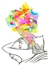
An artist's imaginary depiction of a dream

Oneirology (/ɒnɪˈrɒləi/; from Greek ὄνειρον, oneiron, "dream"; and -λογία, -logia, "the study of") is the scientific study of dreams. Current research seeks correlations between dreaming and current knowledge about the functions of the brain, as well as understanding of how the brain works during dreaming as pertains to memory formation and mental disorders. The study of oneirology can be distinguished from dream interpretation in that the aim is to quantitatively study the process of dreams instead of analyzing the meaning behind them.

History

In the 19th century, two advocates of this discipline were the French sinologists Marquis d'Hervey de Saint Denys and Alfred Maury. The field gained momentum in 1952, when Nathaniel Kleitman and his student Eugene Aserinsky discovered regular cycles. A further experiment by Kleitman and William C. Dement, then another medical student, demonstrated the particular period of sleep during which electrical brain activity, as measured by an electroencephalograph (EEG), closely resembled that of waking, in which the eyes dart about actively. This kind of sleep became known as rapid eye movement (REM) sleep, and Kleitman and Dement's experiment found a correlation of 0.80 between REM sleep and dreaming.

Field of work

Research into dreams includes exploration of the mechanisms of dreaming, the influences on dreaming, and disorders linked to dreaming. Work in oneirology overlaps with neurology and can vary from quantifying dreams, to analyzing brain waves during dreaming, to studying the effects of drugs and neurotransmitters on sleeping or dreaming. Though debate continues about the purpose and origins of dreams, there could be great gains from studying dreams as a function of brain activity. For example, knowledge gained in this area could have implications in the treatment of certain mental illnesses.

Mechanisms of dreaming

Dreaming occurs mainly during REM sleep, and brain scans recording brain activity have witnessed heavy activity in the limbic system and the amygdala during this period. Though current research has reversed the myth that dreaming occurs only during REM sleep, it has also shown that the dreams reported in non-rapid eye movement (NREM) and REM differ qualitatively and quantitatively, suggesting that the mechanisms that control each are different.

During REM sleep, researchers theorize that the brain goes through a process known as synaptic efficacy refreshment. This is observed as brain waves self-firing during sleep, in slow cycles at a rate of around 14 Hz, and is believed to serve the purpose of consolidating recent memories and reinforcing old memories. In this type of brain stimulation, the dreaming that occurs is a by-product of the process.

Stages of sleep

During normal sleep cycles, humans alternate between normal, NREM sleep and REM sleep. The brain waves characteristic of dreaming that are observed during REM sleep are the most commonly studied in dream research because most dreaming occurs during REM sleep.

REM sleep

EEG showing brainwaves during REM sleep

In 1952, Eugene Aserinsky discovered REM sleep while working in the surgery of his PhD advisor. Aserinsky noticed that the sleepers' eyes fluttered beneath their closed eyelids, later using a polygraph machine to record their brain waves during these periods. In one session, he awakened a subject who was wailing and crying out during REM and confirmed his suspicion that dreaming was occurring. In 1953, Aserinsky and his advisor published the ground-breaking study in Science.

Accumulated observation shows that dreams are strongly associated with REM sleep, during which an electroencephalogram shows brain activity to be most like wakefulness. Participant-nonremembered dreams during NREM are normally more mundane in comparison. During a typical lifespan, a human spends a total of about six years dreaming (which is about two hours each night). Most dreams last only 5 to 20 minutes. It is unknown where in the brain dreams originate, if there is a single origin for dreams, if multiple portions of the brain are involved, or what the purpose of dreaming is for the body or mind.

During REM sleep, the release of certain neurotransmitters is completely suppressed. As a result, motor neurons are not stimulated, a condition known as REM atonia. This prevents dreams from resulting in dangerous movements of the body.

Animals have complex dreams and are able to retain and recall long sequences of events while they are asleep. Studies show that various species of mammals and birds experience REM during sleep, and follow the same series of sleeping states as humans.

The discovery that dreams take place primarily during a distinctive electrophysiological state of sleep (REM), which can be identified by objective criteria, led to rebirth of interest in this phenomenon. When REM sleep episodes were timed for their duration and subjects awakened to make reports before major editing or forgetting could take place, it was determined that subjects accurately matched the length of time they judged the dream narrative to occupy with the length of REM sleep that preceded the awakening. This close correlation of REM sleep and dream experience was the basis of the first series of reports describing the nature of dreaming: that it is a regular nightly occurrence, rather than an occasional phenomenon, and that it is a high-frequency activity within each sleep period occurring at predictable intervals of approximately every 60–90 minutes in all humans throughout the life span.

REM sleep episodes and the dreams that accompany them lengthen progressively across the night, with the first episode the shortest, of approximately 10–12 minutes duration, and the second and third episodes increasing to 15–20 minutes. Dreams at the end of the night may last typically 15 minutes, although these may be experienced as several distinct stories due to momentary arousals interrupting sleep as the night ends.

Dream reports can normally be made 50% of the time when an awakening occurs prior to the end of the first REM period. This rate of retrieval is increased to about 99% when awakenings occur during the last REM period of the night. This increase in the ability to recall appears to be related to intensification across the night in the vividness of dream imagery, colors and emotions. The dream story itself in the last REM period is farthest from reality, containing more bizarre elements, and it is these properties, coupled with the increased likelihood of morning waking review to take place, that heighten the chance of recall of the last dream.

Definition of a dream

The definition of dream used in quantitative research is defined through four base components: 1) a form of thinking that occurs under minimal brain direction, external stimuli are blocked, and the part of the brain that recognizes self shuts down; 2) a form of experience that we believed we experience through our senses; 3) something memorable; 4) have some interpretation of experience by self. In summary, a dream, as defined by G. William Domhoff and Adam Schneider, is "a report of a memory of a cognitive experience that happens under the kinds of conditions that are most frequently produced in a state called 'sleep.' "

Commonplace bizarreness in dreaming

Certain kinds of bizarre cognitions, such as disjunctive cognitions and interobjects, are common in dreams.

Interobject

Interobjects, like disjunctive cognitions, are a commonplace bizarreness of dreamlife. Interobjects are a kind of dream condensation that creates a new object that could not occur in waking life. It may have a vague structure that is described as "something between an X and a Y". Hobson dreamt of "a piece of hardware, something like the lock of a door or perhaps a pair of paint-frozen hinges."

Authentic dreaming

Authentic dreams are defined by their tendency to occur "within the realm of experience" and reflect actual memories or experiences the dreamer can relate to. Authentic dreams are believed to be the side effect of synaptic efficacy refreshment that occurs without errors. Research suggests that the brain stimulation that occurs during dreaming authentic dreams is significant in reinforcing neurological pathways, serving as a method for the mind to "rehearse" certain things during sleep.

Illusory dreaming

Illusory dreams are defined as dreams that contain impossible, incongruent, or bizarre content and are hypothesized to stem from memory circuits accumulating efficacy errors. In theory, old memories having undergone synaptic efficacy refreshment multiple times throughout one's lifetime result in accumulating errors that manifest as illusory dreams when stimulated. Qualities of illusory dreaming have been linked to delusions observed in mental disorders. Illusory dreams are believed to most likely stem from older memories that experience this accumulation of errors in contrast to authentic dreams that stem from more recent experiences.

Influences on dreaming

One aspect of dreaming studied is the capability to externally influence the contents of dreams with various stimuli. One such successful connection was made to the olfactory, influencing the emotions of dreams through a smell stimulus. Their research has shown that the introduction of a positive smelling stimulus (roses) induced positive dreams while negative smelling stimulus (rotten eggs) induced negative dreams.

Memories and experience

Though there is much debate within the field about the purpose of dreaming, a leading theory involves the consolidation of memories and experiences that occurs during REM sleep. The electric involuntary stimulus the brain undergoes during sleep is believed to be a basis for a majority of dreaming.

The link between memory, sleep, and dreams becomes more significant in studies analyzing memory consolidation during sleep. Research has shown that NREM sleep is responsible for the consolidation of facts and episodes in contrast to REM sleep that consolidates more emotionally related aspects of memory. The correlation between REM and emotional consolidation could be interpreted as the reason why dreams are of such an emotional nature and produce strong reactions from humans.

Interpersonal attachment

In addition to the conscious role people are aware of memory and experience playing in dreaming, unconscious effects such as health of relationships factor into the types of dreams the brain produces. Of the people analyzed, those suffering from "insecure attachments" were found to dream with more frequency and more vividly than those who were evaluated to have "secure attachments".

Drugs affecting dreaming

Correlations between the usage of drugs and dreaming have been documented, particularly the use of drugs, such as sedatives, and the suppression of dreaming because of drugging effects on the cycles and stages of sleep while not allowing the user to reach REM. Drugs used for their stimulating properties (cocaine, methamphetamine, and ecstasy) have been shown to also decrease the restorative properties of REM sleep and its duration.

Dreaming disorders

Dreaming disorders are difficult to quantify due to the ambiguous nature of dreaming. However, dreaming disorders can be linked to psychological disorders such as post-traumatic stress disorder expressed as nightmares. Research into dreaming also suggests similarity and links in illusory dreaming and delusions.

Post-traumatic stress disorder

Diagnostic symptoms include re-experiencing original trauma(s), by means of flashbacks or nightmares; avoidance of stimuli associated with the trauma; and increased arousal, such as difficulty falling or staying asleep, anger, and hypervigilance.

Links to post-traumatic stress disorder (PTSD) and dreaming have been made in studying the flashbacks or nightmares the victims would suffer. Measurement of the brain waves exhibited by the subjects experiencing these episodes showed great similarity between those of dreaming. The drugs used to treat those suffering from these symptoms of flashbacks and nightmares would suppress not only these traumatic episodes but also any other sort of dreaming function.

Schizophrenia

The symptoms of schizophrenia involve abnormalities in the perception or expression of reality primarily focused on delusions and hallucinations.

The delusions experienced by those with schizophrenia have been likened to the experience of illusory dreams that have come to be interpreted by the subject as actual experiences. Additional research into medication to suppress symptoms of schizophrenia have also shown to influence the REM cycle of those taking the medication and as a result influence the patterns of sleep and dreaming in the subjects.

False awakening

From Wikipedia, the free encyclopedia

A false awakening is a vivid and convincing dream about awakening from sleep, while the dreamer in reality continues to sleep. After a false awakening, subjects often dream they are performing daily morning routine such as showering, cooking, cleaning, eating, and using the bathroom. False awakenings, mainly those in which one dreams that they have awoken from a sleep that featured dreams, take on aspects of a double dream or a dream within a dream. A classic example is the double false awakening of the protagonist in Gogol's Portrait (1835).

Related concepts

Lucidity

A false awakening may occur following a dream or following a lucid dream (one in which the dreamer has been aware of dreaming). Particularly, if the false awakening follows a lucid dream, the false awakening may turn into a "pre-lucid dream", that is, one in which the dreamer may start to wonder if they are really awake and may or may not come to the correct conclusion. In a study by Harvard psychologist Deirdre Barrett, 2,000 dreams from 200 subjects were examined and it was found that false awakenings and lucidity were significantly more likely to occur within the same dream or within different dreams of the same night. False awakenings often preceded lucidity as a cue, but they could also follow the realization of lucidity, often losing it in the process.

Loop

A false awakening loop is when a subject dreams about waking up over and over again, sometimes even up to 10 times or more without knowing which time they are actually awake. At times the individual can perform actions unknowingly. The movie A Nightmare on Elm Street popularized this phenomenon. This phenomenon can be related to that of sleepwalking or carrying out actions in a state of unconsciousness.

Symptoms

Realism and non-realism

Certain aspects of life may be dramatized or out of place in false awakenings. Things may seem wrong: details, like the painting on a wall, not being able to talk or difficulty reading (reportedly, reading in lucid dreams is often difficult or impossible). In some experiences, the subject's senses are heightened, or changed.

Repetition

Because the mind still dreams after a false awakening, there may be more than one false awakening in a single dream. Subjects may dream they wake up, eat breakfast, brush their teeth, and so on; suddenly awake again in bed (still in a dream), begin morning rituals again, awaken again, and so forth. The philosopher Bertrand Russell claimed to have experienced "about a hundred" false awakenings in succession while coming around from a general anesthetic.

Types

Celia Green suggested a distinction should be made between two types of false awakening:

Type 1

Type 1 is the more common, in which the dreamer seems to wake up, but not necessarily in realistic surroundings, that is, not in their own bedroom. A pre-lucid dream may ensue. More commonly, dreamers will believe they have awakened, and then either genuinely wake up in their own bed or "fall back asleep" in the dream.

A common false awakening is a "late for work" scenario. A person may "wake up" in a typical room, with most things looking normal, and realize they overslept and missed the start time at work or school. Clocks, if found in the dream, will show time indicating that fact. The resulting panic is often strong enough to truly awaken the dreamer (much like from a nightmare).

Another common Type 1 example of false awakening can result in bedwetting. In this scenario the dreamer has had a false awakening and while in the state of dream has performed all the traditional behaviors that precede urinating – arising from bed, walking to the bathroom, and sitting down on the toilet or walking up to a urinal. The dreamer may then urinate and suddenly wake up to find they have wet themselves.

Type 2

The type 2 false awakening seems to be considerably less common. Green characterized it as follows:

The subject appears to wake up in a realistic manner but to an atmosphere of suspense. ... The dreamer's surroundings may at first appear normal, and they may gradually become aware of something uncanny in the atmosphere, and perhaps of unwanted [unusual] sounds and movements, or they may "awake" immediately to a "stressed" and "stormy" atmosphere. In either case, the end result would appear to be characterized by feelings of suspense, excitement or apprehension.

Charles McCreery draws attention to the similarity between this description and the description by the German psychopathologist Karl Jaspers (1923) of the so-called "primary delusionary experience" (a general feeling that precedes more specific delusory belief). Jaspers wrote:

Patients feel uncanny and that there is something suspicious afoot. Everything gets a new meaning. The environment is somehow different—not to a gross degree—perception is unaltered in itself but there is some change which envelops everything with a subtle, pervasive and strangely uncertain light. ... Something seems in the air which the patient cannot account for, a distrustful, uncomfortable, uncanny tension invades him.

McCreery suggests this phenomenological similarity is not coincidental and results from the idea that both phenomena, the Type 2 false awakening and the primary delusionary experience, are phenomena of sleep. He suggests that the primary delusionary experience, like other phenomena of psychosis such as hallucinations and secondary or specific delusions, represents an intrusion into waking consciousness of processes associated with stage 1 sleep. It is suggested that the reason for these intrusions is that the psychotic subject is in a state of hyper-arousal, a state that can lead to what Ian Oswald called "micro-sleeps" in waking life.

Other researchers doubt that these are clearly distinguished types, as opposed to being points on a subtle spectrum.

Nightmare

From Wikipedia, the free encyclopedia
 
Nightmare
SpecialtyPsychology, Psychiatry
CausesStress, Anxiety, Fever

A nightmare, also known as a bad dream, is an unpleasant dream that can cause a strong emotional response from the mind, typically fear but also despair, anxiety, disgust or sadness. The dream may contain situations of discomfort, psychological or physical terror, or panic. After a nightmare, a person will often awaken in a state of distress and may be unable to return to sleep for a short period of time. Recurrent nightmares may require medical help, as they can interfere with sleeping patterns and cause insomnia.

Nightmares can have physical causes such as sleeping in an uncomfortable position or having a fever, or psychological causes such as stress or anxiety. Eating before going to sleep, which triggers an increase in the body's metabolism and brain activity, can be a potential stimulus for nightmares.

The prevalence of nightmares in children (5–12 years old) is between 20 and 30%, and for adults is between 8 and 30%. In common language, the meaning of nightmare has extended as a metaphor to many bad things, such as a bad situation or a scary monster or person.

Etymology

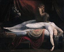
The word nightmare is derived from the Old English mare, a mythological demon or goblin who torments others with frightening dreams. The term has no connection with the Modern English word for a female horse. The word nightmare is cognate with the Dutch term nl:nachtmerrie and German Nachtmahr (dated).

History/Folklore

The sorcerous demons of Iranian mythology known as Divs are likewise associated with the ability to afflict their victims with nightmares. The mare of Germanic and Slavic folklore were thought to ride on people's chests while they sleep, causing nightmares.

Signs and symptoms

Those with nightmares experience abnormal sleep architecture. The impact of having a nightmare during the night has been found to be very similar to that of insomnia. This is thought to be caused by frequent nocturnal awakenings and fear of falling asleep. Nightmare disorder symptoms include repeated awakenings from the major sleep period or naps with detailed recall of extended and extremely frightening dreams, usually involving threats to survival, security, or self-esteem. The awakenings generally occur during the second half of the sleep period.

Classification

According to the International Classification of Sleep Disorders-Third Edition (ICSD-3), the nightmare disorder, together with REM sleep behaviour disorder (RBD) and recurrent isolated sleep paralysis, form the REM-related parasomnias subcategory of the Parasomnias cluster. Nightmares may be idiopathic without any signs of psychopathology or associated with disorders like stress, anxiety, substance abuse, psychiatric illness or PTSD (>80% of PTSD patients report nightmares). As regarding the dream content of the dreams they are usually imprinting negative emotions like sadness, fear or rage. According to the clinical studies the content can include being chased, injury or death of others, falling, natural disasters or accidents. Typical dreams or recurrent dreams may also have some of these topics.

Cause

Scientific research shows that nightmares may have many causes. In a study focusing on children, researchers were able to conclude that nightmares directly correlate with the stress in children's lives. Children who experienced the death of a family member or a close friend or know someone with a chronic illness have more frequent nightmares than those who are only faced with stress from school or stress from social aspects of daily life. A study researching the causes of nightmares focuses on patients who have sleep apnea. The study was conducted to determine whether or not nightmares may be caused by sleep apnea, or being unable to breathe. In the nineteenth century, authors believed that nightmares were caused by not having enough oxygen, therefore it was believed that those with sleep apnea had more frequent nightmares than those without it. The results actually showed that healthy people have more nightmares than sleep apnea patients. Another study supports the hypothesis. In this study, 48 patients (aged 20–85 yrs) with obstructive airways disease (OAD), including 21 with and 27 without asthma, were compared with 149 sex- and age-matched controls without respiratory disease. OAD subjects with asthma reported approximately 3 times as many nightmares as controls or OAD subjects without asthma. The evolutionary purpose of nightmares then could be a mechanism to awaken a person who is in danger.

Lucid-dreaming advocate Stephen LaBerge has outlined a possible reason for how dreams are formulated and why nightmares occur. To LaBerge, a dream starts with an individual thought or scene, such as walking down a dimly lit street. Since dreams are not predetermined, the brain responds to the situation by either thinking a good thought or a bad thought, and the dream framework follows from there. If bad thoughts in a dream are more prominent than good thoughts, the dream may proceed to be a nightmare.

There is a view, possibly featured in the story A Christmas Carol, that eating cheese before sleep can cause nightmares, but there is little scientific evidence for this.

Severe nightmares are also likely to occur when a person has a fever, these nightmares are often referred to as fever dreams.

Treatment

Sigmund Freud and Carl Jung seemed to have shared a belief that people frequently distressed by nightmares could be re-experiencing some stressful event from the past. Both perspectives on dreams suggest that therapy can provide relief from the dilemma of the nightmarish experience.

Halliday (1987) grouped treatment techniques into four classes. Direct nightmare interventions that combine compatible techniques from one or more of these classes may enhance overall treatment effectiveness:

Post-traumatic stress disorder

Recurring post-traumatic stress disorder (PTSD) nightmares in which traumas are re-experienced respond well to a technique called imagery rehearsal. This involves dreamers coming up with alternative, mastery outcomes to the nightmares, mentally rehearsing those outcomes while awake, and then reminding themselves at bedtime that they wish these alternate outcomes should the nightmares reoccur. Research has found that this technique not only reduces the occurrence of nightmares and insomnia, but also improves other daytime PTSD symptoms. The most common variations of imagery rehearsal therapy (IRT) "relate to the number of sessions, duration of treatment, and the degree to which exposure therapy is included in the protocol".

Medication

  • Prazosin (alpha-1 blocker) appears useful in decreasing the number of nightmares and the distress caused by them in people with PTSD.
  • Risperidone (atypical antipsychotic) at a dosage of 2 mg per day, has been shown in case series to remission of nightmares on the first night.
  • Trazodone (antidepressant) has been shown in a case report to treat nightmares associated with a depressed patient.

Trials have included hydrocortisone, gabapentin, paroxetine, tetrahydrocannabinol, eszopiclone, Sodium oxybate, and carvedilol.

Introduction to entropy

From Wikipedia, the free encyclopedia https://en.wikipedia.org/wiki/Introduct...