Search This Blog

Saturday, September 9, 2023

Multiplicative inverse

From Wikipedia, the free encyclopedia
Graph showing the diagrammatic representation of limits approaching infinity
The reciprocal function: y = 1/x. For every x except 0, y represents its multiplicative inverse. The graph forms a rectangular hyperbola.

In mathematics, a multiplicative inverse or reciprocal for a number x, denoted by 1/x or x−1, is a number which when multiplied by x yields the multiplicative identity, 1. The multiplicative inverse of a fraction a/b is b/a. For the multiplicative inverse of a real number, divide 1 by the number. For example, the reciprocal of 5 is one fifth (1/5 or 0.2), and the reciprocal of 0.25 is 1 divided by 0.25, or 4. The reciprocal function, the function f(x) that maps x to 1/x, is one of the simplest examples of a function which is its own inverse (an involution).

Multiplying by a number is the same as dividing by its reciprocal and vice versa. For example, multiplication by 4/5 (or 0.8) will give the same result as division by 5/4 (or 1.25). Therefore, multiplication by a number followed by multiplication by its reciprocal yields the original number (since the product of the number and its reciprocal is 1).

The term reciprocal was in common use at least as far back as the third edition of Encyclopædia Britannica (1797) to describe two numbers whose product is 1; geometrical quantities in inverse proportion are described as reciprocall in a 1570 translation of Euclid's Elements.

In the phrase multiplicative inverse, the qualifier multiplicative is often omitted and then tacitly understood (in contrast to the additive inverse). Multiplicative inverses can be defined over many mathematical domains as well as numbers. In these cases it can happen that abba; then "inverse" typically implies that an element is both a left and right inverse.

The notation f −1 is sometimes also used for the inverse function of the function f, which is for most functions not equal to the multiplicative inverse. For example, the multiplicative inverse 1/(sin x) = (sin x)−1 is the cosecant of x, and not the inverse sine of x denoted by sin−1 x or arcsin x. The terminology difference reciprocal versus inverse is not sufficient to make this distinction, since many authors prefer the opposite naming convention, probably for historical reasons (for example in French, the inverse function is preferably called the bijection réciproque).

Examples and counterexamples

In the real numbers, zero does not have a reciprocal (division by zero is undefined) because no real number multiplied by 0 produces 1 (the product of any number with zero is zero). With the exception of zero, reciprocals of every real number are real, reciprocals of every rational number are rational, and reciprocals of every complex number are complex. The property that every element other than zero has a multiplicative inverse is part of the definition of a field, of which these are all examples. On the other hand, no integer other than 1 and −1 has an integer reciprocal, and so the integers are not a field.

In modular arithmetic, the modular multiplicative inverse of a is also defined: it is the number x such that ax ≡ 1 (mod n). This multiplicative inverse exists if and only if a and n are coprime. For example, the inverse of 3 modulo 11 is 4 because 4 ⋅ 3 ≡ 1 (mod 11). The extended Euclidean algorithm may be used to compute it.

The sedenions are an algebra in which every nonzero element has a multiplicative inverse, but which nonetheless has divisors of zero, that is, nonzero elements x, y such that xy = 0.

A square matrix has an inverse if and only if its determinant has an inverse in the coefficient ring. The linear map that has the matrix A−1 with respect to some base is then the inverse function of the map having A as matrix in the same base. Thus, the two distinct notions of the inverse of a function are strongly related in this case, but they still do not coincide, since the multiplicative inverse of Ax would be (Ax)−1, not A−1x.

These two notions of an inverse function do sometimes coincide, for example for the function where is the principal branch of the complex logarithm and :

.

The trigonometric functions are related by the reciprocal identity: the cotangent is the reciprocal of the tangent; the secant is the reciprocal of the cosine; the cosecant is the reciprocal of the sine.

A ring in which every nonzero element has a multiplicative inverse is a division ring; likewise an algebra in which this holds is a division algebra.

Complex numbers

As mentioned above, the reciprocal of every nonzero complex number z = a + bi is complex. It can be found by multiplying both top and bottom of 1/z by its complex conjugate and using the property that , the absolute value of z squared, which is the real number a2 + b2:

The intuition is that

gives us the complex conjugate with a magnitude reduced to a value of , so dividing again by ensures that the magnitude is now equal to the reciprocal of the original magnitude as well, hence:

In particular, if ||z||=1 (z has unit magnitude), then . Consequently, the imaginary units, ±i, have additive inverse equal to multiplicative inverse, and are the only complex numbers with this property. For example, additive and multiplicative inverses of i are −(i) = −i and 1/i = −i, respectively.

For a complex number in polar form z = r(cos φ + i sin φ), the reciprocal simply takes the reciprocal of the magnitude and the negative of the angle:

Geometric intuition for the integral of 1/x. The three integrals from 1 to 2, from 2 to 4, and from 4 to 8 are all equal. Each region is the previous region halved vertically and doubled horizontally. Extending this, the integral from 1 to 2k is k times the integral from 1 to 2, just as ln 2k = k ln 2.

Calculus

In real calculus, the derivative of 1/x = x−1 is given by the power rule with the power −1:

The power rule for integrals (Cavalieri's quadrature formula) cannot be used to compute the integral of 1/x, because doing so would result in division by 0:

Instead the integral is given by:
where ln is the natural logarithm. To show this, note that , so if and , we have:

Algorithms

The reciprocal may be computed by hand with the use of long division.

Computing the reciprocal is important in many division algorithms, since the quotient a/b can be computed by first computing 1/b and then multiplying it by a. Noting that has a zero at x = 1/b, Newton's method can find that zero, starting with a guess and iterating using the rule:

This continues until the desired precision is reached. For example, suppose we wish to compute 1/17 ≈ 0.0588 with 3 digits of precision. Taking x0 = 0.1, the following sequence is produced:

x1 = 0.1(2 − 17 × 0.1) = 0.03
x2 = 0.03(2 − 17 × 0.03) = 0.0447
x3 = 0.0447(2 − 17 × 0.0447) ≈ 0.0554
x4 = 0.0554(2 − 17 × 0.0554) ≈ 0.0586
x5 = 0.0586(2 − 17 × 0.0586) ≈ 0.0588

A typical initial guess can be found by rounding b to a nearby power of 2, then using bit shifts to compute its reciprocal.

In constructive mathematics, for a real number x to have a reciprocal, it is not sufficient that x ≠ 0. There must instead be given a rational number r such that 0 < r < |x|. In terms of the approximation algorithm described above, this is needed to prove that the change in y will eventually become arbitrarily small.

Graph of f(x) = xx showing the minimum at (1/e, e−1/e).

This iteration can also be generalized to a wider sort of inverses; for example, matrix inverses.

Reciprocals of irrational numbers

Every real or complex number excluding zero has a reciprocal, and reciprocals of certain irrational numbers can have important special properties. Examples include the reciprocal of e (≈ 0.367879) and the golden ratio's reciprocal (≈ 0.618034). The first reciprocal is special because no other positive number can produce a lower number when put to the power of itself; is the global minimum of . The second number is the only positive number that is equal to its reciprocal plus one:. Its additive inverse is the only negative number that is equal to its reciprocal minus one:.

The function gives an infinite number of irrational numbers that differ with their reciprocal by an integer. For example, is the irrational . Its reciprocal is , exactly less. Such irrational numbers share an evident property: they have the same fractional part as their reciprocal, since these numbers differ by an integer.

Further remarks

If the multiplication is associative, an element x with a multiplicative inverse cannot be a zero divisor (x is a zero divisor if some nonzero y, xy = 0). To see this, it is sufficient to multiply the equation xy = 0 by the inverse of x (on the left), and then simplify using associativity. In the absence of associativity, the sedenions provide a counterexample.

The converse does not hold: an element which is not a zero divisor is not guaranteed to have a multiplicative inverse. Within Z, all integers except −1, 0, 1 provide examples; they are not zero divisors nor do they have inverses in Z. If the ring or algebra is finite, however, then all elements a which are not zero divisors do have a (left and right) inverse. For, first observe that the map f(x) = ax must be injective: f(x) = f(y) implies x = y:

Distinct elements map to distinct elements, so the image consists of the same finite number of elements, and the map is necessarily surjective. Specifically, ƒ (namely multiplication by a) must map some element x to 1, ax = 1, so that x is an inverse for a.

Applications

The expansion of the reciprocal 1/q in any base can also act  as a source of pseudo-random numbers, if q is a "suitable" safe prime, a prime of the form 2p + 1 where p is also a prime. A sequence of pseudo-random numbers of length q − 1 will be produced by the expansion.

Creatinine

From Wikipedia, the free encyclopedia
Creatinine
Names
Preferred IUPAC name
2-Amino-1-methyl-5H-imidazol-4-one
Other names
2-Amino-1-methylimidazol-4-ol
Properties
C4H7N3O
Molar mass 113.120 g·mol−1
Appearance White crystals
Density 1.09 g cm−3
Melting point 300 °C (572 °F; 573 K) (decomposes)
1 part per 12

90 mg/mL at 20°C

log P -1.76
Acidity (pKa) 12.309
Basicity (pKb) 1.688
Isoelectric point 11.19
Thermochemistry
138.1 J K−1 mol−1 (at 23.4 °C)
167.4 J K−1 mol−1
−240.81–239.05 kJ mol−1
−2.33539–2.33367 MJ mol−1

Creatinine (/kriˈætɪnɪn, -nn/; from Ancient Greek: κρέας (kréas) 'flesh') is a breakdown product of creatine phosphate from muscle and protein metabolism. It is released at a constant rate by the body (depending on muscle mass).

Biological relevance

Serum creatinine (a blood measurement) is an important indicator of kidney health, because it is an easily measured byproduct of muscle metabolism that is excreted unchanged by the kidneys. Creatinine itself is produced via a biological system involving creatine, phosphocreatine (also known as creatine phosphate), and adenosine triphosphate (ATP, the body's immediate energy supply).

Creatine is synthesized primarily in the liver from the methylation of glycocyamine (guanidino acetate, synthesized in the kidney from the amino acids arginine and glycine) by S-adenosyl methionine. It is then transported through blood to the other organs, muscle, and brain, where, through phosphorylation, it becomes the high-energy compound phosphocreatine. Creatine conversion to phosphocreatine is catalyzed by creatine kinase; spontaneous formation of creatinine occurs during the reaction.

Creatinine is removed from the blood chiefly by the kidneys, primarily by glomerular filtration, but also by proximal tubular secretion. Little or no tubular reabsorption of creatinine occurs. If the filtration in the kidney is deficient, blood creatinine concentrations rise. Therefore, creatinine concentrations in blood and urine may be used to calculate the creatinine clearance (CrCl), which correlates approximately with the glomerular filtration rate (GFR). Blood creatinine concentrations may also be used alone to calculate the estimated GFR (eGFR).

The GFR is clinically important as a measurement of kidney function. In cases of severe kidney dysfunction, though, the CrCl rate will overestimate the GFR because hypersecretion of creatinine by the proximal tubules will account for a larger fraction of the total creatinine cleared. Ketoacids, cimetidine, and trimethoprim reduce creatinine tubular secretion and, therefore, increase the accuracy of the GFR estimate, in particular in severe kidney dysfunction. (In the absence of secretion, creatinine behaves like inulin.)

An alternate estimation of kidney function can be made when interpreting the blood plasma concentration of creatinine along with that of urea. BUN-to-creatinine ratio (the ratio of blood urea nitrogen to creatinine) can indicate other problems besides those intrinsic to the kidney; for example, a urea concentration raised out of proportion to the creatinine may indicate a prerenal problem such as volume depletion.

Counterintuitively, supporting the observation of higher creatinine production in women compared to men, and putting into question the algorithms for GFR that do not distinguish for sex accordingly, women have higher muscle protein synthesis and higher muscle protein turnover across the life span. As HDL supports muscle anabolism, higher muscle protein turnover links increased creatine to the generally higher serum HDL in women as compared to serum HDL in men and the HDL associated benefits like reduced incidence of cardiovascular complications and reduced COVID-19 severity.

Antibacterial and potential immunosuppressive properties

Studies indicate creatinine can be effective at killing bacteria of many species in both the Gram positive and Gram negative as well as diverse antibiotic resistant bacterial strains. Creatinine appears not to affect growth of fungi and yeast; this can be used to isolate slower growing fungi free from the normal bacterial populations found in most environmental samples. The mechanism by which creatinine kills bacteria is not presently known. A recent report also suggests that creatinine may have immunosuppressive properties.

Diagnostic use

Serum creatinine is the most commonly used indicator (but not direct measure) of renal function. Elevated creatinine is not always representative of a true reduction in GFR. A high reading may be due to increased production of creatinine not due to decreased kidney function, to interference with the assay, or to decreased tubular secretion of creatinine. An increase in serum creatinine can be due to increased ingestion of cooked meat (which contains creatinine converted from creatine by the heat from cooking) or excessive intake of protein and creatine supplements, taken to enhance athletic performance. Intense exercise can increase creatinine by increasing muscle breakdown. Dehydration secondary to an inflammatory process with fever may cause a false increase in creatinine concentrations not related to an actual kidney injury, as in some cases with cholecystitis. Several medications and chromogens can interfere with the assay. Creatinine secretion by the tubules can be blocked by some medications, again increasing measured creatinine.

Serum creatinine

Diagnostic serum creatinine studies are used to determine renal function. The reference interval is 0.6–1.3 mg/dL (53–115 μmol/L). Measuring serum creatinine is a simple test, and it is the most commonly used indicator of renal function.

A rise in blood creatinine concentration is a late marker, observed only with marked damage to functioning nephrons. Therefore, this test is unsuitable for detecting early-stage kidney disease. A better estimation of kidney function is given by calculating the estimated glomerular filtration rate (eGFR). eGFR can be accurately calculated without a 24-hour urine collection using serum creatinine concentration and some or all of the following variables: sex, age, weight, and race, as suggested by the American Diabetes Association. Many laboratories will automatically calculate eGFR when a creatinine test is requested. Algorithms to estimate GFR from creatinine concentration and other parameters are discussed in the renal function article.

A concern as of late 2010 relates to the adoption of a new analytical methodology, and a possible impact this may have in clinical medicine. Most clinical laboratories now align their creatinine measurements against a new standardized isotope dilution mass spectrometry (IDMS) method to measure serum creatinine. IDMS appears to give lower values than older methods when the serum creatinine values are relatively low, for example 0.7 mg/dL. The IDMS method would result in a comparative overestimation of the corresponding calculated GFR in some patients with normal renal function. A few medicines are dosed even in normal renal function on that derived GFR. The dose, unless further modified, could now be higher than desired, potentially causing increased drug-related toxicity. To counter the effect of changing to IDMS, new FDA guidelines have suggested limiting doses to specified maxima with carboplatin, a chemotherapy drug.

A 2009 Japanese study found a lower serum creatinine concentration to be associated with an increased risk for the development of type 2 diabetes in Japanese men.

Urine creatinine

Males produce approximately 150 μmol to 200 μmol of creatinine per kilogram of body weight per 24 h while females produce approximately 100 μmol/kg/24 h to 150 μmol/kg/24 h. In normal circumstances, all this daily creatinine production is excreted in the urine.

Creatinine concentration is checked during standard urine drug tests. An expected creatinine concentration indicates the test sample is undiluted, whereas low amounts of creatinine in the urine indicate either a manipulated test or low initial baseline creatinine concentrations. Test samples considered manipulated due to low creatinine are not tested, and the test is sometimes considered failed.

Interpretation

In the United States and in most European countries creatinine is usually reported in mg/dL, whereas in Canada, Australia, and a few European countries, μmol/L is the usual unit. One mg/dL of creatinine is 88.4 μmol/L.

The typical human reference ranges for serum creatinine are 0.5 mg/dL to 1.0 mg/dL (about 45 μmol/L to 90 μmol/L) for women and 0.7 mg/dL to 1.2 mg/dL (60 μmol/L to 110 μmol/L) for men. The significance of a single creatinine value must be interpreted in light of the patient's muscle mass. Patients with greater muscle mass have higher creatinine concentrations.

Reference ranges for blood tests, comparing blood content of creatinine (shown in apple green) with other constituents

The trend of serum creatinine concentrations over time is more important than absolute creatinine concentration.

Serum creatinine concentrations may increase when an ACE inhibitor (ACEI) is taken for heart failure and chronic kidney disease. ACE inhibitors provide survival benefits for patients with heart failure and slow the disease progression in patients with chronic kidney disease. An increase not exceeding 30% is to be expected with ACEI use. Therefore, usage of ACEI should not be stopped unless an increase of serum creatinine exceeded 30% or hyperkalemia develops.

Chemistry

In chemical terms, creatinine is a lactam and an imidazolidinone, so a spontaneously formed cyclic derivative of creatine.

Several tautomers of creatinine exist; ordered by contribution, they are:

  • 2-Amino-1-methyl-1H-imidazol-4-ol (or 2-amino-1-methylimidazol-4-ol)
  • 2-Amino-1-methyl-4,5-dihydro-1H-imidazol-4-one
  • 2-Imino-1-methyl-2,3-dihydro-1H-imidazol-4-ol (or 2-imino-1-methyl-3H-imidazol-4-ol)
  • 2-Imino-1-methylimidazolidin-4-one
  • 2-Imino-1-methyl-2,5-dihydro-1H-imidazol-4-ol (or 2-imino-1-methyl-5H-imidazol-4-ol)

Creatinine starts to decompose around 300 °C.

Cancer biomarker

From Wikipedia, the free encyclopedia
text
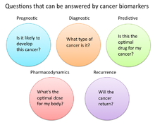
Questions that can be answered by biomarkers

A cancer biomarker refers to a substance or process that is indicative of the presence of cancer in the body. A biomarker may be a molecule secreted by a tumor or a specific response of the body to the presence of cancer. Genetic, epigenetic, proteomic, glycomic, and imaging biomarkers can be used for cancer diagnosis, prognosis, and epidemiology. Ideally, such biomarkers can be assayed in non-invasively collected biofluids like blood or serum.

Cancer is a disease that affects society at a world-wide level. By testing for biomarkers, early diagnosis can be given to prevent deaths.

While numerous challenges exist in translating biomarker research into the clinical space; a number of gene and protein based biomarkers have already been used at some point in patient care; including, AFP (liver cancer), BCR-ABL (chronic myeloid leukemia), BRCA1 / BRCA2 (breast/ovarian cancer), BRAF V600E (melanoma/colorectal cancer), CA-125 (ovarian cancer), CA19.9 (pancreatic cancer), CEA (colorectal cancer), EGFR (Non-small-cell lung carcinoma), HER-2 (Breast Cancer), KIT (gastrointestinal stromal tumor), PSA (prostate specific antigen) (prostate cancer), S100 (melanoma), and many others. Mutant proteins themselves detected by selected reaction monitoring (SRM) have been reported to be the most specific biomarkers for cancers because they can only come from an existing tumor. About 40% of cancers can be cured if detected early through examinations.

Definitions of cancer biomarkers

Organizations and publications vary in their definition of biomarker. In many areas of medicine, biomarkers are limited to proteins identifiable or measurable in the blood or urine. However, the term is often used to cover any molecular, biochemical, physiological, or anatomical property that can be quantified or measured.

The National Cancer Institute (NCI), in particular, defines biomarker as a: “A biological molecule found in blood, other body fluids, or tissues that is a sign of a normal or abnormal process, or of a condition or disease. A biomarker may be used to see how well the body responds to a treatment for a disease or condition. Also called molecular marker and signature molecule."

In cancer research and medicine, biomarkers are used in three primary ways:

  1. To help diagnose conditions, as in the case of identifying early stage cancers (diagnostic)
  2. To forecast how aggressive a condition is, as in the case of determining a patient's ability to fare in the absence of treatment (prognostic)
  3. To predict how well a patient will respond to treatment (predictive)

Role of biomarkers in cancer research and medicine

Uses of biomarkers in cancer medicine

Risk assessment

Cancer biomarkers, particular those associated with genetic mutations or epigenetic alterations, often offer a quantitative way to determine when individuals are predisposed to particular types of cancers. Notable examples of potentially predictive cancer biomarkers include mutations on genes KRAS, p53, EGFR, erbB2 for colorectal, esophageal, liver, and pancreatic cancer; mutations of genes BRCA1 and BRCA2 for breast and ovarian cancer; abnormal methylation of tumor suppressor genes p16, CDKN2B, and p14ARF for brain cancer; hypermethylation of MYOD1, CDH1, and CDH13 for cervical cancer; and hypermethylation of p16, p14, and RB1, for oral cancer.

Diagnosis

Cancer biomarkers can also be useful in establishing a specific diagnosis. This is particularly the case when there is a need to determine whether tumors are of primary or metastatic origin. To make this distinction, researchers can screen the chromosomal alterations found on cells located in the primary tumor site against those found in the secondary site. If the alterations match, the secondary tumor can be identified as metastatic; whereas if the alterations differ, the secondary tumor can be identified as a distinct primary tumor. For example, people with tumors have high levels of circulating tumor DNA (ctDNA) due to tumor cells that have gone through apoptosis. This tumor marker can be detected in the blood, saliva, or urine. The possibility of identifying an effective biomarker for early cancer diagnosis has recently been questioned, in light of the high molecular heterogeneity of tumors observed by next-generation sequencing studies.

Prognosis and treatment predictions

Another use of biomarkers in cancer medicine is for disease prognosis, which take place after an individual has been diagnosed with cancer. Here biomarkers can be useful in determining the aggressiveness of an identified cancer as well as its likelihood of responding to a given treatment. In part, this is because tumors exhibiting particular biomarkers may be responsive to treatments tied to that biomarker's expression or presence. Examples of such prognostic biomarkers include elevated levels of metallopeptidase inhibitor 1 (TIMP1), a marker associated with more aggressive forms of multiple myeloma, elevated estrogen receptor (ER) and/or progesterone receptor (PR) expression, markers associated with better overall survival in patients with breast cancer; HER2/neu gene amplification, a marker indicating a breast cancer will likely respond to trastuzumab treatment; a mutation in exon 11 of the proto-oncogene c-KIT, a marker indicating a gastrointestinal stromal tumor (GIST) will likely respond to imatinib treatment; and mutations in the tyrosine kinase domain of EGFR1, a marker indicating a patient's non-small-cell lung carcinoma (NSCLC) will likely respond to gefitinib or erlotinib treatment.

Pharmacodynamics and pharmacokinetics

Cancer biomarkers can also be used to determine the most effective treatment regime for a particular person's cancer. Because of differences in each person's genetic makeup, some people metabolize or change the chemical structure of drugs differently. In some cases, decreased metabolism of certain drugs can create dangerous conditions in which high levels of the drug accumulate in the body. As such, drug dosing decisions in particular cancer treatments can benefit from screening for such biomarkers. An example is the gene encoding the enzyme thiopurine methyl-transferase (TPMPT). Individuals with mutations in the TPMT gene are unable to metabolize large amounts of the leukemia drug, mercaptopurine, which potentially causes a fatal drop in white blood count for such patients. Patients with TPMT mutations are thus recommended to be given a lower dose of mercaptopurine for safety considerations.

Monitoring treatment response

Cancer biomarkers have also shown utility in monitoring how well a treatment is working over time. Much research is going into this particular area, since successful biomarkers have the potential of providing significant cost reduction in patient care, as the current image-based tests such as CT and MRI for monitoring tumor status are highly costly.

One notable biomarker garnering significant attention is the protein biomarker S100-beta in monitoring the response of malignant melanoma. In such melanomas, melanocytes, the cells that make pigment in our skin, produce the protein S100-beta in high concentrations dependent on the number of cancer cells. Response to treatment is thus associated with reduced levels of S100-beta in the blood of such individuals.

Similarly, additional laboratory research has shown that tumor cells undergoing apoptosis can release cellular components such as cytochrome c, nucleosomes, cleaved cytokeratin-18, and E-cadherin. Studies have found that these macromolecules and others can be found in circulation during cancer therapy, providing a potential source of clinical metrics for monitoring treatment.

Recurrence

Cancer biomarkers can also offer value in predicting or monitoring cancer recurrence. The Oncotype DX® breast cancer assay is one such test used to predict the likelihood of breast cancer recurrence. This test is intended for women with early-stage (Stage I or II), node-negative, estrogen receptor-positive (ER+) invasive breast cancer who will be treated with hormone therapy. Oncotype DX looks at a panel of 21 genes in cells taken during tumor biopsy. The results of the test are given in the form of a recurrence score that indicates likelihood of recurrence at 10 years.

Uses of biomarkers in cancer research

Developing drug targets

In addition to their use in cancer medicine, biomarkers are often used throughout the cancer drug discovery process. For instance, in the 1960s, researchers discovered the majority of patients with chronic myelogenous leukemia possessed a particular genetic abnormality on chromosomes 9 and 22 dubbed the Philadelphia chromosome. When these two chromosomes combine they create a cancer-causing gene known as BCR-ABL. In such patients, this gene acts as the principle initial point in all of the physiological manifestations of the leukemia. For many years, the BCR-ABL was simply used as a biomarker to stratify a certain subtype of leukemia. However, drug developers were eventually able to develop imatinib, a powerful drug that effectively inhibited this protein and significantly decreased production of cells containing the Philadelphia chromosome.

Surrogate endpoints

Another promising area of biomarker application is in the area of surrogate endpoints. In this application, biomarkers act as stand-ins for the effects of a drug on cancer progression and survival. Ideally, the use of validated biomarkers would prevent patients from having to undergo tumor biopsies and lengthy clinical trials to determine if a new drug worked. In the current standard of care, the metric for determining a drug's effectiveness is to check if it has decreased cancer progression in humans and ultimately whether it prolongs survival. However, successful biomarker surrogates could save substantial time, effort, and money if failing drugs could be eliminated from the development pipeline before being brought to clinical trials.

Some ideal characteristics of surrogate endpoint biomarkers include:

  • Biomarker should be involved in process that causes the cancer
  • Changes in biomarker should correlate with changes in the disease
  • Levels of biomarkers should be high enough that they can be measured easily and reliably
  • Levels or presence of biomarker should readily distinguish between normal, cancerous, and precancerous tissue
  • Effective treatment of the cancer should change the level of the biomarker
  • Level of the biomarker should not change spontaneously or in response to other factors not related to the successful treatment of the cancer

Two areas in particular that are receiving attention as surrogate markers include circulating tumor cells (CTCs) and circulating miRNAs. Both these markers are associated with the number of tumor cells present in the blood, and as such, are hoped to provide a surrogate for tumor progression and metastasis. However, significant barriers to their adoption include the difficulty of enriching, identifying, and measuring CTC and miRNA levels in blood. New technologies and research are likely necessary for their translation into clinical care.

Types of cancer biomarkers

Molecular cancer biomarkers

Tumor type Biomarker
Breast ER/PR (estrogen receptor/progesteron receptor)
HER-2/neu
Colorectal EGFR
KRAS
UGT1A1
Gastric HER-2/neu 
GIST c-KIT
Leukemia/lymphoma CD20
CD30
FIP1L1-PDGFRalpha
PDGFR
Philadelphia chromosome (BCR/ABL
PML/RAR-alpha
TPMT
UGT1A1 
Lung EML4/ALK
EGFR 
KRAS 
Melanoma BRAF
Pancreas Elevated levels of leucine, isoleucine and valine
Ovaries CA-125

Other examples of biomarkers:

Cancer biomarkers without specificity

Not all cancer biomarkers have to be specific to types of cancer. Some biomarkers found in the circulatory system can be used to determine an abnormal growth of cells present in the body. All these types of biomarkers can be identified through diagnostic blood tests, which is one of the main reasons to get regularly health tested. By getting regularly tested, many health issues such as cancer can be discovered at an early stage, preventing many deaths.

The neutrophil-to-lymphocyte ratio has been shown to be a non-specific determinant for many cancers. This ratio focuses on the activity of two components of the immune system that are involved in inflammatory response which is shown to be higher in presence of malignant tumors. Additionally, basic fibroblast growth factor (bFGF) is a protein that is involved in the proliferation of cells. Unfortunately, it has been shown that in the presence of tumors it is highly active which has led to the conclusion that it may help malignant cells reproduce at faster rates. Research has shown that anti-bFGF antibodies can be used to help treat tumors from many origins. Moreover, insulin-like growth factor (IGF-R) is involved in cell proliferation and growth. It has is possible that it is involved in inhibiting apoptosis, programmed cell death due to some defect. Due to this, the levels of IGF-R can be increased when cancer such as breast, prostate, lung, and colorectum is present.


Biomarker Description Biosensor used
NLR (neutrophil-to-lymphocyte ratio) Elevates with inflammation caused by cancer No
Basic Fibroblast Growth Factor (bFGF) This level increases when a tumor is present, helps with the fast reproduction of tumor cells Electrochemical
Insulin-like Growth Factor (IGF-R) High activity in cancer cells, help reproduction Electrochemical Impedance Spectroscopy Sensor

Entropy (information theory)

From Wikipedia, the free encyclopedia https://en.wikipedia.org/wiki/Entropy_(information_theory) In info...