Search This Blog

Saturday, January 13, 2024

Reproductive immunology

From Wikipedia, the free encyclopedia
https://en.wikipedia.org/wiki/Reproductive_immunology

Reproductive immunology refers to a field of medicine that studies interactions (or the absence of them) between the immune system and components related to the reproductive system, such as maternal immune tolerance towards the fetus, or immunological interactions across the blood-testis barrier. The concept has been used by fertility clinics to explain fertility problems, recurrent miscarriages and pregnancy complications observed when this state of immunological tolerance is not successfully achieved. Immunological therapy is a method for treating many cases of previously "unexplained infertility" or recurrent miscarriage.

The immune system and pregnancy

The immunological system of the mother plays an important role in pregnancy considering the embryo's tissue is half foreign and unlike mismatched organ transplant, is not normally rejected. During pregnancy, immunological events that take place within the body of the mother are crucial in determining the healthiness of both the mother and the fetus. In order to provide protection and immunity for both the mother and her fetus without developing rejection reactions, the mother must develop immunotolerance to her fetus since both organisms live in an intimate symbiotic situation. Progesterone-induced-blocking factor 1 (PIBF1) is one of the several known contributing immunomodulatory factors to play a role in immunotolerance during pregnancy.

The placenta also plays an important part in protecting the embryo for the immune attack from the mother's system. Secretory molecules produced by placental trophoblast cells and maternal uterine immune cells, within the decidua, work together to develop a functioning placenta. Studies have proposed that proteins in semen may help a person's immune system prepare for conception and pregnancy. For example, there is substantial evidence for exposure to partner's semen as prevention for pre-eclampsia, a pregnancy disorder, largely due to the absorption of several immune modulating factors present in seminal fluid, such as transforming growth factor beta (TGFβ).

Insufficient immune tolerance

An insufficiency in the maternal immune system where the fetus is treated as a foreign substance in the body can lead to many pregnancy-related complications.

  • Rh disease, or Rh isoimmunization, occurs when the maternal immune system develops antibodies that recognizes fetal red blood cells as foreign. This can lead to a number of potentially dangerous consequences to the fetus including hemolytic disease due to the destruction of red blood cells, kernicterus, or even death. Treatment with anti-D immunoglobulin has been studied extensively on the prevention of Rh disease. However, there has been no conclusive evidence that treatment with anti-D immunoglobulin is beneficial to the mother or fetus when it comes to Rh isoimmunization.
  • Pre-eclampsia is a disorder prevalent in 5% to 10% of all pregnancies that can lead to vascular health issues such as hypertension which can lead to other complications such as seizures, hemolytic disease, damage to the placenta, and inhibition of the growth and development of the fetus. Risk factors for pre-eclampsia include older age at which the mother becomes pregnant, obesity, and history of vascular disease. Monocyte activation in pregnancy is mediated by pregnancy hormones to prevent monocytes from becoming pro-inflammatory by inducing apoptosis. However, if there is dysfunction in this process, the activation of monocytes can potentially lead to damage and dysfunction in endothelial cells, which is thought to lead to the hallmark inflammation that is seen in pre-eclampsia. Prevention for those at risk for pre-eclampsia may include calcium supplementation, Vitamin C and E supplementation, low-dose aspirin, unfractionated heparin (UFH) and low-molecular-weight heparin (LMWH), and magnesium sulfate. Treatment goals include lowering the mother's blood pressure using antihypertensive medications that are safe to administer in pregnancy.
  • According to ESHRE guidelines, recurrent miscarriage is defined as 3 or more pregnancy losses before the third trimester (~22 weeks of gestation) and has many etiologies, including many that stem from immune dysfunction, most of which can be treated with immunosuppressive medications
    • An increase in the prevalence of antiphospholipid antibodies (known as antiphospholipid syndrome) can be found in many recurrent miscarriage patients. However, there is no evidence that the increase in antiphospholipid antibodies harms the pregnancy, but is thought to be indicative of immune dysfunction and proinflammatory responses in regards to the pregnancy.
    • An increase in prevalence of proinflammatory cells and natural killer cells can be found in women experiencing a miscarriage. However, there has been no evidence that the prevalence of these proinflammatory cells can predict pregnancy outcomes, including risk of a miscarriage.
    • Maternal HLA class II allele presence has been found to be potentially linked to predisposed immune attacks against male embryos. Proposed treatments for this immune dysfunction include corticosteroids, allogeneic lymphocyte immunization, intravenous immunoglobulin infusion, and tumor necrosis factor α antagonists.

Microbiology

Uterine natural killer (uNK) cells

The maternal immune system, specifically within the uterus, makes some changes in order to allow for implantation and protect a pregnancy from attack. One of these changes are to the uterine natural killer cells (uNK). NK cells, part of the innate immune system, are cytotoxic and responsible for attacking pathogens and infected cells. However, the number and type of receptors the uNK cells contain during a healthy pregnancy differs compared to an abnormal pregnancy. In the first trimester of pregnancy, uNK cells are among the most abundant leukocytes present, but the number of uNK cells present slowly declines up until term. Despite the fetus containing foreign paternal antigens, uNK cells do not recognize it as "non-self". Therefore, the cytotoxic effects of the uNK cells do not target the developing fetus. It has even been proposed that uNK contributes to the protection of extravillous trophoblast (EVT), important cells that contribute to the growth and development of a fetus. uNK cells secrete transforming growth factor-β (TGF-β) which is believed to have an immunosuppressive effect through modulation of leukocyte response to trophoblasts.

Killer immunoglobulin-like receptors (KIRs) and human leukocyte antigen (HLA)

KIRs are expressed by the uNK cells of the mother. Both polymorphic maternal KIRs and fetal HLA-C molecules are variable and specific to a particular pregnancy. In any pregnancy, the maternal KIR genotype could be AA (no activating KIRs), AB, or BB (1–10 activating KIRs) and the HLA-C ligands for KIRs are divided into two groups: HLA-C1 and HLA-C2. Studies have shown that there is a bad compatibility between specifically maternal KIR AA and fetal HLA-C2 which leads to recurrent miscarriage, preeclampsia and implantation failures. In assisted reproduction, these new insights could have an impact on the selection of single embryo transfer, oocyte, and/or sperm donor selection according to KIRs and HLA in patients with recurrent miscarriages.

Medication exposure during pregnancy

Pharmacokinetics

Pregnant-related anatomical and physiological changes affect pharmacokinetics (absorption, distribution, metabolism, and excretion) of many drugs, which may require drug regimen adjustment. Gastrointestinal motility is affected by delayed gastric emptying and increase gastric pH during pregnancy, which may alter drug absorption. Changes in body composition during pregnancy may change drugs volume of distribution due to increased body weight and fat, increased total plasma volume, and decreased albumin. For drugs susceptible to hepatic elimination are influenced by increased production of estrogen and progesterone. In addition, change in hepatic enzyme activity may increase or decrease drug metabolism based on drug composition, however most hepatic enzymes increase both metabolism and elimination during pregnancy. Also, pregnancy increase glomerular filtration, renal plasma flow, and the activity of transporters, which may require increased drug dosage.

FDA regulations

FDA established labeling request for drugs and biological products with medication risks, allowing informed decision making for pregnant and breastfeeding women and their health care providers. Pregnancy category was required on the drug label for systemically absorbed medications with the risk of fetal injury, which is now replaced with pregnancy and lactation labeling rule (PLLR). In addition to pregnancy category requirements on information of pregnancy, labor and delivery, and nursing mothers, PLLR also includes information on females animals of reproductive potential. The labeling change were effective starting June 30, 2015. The labeling requirements of over-the-counter (OTC) medicines we not affected.

Pharmacologic consideration

The change in medication exposure during pregnancy should concern both mother and fetus independently. For example, within antibiotics, penicillin may be used during pregnancy, whereas tetracycline is not recommended due to potential risk of fetus for a wide range of adverse effects.

Drugs

Medications to reduce risk of miscarriage

Progesterone is a medication often used to prevent threatened miscarriage. A threatened miscarriage is signs or symptoms of miscarriage, most often including bleeding that occurs in the first 20-weeks of a pregnancy. Research has shown that supplementation of progesterone can lower the rate of miscarriage, however, it did not have an effect on lowering the rate of pre-term births and live births. In reference to micronized vaginal progesterone, these results were more prominent for people who were at high risk of miscarriage, including people who have had three or more miscarriages and are currently experiencing bleeding.

The use of low dose aspirin may be linked to increased rates of live births and fewer pregnancy losses for people who have had one or two miscarriages. The National Institute of Health has recently changed their stance on using low dose aspirin, stating "low-dose aspirin therapy before conception and during early pregnancy may increase pregnancy chances and live births among a person who has experienced one or two prior miscarriages." This is a change from the previous stance on aspirin preventing pregnancy loss from the National Institute of Health. The reasoning behind the change was the determination that adherence to the medication and not discontinuing low dose aspirin due to side effects "could improve the odds for pregnancy and live birth in this group of people."

Sulfonamides and their risk of congenital malformations

Some studies have shown that maternal exposure to sulfonamides during pregnancy may have an increased risk of congenital malformations. There has been no evidence that certain types of sulfonamides or doses administered may increase or decrease the risk. Exposure to sulfonamides has been the only direct connection.

Medications to increase live birth rate for persons with antiphospholipid syndrome

Some studies have found that using both aspirin and heparin can increase the rate of live birth in a person with antiphospholipid syndrome. It was also found to increase birth weight and gestation age when using heparin and aspirin together. It was also found that people with antiphospholipid syndrome had an increased live birth rate when low molecular weight heparin was substituted in for heparin and co-administered with aspirin.

Sperm cells within a male

The presence of anti-sperm antibodies in infertile men was first reported in 1954 by Rumke and Wilson. It has been noticed that the number of cases of sperm autoimmunity is higher in the infertile population leading to the idea that autoimmunity could be a cause of infertility. Anti sperm antigen has been described as three immunoglobulin isotopes (IgG, IgA, IgM) each of which targets different part of the spermatozoa. If more than 10% of the sperm are bound to anti-sperm antibodies (ASA), then infertility is suspected. The blood-testis barrier separates the immune system and the developing spermatozoa. The tight junction between the Sertoli cells form the blood-testis barrier but it is usually breached by physiological leakage. Not all sperms are protected by the barrier because spermatogonia and early spermatocytes are located below the junction. They are protected by other means like immunologic tolerance and immunomodulation.

Infertility after anti-sperm antibody binding can be caused by autoagglutination, sperm cytotoxicity, blockage of sperm-ovum interaction, and inadequate motility. Each presents itself depending on the binding site of ASA.

Immunocontraceptive vaccine

Immunocontraceptive vaccines with a variety of proposed intervention strategies have been in development and under investigation since the 1970s. One approach is a vaccine designed to inhibit the fusing of spermatozoa to the zona pellucida. This vaccine has been tested in animals with a view to use as effective contraceptive for humans. Normally, spermatozoa fuse with the zona pellucida surrounding the mature oocyte; the resulting acrosome reaction breaks down the egg's tough coating so that the sperm can fertilize the ovum. The mechanism of the vaccine is injection with cloned ZP cDNA, therefore this vaccine is a DNA based vaccine. This results in the production of antibodies against the ZP, which stop the sperm from binding to the zona pellucida and ultimately from fertilizing the ovum.

Another vaccine that has been investigated is one against human chorionic gonadotropin (hCG). In phase I and early phase II human clinical trials, an experimental vaccine consisting of a dimer of β-hCG, with the tetanus toxoid (TT) as an adjuvant, produced antibodies against hCG in the small group of women immunized. The anti-hCG antibodies generated were capable of neutralizing the biological activity of hCG. Without active hCG, maintenance of the uterus in a condition receptive for implantation is not possible, thereby forestalling pregnancy. As only 80% of the women in the study had a level of circulating anti-hCG sufficient to prevent pregnancy, further development of this approach will be to enhance the immunogenicity of the vaccine, in order that it produces a reliable and consistent immune response in a higher proportion of women. Towards this goal, vaccine variations using a peptide of β-hCG that is uniquely specific to hCG, while absent in other hormones – luteinizing hormone (LH), follicle-stimulating hormone (FSH), and fhyroid-stimulating hormone (TSH) – are under investigation in animal models, for their possible enhancement of responses.

Research

Studying the female reproductive tract, especially in humans, allows for a better understanding of the immune system, including during pregnancy. However, studying the female reproductive tract has been a challenging area of research due to existing limitations in the in vitro and in vivo tools available. Ethical concerns is another contributing factor in hindering the study of reproductive immunology. Given such limitations, research in this field relies on stem cell culture and technological advancements by allowing scientists to conduct research on organoids instead of living human subjects. In 2018, a Review study concluded that organoids can be used to model organ development and disease. Other studies have concluded that with further technological advancements, it is possible to create a detailed 3D organoid model of the female reproductive tract which introduces a more efficient method to conduct research and collect data in the fields of drug discovery, basic research and essentially reproductive immunology.

Single-cell technologies

The maternal-fetal interface has the ability to protect against pathogens by providing reproductive immunity. Simultaneously, it is remodeling the tissues needed for placentation. This unique feature of the maternal-fetal interface suggests that the decidual immunome, or the immune function of the female reproductive tract, is not fully understood, yet.

In order to have a better understanding of Reproductive Immunology, more data needs to be collected and analyzed. Technological advances allow reproductive immunologists to collect increasingly complex data at a cellular resolution. Polychromatic flow cytometry allows for greater resolution in the identifying novel cell types by surface and intracellular protein. Two examples of methods in data acquisition include:

Reproductive immunology remains an open area of research as not enough data is available to introduce a significant finding.

Cytokine profiling

Maternal immune activation can be assessed by measuring multiple cytokines (cytokine profiling) in serum or plasma. This method is safe for the fetus since it only requires a peripheral blood sample from the mother and has been used to map maternal immune development throughout normal pregnancies as well as studying the relationship between immune activation and pregnancy complications or abnormal development of the fetus. Unfortunately, the method itself is unable to determine the sources and the targets of the cytokines and only shows systemic immune activation (as long as peripheral blood is analyzed), and the cytokine profile may vary rapidly as cytokines are short-lived proteins. It is also difficult to establish the exact relation between a cytokine profile and the underlying immunological processes.

The impact of unfavorable immune activation on fetal development and the risk of pregnancy complications is an active field of research. Many studies have reported an association between cytokine levels, especially for inflammatory cytokines, and the risk of developing preeclampsia, although the findings are mixed. However, decreased cytokine levels in early pregnancy has been associated to impaired fetal growth. Increased maternal cytokine levels have also been found to increase the risk of neurodevelopmental disorders such as autism spectrum disorders and depression in the offspring. However, more research is needed before these associations are fully understood.

Placental disease

From Wikipedia, the free encyclopedia
Placental disease
Micrograph of a chorangioma. H&E stain.
SpecialtyGynecology

A placental disease is any disease, disorder, or pathology of the placenta.

Ischemic placental disease leads to the attachment of the placenta to the uterine wall to become under-perfused, causing uteroplacental ischemia. Where the term overarches the pathology associated with preeclampsia, placental abruptions and intrauterine growth restriction (IUGR). These factors are known to be the primary pathophysiology cause placental disease. Which is considered to be associated with more than half of premature births.

Abnormalities present within the spiral arteries lead to higher velocities in blood, in turn causes the maternal villi to shred. Which trigger pro-coagulator molecules to be released into the blood stream causing action of the coagulator cascade, eventually leading to placental infarction. Risk factors such as diabetes, chronic blood pressure and multiple pregnancies can increase the risk of developing placental disease. Also, exposure to sudden trauma can increase the risk of placental abruption which coincides with placental disease.

There is no target treatment available for placental disease. Associative prevention mechanisms can be a method of minimising the risk of developing the disease, within early stages of pregnancy.

Placental syndromes include pregnancy loss, fetal growth restriction, preeclampsia, preterm delivery, premature rupture of membranes, placental abruption and intrauterine fetal demise.

Signs and symptoms

The abnormal spiral arteries lead decreased level of oxygen diffusion through the placental villus, which cause chronic hypoxia. The abnormal trophoblast invasion, lead to overall uteroplacental insufficiencies and uteroplacental underperfusion. It is due to the decreased vascularisation, there are reduced levels of nutrient delivery to the foetus. Also, cases of still births can be associated with placental disease.

Causes

Preeclampsia is considered to be linked with Placental Disease, as well as intrauterine growth restriction (IUGR) and placental abruptions are risk factors that lead to placental disease. Especially when these symptoms are evident at early stages of pregnancy. The abnormal invasion of the trophoblast cells, lack of important growth factors such as vascular endothelial growth factor (VEGF) and placental growth factor (PlGF), has an association with the onset of placental disease.

Risk factors

Risk factors associated with placental disease are as follows:

  • Smoking cigarettes and use other forms of drugs such as cocaine.
  • Diabetes mellitus
  • Maternal age less than 20 years or over the age of 35
  • Multiple pregnancies
  • Chronic high blood pressure
  • Being underweight or obese

Also, chronic renal disease, collagen vascular disease, thrombophilia, and cardiovascular disease increase the risk of developing placental disease. Moreover, being exposed to severe trauma within the pregnancy period, rapid acceleration and deceleration and uterine compression increase the risk of placental abruption, in turn leading to placental disease.

Adherence/penetration

Inflammatory/infectious

Placental development

Obstruction of os

Vascular

Neoplastic

Trophoblastic neoplasms derive from trophoblastic tissue. Examples include:

Mechanism

In placental disease, there's abnormalities present within the spiral arties of the uterus, where the terminal part of the spinal arteries does not dilate. This leads to decrease oxygen carried past the maternal villi into the intervillus space. The lack of terminal dilation and inclining blood velocity causes shredding of the villi into the maternal blood, releasing blood coagulants activating the coagulation cascade. Which then leads to blocking of the blood vessels causing placental infarction.

Diagnosis

Placental Disease can be diagnosed through technologies such as, Prenatal ultrasound evaluation and invasive foetal testing. The size of the foetus is taken into account through ultrasonography in terms of intrauterine growth restriction (IUGR). In conjunction with taking into account the maternal history. Suspicions may be confirmed by postpartum examination of the placenta.

Prevention

The following factors can be linked with reducing the likelihood of developing placental disease:

  • Use of aspirin, can reduce the risks associated with preeclampsia
  • Low calcium intake can reduce the risk of preeclampsia
  • Reducing oxidative stress present within the body
  • Intake of prenatal multivitamins

Treatment

Treatment of placental disease would require a premature birth, in order to avoid a still birth.

Epidemiology

Placental disease is more common in preterm gestation than with full term. Which leads to serious injuries to both the mother and the new-born. Women who endured placental disease within the first pregnancy has an increased risk of the disease progressing within future pregnancies. The onset of the disease within the first trimester leads to preterm delivery of a premature baby. Preeclampsia is diagnosed in 3-5% of pregnancies that place them at risk of developing placental disease. Ischemic placental disease is linked with approximately more than half of premature births.

Chain gang

From Wikipedia, the free encyclopedia
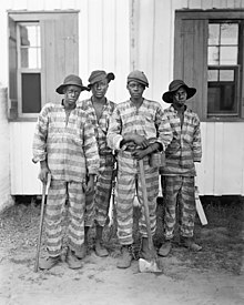
Chain gang street sweepers, Washington, D. C. 1909
Female convicts in Dar es Salaam chained together by their necks, c. 1890–1927

A chain gang or road gang is a group of prisoners chained together to perform menial or physically challenging work as a form of punishment. Such punishment might include repairing buildings, building roads, or clearing land. The system was notably used in the convict era of Australia and in the Southern United States. By 1955 it had largely been phased out in the U.S., with Georgia among the last states to abandon the practice. North Carolina continued to use chain gangs into the 1970s. Chain gangs were reintroduced by a few states during the "get tough on crime" 1990s: In 1995, Alabama was the first state to revive them. The experiment ended after about one year in all states except Arizona, where in Maricopa County inmates can still volunteer for a chain gang to earn credit toward a high school diploma or avoid disciplinary lockdowns for rule infractions.

Synonyms and disambiguation

A single ankle shackle with a short length of chain attached to a heavy ball is known as a ball and chain. It limited prisoner movement and impeded escape.

Two ankle shackles attached to each other by a short length of chain are known as a hobble or as leg irons. These could be chained to a much longer chain with several other prisoners, creating a work crew known as a chain gang. The walk required to avoid tripping while in leg irons is known as the convict shuffle.

Parchman Farm chain gang, 1911

A group of prisoners working outside prison walls under close supervision, but without chains, is a work gang. Their distinctive attire (stripe wear or orange vests or jumpsuits) and shaven heads served the purpose of displaying their punishment to the public, as well as making them identifiable if they attempted to escape. However, the public was often brutal, swearing at convicts and even throwing things at them.

The use of chains could be hazardous. Some of the chains used in the Georgia system in the first half of the twentieth century weighed 20 pounds (9 kg). Some prisoners suffered from shackle sores—ulcers where the iron ground against their skin. Gangrene and other infections were serious risks. Falls could imperil several individuals at once.

Modern prisoners are sometimes put into handcuffs or wrist manacles (similar to handcuffs, but with a longer length of chain) and leg irons, with both sets of manacles (wrist and ankle) being chained to a belly chain. This form of restraint is most often used on prisoners expected to be violent, or prisoners appearing in a setting where they may be near the public (a courthouse) or have an opportunity to flee (being transferred from a prison to a court). Although prisoners in these restraints are sometimes chained to one another during transport or other movement, this is not a chain gang—although reporters may refer to it as such—because the restraints make any kind of manual work impossible.

Purpose

1894 illustration of chain gang performing manual labor

Various claims as to the purpose of chain gangs have been offered. These include:

  • punishment
  • societal restitution for the cost of housing, feeding, and guarding the inmates. The money earned by work performed goes to offset prison expenses by providing a large workforce at no cost for government projects, and at minimal convict leasing cost for private businesses
  • a way of perpetuating African-American servitude after the Thirteenth Amendment to the United States Constitution ended slavery outside of the context of punishment for a crime
  • reducing inmates' idleness
  • to serve as a deterrent to crime
  • to satisfy the needs of politicians to appear "tough on crime"
  • to accomplish undesirable and difficult tasks

History

Australia

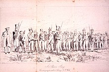
Illustration of a chain gang of 21 convicts in Hobart, Tasmania, in 1833, alongside six uniformed soldiers and a well-dressed man who is possibly a magistrate.

In the Australian penal colonies, chain gangs were also referred to as "iron gangs". They were used as a punishment for convicts who reoffended after being transported. Iron gangs were frequently employed on the construction of roads in remote areas where escape was a possibility, such as on the Great North Road from Sydney to the Hunter Valley and the road from Sydney to Bathurst over the Blue Mountains. The leg irons were installed by blacksmiths using hot rivets, and then attached to a single "gang chain" to allow for control by an overseer. The irons and chains could weigh as much as 4.5 kilograms (9.9 lb) or more. Some of the convicts on iron gangs were as young as 11 years old.

The use of iron gangs in the Colony of New South Wales was expanded by Governor Ralph Darling as part of his infrastructure program. Their tasks included "breaking rocks, clearing trees, constructing stone culverts and bridges". In 1828, the colony's chief surveyor Edmund Lockyer directed that each iron gang could contain up to 60 men, supervised by one main overseer and three assistants. The iron gangs "received the worst and least trustworthy characters, together with the strictest security measures". Better-behaved convicts still worked in gangs but were unshackled. Convicts who escaped from iron gangs were described as "bolters" and became some of the first bushrangers.

United States

A chain gang in the southern US, 1903

The introduction of chain gangs into the United States began after the American Civil War. The southern states needed finances and public works to be performed. Prisoners were a free way for these works to be achieved.

The use of chain gangs for prison labor was the preferred method of punishment in some southern states like Florida, Georgia, Louisiana, Virginia, North Carolina, Arkansas, Texas, Mississippi, and Alabama.

Abuses in chain gangs led to reform and to their general elimination by 1955. There were still chain gangs in the South in December 1955.

Chain gangs experienced a resurgence when Alabama began to use them again in 1995; they still existed in 1997.

Reintroduction

Several jurisdictions in the United States have re-introduced prison labor. In 1995 Sheriff Joe Arpaio reintroduced chain gangs in Arizona.

A year after reintroducing the chain gang in 1995, Alabama was forced to again abandon the practice pending a lawsuit from the Southern Poverty Law Center, among other organizations. The SPLC's attorney J. Richard Cohen said, "they realized that chaining them together was inefficient; that it was unsafe". Alabama Prison Commissioner Ron Jones was fired in 1996 for trying to put female prisoners on chain gangs. However, as late as 2000, Jones had proposed reintroducing the chain gang.

Chain gang of juvenile convicts in the US, 2006
Chain gang of juvenile convicts in the US, 1903

In 2011, Tim Hudak, former leader of the Progressive Conservative Party of Ontario in Canada, campaigned on introducing penal labour in the province, referred to by many as chain gangs. He lost seats to the provincial Liberals which formed another majority government in the subsequent general election.

According to their own policies, Britain First (a British far-right political organization) want to re-introduce chain gangs "to provide labour for national public works". This is part of their aim to turn prisons from "cosy holiday camps" into "a place of hard labour".

In 2013, Brevard County Jail in Sharpes, Florida reintroduced chain gangs as a deterrent on crime in a pilot project. Ex-convict Larry Lawton, critical of this move, said "Chain gangs send a bad message about our county", adding "I don't think people want to come to this county as a tourist or a beach person and see people in chains." Instead he proposed a better use of law enforcement resources would be to combat drug addiction because he says it is a "contributing factor" to criminal activity.

Synapsid

From Wikipedia, the free encyclopedia

Synapsids
Temporal range: PennsylvanianHolocene, 318–0 Ma
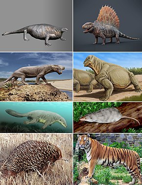
Examples of synapsids (left to right, top to bottom): Cotylorhynchus, Dimetrodon, Inostrancevia, Moschops, Castorocauda, Adelobasileus, Tachyglossus aculeatus, and Panthera tigris

Synapsids are one of the two major clades of vertebrate animals in the group Amniota, the other being the sauropsids, which include reptiles (turtles, crocodilians and lepidosaurs) and birds. The synapsids were the dominant land animals in the late Paleozoic and early Mesozoic, but the only extant group that survived into the Cenozoic are the mammals. Unlike other amniotes, synapsids have a single temporal fenestra, an opening low in the skull roof behind each eye orbit, leaving a bony arch beneath each; this accounts for their name. The distinctive temporal fenestra developed about 318 million years ago during the Late Carboniferous period, when synapsids and sauropsids diverged, but was subsequently merged with the orbit in early mammals.

The animals (basal amniotes) from which non-mammalian synapsids evolved were traditionally called "reptiles". Therefore, synapsids were described as mammal-like reptiles in classical systematics, and non-therapsid synapsids were also referred to as pelycosaurs, or pelycosaur-grade synapsids. These paraphyletic terms have now fallen out of favor and are only used informally (if at all) in modern literature. It is now known that all extant animals traditionally called "reptiles" are more closely related to each other than to synapsids, so the word "reptile" has been re-defined to mean only members of Sauropsida (bird-line Amniota) or even just an under-clade thereof, and synapsids are not part of the sauropsid lineage in a cladistical sense. Therefore, calling synapsids "mammal-like reptiles" is incorrect under the new definition of "reptile", so they are now referred to as stem mammals, and sometimes as proto-mammals, or paramammals.

Synapsids were the largest terrestrial vertebrates in the Permian period, 299 to 251 million years ago, equalled only by some large pareiasaurs at the end of the Permian. Most lineages of pelycosaur-grade synapsids were replaced at the end of the Early Permian by the more advanced therapsids. Synapsid numbers and variety were severely reduced by the Permian–Triassic extinction event. Only the therapsid dicynodonts and eutheriodonts (consisting of Therocephalia and Cynodontia) are known to have continued into the Triassic period. The cynodont group Probainognathia, which includes Mammaliaformes (mammals and their closer ancestors), were the only synapsids to survive beyond the Triassic.

During the Triassic, the sauropsid archosaurs became some of the largest and most numerous land vertebrates, only rivaled in size by kannemeyeriiform dicynodonts, and gave rise to the dinosaurs. When all non-avian dinosaurs were wiped out by the Cretaceous–Paleogene extinction event, the mammalian synapsids diversified again to become the largest land and marine animals on Earth.

Linnaean and cladistic classifications

At the turn of the 20th century synapsids were thought to be one of the four main subclasses of reptiles. However, this notion was disproved upon closer inspection of skeletal remains, as synapsids are differentiated from reptiles by their distinctive temporal openings. These openings in the skull bones allowed the attachment of larger jaw muscles, hence a more efficient bite.

Synapsids were subsequently considered to be a later reptilian lineage that became mammals by gradually evolving increasingly mammalian features, hence the name "mammal-like reptiles" (also known as pelycosaurs). These became the traditional terms for all Paleozoic (early) synapsids. More recent studies have debunked this notion as well, and reptiles are now classified within Sauropsida (sauropsids), the sister group to synapsids, thus making synapsids their own taxonomic group.

As a result, the paraphyletic terms "mammal-like reptile" and "pelycosaur" are seen as outdated and disfavored in technical literature, and the term stem mammal (or sometimes protomammal or paramammal) is used instead. Phylogenetically, it is now understood that synapsids comprise an independent branch of the tree of life. The monophyly of Synapsida is not in doubt, and the expressions such as "Synapsida contains the mammals" and "synapsids gave rise to the mammals" both express the same phylogenetic hypothesis. This terminology reflects the modern cladistical approach to animal relationships, according to which the only valid groups are those that include all of the descendants of a common ancestor: these are known as monophyletic groups, or clades.

Additionally, Reptilia (reptiles) has been revised into a monophyletic group and is considered entirely distinct from Synapsida, falling within Sauropsida, the sister group of Synapsida within Amniota.

Primitive and advanced synapsids

The synapsids are traditionally divided for convenience, into therapsids, an advanced group of synapsids and the branch within which mammals evolved, and stem mammals, (previously known as pelycosaurs), comprising the other six more primitive families of synapsids. Stem mammals were all rather lizard-like, with sprawling gait and possibly horny scutes, while therapsids tended to have a more erect pose and possibly hair, at least in some forms. In traditional taxonomy, the Synapsida encompasses two distinct grades: the low-slung stem mammals have given rise to the more erect therapsids, who in their turn have given rise to the mammals. In traditional vertebrate classification, the stem mammals and therapsids were both considered orders of the subclass Synapsida.

Practical versus phylogenetic usage of "synapsid" and "therapsid"

In phylogenetic nomenclature, the terms are used somewhat differently, as the daughter clades are included. Most papers published during the 21st century have treated "Pelycosaur" as an informal grouping of primitive members. Therapsida has remained in use as a clade containing both the traditional therapsid families and mammals.

Although Synapsida and Therapsida includes modern mammals, in practical usage, those two terms are used almost exclusively when referring to the more basal members that lie outside of Mammaliaformes.

Characteristics

Temporal openings

The synapsids are distinguished by a single hole, known as the temporal fenestra, in the skull behind each eye. This schematic shows the skull viewed from the left side. The middle opening is the orbit of the eye; the opening to the right of it is the temporal fenestra.

Synapsids evolved a temporal fenestra behind each eye orbit on the lateral surface of the skull. It may have provided new attachment sites for jaw muscles. A similar development took place in the diapsids, which evolved two rather than one opening behind each eye. Originally, the openings in the skull left the inner cranium covered only by the jaw muscles, but in higher therapsids and mammals, the sphenoid bone has expanded to close the opening. This has left the lower margin of the opening as an arch extending from the lower edges of the braincase.

Teeth

Eothyris, an early synapsid with multiple canines

Synapsids are characterized by having differentiated teeth. These include the canines, molars, and incisors. The trend towards differentiation is found in some labyrinthodonts and early anapsid reptilians in the form of enlargement of the first teeth on the maxilla, forming a form of protocanines. This trait was subsequently lost in the diapsid line, but developed further in the synapsids. Early synapsids could have two or even three enlarged "canines", but in the therapsids, the pattern had settled to one canine in each upper jaw half. The lower canines developed later.

Jaw

The jaw transition is a good classification tool, as most other fossilized features that make a chronological progression from a reptile-like to a mammalian condition follow the progression of the jaw transition. The mandible, or lower jaw, consists of a single, tooth-bearing bone in mammals (the dentary), whereas the lower jaw of modern and prehistoric reptiles consists of a conglomeration of smaller bones (including the dentary, articular, and others). As they evolved in synapsids, these jaw bones were reduced in size and either lost or, in the case of the articular, gradually moved into the ear, forming one of the middle ear bones: while modern mammals possess the malleus, incus and stapes, basal synapsids (like all other tetrapods) possess only a stapes. The malleus is derived from the articular (a lower jaw bone), while the incus is derived from the quadrate (a cranial bone).

Mammalian jaw structures are also set apart by the dentary-squamosal jaw joint. In this form of jaw joint, the dentary forms a connection with a depression in the squamosal known as the glenoid cavity. In contrast, all other jawed vertebrates, including reptiles and nonmammalian synapsids, possess a jaw joint in which one of the smaller bones of the lower jaw, the articular, makes a connection with a bone of the cranium called the quadrate bone to form the articular-quadrate jaw joint. In forms transitional to mammals, the jaw joint is composed of a large, lower jaw bone (similar to the dentary found in mammals) that does not connect to the squamosal, but connects to the quadrate with a receding articular bone.

Palate

Over time, as synapsids became more mammalian and less 'reptilian', they began to develop a secondary palate, separating the mouth and nasal cavity. In early synapsids, a secondary palate began to form on the sides of the maxilla, still leaving the mouth and nostril connected.

Eventually, the two sides of the palate began to curve together, forming a U shape instead of a C shape. The palate also began to extend back toward the throat, securing the entire mouth and creating a full palatine bone. The maxilla is also closed completely. In fossils of one of the first eutheriodonts, the beginnings of a palate are clearly visible. The later Thrinaxodon has a full and completely closed palate, forming a clear progression.

Skin and fur

The sea otter has the densest fur of modern mammals.

In addition to the glandular skin covered in fur found in most modern mammals, modern and extinct synapsids possess a variety of modified skin coverings, including osteoderms (bony armor embedded in the skin), scutes (protective structures of the dermis often with a horny covering), hair or fur, and scale-like structures (often formed from modified hair, as in pangolins and some rodents). While the skin of reptiles is rather thin, that of mammals has a thick dermal layer.

The ancestral skin type of synapsids has been subject to discussion. Among the early synapsids, only two species of small varanopids have been found to possess osteoderms; fossilized rows of osteoderms indicate bony armour on the neck and back. However, some recent studies have cast doubt on the placement of Varanopidae in Synapsida, while others have countered and lean towards this traditional placement. Skin impressions indicate some early synapsids basal possessed rectangular scutes on their undersides and tails. The pelycosaur scutes probably were nonoverlapping dermal structures with a horny overlay, like those found in modern crocodiles and turtles. These differed in structure from the scales of lizards and snakes, which are an epidermal feature (like mammalian hair or avian feathers). Recently, skin impressions from the genus Ascendonanus suggest that at least varanopsids developed scales similar to those of squamates.

It is currently unknown exactly when mammalian characteristics such as body hair and mammary glands first appeared, as the fossils only rarely provide direct evidence for soft tissues. An exceptionally well-preserved skull of Estemmenosuchus, a therapsid from the Upper Permian, preserves smooth skin with what appear to be glandular depressions, an animal noted as being semi-aquatic. The oldest known fossil showing unambiguous imprints of hair is the Callovian (late middle Jurassic) Castorocauda and several contemporary haramiyidans, both non-mammalian mammaliaform (see below, however). More primitive members of the Cynodontia are also hypothesized to have had fur or a fur-like covering based on their inferred warm-blooded metabolism. While more direct evidence of fur in early cynodonts has been proposed in the form of small pits on the snout possibly associated with whiskers, such pits are also found in some reptiles that lack whiskers. There is evidence that some other non-mammalian cynodonts more basal than Castorocauda, such as Morganucodon, had Harderian glands, which are associated with the grooming and maintenance of fur. The apparent absence of these glands in non-mammaliaformes may suggest that fur did not originate until that point in synapsid evolution. It is possible that fur and associated features of true warm-bloodedness did not appear until some synapsids became extremely small and nocturnal, necessitating a higher metabolism. The oldest examples of nocturnality in synapsids is believed to have been in species that lived more than 300 million years ago.

However, Permian coprolites from Russia showcase that at least some synapsids did already have fur in this epoch. These are the oldest impressions of hair on synapsids.

Mammary glands

Early synapsids, as far back as their known evolutionary debut in the Late Carboniferous period, may have laid parchment-shelled (leathery) eggs, which lacked a calcified layer, as most modern reptiles and monotremes do. This may also explain why there is no fossil evidence for synapsid eggs to date. Because they were vulnerable to desiccation, secretions from apocrine-like glands may have helped keep the eggs moist.

According to Oftedal, early synapsids may have buried the eggs into moisture laden soil, hydrating them with contact with the moist skin, or may have carried them in a moist pouch, similar to that of monotremes (echidnas carry their eggs and offspring via a temporary pouch), though this would limit the mobility of the parent. The latter may have been the primitive form of egg care in synapsids rather than simply burying the eggs, and the constraint on the parent's mobility would have been solved by having the eggs "parked" in nests during foraging or other activities and periodically be hydrated, allowing higher clutch sizes than could fit inside a pouch (or pouches) at once, and large eggs, which would be cumbersome to carry in a pouch, would be easier to care for. The basis of Oftedal's speculation is the fact that many species of anurans can carry eggs or tadpoles attached to the skin, or embedded within cutaneous "pouches" and how most salamanders curl around their eggs to keep them moist, both groups also having glandular skin.

The glands involved in this mechanism would later evolve into true mammary glands with multiple modes of secretion in association with hair follicles. Comparative analyses of the evolutionary origin of milk constituents support a scenario in which the secretions from these glands evolved into a complex, nutrient-rich milk long before true mammals arose (with some of the constituents possibly predating the split between the synapsid and sauropsid lines). Cynodonts were almost certainly able to produce this, which allowed a progressive decline of yolk mass and thus egg size, resulting in increasingly altricial hatchlings as milk became the primary source of nutrition, which is all evidenced by the small body size, the presence of epipubic bones, and limited tooth replacement in advanced cynodonts, as well as in mammaliaforms.

Patagia

Aerial locomotion first began in non-mammalian haramiyidan cynodonts, with Arboroharamiya, Xianshou, Maiopatagium and Vilevolodon bearing exquisitely preserved, fur-covered wing membranes that stretch across the limbs and tail. Their fingers are elongated, similar to those of bats and colugos and likely sharing similar roles both as wing supports and to hang on tree branches.

Within true mammals, aerial locomotion first occurs in volaticotherian eutriconodonts. A fossil Volaticotherium has an exquisitely preserved furry patagium with delicate wrinkles and that is very extensive, "sandwiching" the poorly preserved hands and feet and extending to the base of the tail. Argentoconodon, a close relative, shares a similar femur adapted for flight stresses, indicating a similar lifestyle.

Therian mammals would only achieve powered flight and gliding long after these early aeronauts became extinct, with the earliest-known gliding metatherians and bats evolving in the Paleocene.

Metabolism

Recently, it has been found that endothermy was developed as early as Ophiacodon in the late Carboniferous. The presence of fibrolamellar, a specialised type of bone that can grow quickly while maintaining a stable structure, shows that Ophiacodon would have used its high internal body temperature to fuel a fast growth comparable to modern endotherms.

Evolutionary history

Archaeothyris, one of the oldest synapsids found
Cotylorhynchus (background), Ophiacodon and Varanops were early synapsids that lived until the Early Permian.

Over the course of synapsid evolution, progenitor taxa at the start of adaptive radiations have tended to be derived carnivores. Synapsid adaptive radiations have generally occurred after extinction events that depleted the biosphere and left vacant niches open to be filled by newly evolved taxa. In non-mammaliaform synapsids, those taxa that gave rise to rapidly diversifying lineages have been both small and large in body size, although after the Late Triassic, progenitors of new synapsid lineages have generally been small, unspecialised generalists.

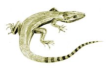
Asaphestera, Archaeothyris and Clepsydrops, the earliest-known synapsids, lived in the Pennsylvanian subperiod (323–299 mya) of the Carboniferous period and were one of many types of primitive synapsids that are now informally grouped together as stem mammals or sometimes as protomammals (previously known as pelycosaurs). The early synapsids spread and diversified, becoming the largest terrestrial animals in the latest Carboniferous and Early Permian periods, ranging up to 6 metres (20 ft) in length. They were sprawling, bulky, possibly cold-blooded, and had small brains. Some, such as Dimetrodon, had large sails that might have helped raise their body temperature. A few relict groups lasted into the later Permian but, by the middle of the Late Permian, all had either died off or evolved into their successors, the therapsids.

Moschops was a tapinocephalian from the Middle Permian of South Africa.

The therapsids, a more advanced group of synapsids, appeared during the Middle Permian and included the largest terrestrial animals in the Middle and Late Permian. They included herbivores and carnivores, ranging from small animals the size of a rat (e.g.: Robertia), to large, bulky herbivores a ton or more in weight (e.g.: Moschops). After flourishing for many millions of years, these successful animals were all but wiped out by the Permian–Triassic mass extinction about 250 mya, the largest known extinction in Earth's history, possibly related to the Siberian Traps volcanic event.

Nikkasaurus was an enigmatic synapsid from the Middle Permian of Russia.
Lystrosaurus was the most common synapsid shortly after the Permian–Triassic extinction event.

Only a few therapsids went on to be successful in the new early Triassic landscape; they include Lystrosaurus and Cynognathus, the latter of which appeared later in the early Triassic. However, they were accompanied by the early archosaurs (soon to give rise to the dinosaurs). Some of these archosaurs, such as Euparkeria, were small and lightly built, while others, such as Erythrosuchus, were as big as or bigger than the largest therapsids.

After the Permian extinction, the synapsids did not count more than three surviving clades. The first comprised the therocephalians, which only lasted the first 20 million years of the Triassic period. The second were specialised, beaked herbivores known as dicynodonts (such as the Kannemeyeriidae), which contained some members that reached large size (up to a tonne or more). And finally there were the increasingly mammal-like carnivorous, herbivorous, and insectivorous cynodonts, including the eucynodonts from the Olenekian age, an early representative of which was Cynognathus.

Cynognathus was the largest predatory cynodont of the Triassic.

Unlike the dicynodonts, which were large, the cynodonts became progressively smaller and more mammal-like as the Triassic progressed, though some forms like Trucidocynodon remained large. The first mammaliaforms evolved from the cynodonts during the early Norian age of the Late Triassic, about 225 mya.

During the evolutionary succession from early therapsid to cynodont to eucynodont to mammal, the main lower jaw bone, the dentary, replaced the adjacent bones. Thus, the lower jaw gradually became just one large bone, with several of the smaller jaw bones migrating into the inner ear and allowing sophisticated hearing.

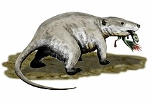
Repenomamus was the largest mammal of the Mesozoic.

Whether through climate change, vegetation change, ecological competition, or a combination of factors, most of the remaining large cynodonts (belonging to the Traversodontidae) and dicynodonts (of the family Kannemeyeriidae) had disappeared by the Rhaetian age, even before the Triassic–Jurassic extinction event that killed off most of the large non-dinosaurian archosaurs. The remaining Mesozoic synapsids were small, ranging from the size of a shrew to the badger-like mammal Repenomamus.

Tritylodon was a cynodont that lived in the Early Jurassic.

During the Jurassic and Cretaceous, the remaining non-mammalian cynodonts were small, such as Tritylodon. No cynodont grew larger than a cat. Most Jurassic and Cretaceous cynodonts were herbivorous, though some were carnivorous. The family Tritheledontidae, which first appeared near the end of the Triassic, was carnivorous and persisted well into the Middle Jurassic. The other, Tritylodontidae, first appeared at the same time as the tritheledonts, but was herbivorous. This group became extinct at the end of the Early Cretaceous epoch. Dicynodonts are generally thought to have become extinct near the end of the Triassic period, but there was evidence this group survived, in the form of six fragments of fossil bone that were found in Cretaceous rocks of Queensland, Australia. If true, it would mean there is a significant ghost lineage of Dicynodonts in Gondwana. However, these fossils were re-described in 2019 as being Pleistocene in age, and possibly belonging to a diprotodontid marsupial.

Today, the 5,500 species of living synapsids, known as the mammals, include both aquatic (whales) and flying (bats) species, and the largest animal ever known to have existed (the blue whale). Humans are synapsids, as well. Most mammals are viviparous and give birth to live young rather than laying eggs with the exception being the monotremes.

Triassic and Jurassic ancestors of living mammals, along with their close relatives, had high metabolic rates. This meant consuming food (generally thought to be insects) in much greater quantity. To facilitate rapid digestion, these synapsids evolved mastication (chewing) and specialized teeth that aided chewing. Limbs also evolved to move under the body instead of to the side, allowing them to breathe more efficiently during locomotion. This helped make it possible to support their higher metabolic demands.

Relationships

Below is a cladogram of the most commonly accepted phylogeny of synapsids, showing a long stem lineage including Mammalia and successively more basal clades such as Theriodontia, Therapsida and Sphenacodontia:

Most uncertainty in the phylogeny of synapsids lies among the earliest members of the group, including forms traditionally placed within Pelycosauria. As one of the earliest phylogenetic analyses, Brinkman & Eberth (1983) placed the family Varanopidae with Caseasauria as the most basal offshoot of the synapsid lineage. Reisz (1986) removed Varanopidae from Caseasauria, placing it in a more derived position on the stem. While most analyses find Caseasauria to be the most basal synapsid clade, Benson's analysis (2012) placed a clade containing Ophiacodontidae and Varanopidae as the most basal synapsids, with Caseasauria occupying a more derived position. Benson attributed this revised phylogeny to the inclusion of postcranial characteristics, or features of the skeleton other than the skull, in his analysis. When only cranial or skull features were included, Caseasauria remained the most basal synapsid clade.

However, more recent examination of the phylogeny of basal synapsids, incorporating newly described basal caseids and eothyridids, returned Caseasauria to its position as the sister to all other synapsids. Brocklehurst et al. (2016) demonstrated that many of the postcranial characters used by Benson (2012) to unite Caseasauria with Sphenacodontidae and Edaphosauridae were absent in the newly discovered postcranial material of eothyridids, and were therefore acquired convergently.

Entropy (information theory)

From Wikipedia, the free encyclopedia https://en.wikipedia.org/wiki/Entropy_(information_theory) In info...