Search This Blog

Wednesday, August 28, 2024

Benign tumor

From Wikipedia, the free encyclopedia
 
Benign tumor
Other namesnon-cancerous tumor
Normal epidermis and dermis with intradermal nevus, 10x-cropped
SpecialtyOncology, Pathology

A benign tumor is a mass of cells (tumor) that does not invade neighboring tissue or metastasize (spread throughout the body). Compared to malignant (cancerous) tumors, benign tumors generally have a slower growth rate. Benign tumors have relatively well differentiated cells. They are often surrounded by an outer surface (fibrous sheath of connective tissue) or stay contained within the epithelium. Common examples of benign tumors include moles and uterine fibroids.

Some forms of benign tumors may be harmful to health. Benign tumor growth causes a mass effect that can compress neighboring tissues. This can lead to nerve damage, blood flow reduction (ischemia), tissue death (necrosis), or organ damage. The health effects of benign tumor growth may be more prominent if the tumor is contained within an enclosed space such as the cranium, respiratory tract, sinus, or bones. For example, unlike most benign tumors elsewhere in the body, benign brain tumors can be life-threatening. Tumors may exhibit behaviors characteristic of their cell type of origin; as an example, endocrine tumors such as thyroid adenomas and adrenocortical adenomas may overproduce certain hormones.

The word "benign" means "favourable, kind, fortunate, salutary, propitious". However, a benign tumour is not benign in the usual sense; the name merely specifies that it is not "malignant", i.e. cancerous. While benign tumours usually do not pose a serious health risk, they can be harmful or fatal. Many types of benign tumors have the potential to become cancerous (malignant) through a process known as tumor progression. For this reason and other possible harms, some benign tumors are removed by surgery. When removed, benign tumors usually do not return. Exceptions to this rule may indicate malignant transformation.

Signs and symptoms

Benign tumors are very diverse; they may be asymptomatic or may cause specific symptoms, depending on their anatomic location and tissue type. They grow outward, producing large, rounded masses which can cause what is known as a "mass effect". This growth can cause compression of local tissues or organs, leading to many effects, such as blockage of ducts, reduced blood flow (ischaemia), tissue death (necrosis) and nerve pain or damage. Some tumors also produce hormones that can lead to life-threatening situations. Insulinomas can produce large amounts of insulin, causing hypoglycemia. Pituitary adenomas can cause elevated levels of hormones such as growth hormone and insulin-like growth factor-1, which cause acromegaly; prolactin; ACTH and cortisol, which cause Cushing's disease; TSH, which causes hyperthyroidism; and FSH and LH. Bowel intussusception can occur with various benign colonic tumors. Cosmetic effects can be caused by tumors, especially those of the skin, possibly causing psychological or social discomfort for the person with the tumor. Vascular tissue tumors can bleed, in some cases leading to anemia.

Causes

PTEN hamartoma syndrome

PTEN hamartoma syndrome encompasses hamartomatous disorders characterized by genetic mutations in the PTEN tumor suppressor gene, including Cowden syndrome, Bannayan–Riley–Ruvalcaba syndrome, Proteus syndrome and Proteus-like syndrome. Absent or dysfunctional PTEN protein allows cells to over-proliferate, causing hamartomas. Cowden syndrome is an autosomal dominant genetic disorder characterized by multiple benign hamartomas (trichilemmomas and mucocutaneous papillomatous papules) as well as a predisposition for cancers of multiple organs including the breast and thyroid. Bannayan–Riley–Ruvalcaba syndrome is a congenital disorder characterized by hamartomatous intestinal polyposis, macrocephaly, lipomatosis, hemangiomatosis and glans penis macules. Proteus syndrome is characterized by nevi, asymmetric overgrowth of various body parts, adipose tissue dysregulation, cystadenomas, adenomas, vascular malformation.

Endoscopic image of sigmoid colon of a patient with familial adenomatous polyposis.

Familial adenomatous polyposis

Familial adenomatous polyposis (FAP) is a familial cancer syndrome caused by mutations in the APC gene. In FAP, adenomatous polyps are present in the colon. The polyps progress into colon cancer unless removed. The APC gene is a tumor suppressor. Its protein product is involved in many cellular processes. Inactivation of the APC gene leads to the buildup of a protein called β-catenin. This protein activates two transcription factors: T-cell factor (TCF) and lymphoid enhancer factor (LEF). These factors cause the upregulation of many genes involved in cell proliferation, differentiation, migration and apoptosis (programmed cell death), causing the growth of benign tumors.

Tuberous sclerosis complex

Tuberous sclerosis complex (TSC) is an autosomal dominant genetic disorder caused by mutations in the genes TSC1 and TSC2. TSC1 produces the protein hamartin. TSC2 produces the protein tuberin. This disorder presents with many benign hamartomatous tumors including angiofibromas, renal angiomyolipomas, and pulmonary lymphangiomyomatosis. Tuberin and hamartin inhibit the mTOR protein in normal cellular physiology. Inactivation of the TSC tumor suppressors causes an increase in mTOR activity. This leads to the activation of genes and the production of proteins that increase cell growth.

Von Hippel–Lindau disease

Von Hippel–Lindau disease is a dominantly inherited cancer syndrome that significantly increases the risk of various tumors. This includes benign hemangioblastomas and malignant pheochromocytomas, renal cell carcinomas, pancreatic endocrine tumors, and endolymphatic sac tumors. It is caused by genetic mutations in the Von Hippel–Lindau tumor suppressor gene. The VHL protein (pVHL) is involved in cellular signaling in oxygen starved (hypoxic) cells. One role of pVHL is to cause the cellular degradation of another protein, HIF1α. Dysfunctional pVHL leads to accumulation of HIF1α. This activates several genes responsible for the production of substances involved in cell growth and blood vessel production: VEGF, PDGFβ, TGFα and erythropoietin.

Bone tumors

Benign tumors of bone can be similar macroscopically and require a combination of a clinical history with cytogenetic, molecular, and radiologic tests for diagnosis. Three common forms of benign bone tumors with are giant cell tumor of bone, osteochondroma, and enchondroma; other forms of benign bone tumors exist but may be less prevalent.

Giant cell tumors

Giant cell tumors of bone frequently occur in long bone epiphyses of the appendicular skeleton or the sacrum of the axial skeleton. Local growth can cause destruction of neighboring cortical bone and soft tissue, leading to pain and limiting range of motion. The characteristic radiologic finding of giant cell tumors of bone is a lytic lesion that does not have marginal sclerosis of bone. On histology, giant cells of fused osteoclasts are seen as a response to neoplastic mononucleated cells. Notably, giant cells are not unique among benign bone tumors to giant cell tumors of bone. Molecular characteristics of the neoplastic cells causing giant cell tumors of bone indicate an origin of pluripotent mesenchymal stem cells that adopt preosteoblastic markers. Cytogenetic causes of giant cell tumors of bone involve telomeres. Treatment involves surgical curettage with adjuvant bisphosphonates.

Osteochondroma

Osteochondromas form cartilage-capped projections of bone. Structures such as the marrow cavity and cortical bone of the osteochondroma are contiguous to those of the originating bone. Sites of origin often involve metaphyses of long bones. While many osteochondromas occur spontaneously, there are cases in which several osteochondromas can occur in the same individual; these may be linked to a genetic condition known as hereditary multiple osteochondromas. Osteochondroma appears on X-ray as a projecting mass that often points away from joints. These tumors stop growing with the closure of the parental bone's growth plates. Failure to stop growth can be indicative of transformation to malignant chondrosarcoma. Treatment is not indicated unless symptomatic. In that case, surgical excision is often curative.

Enchondroma

Enchondromas are benign tumors of hyaline cartilage. Within a bone, enchondromas are often found in metaphyses. They can be found in many types of bone, including small bones, long bones, and the axial skeleton. X-ray of enchondromas shows well-defined borders and a stippled appearance. Presentation of multiple enchondromas is consistent with multiple enchondromatosis (Ollier Disease). Treatment of enchondromas involves surgical curettage and grafting.

Benign soft tissue tumors

Lipomas

Lipomas are benign, subcutaneous tumors of fat cells (adipocytes). They are usually painless, slow-growing, and mobile masses that can occur anywhere in the body where there are fat cells, but are typically found on the trunk and upper extremities.

 Although lipomas can develop at any age, they more commonly appear between the ages of 40 and 60. Lipomas affect about 1% of the population, with no documented sex bias, and about 1 in every 1000 people will have a lipoma within their lifetime. The cause of lipomas is not well defined. Genetic or inherited causes of lipomas play a role in around 2-3% of patients. In individuals with inherited familial syndromes such as Proteus syndrome or Familial multiple lipomatosis, it is common to see multiple lipomas across the body. These syndromes are also associated with specific symptoms and sub-populations. Mutations in chromosome 12 have been identified in around 65% of lipoma cases. Lipomas have also been shown to be increased in those with obesity, hyperlipidemia, and diabetes mellitus.

Lipomas are usually diagnosed clinically, although imaging (ultrasound, computed tomography, or magnetic resonance imaging) may be utilized to assist with the diagnosis of lipomas in atypical locations. The main treatment for lipomas is surgical excision, after which the tumor is examined with histopathology to confirm the diagnosis. The prognosis for benign lipomas is excellent and recurrence after excision is rare, but may occur if the removal was incomplete.

Mechanism

Benign (L) vs Malignant tumor (R).

Benign vs malignant

Diagram showing two epithelial tumors. The upper tumor is a benign tumor that is non-invasive. Benign tumors are usually round in shape and encapsulated by fibrous connective tissue. The lower picture depicts a malignant tumor. It is irregularly shaped, vascular, and it is invasive, crossing the basement membrane.

One of the most important factors in classifying a tumor as benign or malignant is its invasive potential. If a tumor lacks the ability to invade adjacent tissues or spread to distant sites by metastasizing then it is benign, whereas invasive or metastatic tumors are malignant. For this reason, benign tumors are not classed as cancer. Benign tumors will grow in a contained area usually encapsulated in a fibrous connective tissue capsule. The growth rates of benign and malignant tumors also differ; benign tumors generally grow more slowly than malignant tumors. Although benign tumors pose a lower health risk than malignant tumors, they both can be life-threatening in certain situations. There are many general characteristics which apply to either benign or malignant tumors, but sometimes one type may show characteristics of the other. For example, benign tumors are mostly well differentiated and malignant tumors are often undifferentiated. However, undifferentiated benign tumors and differentiated malignant tumors can occur. Although benign tumors generally grow slowly, cases of fast-growing benign tumors have also been documented. Some malignant tumors are mostly non-metastatic such as in the case of basal-cell carcinoma. CT and chest radiography can be a useful diagnostic exam in visualizing a benign tumor and differentiating it from a malignant tumor. The smaller the tumor on a radiograph the more likely it is to be benign as 80% of lung nodules less than 2 cm in diameter are benign. Most benign nodules are smoothed radiopaque densities with clear margins but these are not exclusive signs of benign tumors.

Multistage carcinogenesis

Tumors are formed by carcinogenesis, a process in which cellular alterations lead to the formation of cancer. Multistage carcinogenesis involves the sequential genetic or epigenetic changes to a cell's DNA, where each step produces a more advanced tumor. It is often broken down into three stages; initiation, promotion and progression, and several mutations may occur at each stage. Initiation is where the first genetic mutation occurs in a cell. Promotion is the clonal expansion (repeated division) of this transformed cell into a visible tumor that is usually benign. Following promotion, progression may take place where more genetic mutations are acquired in a sub-population of tumor cells. Progression changes the benign tumor into a malignant tumor. A prominent and well studied example of this phenomenon is the tubular adenoma, a common type of colon polyp which is an important precursor to colon cancer. The cells in tubular adenomas, like most tumors that frequently progress to cancer, show certain abnormalities of cell maturation and appearance collectively known as dysplasia. These cellular abnormalities are not seen in benign tumors that rarely or never turn cancerous, but are seen in other pre-cancerous tissue abnormalities which do not form discrete masses, such as pre-cancerous lesions of the uterine cervix.

Diagnosis

Classification

Cell origin Cell type Tumor
Endodermal Biliary tree Cholangioma
Colon Colonic polyp
Glandular Adenoma
Papilloma
Cystadenoma
Liver Liver cell adenoma
Placental Hydatiform mole
Renal Renal tubular adenoma
Squamous Squamous cell papilloma
Stomach Gastric polyp
Mesenchymal Blood vessel Hemangioma, Cardiac myxoma
Bone Osteoma
Cartilage Chondroma
Fat tissue Lipoma
Fibrous tissue Fibroma
Lymphatic vessel Lymphangioma
Smooth muscle Leiomyoma
Striated muscle Rhabdomyoma
Ectodermal Glia Astrocytoma, Schwannoma
Melanocytes Nevus
Meninges Meningioma
Nerve cells Ganglioneuroma

Benign neoplasms are typically, but not always, composed of cells which bear a strong resemblance to a normal cell type in their organ of origin. These tumors are named for the cell or tissue type from which they originate. The suffix "-oma" (but not -carcinoma, -sarcoma, or -blastoma, which are generally cancers) is applied to indicate a benign tumor. For example, a lipoma is a common benign tumor of fat cells (lipocytes), and a chondroma is a benign tumor of cartilage-forming cells (chondrocytes). Adenomas are benign tumors of gland-forming cells, and are usually specified further by their cell or organ of origin, as in hepatic adenoma (a benign tumor of hepatocytes, or liver cells). Teratomas contain many cell types such as skin, nerve, brain and thyroid, among others, because they are derived from germ cells. Hamartomas are a group of benign tumors that have relatively normal cellular differentiation but exhibit disorganized tissue organization.

Exceptions to the nomenclature rules exist for historical reasons; malignant examples include melanoma (a cancer of pigmented skin cells, or melanocytes) and seminoma (a cancer of male reproductive cells).

Benign tumors do not encompass all benign growths. Skin tags, vocal chord polyps, and hyperplastic polyps of the colon are often referred to as benign, but they are overgrowths of normal tissue rather than neoplasms.

Treatment

Benign tumors typically need no treatment unless if they cause problems such as seizures, discomfort or cosmetic concerns. Surgery is usually the most effective approach and is used to treat most benign tumors. In some cases, other treatments may be used. Adenomas of the rectum may be treated with sclerotherapy, in which chemicals are used to shrink blood vessels in order to cut off the blood supply. Most benign tumors do not respond to chemotherapy or radiation therapy, although there are exceptions; benign intercranial tumors are sometimes treated with radiation therapy and chemotherapy under certain circumstances. Radiation can also be used to treat hemangiomas in the rectum. Benign skin tumors are usually surgically resected but other treatments such as cryotherapy, curettage, electrodesiccation, laser therapy, dermabrasion, chemical peels and topical medication are used.

Metastasis

From Wikipedia, the free encyclopedia
 
Metastasis
Other namesMetastatic disease  
Illustration showing hematogenous metastasis
Pronunciation
SpecialtyOncology

Metastasis is a pathogenic agent's spread from an initial or primary site to a different or secondary site within the host's body; the term is typically used when referring to metastasis by a cancerous tumor. The newly pathological sites, then, are metastases (mets). It is generally distinguished from cancer invasion, which is the direct extension and penetration by cancer cells into neighboring tissues.

Cancer occurs after cells are genetically altered to proliferate rapidly and indefinitely. This uncontrolled proliferation by mitosis produces a primary heterogeneic tumour. The cells which constitute the tumor eventually undergo metaplasia, followed by dysplasia then anaplasia, resulting in a malignant phenotype. This malignancy allows for invasion into the circulation, followed by invasion to a second site for tumorigenesis.

Some cancer cells known as circulating tumor cells acquire the ability to penetrate the walls of lymphatic or blood vessels, after which they are able to circulate through the bloodstream to other sites and tissues in the body. This process is known (respectively) as lymphatic or hematogenous spread. After the tumor cells come to rest at another site, they re-penetrate the vessel or walls and continue to multiply, eventually forming another clinically detectable tumor. This new tumor is known as a metastatic (or secondary) tumor. Metastasis is one of the hallmarks of cancer, distinguishing it from benign tumors. Most cancers can metastasize, although in varying degrees. Basal cell carcinoma for example rarely metastasizes.

When tumor cells metastasize, the new tumor is called a secondary or metastatic tumor, and its cells are similar to those in the original or primary tumor. This means that if breast cancer metastasizes to the lungs, the secondary tumor is made up of abnormal breast cells, not of abnormal lung cells. The tumor in the lung is then called metastatic breast cancer, not lung cancer. Metastasis is a key element in cancer staging systems such as the TNM staging system, where it represents the "M". In overall stage grouping, metastasis places a cancer in Stage IV. The possibilities of curative treatment are greatly reduced, or often entirely removed when a cancer has metastasized.

Signs and symptoms

Cut surface of a liver showing multiple paler metastatic nodules originating from pancreatic cancer

Initially, nearby lymph nodes are struck early. The lungs, liver, brain, and bones are the most common metastasis locations from solid tumors.

Although advanced cancer may cause pain, it is often not the first symptom.

Some patients, however, do not show any symptoms. When the organ gets a metastatic disease it begins to shrink until its lymph nodes burst, or undergo lysis.

Pathophysiology

Metastatic tumors are very common in the late stages of cancer. The spread of metastasis may occur via the blood or the lymphatics or through both routes. The most common sites of metastases are the lungs, liver, brain, and the bones.

Currently, three main theories have been proposed to explain the metastatic pathway of cancer: the epithelial-mesenchymal transition (EMT) and mesenchymal-epithelial transition (MET) hypothesis (1), the cancer stem cell hypothesis (2), and the macrophage–cancer cell fusion hybrid hypothesis (3). Some new hypotheses were suggested as well, i.e., under the effect of particular biochemical and/or physical stressors, cancer cells can undergo nuclear expulsion with subsequent macrophage engulfment and fusion, with the formation of cancer fusion cells (CFCs). Understanding the enigma of cancer cell spread to distant sites, which accounts for over 90% of cancer-related deaths, necessitates comprehensive investigation. Key outstanding questions revolve around the survival and migration of cancer cells, such as the nucleus, as they face challenges in passage through capillary valves and hydrodynamic shear forces in the circulation system, making CTCs an unlikely source of metastasis. Moreover, understanding how cancer cells adapt to the metastatic niche and remain dormant (tumor dormancy) for extended periods presents difficult questions that require further investigation.

Factors involved

Metastasis involves a complex series of steps in which cancer cells leave the original tumor site and migrate to other parts of the body via the bloodstream, via the lymphatic system, or by direct extension. To do so, malignant cells break away from the primary tumor and attach to and degrade proteins that make up the surrounding extracellular matrix (ECM), which separates the tumor from adjoining tissues. By degrading these proteins, cancer cells are able to breach the ECM and escape. The location of the metastases is not always random, with different types of cancer tending to spread to particular organs and tissues at a rate that is higher than expected by statistical chance alone. Breast cancer, for example, tends to metastasize to the bones and lungs. This specificity seems to be mediated by soluble signal molecules such as chemokines and transforming growth factor beta. The body resists metastasis by a variety of mechanisms through the actions of a class of proteins known as metastasis suppressors, of which about a dozen are known.

Human cells exhibit different kinds of motion: collective motility, mesenchymal-type movement, and amoeboid movement. Cancer cells often opportunistically switch between different kinds of motion. Some cancer researchers hope to find treatments that can stop or at least slow down the spread of cancer by somehow blocking some necessary step in one or more kinds of motion.

All steps of the metastatic cascade involve a number of physical processes. Cell migration requires the generation of forces, and when cancer cells transmigrate through the vasculature, this requires physical gaps in the blood vessels to form. Besides forces, the regulation of various types of cell-cell and cell-matrix adhesions is crucial during metastasis.

The metastatic steps are critically regulated by various cell types, including the blood vessel cells (endothelial cells), immune cells or stromal cells. The growth of a new network of blood vessels, called tumor angiogenesis, is a crucial hallmark of cancer. It has therefore been suggested that angiogenesis inhibitors would prevent the growth of metastases. Endothelial progenitor cells have been shown to have a strong influence on metastasis and angiogenesis. Endothelial progenitor cells are important in tumor growth, angiogenesis and metastasis, and can be marked using the Inhibitor of DNA Binding 1 (ID1). This novel finding meant that investigators gained the ability to track endothelial progenitor cells from the bone marrow to the blood to the tumor-stroma and even incorporated in tumor vasculature. Endothelial progenitor cells incorporated in tumor vasculature suggests that this cell type in blood-vessel development is important in a tumor setting and metastasis. Furthermore, ablation of the endothelial progenitor cells in the bone marrow can lead to a significant decrease in tumor growth and vasculature development. Therefore, endothelial progenitor cells are important in tumor biology and present novel therapeutic targets. The immune system is typically deregulated in cancer and affects many stages of tumor progression, including metastasis.

Epigenetic regulation also plays an important role in the metastatic outgrowth of disseminated tumor cells. Metastases display alterations in histone modifications, such as H3K4-methylation and H3K9-methylation, when compared to matching primary tumors. These epigenetic modifications in metastases may allow the proliferation and survival of disseminated tumor cells in distant organs.

A recent study shows that PKC-iota promotes melanoma cell invasion by activating Vimentin during EMT. PKC-iota inhibition or knockdown resulted in an increase in E-cadherin and RhoA levels while decreasing total Vimentin, phosphorylated Vimentin (S39) and Par6 in metastatic melanoma cells. These results suggested that PKC-ι is involved in signaling pathways which upregulate EMT in melanoma thereby directly stimulates metastasis.

Recently, a series of high-profile experiments suggests that the co-option of intercellular cross-talk mediated by exosome vesicles is a critical factor involved in all steps of the invasion-metastasis cascade.

Routes

Metastasis occurs by the following four routes:

Transcoelomic

The spread of a malignancy into body cavities can occur via penetrating the surface of the peritoneal, pleural, pericardial, or subarachnoid spaces. For example, ovarian tumors can spread transperitoneally to the surface of the liver.

Lymphatic spread

Lymphatic spread allows the transport of tumor cells to regional lymph nodes near the primary tumor and ultimately, to other parts of the body. This is called nodal involvement, positive nodes, or regional disease. "Positive nodes" is a term that would be used by medical specialists to describe regional lymph nodes that tested positive for malignancy. It is common medical practice to test by biopsy at least one lymph node near a tumor site when carrying out surgery to examine or remove a tumor. This lymph node is then called a sentinel lymph node. Lymphatic spread is the most common route of initial metastasis for carcinomas. In contrast, it is uncommon for a sarcoma to metastasize via this route. Localized spread to regional lymph nodes near the primary tumor is not normally counted as a metastasis, although this is a sign of a worse outcome. The lymphatic system does eventually drain from the thoracic duct and right lymphatic duct into the systemic venous system at the venous angle and into the brachiocephalic veins, and therefore these metastatic cells can also eventually spread through the haematogenous route.

Lymph node with almost complete replacement by metastatic melanoma. The brown pigment is focal deposition of melanin

Hematogenous spread

This is typical route of metastasis for sarcomas, but it is also the favored route for certain types of carcinoma, such as renal cell carcinoma originating in the kidney and follicular carcinomas of the thyroid. Because of their thinner walls, veins are more frequently invaded than are arteries, and metastasis tends to follow the pattern of venous flow. That is, hematogenous spread often follows distinct patterns depending on the location of the primary tumor. For example, colorectal cancer spreads primarily through the portal vein to the liver.

Canalicular spread

Some tumors, especially carcinomas may metastasize along anatomical canalicular spaces. These spaces include for example the bile ducts, the urinary system, the airways and the subarachnoid space. The process is similar to that of transcoelomic spread. However, often it remains unclear whether simultaneously diagnosed tumors of a canalicular system are one metastatic process or in fact independent tumors caused by the same agent (field cancerization).

Organ-specific targets

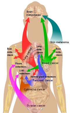
Main sites of metastases for some common cancer types. Primary cancers are denoted by "...cancer" and their main metastasis sites are denoted by "...metastases".

There is a propensity for certain tumors to seed in particular organs. This was first discussed as the "seed and soil" theory by Stephen Paget in 1889. The propensity for a metastatic cell to spread to a particular organ is termed 'organotropism'. For example, prostate cancer usually metastasizes to the bones. In a similar manner, colon cancer has a tendency to metastasize to the liver. Stomach cancer often metastasises to the ovary in women, when it is called a Krukenberg tumor.

According to the "seed and soil" theory, it is difficult for cancer cells to survive outside their region of origin, so in order to metastasize they must find a location with similar characteristics. For example, breast tumor cells, which gather calcium ions from breast milk, metastasize to bone tissue, where they can gather calcium ions from bone. Malignant melanoma spreads to the brain, presumably because neural tissue and melanocytes arise from the same cell line in the embryo.

In 1928, James Ewing challenged the "seed and soil" theory and proposed that metastasis occurs purely by anatomic and mechanical routes. This hypothesis has been recently utilized to suggest several hypotheses about the life cycle of circulating tumor cells (CTCs) and to postulate that the patterns of spread could be better understood through a 'filter and flow' perspective. However, contemporary evidences indicate that the primary tumour may dictate organotropic metastases by inducing the formation of pre-metastatic niches at distant sites, where incoming metastatic cells may engraft and colonise. Specifically, exosome vesicles secreted by tumours have been shown to home to pre-metastatic sites, where they activate pro-metastatic processes such as angiogenesis and modify the immune contexture, so as to foster a favourable microenvironment for secondary tumour growth.

Metastasis and primary cancer

It is theorized that metastasis always coincides with a primary cancer, and, as such, is a tumor that started from a cancer cell or cells in another part of the body. However, over 10% of patients presenting to oncology units will have metastases without a primary tumor found. In these cases, doctors refer to the primary tumor as "unknown" or "occult," and the patient is said to have cancer of unknown primary origin (CUP) or unknown primary tumors (UPT). It is estimated that 3% of all cancers are of unknown primary origin. Studies have shown that, if simple questioning does not reveal the cancer's source (coughing up blood—"probably lung", urinating blood—"probably bladder"), complex imaging will not either. In some of these cases a primary tumor may appear later.

The use of immunohistochemistry has permitted pathologists to give an identity to many of these metastases. However, imaging of the indicated area only occasionally reveals a primary. In rare cases (e.g., of melanoma), no primary tumor is found, even on autopsy. It is therefore thought that some primary tumors can regress completely, but leave their metastases behind. In other cases, the tumor might just be too small and/or in an unusual location to be diagnosed.

Diagnosis

Pulmonary metastases shown on Chest X-Ray

The cells in a metastatic tumor resemble those in the primary tumor. Once the cancerous tissue is examined under a microscope to determine the cell type, a doctor can usually tell whether that type of cell is normally found in the part of the body from which the tissue sample was taken.

For instance, breast cancer cells look the same whether they are found in the breast or have spread to another part of the body. So, if a tissue sample taken from a tumor in the lung contains cells that look like breast cells, the doctor determines that the lung tumor is a secondary tumor. Still, the determination of the primary tumor can often be very difficult, and the pathologist may have to use several adjuvant techniques, such as immunohistochemistry, FISH (fluorescent in situ hybridization), and others. Despite the use of techniques, in some cases the primary tumor remains unidentified.

Metastatic cancers may be found at the same time as the primary tumor, or months or years later. When a second tumor is found in a patient that has been treated for cancer in the past, it is more often a metastasis than another primary tumor.

It was previously thought that most cancer cells have a low metastatic potential and that there are rare cells that develop the ability to metastasize through the development of somatic mutations. According to this theory, diagnosis of metastatic cancers is only possible after the event of metastasis. Traditional means of diagnosing cancer (e.g. a biopsy) would only investigate a subpopulation of the cancer cells and would very likely not sample from the subpopulation with metastatic potential.

The somatic mutation theory of metastasis development has not been substantiated in human cancers. Rather, it seems that the genetic state of the primary tumor reflects the ability of that cancer to metastasize. Research comparing gene expression between primary and metastatic adenocarcinomas identified a subset of genes whose expression could distinguish primary tumors from metastatic tumors, dubbed a "metastatic signature." Up-regulated genes in the signature include: SNRPF, HNRPAB, DHPS and securin. Actin, myosin and MHC class II down-regulation was also associated with the signature. Additionally, the metastatic-associated expression of these genes was also observed in some primary tumors, indicating that cells with the potential to metastasize could be identified concurrently with diagnosis of the primary tumor. Recent work identified a form of genetic instability in cancer called chromosome instability (CIN) as a driver of metastasis. In aggressive cancer cells, loose DNA fragments from unstable chromosomes spill in the cytosol leading to the chronic activation of innate immune pathways, which are hijacked by cancer cells to spread to distant organs.

Expression of this metastatic signature has been correlated with a poor prognosis and has been shown to be consistent in several types of cancer. Prognosis was shown to be worse for individuals whose primary tumors expressed the metastatic signature. Additionally, the expression of these metastatic-associated genes was shown to apply to other cancer types in addition to adenocarcinoma. Metastases of breast cancer, medulloblastoma and prostate cancer all had similar expression patterns of these metastasis-associated genes.

The identification of this metastasis-associated signature provides promise for identifying cells with metastatic potential within the primary tumor and hope for improving the prognosis of these metastatic-associated cancers. Additionally, identifying the genes whose expression is changed in metastasis offers potential targets to inhibit metastasis.

Management

Treatment and survival is determined, to a great extent, by whether or not a cancer remains localized or spreads to other locations in the body. If the cancer metastasizes to other tissues or organs it usually dramatically increases a patient's likelihood of death. Some cancers—such as some forms of leukemia, a cancer of the blood, or malignancies in the brain—can kill without spreading at all.

Once a cancer has metastasized it may still be treated with radiosurgery, chemotherapy, radiation therapy, biological therapy, hormone therapy, surgery, or a combination of these interventions ("multimodal therapy"). The choice of treatment depends on many factors, including the type of primary cancer, the size and location of the metastases, the patient's age and general health, and the types of treatments used previously. In patients diagnosed with CUP it is often still possible to treat the disease even when the primary tumor cannot be located.

Current treatments are rarely able to cure metastatic cancer though some tumors, such as testicular cancer and thyroid cancer, are usually curable.

Palliative care, care aimed at improving the quality of life of people with major illness, has been recommended as part of management programs for metastasis. Results from a systematic review of the literature on radiation therapy for brain metastases found that there is little evidence to inform comparative effectiveness and patient-centered outcomes on quality of life, functional status, and cognitive effects.

Research

Although metastasis is widely accepted to be the result of the tumor cells migration, there is a hypothesis saying that some metastases are the result of inflammatory processes by abnormal immune cells. The existence of metastatic cancers in the absence of primary tumors also suggests that metastasis is not always caused by malignant cells that leave primary tumors.

The research done by Sarna's team proved that heavily pigmented melanoma cells have Young's modulus about 4.93, when in non-pigmented ones it was only 0.98. In another experiment they found that elasticity of melanoma cells is important for its metastasis and growth: non-pigmented tumors were bigger than pigmented and it was much easier for them to spread. They shown that there are both pigmented and non-pigmented cells in melanoma tumors, so that they can both be drug-resistant and metastatic.

History

The first physician to report the possibility of local metastasis from a primary cancerous source to nearby tissues was Ibn Sina. He described a case of breast cancer and metastatic condition in The Canon of Medicine. His hypothesis was based on clinical course of the patient.

In March 2014 researchers discovered the oldest complete example of a human with metastatic cancer. The tumors had developed in a 3,000-year-old skeleton found in 2013 in a tomb in Sudan dating back to 1200 BC. The skeleton was analyzed using radiography and a scanning electron microscope. These findings were published in the Public Library of Science journal.

Etymology

Metastasis is a Greek word meaning "displacement", from μετά, meta, "next", and στάσις, stasis, "placement".

Earliest known life forms

From Wikipedia, the free encyclopedia
https://en.wikipedia.org/wiki/Earliest_known_life_forms
Evidence of possibly the oldest forms of life on Earth has been found in hydrothermal vent precipitates.

The earliest known life forms on Earth may be as old as 4.1 billion years old (or Ga) according to biologically fractionated graphite inside a single zircon grain in the Jack Hills range of Australia. The earliest evidence of life found in a stratigraphic unit, not just a single mineral grain, is the 3.7 Ga metasedimentary rocks containing graphite from the Isua Supracrustal Belt in Greenland. The earliest direct known life on land may be stromatolites which have been found in 3.480-billion-year-old geyserite uncovered in the Dresser Formation of the Pilbara Craton of Western Australia. Various microfossils of microorganisms have been found in 3.4 Ga rocks, including 3.465-billion-year-old Apex chert rocks from the same Australian craton region, and in 3.42 Ga hydrothermal vent precipitates from Barberton, South Africa. Much later in the geologic record, likely starting in 1.73 Ga, preserved molecular compounds of biologic origin are indicative of aerobic life. Therefore, the earliest time for the origin of life on Earth is at least 3.5 billion years ago, possibly as early as 4.1 billion years ago — not long after the oceans formed 4.5 billion years ago and after the formation of the Earth 4.54 billion years ago.

Biospheres

Earth is the only place in the universe known to harbor life, where it exists in multiple environments. The origin of life on Earth was at least 3.5 billion years ago, possibly as early as 3.8-4.1 billion years ago. Since its emergence, life has persisted in several geological environments. The Earth's biosphere extends down to at least 10 km (6.2 mi) below the seafloor, up to 41–77 km (25–48 mi) into the atmosphere, and includes soil, hydrothermal vents, and rock. Further, the biosphere has been found to extend at least 914.4 m (3,000 ft; 0.5682 mi) below the ice of Antarctica and includes the deepest parts of the ocean. In July 2020, marine biologists reported that aerobic microorganisms (mainly) in "quasi-suspended animation" were found in organically poor sediment 76.2 m (250 ft) below the seafloor in the South Pacific Gyre (SPG) ("the deadest spot in the ocean"). Microbes have been found in the Atacama Desert in Chile, one of the driest places on Earth, and in deep-sea hydrothermal vent environments which can reach temperatures over 400°C. Microbial communities can also survive in cold permafrost conditions down to -25°C. Under certain test conditions, life forms have been observed to survive in the vacuum of outer space. More recently, studies conducted on the International Space Station found that bacteria could survive in outer space. In February 2023, findings of a "dark microbiome" of unfamiliar microorganisms in the Atacama Desert in Chile, a Mars-like region of planet Earth, were reported.

Geochemical evidence

The age of Earth is about 4.54 billion years; the earliest undisputed evidence of life on Earth dates from at least 3.5 billion years ago according to the stromatolite record. Some computer models suggest life began as early as 4.5 billion years ago. The oldest evidence of life is indirect in the form of isotopic fractionation. Microorganisms will preferentially use the lighter isotope of an atom to build biomass, as it takes less energy to break the bonds for metabolic processes. Biologic material will often have a composition that is enriched in lighter isotopes compared to the surrounding rock it's found in. Carbon isotopes, expressed scientifically in parts per thousand difference from a standard as δ13C, are frequently used to detect carbon fixation by organisms and assess if purported early life evidence has biological origins. Typically, life will preferentially metabolize the isotopically light 12C isotope instead of the heavier 13C isotope. Biologic material can record this fractionation of carbon.

Zircons in metaconglomerates from the Jack Hills in Australia show carbon isotopic evidence for early life.

The oldest disputed geochemical evidence of life is isotopically light graphite inside a single zircon grain from the Jack Hills in Western Australia. The graphite showed a δ13C signature consistent with biogenic carbon on Earth. Other early evidence of life is found in rocks both from the Akilia Sequence and the Isua Supracrustal Belt (ISB) in Greenland. These 3.7 Ga metasedimentary rocks also contain graphite or graphite inclusions with carbon isotope signatures that suggest biological fractionation.

The primary issue with isotopic evidence of life is that abiotic processes can fractionate isotopes and produce similar signatures to biotic processes. Reassessment of the Akilia graphite show that metamorphism, Fischer-Tropsch mechanisms in hydrothermal environments, and volcanic processes may be responsible for enrichment lighter carbon isotopes. The ISB rocks that contain the graphite may have experienced a change in composition from hot fluids, i.e. metasomatism, thus the graphite may have been formed by abiotic chemical reactions. However, the ISB's graphite is generally more accepted as biologic in origin after further spectral analysis.

Metasedimentary rocks from the 3.5 Ga Dresser Formation, which experienced less metamorphism than the sequences in Greenland, contain better preserved geochemical evidence. Carbon isotopes as well as sulfur isotopes found in barite, which are fractionated by microbial metabolisms during sulfate reduction, are consistent with biological processes. However, the Dresser formation was deposited in an active volcanic and hydrothermal environment, and abiotic processes could still be responsible for these fractionations. Many of these findings are supplemented by direct evidence, typically by the presence of microfossils, however.

Fossil evidence

Fossils are direct evidence of life. In the search for the earliest life, fossils are often supplemented by geochemical evidence. The fossil record does not extend as far back as the geochemical record due to metamorphic processes that erase fossils from geologic units.

Stromatolites

Stromatolites are laminated sedimentary structures created by photosynthetic organisms as they establish a microbial mat on a sediment surface. An important distinction for biogenicity is their convex-up structures and wavy laminations, which are typical of microbial communities who build preferentially toward the sun. A disputed report of stromatolites is from the 3.7 Ga Isua metasediments that show convex-up, conical, and domical morphologies. Further mineralogical analysis disagrees with the initial findings of internal convex-up laminae, a critical criteria for stromatolite identification, suggesting that the structures may be deformation features (i.e. boudins) caused by extensional tectonics in the Isua Supracrustal Belt.

Stromatolite fossil showing convex-up structures.

The earliest direct evidence of life are stromatolites found in 3.48 billion-year-old chert in the Dresser formation of the Pilbara Craton in Western Australia. Several features in these fossils are difficult to explain with abiotic processes, for example, the thickening of laminae over flexure crests that is expected from more sunlight. Sulfur isotopes from barite veins in the stromatolites also favor a biologic origin. However, while most scientists accept their biogenicity, abiotic explanations for these fossils cannot be fully discarded due to their hydrothermal depositional environment and debated geochemical evidence.

Most archean stromatolites older than 3.0 Ga are found in Australia or South Africa. Stratiform stromatolites from the Pilbara Craton have been identified in the 3.47 Ga Mount Ada Basalt. Barberton, South Africa hosts stratiform stromatolites in the 3.46 Hooggenoeg, 3.42 Kromberg and 3.33 Ga Mendon Formations of the Onverwacht Group. The 3.43 Ga Strelley Pool Formation in Western Australia hosts stromatolites that demonstrate vertical and horizontal changes that may demonstrate microbial communities responding to transient environmental conditions. Thus, it is likely anoxygenic or oxygenic photosynthesis has been occurring since at least 3.43 Ga Strelley Pool Formation.

Microfossils

Claims of the earliest life using fossilized microorganisms (microfossils) are from hydrothermal vent precipitates from an ancient sea-bed in the Nuvvuagittuq Belt of Quebec, Canada. These may be as old as 4.28 billion years, which would make it the oldest evidence of life on Earth, suggesting "an almost instantaneous emergence of life" after ocean formation 4.41 billion years ago. These findings may be better explained by abiotic processes: for example, silica-rich waters, "chemical gardens," circulating hydrothermal fluids, and volcanic ejecta can produce morphologies similar to those presented in Nuvvuagittuq.

Archaea (prokaryotic microbes) were first found in extreme environments, such as hydrothermal vents.

The 3.48 Ga Dresser formation hosts microfossils of prokaryotic filaments in silica veins, the earliest fossil evidence of life on Earth, but their origins may be volcanic. 3.465-billion-year-old Australian Apex chert rocks may once have contained microorganisms, although the validity of these findings has been contested. "Putative filamentous microfossils," possibly of methanogens and/or methanotrophs that lived about 3.42-billion-year-old in "a paleo-subseafloor hydrothermal vein system of the Barberton greenstone belt, have been identified in South Africa." A diverse set of microfossil morphologies have been found in the 3.43 Ga Strelley Pool Formation including spheroid, lenticular, and film-like microstructures. Their biogenicity are strengthened by their observed chemical preservation. The early lithification of these structures allowed important chemical tracers, such as the carbon-to-nitrogen ratio, to be retained at levels higher than is typical in older, metamorphosed rock units.

Molecular Biomarkers

Biomarkers are compounds of biologic origin found in the geologic record that can be linked to past life. Although they aren't preserved until the late Archean, they are important indicators of early photosynthetic life. Lipids are particularly useful biomarkers because they can survive for long periods of geologic time and reconstruct past environments.

Lipids are commonly used in geologic studies to find evidence of oxygenic photosynthesis.

Fossilized lipids were reported from 2.7 Ga laminated shales from the Pilbara Craton and the 2.67 Ga Kaapvaal Craton in South Africa. However, the age of these biomarkers and whether their deposition was synchronous with their host rocks were debated, and further work showed that the lipids were contaminants. The oldest "clearly indigenous" biomarkers are from the 1.64 Ga Barney Creek Formation in the McArthur Basin in Northern Australia, but hydrocarbons from the 1.73 Ga Wollogorang Formation in the same basin have also been detected.

Other indigenous biomarkers can be dated to the Mesoproterozoic era (1.6-1.0 Ga). The 1.4 Ga Hongshuizhuang Formation in the North China Craton contains hydrocarbons in shales that were likely sourced from prokaryotes. Biomarkers were found in siltstones from the 1.38 Ga Roper Group of the McArthur Basin. Hydrocarbons possibly derived from bacteria and algae were reported in 1.37 Ga Xiamaling Formation of the NCC. The 1.1 Ga Atar/El Mreïti Group in the Taoudeni Basin, Mauritania show indigenous biomarkers in black shales.

Genomic evidence

By comparing the genomes of modern organisms (in the domains Bacteria and Archaea), it is evident that there was a last universal common ancestor (LUCA). LUCA is not thought to be the first life on Earth, but rather the only type of organism of its time to still have living descendants. In 2016, M. C. Weiss and colleagues proposed a minimal set of genes that each occurred in at least two groups of Bacteria and two groups of Archaea. They argued that such a distribution of genes would be unlikely to arise by horizontal gene transfer, and so any such genes must have derived from the LUCA. A molecular clock model suggests that the LUCA may have lived 4.477—4.519 billion years ago, within the Hadean eon.

RNA replicators

Model Hadean-like geothermal microenvironments were demonstrated to have the potential to support the synthesis and replication of RNA and thus possibly the evolution of primitive life. Porous rock systems, comprising heated air-water interfaces, were shown to facilitate ribozyme catalyzed RNA replication of sense and antisense strands and then subsequent strand-dissociation. This enabled combined synthesis, release and folding of active ribozymes.

Further work on early life

Extraterrestrial origin for early life?

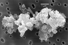
The theory of panspermia speculates that life on Earth may have come from biological matter carried by space dust or meteorites.

While current geochemical evidence dates the origin of life to possibly as early as 4.1 Ga, and fossil evidence shows life at 3.5 Ga, some researchers speculate that life may have started nearly 4.5 billion years ago. According to biologist Stephen Blair Hedges, "If life arose relatively quickly on Earth ... then it could be common in the universe." The possibility that terrestrial life forms may have been seeded from outer space has been considered. In January 2018, a study found that 4.5 billion-year-old meteorites found on Earth contained liquid water along with prebiotic complex organic substances that may be ingredients for life.

Early life on land

As for life on land, in 2019 scientists reported the discovery of a fossilized fungus, named Ourasphaira giraldae, in the Canadian Arctic, that may have grown on land a billion years ago, well before plants are thought to have been living on land. The earliest life on land may have been bacteria 3.22 billion years ago. Evidence of microbial life on land may have been found in 3.48 billion-year-old geyserite in the Pilbara Craton of Western Australia.

Lie group

From Wikipedia, the free encyclopedia https://en.wikipedia.org/wiki/Lie_group In mathematics , a Lie gro...