Search This Blog

Monday, April 29, 2019

Fatty acid

From Wikipedia, the free encyclopedia

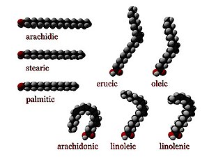
Three-dimensional representations of several fatty acids. Saturated fatty acids have perfectly straight chain structure. Unsaturated ones are typically bent, unless they have a trans configuration.
 
In chemistry, particularly in biochemistry, a fatty acid is a carboxylic acid with a long aliphatic chain, which is either saturated or unsaturated. Most naturally occurring fatty acids have an unbranched chain of an even number of carbon atoms, from 4 to 28. Fatty acids are usually not found in organisms, but instead as three main classes of esters: triglycerides, phospholipids, and cholesterol esters. In any of these forms, fatty acids are both important dietary sources of fuel for animals and they are important structural components for cells.

History

The concept of fatty acid (acide gras) was introduced by Michel Eugène Chevreul, though he initially used some variant terms: graisse acide and acide huileux ("acid fat" and "oily acid").

Types of fatty acids

Comparison of the trans isomer Elaidic acid (top) and the cis isomer oleic acid (bottom).

Length of fatty acids

Fatty acids differ by length, often categorized as short to very long.

Saturated fatty acids

Saturated fatty acids have no C=C double bonds. They have the same formula CH3(CH2)nCOOH, with variations in "n". An important saturated fatty acid is stearic acid (n = 16), which when neutralized with lye is the most common form of soap

Arachidic acid, a saturated fatty acid.
 
Examples of Saturated Fatty Acids
Common name Chemical structure C:D
Caprylic acid CH3(CH2)6COOH 8:0
Capric acid CH3(CH2)8COOH 10:0
Lauric acid CH3(CH2)10COOH 12:0
Myristic acid CH3(CH2)12COOH 14:0
Palmitic acid CH3(CH2)14COOH 16:0
Stearic acid CH3(CH2)16COOH 18:0
Arachidic acid CH3(CH2)18COOH 20:0
Behenic acid CH3(CH2)20COOH 22:0
Lignoceric acid CH3(CH2)22COOH 24:0
Cerotic acid CH3(CH2)24COOH 26:0

Unsaturated fatty acids

Unsaturated fatty acids have one or more C=C double bonds. The C=C double bonds can give either cis or trans isomers.
cis 
A cis configuration means that the two hydrogen atoms adjacent to the double bond stick out on the same side of the chain. The rigidity of the double bond freezes its conformation and, in the case of the cis isomer, causes the chain to bend and restricts the conformational freedom of the fatty acid. The more double bonds the chain has in the cis configuration, the less flexibility it has. When a chain has many cis bonds, it becomes quite curved in its most accessible conformations. For example, oleic acid, with one double bond, has a "kink" in it, whereas linoleic acid, with two double bonds, has a more pronounced bend. α-Linolenic acid, with three double bonds, favors a hooked shape. The effect of this is that, in restricted environments, such as when fatty acids are part of a phospholipid in a lipid bilayer, or triglycerides in lipid droplets, cis bonds limit the ability of fatty acids to be closely packed, and therefore can affect the melting temperature of the membrane or of the fat.
trans
A trans configuration, by contrast, means that the adjacent two hydrogen atoms lie on opposite sides of the chain. As a result, they do not cause the chain to bend much, and their shape is similar to straight saturated fatty acids.
In most naturally occurring unsaturated fatty acids, each double bond has three n carbon atoms after it, for some n, and all are cis bonds. Most fatty acids in the trans configuration (trans fats) are not found in nature and are the result of human processing (e.g., hydrogenation). 

The differences in geometry between the various types of unsaturated fatty acids, as well as between saturated and unsaturated fatty acids, play an important role in biological processes, and in the construction of biological structures (such as cell membranes).

Nomenclature

Numbering of the carbon atoms in a fatty acid

Numbering of carbon atoms
 
The position of the carbon atoms in a fatty acid can be indicated from the −COOH (or carboxy) end, or from the −CH3 (or methyl) end. If indicated from the −COOH end, then the C-1, C-2, C-3, ….(etc.) notation is used (blue numerals in the diagram on the right, where C-1 is the −COOH carbon). If the position is counted from the other, −CH3, end then the position is indicated by the ω-n notation (numerals in red, where ω-1 refers to the methyl carbon).

The positions of the double bonds in a fatty acid chain can, therefore, be indicated in two ways, using the C-n or the ω-n notation. Thus, in an 18 carbon fatty acid, a double bond between C-12 (or ω-7) and C-13 (or ω-6) is reported either as Δ12 if counted from the −COOH end (indicating only the “beginning” of the double bond), or as ω-6 (or omega-6) if counting from the −CH3 end. The “Δ” is the Greek letter delta, which translates into “D” ( for Double bond) in the Roman alphabet. Omega (ω) is the last letter in the Greek alphabet, and is therefore used to indicate the “last” carbon atom in the fatty acid chain. Since the ω-n notation is used almost exclusively to indicate the positions of the double bonds close to the −CH3 end in essential fatty acids, there is no necessity for an equivalent “Δ”-like notation - the use of the “ω-n” notation always refers to the position of a double bond.

Fatty acids with an odd number of carbon atoms are called odd-chain fatty acids, whereas the rest are even-chain fatty acids. The difference is relevant to gluconeogenesis.

Naming of fatty acids

The following table describes the most common systems of naming fatty acids.

System Example Explanation
Trivial nomenclature Palmitoleic acid Trivial names (or common names) are non-systematic historical names, which are the most frequent naming system used in literature. Most common fatty acids have trivial names in addition to their systematic names (see below). These names frequently do not follow any pattern, but they are concise and often unambiguous.
Systematic nomenclature (9Z)-octadec-9-enoic acid Systematic names (or IUPAC names) derive from the standard IUPAC Rules for the Nomenclature of Organic Chemistry, published in 1979, along with a recommendation published specifically for lipids in 1977. Counting begins from the carboxylic acid end. Double bonds are labelled with cis-/trans- notation or E-/Z- notation, where appropriate. This notation is generally more verbose than common nomenclature, but has the advantage of being more technically clear and descriptive.
Δx nomenclature cis,cis912 octadecadienoic acid In Δx (or delta-x) nomenclature, each double bond is indicated by Δx, where the double bond is located on the xth carbon–carbon bond, counting from the carboxylic acid end. Each double bond is preceded by a cis- or trans- prefix, indicating the configuration of the molecule around the bond. For example, linoleic acid is designated "cis9, cis12 octadecadienoic acid". This nomenclature has the advantage of being less verbose than systematic nomenclature, but is no more technically clear or descriptive.
nx nomenclature n−3 nx (n minus x; also ω−x or omega-x) nomenclature both provides names for individual compounds and classifies them by their likely biosynthetic properties in animals. A double bond is located on the xth carbon–carbon bond, counting from the terminal methyl carbon (designated as n or ω) toward the carbonyl carbon. For example, α-Linolenic acid is classified as a n−3 or omega-3 fatty acid, and so it is likely to share a biosynthetic pathway with other compounds of this type. The ω−x, omega-x, or "omega" notation is common in popular nutritional literature, but IUPAC has deprecated it in favor of nx notation in technical documents. The most commonly researched fatty acid biosynthetic pathways are n−3 and n−6.
Lipid numbers 18:3
18:3ω6
18:3, cis,cis,cis91215
18:3(9,12,15)
Lipid numbers take the form C:D, where C is the number of carbon atoms in the fatty acid and D is the number of double bonds in the fatty acid (if more than one, the double bonds are assumed to be interrupted by CH
2
units
, i.e., at intervals of 3 carbon atoms along the chain). This notation can be ambiguous, as some different fatty acids can have the same numbers. Consequently, when ambiguity exists this notation is usually paired with either a Δx or nx term. IUPAC nomenclature of lipids recommendations use the first mentioned notation with a list of double bond positions in parentheses appended.

Free fatty acids

When circulating in the plasma (plasma fatty acids) are not in their ester, fatty acids are known as non-esterified fatty acids (NEFAs) or free fatty acids (FFAs). FFAs are always bound to a transport protein, such as albumin.

Production

Industrial

Fatty acids are usually produced industrially by the hydrolysis of triglycerides, with the removal of glycerol (see oleochemicals). Phospholipids represent another source. Some fatty acids are produced synthetically by hydrocarboxylation of alkenes.

By animals

In animals, fatty acids are formed from carbohydrates predominantly in the liver, adipose tissue, and the mammary glands during lactation.

Carbohydrates are converted into pyruvate by glycolysis as the first important step in the conversion of carbohydrates into fatty acids. Pyruvate is then decarboxylated to form acetyl-CoA in the mitochondrion. However, this acetyl CoA needs to be transported into cytosol where the synthesis of fatty acids occurs. This cannot occur directly. To obtain cytosolic acetyl-CoA, citrate (produced by the condensation of acetyl-CoA with oxaloacetate) is removed from the citric acid cycle and carried across the inner mitochondrial membrane into the cytosol. There it is cleaved by ATP citrate lyase into acetyl-CoA and oxaloacetate. The oxaloacetate is returned to the mitochondrion as malate. The cytosolic acetyl-CoA is carboxylated by acetyl CoA carboxylase into malonyl-CoA, the first committed step in the synthesis of fatty acids.

Malonyl-CoA is then involved in a repeating series of reactions that lengthens the growing fatty acid chain by two carbons at a time. Almost all natural fatty acids, therefore, have even numbers of carbon atoms. When synthesis is complete the free fatty acids are nearly always combined with glycerol (three fatty acids to one glycerol molecule) to form triglycerides, the main storage form of fatty acids, and thus of energy in animals. However, fatty acids are also important components of the phospholipids that form the phospholipid bilayers out of which all the membranes of the cell are constructed (the cell wall, and the membranes that enclose all the organelles within the cells, such as the nucleus, the mitochondria, endoplasmic reticulum, and the Golgi apparatus).

The "uncombined fatty acids" or "free fatty acids" found in the circulation of animals come from the breakdown (or lipolysis) of stored triglycerides. Because they are insoluble in water, these fatty acids are transported bound to plasma albumin. The levels of "free fatty acids" in the blood are limited by the availability of albumin binding sites. They can be taken up from the blood by all cells that have mitochondria (with the exception of the cells of the central nervous system). Fatty acids can only be broken down in mitochondria, by means of beta-oxidation followed by further combustion in the citric acid cycle to CO2 and water. Cells in the central nervous system, which, although they possess mitochondria, cannot take free fatty acids up from the blood, as the blood-brain barrier is impervious to most free fatty acids, excluding short-chain fatty acids and medium-chain fatty acids. These cells have to manufacture their own fatty acids from carbohydrates, as described above, in order to produce and maintain the phospholipids of their cell membranes, and those of their organelles.

Fatty acids in dietary fats

The following table gives the fatty acid, vitamin E and cholesterol composition of some common dietary fats.

Saturated Monounsaturated Polyunsaturated Cholesterol Vitamin E

g/100g g/100g g/100g mg/100g mg/100g
Animal fats
Duck fat 33.2 49.3 12.9 100 2.70
Lard 40.8 43.8 9.6 93 0.60
Tallow 49.8 41.8 4.0 109 2.70
Butter 54.0 19.8 2.6 230 2.00
Vegetable fats
Coconut oil 85.2 6.6 1.7 0 .66
Cocoa butter 60.0 32.9 3.0 0 1.8
Palm kernel oil 81.5 11.4 1.6 0 3.80
Palm oil 45.3 41.6 8.3 0 33.12
Cottonseed oil 25.5 21.3 48.1 0 42.77
Wheat germ oil 18.8 15.9 60.7 0 136.65
Soybean oil 14.5 23.2 56.5 0 16.29
Olive oil 14.0 69.7 11.2 0 5.10
Corn oil 12.7 24.7 57.8 0 17.24
Sunflower oil 11.9 20.2 63.0 0 49.00
Safflower oil 10.2 12.6 72.1 0 40.68
Hemp oil 10 15 75 0 12.34
Canola/Rapeseed oil 5.3 64.3 24.8 0 22.21

Reactions of fatty acids

Fatty acids exhibit reactions like other carboxylic acids, i.e. they undergo esterification and acid-base reactions.

Acidity

Fatty acids do not show a great variation in their acidities, as indicated by their respective pKa. Nonanoic acid, for example, has a pKa of 4.96, being only slightly weaker than acetic acid (4.76). As the chain length increases, the solubility of the fatty acids in water decreases, so that the longer-chain fatty acids have minimal effect on the pH of an aqueous solution. Even those fatty acids that are insoluble in water will dissolve in warm ethanol, and can be titrated with sodium hydroxide solution using phenolphthalein as an indicator. This analysis is used to determine the free fatty acid content of fats; i.e., the proportion of the triglycerides that have been hydrolyzed

Neutralization of fatty acids, i.e. saponification, is a widely practiced route to metallic soaps.

Hydrogenation and hardening

Hydrogenation of unsaturated fatty acids is widely practiced. Typical conditions involve 2.0–3.0 MPa of H2 pressure, 150 °C, and nickel supported on silica as a catalyst. This treatment affords saturated fatty acids. The extent of hydrogenation is indicated by the iodine number. Hydrogenated fatty acids are less prone toward rancidification. Since the saturated fatty acids are higher melting than the unsaturated precursors, the process is called hardening. Related technology is used to convert vegetable oils into margarine. The hydrogenation of triglycerides (vs fatty acids) is advantageous because the carboxylic acids degrade the nickel catalysts, affording nickel soaps. During partial hydrogenation, unsaturated fatty acids can be isomerized from cis to trans configuration.

More forcing hydrogenation, i.e. using higher pressures of H2 and higher temperatures, converts fatty acids into fatty alcohols. Fatty alcohols are, however, more easily produced from fatty acid esters.

In the Varrentrapp reaction certain unsaturated fatty acids are cleaved in molten alkali, a reaction which was, at one point of time, relevant to structure elucidation.

Auto-oxidation and rancidity

Unsaturated fatty acids undergo a chemical change known as auto-oxidation. The process requires oxygen (air) and is accelerated by the presence of trace metals. Vegetable oils resist this process to a small degree because they contain antioxidants, such as tocopherol. Fats and oils often are treated with chelating agents such as citric acid to remove the metal catalysts.

Ozonolysis

Unsaturated fatty acids are susceptible to degradation by ozone. This reaction is practiced in the production of azelaic acid ((CH2)7(CO2H)2) from oleic acid.

Analysis

In chemical analysis, fatty acids are separated by gas chromatography of methyl esters; additionally, a separation of unsaturated isomers is possible by argentation thin-layer chromatography.

Circulation

Digestion and intake

Short- and medium-chain fatty acids are absorbed directly into the blood via intestine capillaries and travel through the portal vein just as other absorbed nutrients do. However, long-chain fatty acids are not directly released into the intestinal capillaries. Instead they are absorbed into the fatty walls of the intestine villi and reassembled again into triglycerides. The triglycerides are coated with cholesterol and protein (protein coat) into a compound called a chylomicron.

From within the cell, the chylomicron is released into a lymphatic capillary called a lacteal, which merges into larger lymphatic vessels. It is transported via the lymphatic system and the thoracic duct up to a location near the heart (where the arteries and veins are larger). The thoracic duct empties the chylomicrons into the bloodstream via the left subclavian vein. At this point the chylomicrons can transport the triglycerides to tissues where they are stored or metabolized for energy.

Metabolism

When metabolized, fatty acids yield large quantities of ATP. Many cell types can use either glucose or fatty acids for this purpose. Fatty acids (provided either by ingestion or by drawing on triglycerides stored in fatty tissues) are distributed to cells to serve as a fuel for muscular contraction and general metabolism. They are broken down to CO2 and water by the intra-cellular mitochondria, releasing large amounts of energy, captured in the form of ATP through beta oxidation and the citric acid cycle.

Essential fatty acids

Fatty acids that are required for good health but cannot be made in sufficient quantity from other substrates, and therefore must be obtained from food, are called essential fatty acids. There are two series of essential fatty acids: one has a double bond three carbon atoms away from the methyl end; the other has a double bond six carbon atoms away from the methyl end. Humans lack the ability to introduce double bonds in fatty acids beyond carbons 9 and 10, as counted from the carboxylic acid side. Two essential fatty acids are linoleic acid (LA) and alpha-linolenic acid (ALA). These fatty acids are widely distributed in plant oils. The human body has a limited ability to convert ALA into the longer-chain omega-3 fatty acidseicosapentaenoic acid (EPA) and docosahexaenoic acid (DHA), which can also be obtained from fish. Omega-3 and omega-6 fatty acids are biosynthetic precursors to endocannabinoids with antinociceptive, anxiolytic, and neurogenic properties.

Distribution

Blood fatty acids are in different forms in different stages in the blood circulation. They are taken in through the intestine in chylomicrons, but also exist in very low density lipoproteins (VLDL) and low density lipoproteins (LDL) after processing in the liver. In addition, when released from adipocytes, fatty acids exist in the blood as free fatty acids.

It is proposed that the blend of fatty acids exuded by mammalian skin, together with lactic acid and pyruvic acid, is distinctive and enables animals with a keen sense of smell to differentiate individuals.

Industrial uses

Fatty acids are mainly used in the production of soap, both for cosmetic purposes and, in the case of metallic soaps, as lubricants. Fatty acids are also converted, via their methyl esters, to fatty alcohols and fatty amines, which are precursors to surfactants, detergents, and lubricants. Other applications include their use as emulsifiers, texturizing agents, wetting agents, anti-foam agents, or stabilizing agents.

Esters of fatty acids with simpler alcohols (such as methyl-, ethyl-, n-propyl-, isopropyl- and butyl esters) are used as emollients in cosmetics and other personal care products and as synthetic lubricants. Esters of fatty acids with more complex alcohols, such as sorbitol, ethylene glycol, diethylene glycol, and polyethylene glycol are consumed in food, or used for personal care and water treatment, or used as synthetic lubricants or fluids for metal working.
Beta oxidation

From Wikipedia, the free encyclopedia

In biochemistry and metabolism, beta-oxidation is the catabolic process by which fatty acid molecules are broken down in the cytosol in prokaryotes and in the mitochondria in eukaryotes to generate acetyl-CoA, which enters the citric acid cycle, and NADH and FADH2, which are co-enzymes used in the electron transport chain. It is named as such because the beta carbon of the fatty acid undergoes oxidation to a carbonyl group. Beta-oxidation is primarily facilitated by the mitochondrial trifunctional protein, an enzyme complex associated with the inner mitochondrial membrane, although very long chain fatty acids are oxidized in peroxisomes

The overall reaction for one cycle of beta oxidation is:
Cn-acyl-CoA + FAD + NAD+ + H
2
O
+ CoA → 
Cn-2-acyl-CoA + FADH
2
+ NADH + H+ + acetyl-CoA

Overview

Fatty acid catabolism consists of:
  1. Activation and membrane transport of free fatty acids by binding to coenzyme A.
  2. Oxidation of the beta carbon to a carbonyl group.
  3. Cleavage of two-carbon segments resulting in acetyl-CoA.
  4. Oxidation of acetyl-CoA to carbon dioxide in the citric acid cycle.
  5. Electron transfer from electron carriers to the electron transport chain in oxidative phosphorylation.

Activation and membrane transport

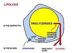
A diagrammatic illustration of the process of lipolysis (in a fat cell) induced by high epinephrine and low insulin levels in the blood. Epinephrine binds to a beta-adrenergic receptor in the cell wall of the adipocyte, which causes cAMP to be generated inside the cell. The cAMP activates a protein kinase, which phosphorylates and thus, in turn, activates a hormone-sensitive lipase in the fat cell. This lipase cleaves free fatty acids from their attachment to glycerol in the fat stored in the fat droplet of the adipocyte. The free fatty acids and glycerol are then released into the blood.
 
A diagrammatic illustration of the transport of free fatty acids in the blood attached to plasma albumin, its diffusion across the cell membrane using a protein transporter, and its activation, using ATP, to form acyl-CoA in the cytosol. The illustration is, for diagrammatic purposes, of a 12 carbon fatty acid. Most fatty acids in human plasma are 16 or 18 carbon atoms long.
 
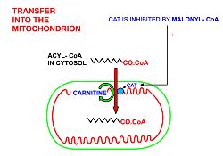
A diagrammatic illustration of the transfer of an acyl-CoA molecule across the inner membrane of the mitochondrion by carnitine-acyl-CoA transferase (CAT). The illustrated acyl chain is, for diagrammatic purposes, only 12 carbon atoms long. Most fatty acids in human plasma are 16 or 18 carbon atoms long. CAT is inhibited by high concentrations of malonyl-CoA (the first committed step in fatty acid synthesis) in the cytoplasm. This means that fatty acid synthesis and fatty acid catabolism cannot occur simultaneously in any given cell.
 
A diagrammatic illustration of the process of the beta-oxidation of an acyl-CoA molecule in the mitochodrial matrix. During this process an acyl-CoA molecule which is 2 carbons shorter than it was at the beginning of the process is formed. Acetyl-CoA, water and 5 ATP molecules are the other products of each beta-oxidative event, until the entire acyl-CoA molecule has been reduced to a set of acetyl-CoA molecules.
 
Free fatty acids cannot penetrate any biological membrane due to their negative charge. Free fatty acids must cross the cell membrane through specific transport proteins, such as the SLC27 family fatty acid transport protein. Once in the cytosol, the following processes bring fatty acids into the mitochondrial matrix so that beta-oxidation can take place.
  1. Long-chain-fatty-acid—CoA ligase catalyzes the reaction between a fatty acid with ATP to give a fatty acyl adenylate, plus inorganic pyrophosphate, which then reacts with free coenzyme A to give a fatty acyl-CoA ester and AMP.
  2. If the fatty acyl-CoA has a long chain, then the carnitine shuttle must be utilized:
    1. Acyl-CoA is transferred to the hydroxyl group of carnitine by carnitine palmitoyltransferase I, located on the cytosolic faces of the outer and inner mitochondrial membranes.
    2. Acyl-carnitine is shuttled inside by a carnitine-acylcarnitine translocase, as a carnitine is shuttled outside.
    3. Acyl-carnitine is converted back to acyl-CoA by carnitine palmitoyltransferase II, located on the interior face of the inner mitochondrial membrane. The liberated carnitine is shuttled back to the cytosol, as an acyl-carnitine is shuttled into the matrix.
  3. If the fatty acyl-CoA contains a short chain, these short-chain fatty acids can simply diffuse through the inner mitochondrial membrane.

General mechanism

Once the fatty acid is inside the mitochondrial matrix, beta-oxidation occurs by cleaving two carbons every cycle to form acetyl-CoA. The process consists of 4 steps.
  1. A long-chain fatty acid is dehydrogenated to create a trans double bond between C2 and C3. This is catalyzed by acyl CoA dehydrogenase to produce trans-delta 2-enoyl CoA. It uses FAD as an electron acceptor and it is reduced to FADH2.
  2. Trans-delta2-enoyl CoA is hydrated at the double bond to produce L-3-hydroxyacyl CoA by enoyl-CoA hydratase.
  3. L-3-hydroxyacyl CoA is dehydrogenated again to create 3-ketoacyl CoA by 3-hydroxyacyl CoA dehydrogenase. This enzyme uses NAD as an electron acceptor.
  4. Thiolysis occurs between C2 and C3 (alpha and beta carbons) of 3-ketoacyl CoA. Thiolase enzyme catalyzes the reaction when a new molecule of coenzyme A breaks the bond by nucleophilic attack on C3. This releases the first two carbon units, as acetyl CoA, and a fatty acyl CoA minus two carbons. The process continues until all of the carbons in the fatty acid are turned into acetyl CoA.
Fatty acids are oxidized by most of the tissues in the body. However, some tissues such as the red blood cells of mammals (which do not contain mitochondria), and cells of the central nervous system do not use fatty acids for their energy requirements, but instead use carbohydrates (red blood cells and neurons) or ketone bodies (neurons only).

Because many fatty acids are not fully saturated or do not have an even number of carbons, several different mechanisms have evolved, described below.

Even-numbered saturated fatty acids

Once inside the mitochondria, each cycle of β-oxidation, liberating a two carbon unit (acetyl-CoA), occurs in a sequence of four reactions. This process continues until the entire chain is cleaved into acetyl CoA units. The final cycle produces two separate acetyl CoAs, instead of one acyl CoA and one acetyl CoA. For every cycle, the Acyl CoA unit is shortened by two carbon atoms. Concomitantly, one molecule of FADH2, NADH and acetyl CoA are formed.

Odd-numbered saturated fatty acids

In general, fatty acids with an odd number of carbons are found in the lipids of plants and some marine organisms. Many ruminant animals form a large amount of 3-carbon propionate during the fermentation of carbohydrates in the rumen. Long-chain fatty acids with an odd number of carbon atoms are found particularly in ruminant fat and milk.

Chains with an odd-number of carbons are oxidized in the same manner as even-numbered chains, but the final products are propionyl-CoA and Acetyl-CoA.

Propionyl-CoA is first carboxylated using a bicarbonate ion into D-stereoisomer of methylmalonyl-CoA, in a reaction that involves a biotin co-factor, ATP, and the enzyme propionyl-CoA carboxylase. The bicarbonate ion's carbon is added to the middle carbon of propionyl-CoA, forming a D-methylmalonyl-CoA. However, the D conformation is enzymatically converted into the L conformation by methylmalonyl-CoA epimerase, then it undergoes intramolecular rearrangement, which is catalyzed by methylmalonyl-CoA mutase (requiring B12 as a coenzyme) to form succinyl-CoA. The succinyl-CoA formed can then enter the citric acid cycle

However, whereas acetyl-CoA enters the citric acid cycle by condensing with an existing molecule of oxaloacetate, succinyl-CoA enters the cycle as a principal in its own right. Thus the succinate just adds to the population of circulating molecules in the cycle and undergoes no net metabolization while in it. When this infusion of citric acid cycle intermediates exceeds cataplerotic demand (such as for aspartate or glutamate synthesis), some of them can be extracted to the gluconeogenesis pathway, in the liver and kidneys, through phosphoenolpyruvate carboxykinase, and converted to free glucose.

Unsaturated fatty acids

β-Oxidation of unsaturated fatty acids poses a problem since the location of a cis bond can prevent the formation of a trans-Δ2 bond. These situations are handled by an additional two enzymes, Enoyl CoA isomerase or 2,4 Dienoyl CoA reductase

Complete beta oxidation of linoleic acid (an unsaturated fatty acid).
 
Whatever the conformation of the hydrocarbon chain, β-oxidation occurs normally until the acyl CoA (because of the presence of a double bond) is not an appropriate substrate for acyl CoA dehydrogenase, or enoyl CoA hydratase:
  • If the acyl CoA contains a cis-Δ3 bond, then cis-Δ3-Enoyl CoA isomerase will convert the bond to a trans-Δ2 bond, which is a regular substrate.
  • If the acyl CoA contains a cis-Δ4 double bond, then its dehydrogenation yields a 2,4-dienoyl intermediate, which is not a substrate for enoyl CoA hydratase. However, the enzyme 2,4 Dienoyl CoA reductase reduces the intermediate, using NADPH, into trans-Δ3-enoyl CoA. As in the above case, this compound is converted into a suitable intermediate by 3,2-Enoyl CoA isomerase.
To summarize:
  • Odd-numbered double bonds are handled by the isomerase;
  • Even-numbered double bonds by the reductase (which creates an odd-numbered double bond).

Peroxisomal beta-oxidation

Fatty acid oxidation also occurs in peroxisomes when the fatty acid chains are too long to be handled by the mitochondria. The same enzymes are used in peroxisomes as in the mitochondrial matrix, and acetyl-CoA is generated. It is believed that very long chain (greater than C-22) fatty acids, branched fatty acids, some prostaglandins and leukotrienes undergo initial oxidation in peroxisomes until octanoyl-CoA is formed, at which point it undergoes mitochondrial oxidation.

One significant difference is that oxidation in peroxisomes is not coupled to ATP synthesis. Instead, the high-potential electrons are transferred to O2, which yields H2O2. It does generate heat however. The enzyme catalase, found exclusively in peroxisomes, converts the hydrogen peroxide into water and oxygen

Peroxisomal β-oxidation also requires enzymes specific to the peroxisome and to very long fatty acids. There are three key differences between the enzymes used for mitochondrial and peroxisomal β-oxidation:
  1. The NADH formed in the third oxidative step cannot be reoxidized in the peroxisome, so reducing equivalents are exported to the cytosol.
  2. β-oxidation in the peroxisome requires the use of a peroxisomal carnitine acyltransferase (instead of carnitine acyltransferase I and II used by the mitochondria) for transport of the activated acyl group into the mitochondria for further breakdown.
  3. The first oxidation step in the peroxisome is catalyzed by the enzyme acyl-CoA oxidase.
  4. The β-ketothiolase used in peroxisomal β-oxidation has an altered substrate specificity, different from the mitochondrial β-ketothiolase.
Peroxisomal oxidation is induced by a high-fat diet and administration of hypolipidemic drugs like clofibrate.

Energy yield

The ATP yield for every oxidation cycle is theoretically a maximum yield of 17, as NADH produces 2.5 ATP, FADH2 produces 1.5 and a full rotation of the citric acid cycle produces 10. In practice it is closer to 14 ATP for a full oxidation cycle as the theoretical yield is not attained - it is generally closer to 2.5 ATP per NADH molecule produced, 1.5 for each FADH2 molecule produced and this equates to 10 per cycle of the TCA (according to the P/O ratio), broken down as follows:

Source ATP Total
1 FADH2 x 1.5 ATP = 1.5 ATP (Theoretically 2 ATP)
1 NADH x 2.5 ATP = 2.5 ATP (Theoretically 3 ATP)
1 acetyl CoA x 10 ATP = 10 ATP (Theoretically 12 ATP)
TOTAL
= 14 ATP

For an even-numbered saturated fat (C2n), n - 1 oxidations are necessary, and the final process yields an additional acetyl CoA. In addition, two equivalents of ATP are lost during the activation of the fatty acid. Therefore, the total ATP yield can be stated as:
(n - 1) * 14 + 10 - 2 = total ATP
or
14n-6 (alternatively)
For instance, the ATP yield of palmitate (C16, n = 8) is:
(8 - 1) * 14 + 10 - 2 = 106 ATP
Represented in table form: 

Source ATP Total
7 FADH2 x 1.5 ATP = 10.5 ATP
7 NADH x 2.5 ATP = 17.5 ATP
8 acetyl CoA x 10 ATP = 80 ATP
Activation
= -2 ATP
NET
= 106 ATP

For sources that use the larger ATP production numbers described above, the total would be 129 ATP ={(8-1)*17+12-2} equivalents per palmitate. 

Beta-oxidation of unsaturated fatty acids changes the ATP yield due to the requirement of two possible additional enzymes.

Similarities between beta-oxidation and citric acid cycle

The reactions of beta oxidation and part of citric acid cycle present structural similarities in three of four reactions of the beta oxidation: the oxidation by FAD, the hydration, and the oxidation by NAD+. Each enzyme of these metabolic pathways presents structural similarity.

Clinical significance

There are at least 25 enzymes and specific transport proteins in the β-oxidation pathway. Of these, 18 have been associated with human disease as inborn errors of metabolism.

Cooperativity

From Wikipedia, the free encyclopedia

Cooperativity is a phenomenon displayed by systems involving identical or near-identical elements, which act dependently of each other, relative to a hypothetical standard non-interacting system in which the individual elements are acting independently. One manifestation of this is enzymes or receptors that have multiple binding sites where the affinity of the binding sites for a ligand is apparently increased, positive cooperativity, or decreased, negative cooperativity, upon the binding of a ligand to a binding site. For example, when an oxygen atom binds to one of hemoglobin's four binding sites, the affinity to oxygen of the three remaining available binding sites increases; i.e. oxygen is more likely to bind to a hemoglobin bound to one oxygen than to an unbound hemoglobin. This is referred to as cooperative binding.

We also see cooperativity in large chain molecules made of many identical (or nearly identical) subunits (such as DNA, proteins, and phospholipids), when such molecules undergo phase transitions such as melting, unfolding or unwinding. This is referred to as subunit cooperativity. However, the definition of cooperativity based on apparent increase or decrease in affinity to successive ligand binding steps is problematic, as the concept of "energy" must always be defined relative to a standard state. When we say that the affinity is increased upon binding of one ligand, it is empirically unclear what we mean since a non-cooperative binding curve is required to rigorously define binding energy and hence also affinity. A much more general and useful definition of positive cooperativity is: A process involving multiple identical incremental steps, in which intermediate states are statistically underrepresented relative to a hypothetical standard system (null hypothesis) where the steps occur independently of each other.

Likewise, a definition of negative cooperativity would be a process involving multiple identical incremental steps, in which the intermediate states are overrepresented relative to a hypothetical standard state in which individual steps occur independently. These latter definitions for positive and negative cooperativity easily encompass all processes which we call "cooperative", including conformational transitions in large molecules (such as proteins) and even psychological phenomena of large numbers of people (which can act independently of each other, or in a co-operative fashion).

Cooperative binding

When a substrate binds to one enzymatic subunit, the rest of the subunits are stimulated and become active. Ligands can either have positive cooperativity, negative cooperativity, or non-cooperativity. 

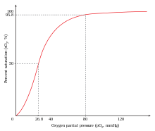
The sigmoidal shape of hemoglobin's oxygen-dissociation curve results from cooperative binding of oxygen to hemoglobin.
 
An example of positive cooperativity is the binding of oxygen to hemoglobin. One oxygen molecule can bind to the ferrous iron of a heme molecule in each of the four chains of a hemoglobin molecule. Deoxy-hemoglobin has a relatively low affinity for oxygen, but when one molecule binds to a single heme, the oxygen affinity increases, allowing the second molecule to bind more easily, and the third and fourth even more easily. The oxygen affinity of 3-oxy-hemoglobin is ~300 times greater than that of deoxy-hemoglobin. This behavior leads the affinity curve of hemoglobin to be sigmoidal, rather than hyperbolic as with the monomeric myoglobin. By the same process, the ability for hemoglobin to lose oxygen increases as fewer oxygen molecules are bound.

Negative cooperativity means that the opposite will be true; as ligands bind to the protein, the protein's affinity for the ligand will decrease, i.e. it becomes less likely for the ligand to bind to the protein. An example of this occurring is the relationship between glyceraldehyde-3-phosphate and the enzyme glyceraldehyde-3-phosphate dehydrogenase.

Homotropic cooperativity refers to the fact that the molecule causing the cooperativity is the one that will be affected by it. Heterotropic cooperativity is where a third party substance causes the change in affinity. Homotropic or heterotropic cooperativity could be of both positives as well as negative types depend upon whether it support or oppose further binding of the ligand molecules to the enzymes.

Subunit cooperativity

Cooperativity is not only a phenomenon of ligand binding, but also applies anytime energetic interactions make it easier or more difficult for something to happen involving multiple units as opposed to with single units. (That is, easier or more difficult compared with what is expected when only accounting for the addition of multiple units). For example, unwinding of DNA involves cooperativity: Portions of DNA must unwind in order for DNA to carry out replication, transcription and recombination. Positive cooperativity among adjacent DNA nucleotides makes it easier to unwind a whole group of adjacent nucleotides than it is to unwind the same number of nucleotides spread out along the DNA chain. The cooperative unit size is the number of adjacent bases that tend to unwind as a single unit due to the effects of positive cooperativity. This phenomenon applies to other types of chain molecules as well, such as the folding and unfolding of proteins and in the "melting" of phospholipid chains that make up the membranes of cells. Subunit cooperativity is measured on the relative scale known as Hill's Constant.

Hill equation

A simple and widely used model for molecular interactions is the Hill equation, which provides a way to quantify cooperative binding by describing the fraction of saturated ligand binding sites as a function of the ligand concentration.

Hill Coefficient

The Hill coefficient is a measure of ultrasensitivity (i.e. how steep is the response curve).

From an operational point of view the Hill coefficient can be calculated as:
.
where and are the input values needed to produce the 10% and 90% of the maximal response, respectively.

Response Coefficient

Global sensitivity measure such as Hill coefficient do not characterise the local behaviours of the s-shaped curves. Instead, these features are well captured by the response coefficient measure  defined as:

Link between Hill Coefficient and Response coefficient

Altszyler et al. (2017) have shown that these ultrasensitivity measures can be linked by the following equation:
where denoted the mean value of the variable x over the range [a,b].

Ultrasensitivity in function composition

Consider two coupled ultrasensitive modules, disregarding effects of sequestration of molecular components between layers. In this case, the expression for the system's dose-response curve, F, results from the mathematical composition of the functions, , which describe the input/output relationship of isolated modules :
Brown et al. (1997) have shown that the local ultrasensitivity of the different layers combines multiplicatively:
.
In connection with this result, Ferrell et al. (1997) showed, for Hill-type modules, that the overall cascade global ultrasensitivity had to be less than or equal to the product of the global ultrasensitivity estimations of each cascade's layer,
,
where and are the Hill coefficient of modules 1 and 2 respectively. 

Altszyler et al. (2017) have shown that the cascade's global ultrasensitivity can be analytically calculated:
where and delimited the Hill input's working range of the composite system, i.e. the input values for the i-layer so that the last layer (corresponding to in this case) reached the 10% and 90% of it maximal output level. It followed this equation that the system's Hill coefficient n could be written as the product of two factors, and , which characterized local average sensitivities over the relevant input region for each layer: , with in this case.

For the more general case of a cascade of N modules, the Hill Coefficient can be expressed as:
,

Supramultiplicativity

Several authors have reported the existence of supramultiplicative behavior in signaling cascades (i.e. the ultrasensitivity of the combination of layers is higher than the product of individual ultrasensitivities), but in many cases the ultimate origin of supramultiplicativity remained elusive. Altszyler et al. (2017) framework naturally suggested a general scenario where supramultiplicative behavior could take place. This could occur when, for a given module, the corresponding Hill's input working range was located in an input region with local ultrasensitivities higher than the global ultrasensitivity of the respective dose-response curve.

Entropy and cooperativity

In all of the above types of cooperativity, entropy plays a role. For example, in the case of oxygen binding to hemoglobin, the first oxygen has four different available binding sites. This represents a state of higher entropy compared to a fourth oxygen having one available binding site. Thus, in transition from the unbound to the bound state, the first oxygen must overcome a larger entropy change than the last oxygen in order to bind to the hemoglobin.

Introduction to entropy

From Wikipedia, the free encyclopedia https://en.wikipedia.org/wiki/Introduct...