Search This Blog

Saturday, March 21, 2020

Curare

Homocysteine

From Wikipedia, the free encyclopedia

Homocysteine
Skeletal formula
Ball-and-stick model
Names
IUPAC name
2-Amino-4-sulfanylbutanoic acid
Identifiers
3D model (JSmol)
ChEBI
ChEMBL
ChemSpider
ECHA InfoCard 100.006.567
EC Number
  • 207-222-9
KEGG
PubChem CID
UNII
Properties
C4H9NO2S
Molar mass 135.18 g/mol
Appearance White crystalline powder
Melting point 234–235 °C (453–455 °F; 507–508 K)[2] (decomposes)
soluble
log P -2.56 [1]
Acidity (pKa) 2.25 [1]
Hazards
GHS pictograms GHS07: Harmful
GHS Signal word Warning
H302
Except where otherwise noted, data are given for materials in their standard state (at 25 °C [77 °F], 100 kPa).

Homocysteine /ˌhmˈsɪstn/ is a non-proteinogenic α-amino acid. It is a homologue of the amino acid cysteine, differing by an additional methylene bridge (-CH2-). It is biosynthesized from methionine by the removal of its terminal Cε methyl group. Homocysteine can be recycled into methionine or converted into cysteine with the aid of certain B-vitamins.

A high level of homocysteine in the blood (hyperhomocysteinemia) makes a person more prone to endothelial cell injury, which leads to inflammation in the blood vessels, which in turn may lead to atherogenesis, which can result in ischemic injury. Hyperhomocysteinemia is therefore a possible risk factor for coronary artery disease. Coronary artery disease occurs when an atherosclerotic plaque blocks blood flow to the coronary arteries, which supply the heart with oxygenated blood.

Hyperhomocysteinemia has been correlated with the occurrence of blood clots, heart attacks and strokes, though it is unclear whether hyperhomocysteinemia is an independent risk factor for these conditions. Hyperhomocysteinemia has also been associated with early pregnancy loss and with neural tube defects.

Structure

Zwitterionic form of (S)-homocysteine (left) and (R)-homocysteine (right)

Homocysteine exists at neutral pH values as a zwitterion.

Biosynthesis and biochemical roles

Two of homocysteine's main biochemical roles. (Homocysteine is seen in the left middle of the image.) It can be synthesized from methionine and then converted back to methionine via the SAM cycle or used to create cysteine and alpha-ketobutyrate.

Homocysteine is not obtained from the diet. Instead, it is biosynthesized from methionine via a multi-step process. First, methionine receives an adenosine group from ATP, a reaction catalyzed by S-adenosyl-methionine synthetase, to give S-adenosyl methionine (SAM). SAM then transfers the methyl group to an acceptor molecule, (e.g., norepinephrine as an acceptor during epinephrine synthesis, DNA methyltransferase as an intermediate acceptor in the process of DNA methylation). The adenosine is then hydrolyzed to yield L-homocysteine. L-Homocysteine has two primary fates: conversion via tetrahydrofolate (THF) back into L-methionine or conversion to L-cysteine.

Biosynthesis of cysteine

Mammals biosynthesize the amino acid cysteine via homocysteine. Cystathionine β-synthase catalyses the condensation of homocysteine and serine to give cystathionine. This reaction uses pyridoxine (vitamin B6) as a cofactor. Cystathionine γ-lyase then converts this double amino acid to cysteine, ammonia, and α-ketobutyrate. Bacteria and plants rely on a different pathway to produce cysteine, relying on O-acetylserine.

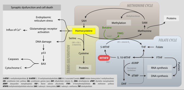
MTHFR metabolism: folate cycle, methionine cycle, trans-sulfuration and hyperhomocysteinemia. 5-MTHF: 5-methyltetrahydrofolate; 5,10-methyltetrahydrofolate; BAX: Bcl-2-associated X protein; BHMT: betaine-homocysteine S-methyltransferase; CBS: cystathionine beta synthase; CGL: cystathionine gamma-lyase; DHF: dihydrofolate (vitamin B9); DMG: dimethylglycine; dTMP: thymidine monophosphate; dUMP: deoxyuridine monophosphate; FAD+ flavine adenine dicucleotide; FTHF: 10-formyltetrahydrofolate; MS: methionine synthase; MTHFR: mehtylenetetrahydrofolate reductase; SAH: S-adenosyl-L-homocysteine; SAME: S-adenosyl-L-methionine; THF: tetrahydrofolate.

Methionine salvage

Homocysteine can be recycled into methionine. This process uses N5-methyl tetrahydrofolate as the methyl donor and cobalamin (vitamin B12)-related enzymes. More detail on these enzymes can be found in the article for methionine synthase.

Other reactions of biochemical significance

Homocysteine can cyclize to give homocysteine thiolactone, a five-membered heterocycle. Because of this "self-looping" reaction, homocysteine-containing peptides tend to cleave themselves by reactions generating oxidative stress.

Homocysteine also acts as an allosteric antagonist at Dopamine D2 receptors. It has been proposed that both homocysteine and its thiolactone may have played a significant role in the appearance of life on the early Earth.

Homocysteine levels

Total plasma homocysteine

Homocysteine levels are typically higher in men than women, and increase with age.

Common levels in Western populations are 10 to 12 μmol/L, and levels of 20 μmol/L are found in populations with low B-vitamin intakes or in the elderly (e.g., Rotterdam, Framingham).

It is decreased with methyl folate trapping, where it is accompanied by decreased methylmalonic acid, increased folate and a decrease in formiminoglutamic acid. This is the opposite of MTHFR C677T mutations, which result in an increase in homocysteine.

Blood reference ranges for homocysteine:
Sex Age Lower
limit
Upper
limit
Unit Elevated Therapeutic
target
Female 12–19 years 3.3 7.2 μmol/L > 10.4 μmol/L
or
> 140 μg/dl
< 6.3 μmol/L
or
< 85 μg/dL
45 100 μg/dL
>60 years 4.9 11.6 μmol/L
66 160 μg/dL
Male 12–19 years 4.3 9.9 μmol/L > 11.4 μmol/L
or
> 150 μg/dL
60 130 μg/dL
>60 years 5.9 15.3 μmol/L
80 210 μg/dL

The ranges above are provided as examples only; test results should always be interpreted using the range provided by the laboratory that produced the result.

Elevated homocysteine

Abnormally high levels of homocysteine in the serum, above 15 µmol/L, are a medical condition called hyperhomocysteinemia. This has been claimed to be a significant risk factor for the development of a wide range of diseases, including thrombosis, neuropsychiatric illness, and fractures. It is also found to be associated with microalbuminuria which is a strong indicator of the risk of future cardiovascular disease and renal dysfunction. Vitamin B12 deficiency, when coupled with high serum folate levels, has been found to increase overall homocysteine concentrations as well.

Choline

From Wikipedia, the free encyclopedia
 
Choline
Skeletal formula
Ball-and-stick model
Names
Preferred IUPAC name
2-Hydroxy-N,N,N-trimethylethan-1-aminium
Other names
2-Hydroxy-N,N,N-trimethylethanaminium
Bilineurine
(2-Hydroxyethyl)trimethylammonium
Identifiers
3D model (JSmol)
1736748
ChEBI
ChEMBL
ChemSpider
DrugBank
ECHA InfoCard 100.000.487
EC Number
  • 200-535-1
324597
KEGG
PubChem CID
UNII
Properties
C5H14NO+
Molar mass 104.17 g/mol
Appearance viscous deliquescent liquid (choline hydroxide)
very soluble (choline hydroxide)
Solubility soluble in ethanol, insoluble in diethylether and chloroform (choline hydroxide)
Hazards
NFPA 704 (fire diamond)
Flammability (red): no hazard codeHealth (blue): no hazard codeReactivity (yellow): no hazard codeSpecial hazard COR: Corrosive; strong acid or base. E.g. sulfuric acid, potassium hydroxideNFPA 704 four-colored diamond
Lethal dose or concentration (LD, LC):
LD50 (median dose)
3–6 g/kg bw, rats, oral
Except where otherwise noted, data are given for materials in their standard state (at 25 °C [77 °F], 100 kPa).

Choline /ˈkəʊln/ occurs as a cation that forms various salts (X in the depicted formula is an undefined counteranion). Choline is an essential nutrient for humans and many other animals. To maintain health, it must be obtained from the diet as choline or as choline phospholipids, like phosphatidylcholine. Humans and most animals make choline de novo, but production is insufficient in humans and most species. Choline is often not classified as a vitamin, but as a nutrient with an amino acid-like metabolism. In most animals, choline phospholipids are necessary components in cell membranes, in the membranes of cell organelles, and in very low-density lipoproteins. Choline is required to produce acetylcholine – a neurotransmitter – and S-adenosyl methionine, a universal methyl donor involved in the synthesis of homocysteine.

Symptomatic choline deficiency – rare in humans – causes nonalcoholic fatty liver disease and muscle damage. Excessive 8–20 gram daily doses of choline can cause low blood pressure, sweating, diarrhea and fish-like body odor due to trimethylamine, which forms in its metabolism. Rich dietary sources of choline and choline phospholipids include hen egg yolk, wheat germ, and meats, especially organ meats, such as beef liver.

Chemistry

Choline is a family of water-soluble quaternary ammonium compounds. Choline hydroxide is known as choline base. It is hygroscopic and thus often encountered as a colorless viscous hydrated syrup that smells of trimethylamine (TMA). Aqueous solutions of choline are stable, but the compound slowly breaks down to ethylene glycol, polyethylene glycols, and TMA.

Choline chloride can be made by treating TMA with 2-chloroethanol:
N(CH3)3 + ClCH2CH2OH → (CH3)3N+CH2CH2OH · Cl
The 2-chloroethanol can be generated from ethylene oxide. Choline has historically been produced from natural sources, e.g., via hydrolysis of lecithin.

Metabolism

Biosynthesis

Biosynthesis of choline in plants

In plants, the first step in de novo biosynthesis of choline is the decarboxylation of serine into ethanolamine, which is catalyzed by a serine decarboxylase. The synthesis of choline from ethanolamine may take place in three parallel pathways, where three consecutive N-methylation steps catalyzed by a methyl transferase are carried out on either the free-base, phospho-bases, or phosphatidyl-bases. The source of the methyl group is S-adenosyl-L-methionine and S-adenosyl-L-homocysteine is generated as a side product.

Main pathways of choline (Chol) metabolism, synthesis and excretion. Click for details. Some of the abbreviations are used in this section.

In humans and most other animals, de novo synthesis of choline is via the phosphatidylethanolamine N-methyltransferase (PEMT) pathway, but biosynthesis is not enough to meet human requirements. In the hepatic PEMT route, 3-phosphoglycerate (3PG) receives 2 acyl groups from acyl-CoA forming a phosphatidic acid. It reacts with cytidine triphosphate to form cytidine diphosphate-diacylglycerol. Its hydroxyl group reacts with serine to form phosphatidylserine which decarboxylates to ethanolamine and phosphatidylethanolamine (PE) forms. A PEMT-enzyme moves three methyl groups from three S-adenosyl methionines (SAM) donors to the ethanolamine group of the PE to form choline in the form of a PC. Three S-adenosyl homocysteines (SAHs) are formed as a byproduct.

Choline can also be released from more complex choline containing molecules. For example, phosphatidylcholines (PC) can be hydrolyzed to choline (Chol) in most cell types. Choline can also be produced by the CDP-choline route, cytosolic choline kinases (CK) phosphorylate choline with ATP to phosphocholine (PChol). This happens in some cell types like liver and kidney. Choline-phosphate cytidylyltransferases (CPCT) transform PChol to CDP-choline (CDP-Chol) with cytidine triphosphate (CTP). CDP-choline and diglyceride are transformed to PC by diacylglycerol cholinephosphotransferase (CPT).

In humans, certain PEMT-enzyme mutations and estrogen deficiency (often due to menopause) increase the dietary need for choline. In rodents, 70% of phosphatidylcholines are formed via the PEMT route and only 30% via the CDP-choline route. In knockout mice, PEMT inactivation makes them completely dependent on dietary choline.

Absorption

In humans, choline is absorbed from the intestines via the SLC44A1 (CTL1) membrane protein via facilitated diffusion governed by the choline concentration gradient and the electrical potential across the enterocyte membranes. The SLC44A1s have limited ability to transport choline: at high concentrations part of it is left unabsorbed. Absorbed choline leaves the enterocytes via the portal vein, passes the liver and enters systemic circulation. Gut microbes degrade the unabsorbed choline to trimethylamine, which is oxidized in the liver to trimethylamine N-oxide.

Phosphocholine and glycerophosphocholines are hydrolyzed via phospholipases to choline, which enters the portal vein. Due to their water solubility, some of them escape unchanged to the portal vein. Fat-soluble choline-containing compounds (phosphatidylcholines and sphingomyelins) are either hydrolyzed by phospholipases or enter the lymph incorporated into chylomicrons.

Transport

In humans, choline is transported as a free molecule in blood. Choline–containing phospholipids and other substances, like glycerophosphocholines, are transported in blood lipoproteins. Blood plasma choline levels in healthy fasting adults is 7–20 micromoles per liter (µmol/l) and 10 µmol/l on average. Levels are regulated, but choline intake and deficiency alters these levels. Levels are elevated for about 3 hours after choline consumption. Phosphatidylcholine levels in the plasma of fasting adults is 1.5–2.5 mmol/l. Its consumption elevates the free choline levels for about 8–12 hours, but does not affect phosphatidylcholine levels significantly.

Choline is a water soluble ion and thus requires transporters to pass through fat-soluble cell membranes. Three types of choline transporters are known:
SLC5A7s are sodium (Na+) and ATP dependent transporters. They have high binding affinity for choline, transport it primarily to neurons and are indirectly associated with the acetylcholine production. Their deficient function causes hereditary weakness in the pulmonary and other muscles in humans via acetylcholine deficiency. In knockout mice, their dysfunction results easily in death with cyanosis and paralysis.

CTL1s have moderate affinity for choline and transport it in almost all tissues: e.g., intestines, the liver, kidneys, the placenta and mitochondria. CTL1s supply choline for phosphatidylcholine and trimethylglycine production. CTL2s occur especially in the mitochondria in the tongue, kidneys, muscles and heart. They are associated with the mitochondrial oxidation of choline to trimethylglycine. CTL1s and CTL2s are not associated with the acetylcholine production, but transport choline together via the blood–brain barrier. Only CTL2s occur on the brain side of the barrier. They also remove excess choline from the neurons back to blood. CTL1s occur only on the blood side of the barrier, but also on the membranes of astrocytes and neurons.

OCT1s and OCT2s are not associated with the acetylcholine production. They transport choline with low affinity. OCT1s transport choline primarily in the liver and kidneys; OCT2s in kidneys and the brain.

Storage

Choline is stored in the cell membranes and organelles as phospholipids, and inside cells as phosphatidylcholines and glycerophosphocholines.

Excretion

Even at choline doses of 2–8 g, little choline is excreted to urine in humans. Excretion happens via transporters that occur within kidneys (see transport). Trimethylglycine is demethylated in the liver and kidneys to dimethylglycine (tetrahydrofolate receives one of the methyl groups). Methylglycine forms, is excreted to urine, or is demethylated to glycine.

Function

Choline and its derivatives have many functions in humans and in other organisms. The most notable function is that choline serves as a synthetic precursor for other essential cell components and signalling molecules, such as phospholipids that form cell membranes, the neurotransmitter acetylcholine, and the osmoregulator trimethylglycine (betaine). Trimethylglycine in turn serves as a source of methyl groups by participating in the biosynthesis of S-adenosylmethionine.

Phospholipid precursor

Choline is transformed to different phospholipids, like phosphatidylcholines and sphingomyelins. These are found in all cell membranes and from the membranes of most cell organelles. Phosphatidylcholines are structurally important part of the cell membranes. In humans 40–50% of their phospholipids are phosphatidylcholines.

Choline phospholipids also form lipid rafts in the cell membranes along with cholesterol. The rafts are centers, e.g., for receptors and receptor signal transduction enzymes.

Phosphatidylcholines are needed for the synthesis of VLDLs: 70–95% of their phospholipids are phosphatidylcholines in humans.

Choline is also needed for the synthesis of pulmonary surfactant, which is a mixture consisting mostly of phosphatidylcholines. The surfactant is responsible for lung elasticity, i.e., for its ability to contract and expand. For example, deficiency of phosphatidylcholines in the lung tissues has been linked to acute respiratory distress syndrome.

Phosphatidylcholines are excreted into bile and work together with bile acid salts as surfactants in it, thus helping with the intestinal absorption of lipids.

Acetylcholine synthesis

Choline is needed to produce acetylcholine. This is a neurotransmitter which plays a necessary role in muscle contraction, memory and neural development, for example. Nonetheless, there is little acetylcholine in the human body relative to other forms of choline. Neurons also store choline in the form of phospholipids to their cell membranes for the production of acetylcholine.

Source of trimethylglycine

In humans, choline is oxidized irreversibly in liver mitochondria to glycine betaine aldehyde by choline oxidases. This is oxidized by mitochondrial or cytosolic betaine-aldehyde dehydrogenases to trimethylglycine. Trimethylglycine is a necessary osmoregulator. It also works as a substrate for the BHMT-enzyme, which methylates homocysteine to methionine. This is a S-adenosyl methionine (SAM) precursor. SAM is a common reagent in biological methylation reactions. For example, it methylates guanidines of DNA and certain lysines of histones. Thus it is part of gene expression and epigenetic regulation. Choline deficiency thus leads to elevated homocysteine levels and decreased S-adenosyl methionine levels in blood.

Content in foods

Choline occurs in foods as a free molecule and in the form of phospholipids, especially as phosphatidylcholines. The total choline content accounting for all of these forms is one of the highest of all foods in hen egg yolk. It has about 670 milligrams of total choline per 100 grams of yolk (mg/100 g). After eggs, content decreases in general and respectively in meats, grains, vegetables, fruits and fats. Cooking oils and other food fats have about 5 mg/100 g of total choline. In the United States, food labels express the amount of choline in a serving as a percentage of daily value (%DV) based on the adequate intake of 550 mg/d. 100% of the daily value means that a serving of food has 550 mg of choline.

Human breast milk is rich in choline. Exclusive breastfeeding corresponds to about 120 mg of choline per day for the baby. Increase in a mother's choline intake raises the choline content of breast milk and low intake decreases it. Infant formulas may or may not contain enough choline. In the EU and the US, it is mandatory to add at least 7 mg of choline per 100 kilocalories (kcal) to every infant formula. In the EU, levels above 50 mg/100 kcal are not allowed.

Trimethylglycine is a functional metabolite of choline. It substitutes for choline nutritionally, but only partially. High amounts of trimethylglycine occur in wheat bran (1339 mg/100 g), toasted wheat germ (1240 mg/100 g) and spinach (600–645 mg/100 g), for example.

Choline content of foods (mg/100 g)
Meats Vegetables
Bacon, cooked 124.89 Bean, snap 13.46
Beef, trim-cut, cooked 78.15 Beetroot 6.01
Beef liver, pan fried 418.22 Broccoli 40.06
Chicken, roasted, with skin 65.83 Brussels sprout 40.61
Chicken, roasted, no skin 78.74 Cabbage 15.45
Chicken liver 290.03 Carrot 8.79
Cod, atlantic 83.63 Cauliflower 39.10
Ground beef, 75-85% lean, broiled 79.32–82.35 Sweetcorn, yellow 21.95
Pork loin cooked 102.76 Cucumber 5.95
Shrimp, canned 70.60 Lettuce, iceberg 6.70
Dairy products (cow) Lettuce, romaine 9.92
Butter, salted 18.77 Pea 27.51
Cheese 16.50–27.21 Sauerkraut 10.39
Cottage cheese 18.42 Spinach 22.08
Milk, whole/skimmed 14.29–16.40 Sweet potato 13.11
Sour cream 20.33 Tomato 6.74
Yogurt, plain 15.20 Zucchini 9.36
Grains Fruits
Oat bran, raw 58.57 Apple 3.44
Oats, plain 7.42 Avocado 14.18
Rice, white 2.08 Banana 9.76
Rice, brown 9.22 Blueberry 6.04
Wheat bran 74.39 Cantaloupe 7.58
Wheat germ, toasted 152.08 Grape 7.53
Others Grapefruit 5.63
Bean, navy 26.93 Orange 8.38
Egg, hen 251.00 Peach 6.10
Olive oil 0.29 Pear 5.11
Peanut 52.47 Prune 9.66
Soybean, raw 115.87 Strawberry 5.65
Tofu, soft 27.37 Watermelon 4.07
  1. Foods are raw unless noted otherwise. Contents are approximate sums of free choline and choline containing phospholipids.

Dietary recommendations

Recommendations are in milligrams per day (mg/d). The European Food Safety Authority (EFSA) recommendations are general recommendations for the EU countries. The EFSA has not set any upper limits for intake. Individual EU countries may have more specific recommendations. The National Academy of Medicine (NAM) recommendations apply in the United States, Australia and New Zealand.

Choline recommendations (mg/d)
Age EFSA adequate intake US NAM adequate intake US NAM tolerable upper intake levels
Infants and children
0–6 months Not established 125 Not established
7–12 months 160 150 Not established
1–3 years (y) 140 200 1000
4–6 y 170 250 1000
7–8 y 250 250 1000
9–10 y 250 375 1000
11–13 y 340 375 2000
Males
14 y 340 550 3000
15+ y 400 550 3500 (3000 for 15–18 y)
Females
14 y 340 400 3000
15–18 y 400 400 3000
19+ y 400 425 3500
If pregnant 480 450 3500 (3000 if ≤ 18 y)
If breastfeeding 520 550 3500 (3000 if ≤ 18 y)

Intake in populations

Twelve surveys undertaken in 9 EU countries between 2000 and 2011 estimated choline intake of adults in these countries to be 269–468 milligrams per day (mg/d). Intake was 269–444 mg/d in adult women and 332–468 mg/d in adult men. Intake was 75–127 mg/d in infants, 151–210 mg/d in 1–3 year olds, 177–304 mg/d in 3–10 year olds and 244–373 mg/d in 10–18 years. The total choline intake mean estimate was 336 mg/day in pregnant adolescents and 356 mg/day in pregnant women.

A study based on the NHANES 2009–2012 survey estimated the choline intake to be too low in some US subpopulations. Intake was 315.2–318.8 mg/d in ≥2 year olds between this time period. Out of ≥2 year olds, only 15.6 ± 0.8% of males and 6.1 ± 0.6% of females exceeded the adequate intake (AI). AI was exceeded by 62.9 ± 3.1% of 2–3 year olds, 45.4 ± 1.6% of 4–8 year olds, 9.0 ± 1.0% of 9–13 year olds, 1.8 ± 0.4% of 14–18 and 6.6 ± 0.5% of over 19 year olds. Upper intake level was not exceeded in any subpopulations.

A 2013–2014 NHANES study of the US population found the choline intake of 2–19 year olds to be 256 ± 3.8 mg/d and 339 ± 3.9 mg/d in ≥20 year olds. Intake was 402 ± 6.1 mg/d in ≥20-year-old men and 278 mg/d in ≥20-year-old women.

Deficiency

Signs and symptoms

Symptomatic choline deficiency is rare in humans. Most obtain sufficient amounts of it from the diet and are able to biosynthesize limited amounts of it. Symptomatic deficiency is often caused by certain diseases or by other indirect causes. Severe deficiency causes muscle damage and non-alcoholic fatty liver disease, which may develop into cirrhosis.

Besides humans, fatty liver is also a typical sign of choline deficiency in other animals. Bleeding in the kidneys can also occur in some species. This is suspected to be due to deficiency of choline derived trimethylglycine, which functions as an osmoregulator.

Causes and mechanisms

Estrogen production is a relevant factor which predisposes individuals to deficiency along with low dietary choline intake. Estrogens activate phosphatidylcholine producing PEMT-enzymes. Women before menopause have lower dietary need for choline than men due to women's higher estrogen production. Without estrogen therapy, the choline needs of post-menopausal women are similar to men's. Some single-nucleotide polymorphisms (i.e., genetic factors) affecting choline and folate metabolism are also relevant. Certain gut microbes also degrade choline more efficiently than others, so they are also relevant.

In deficiency, availability of phosphatidylcholines in the liver are decreased – these are needed for formation of VLDLs. Thus VLDL-mediated fatty acid transport out of the liver decreases leading to fat accumulation in the liver. Other simultaneously occurring mechanisms explaining the observed liver damage have also been suggested. For example, choline phospholipids are also needed in mitochondrial membranes. Their inavailability leads to the inability of mitochondrial membranes to maintain proper electrochemical gradient, which, among other things, is needed for degrading fatty acids via β-oxidation. Fat metabolism within liver therefore decreases.

Excess intake

Excessive doses of choline can have adverse effects. Daily 8–20 gram doses of choline, for example, have been found to cause low blood pressure, nausea, diarrhea and fish-like body odor. The odor is due to trimethylamine (TMA) formed by the gut microbes from the unabsorbed choline (see trimethylaminuria).
The liver oxidizes TMA to trimethylamine N-oxide (TMAO). Elevated levels of TMA and TMAO in the body have been linked to increased risk of atherosclerosis and mortality. Thus, excessive choline intake has been suggested to increase these risks in addition to carnitine, which also forms TMA and TMAO. However, it is plausible that elevated TMA and TMAO levels are just a symptom of other underlying illnesses or genetic factors that predispose individuals for increased mortality. Such factors may have not been properly accounted for in certain studies observing TMA and TMAO level related mortality. Causality may be reverse or confounding and large choline intake might not increase mortality in humans. For example, kidney dysfunction predisposes for cardiovascular diseases, but can also decrease TMA and TMAO excretion.

Health effects

Neural tube closure

Some human studies showed low maternal intake of choline to significantly increase the risk of neural tube defects (NTDs) in newborns. Folate deficiency also causes NTDs. Choline and folate, interacting with vitamin B12, act as methyl donors to homocysteine to form methionine, which can then go on to form SAM (S-adenosyl methionine). SAM is the substrate for almost all methylation reactions in mammals. It has been suggested that disturbed methylation via SAM could be responsible for the relation between folate and NTDs. This may also apply to choline. Certain mutations that disturb choline metabolism increase the prevalence of NTDs in newborns, but the role of dietary choline deficiency remains unclear, as of 2015.

Cardiovascular diseases and cancer

Choline deficiency can cause fatty liver, which increases cancer and cardiovascular disease risk. Choline deficiency also decreases SAM production, which partakes in DNA methylation – this decrease may also contribute to carcinogenesis. Thus, deficiency and its association with such diseases has been studied. However, observational studies of free populations have not convincingly shown an association between low choline intake and cardiovascular diseases or most cancers. Studies on prostate cancer have been contradictory.

Cognition

Studies observing the effect between higher choline intake and cognition have been conducted human adults, with contradictory results. Similar studies on human infants and children have been contradictory and also limited.

Pregnancy and brain development

Both pregnancy and lactation increase demand for choline dramatically. This demand may be met by upregulation of PEMT via increasing estrogen levels to produce more choline de novo, but even with increased PEMT activity, the demand for choline is still so high that bodily stores are generally depleted. This is exemplified by the observation that Pemt -/- mice (mice lacking functional PEMT) will abort at 9–10 days unless fed supplemental choline.

While maternal stores of choline are depleted during pregnancy and lactation, the placenta accumulates choline by pumping choline against the concentration gradient into the tissue, where it is then stored in various forms, mostly as acetylcholine. Choline concentrations in amniotic fluid can be ten times higher than in maternal blood.

Functions in the fetus

Choline is in high demand during pregnancy as a substrate for building cellular membranes, (rapid fetal and mother tissue expansion), increased need for one-carbon moieties (a substrate for addition of methylation to DNA and other functions), raising choline stores in fetal and placental tissues, and for increased production of lipoproteins (proteins containing "fat" portions). In particular, there is interest in the impact of choline consumption on the brain. This stems from choline's use as a material for making cellular membranes, (particularly in making phosphatidylcholine). Human brain growth is most rapid during the third trimester of pregnancy and continues to be rapid to approximately five years of age. During this time, the demand is high for sphingomyelin, which is made from phosphatidyl choline (and thus from choline), because this material is used to myelinate (insulate) nerve fibers. Choline is also in demand for the production of the neurotransmitter acetylcholine, which can influence the structure and organization of brain regions, neurogenesis, myelination, and synapse formation. Acetylcholine is even present in the placenta and may help control cell proliferation/differentiation (increases in cell number and changes of multiuse cells into dedicated cellular functions) and parturition.

Choline uptake into the brain is controlled by a low-affinity transporter located at the blood-brain barrier. Transport occurs when arterial plasma choline concentrations increase above 14 μmol/l, which can occur during a spike in choline concentration after consuming choline-rich foods. Neurons, conversely, acquire choline by both high- and low-affinity transporters. Choline is stored as membrane-bound phosphatidylcholine, which can then be used for acetylcholine neurotransmitter synthesis later. Acetylcholine is formed as needed, travels across the synapse, and transmits the signal to the following neuron. Afterwards, acetylcholinesterase degrades it, and the free choline is taken up by a high-affinity transporter into the neuron again.

Uses

Choline chloride and choline bitartrate are used in dietary supplements. Bitartrate is used more often due to its lower hygroscopicity. Certain choline salts are used to supplement chicken, turkey and some other animal feeds. Some salts are also used as industrial chemicals: for example, in photolitography to remove photoresist. Choline theophyllinate and choline salicylate are used as a medicines, as well as structural analogs, like methacholine and carbachol. Radiolabeled cholines, like carbon-11-choline, are used in medical imaging. Other commercially used salts include tricholine citrate and choline bicarbonate.

Antagonists and inhibitors

Hundreds of choline antagonists and enzyme inhibitors have been developed for research purposes. Aminomethyl propanol is among the first ones used as a research tool. It inhibits choline and trimethylglycine synthesis. It is able to induce choline deficiency that in turn results in fatty liver in rodents. Diethanolamine is another such compound, but also an environmental pollutant. N-cyclohexylcholine inhibits choline uptake primarily in brains. Hemicholinium-3 is a more general inhibitor, but also moderately inhibits choline kinases. More specific choline kinase inhibitors have also been developed. Trimethylglycine synthesis inhibitors also exists: carboxybutylhomocysteine is an example of a specific BHMT inhibitor.

The cholinergic hypothesis of dementia has not only lead to medicinal acetylcholinesterase inhibitors, but also to a variety of acetylcholine inhibitors. Examples of such inhibiting research chemicals include triethylcholine, homocholine and many other N-ethyl derivates of choline, which are false neurotransmitter analogs of acetylcholine. Choline acetyltransferase inhibitors have also been developed.

History

Discovery

In 1849, Adolph Strecker was the first to isolate choline from pig bile. In 1852, L. Babo and M. Hirschbrunn extracted choline from white mustard seeds and named it sinkaline. In 1862, Strecker repeated his experiment with pig and ox bile, calling the substance choline for the first time after the Greek word for bile, chole, and identifying it with the chemical formula C5H13NO. In 1850, Theodore Nicolas Gobley extracted from the brains and roe of carps a substance he named lecithin after the Greek word for egg yolk, lekithos, showing in 1874 that it was a mixture of phosphatidylcholines.

In 1865, Oscar Liebreich isolated "neurine" from animal brains. The structural formulas of acetylcholine and Liereich's "neurine" were resolved by Adolf von Baeyer in 1867. Later that year "neurine" and sinkaline were shown to be the same substances as Strecker's choline. The compound now known as neurine is unrelated to choline.

Discovery as a nutrient

In the early 1930s, Charles Best and colleagues noted that fatty liver in rats on a special diet and diabetic dogs could be prevented by feeding them lecithin, proving in 1932 that choline in lecithin was solely responsible for this preventive effect. In 1998, the US National Academy of Medicine reported their first recommendations for choline in the human diet.

Representation of a Lie group

From Wikipedia, the free encyclopedia https://en.wikipedia.org/wiki/Representation_of_a_Lie_group...