Search This Blog

Sunday, March 31, 2019

Myasthenia gravis

From Wikipedia, the free encyclopedia

Myasthenia gravis
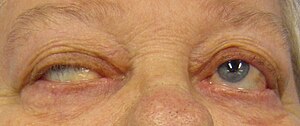
DiplopiaMG1.jpg
Eye deviation and a drooping eyelid in a person with myasthenia gravis trying to open her eyes
SpecialtyNeurology
SymptomsVarying degrees muscle weakness, double vision, drooping eyelids, trouble talking, trouble walking
Usual onsetWomen under 40, men over 60
DurationLong term
CausesAutoimmune disease
Diagnostic methodBlood tests for specific antibodies, edrophonium test, nerve conduction studies
Differential diagnosisGuillain-Barre syndrome, botulism, organophosphate poisoning, brainstem stroke
TreatmentMedications, surgical removal of the thymus, plasmapheresis
MedicationAcetylcholinesterase inhibitors (neostigmine, pyridostigmine), immunosuppressants
Frequency50 to 200 per million

Myasthenia gravis (MG) is a long-term neuromuscular disease that leads to varying degrees of skeletal muscle weakness. The most commonly affected muscles are those of the eyes, face, and swallowing. It can result in double vision, drooping eyelids, trouble talking, and trouble walking. Onset can be sudden. Those affected often have a large thymus or develop a thymoma.

Myasthenia gravis is an autoimmune disease which results from antibodies that block or destroy nicotinic acetylcholine receptors at the junction between the nerve and muscle. This prevents nerve impulses from triggering muscle contractions. Rarely, an inherited genetic defect in the neuromuscular junction results in a similar condition known as congenital myasthenia. Babies of mothers with myasthenia may have symptoms during their first few months of life, known as neonatal myasthenia. Diagnosis can be supported by blood tests for specific antibodies, the edrophonium test, or a nerve conduction study.

Myasthenia gravis is generally treated with medications known as acetylcholinesterase inhibitors such as neostigmine and pyridostigmine. Immunosuppressants, such as prednisone or azathioprine, may also be used. The surgical removal of the thymus may improve symptoms in certain cases. Plasmapheresis and high dose intravenous immunoglobulin may be used during sudden flares of the condition. If the breathing muscles become significantly weak, mechanical ventilation may be required.

MG affects 50 to 200 per million people. It is newly diagnosed in three to 30 per million people each year. Diagnosis is becoming more common due to increased awareness. It most commonly occurs in women under the age of 40 and in men over the age of 60. It is uncommon in children. With treatment, most of those affected lead relatively normal lives and have a normal life expectancy. The word is from the Greek mys "muscle" and astheneia "weakness", and the Latin: gravis "serious".

Signs and symptoms

The initial, main symptom in MG is painless weakness of specific muscles, not fatigue. The muscle weakness becomes progressively worse during periods of physical activity and improves after periods of rest. Typically, the weakness and fatigue are worse toward the end of the day. MG generally starts with ocular (eye) weakness; it might then progress to a more severe generalized form, characterized by weakness in the extremities or in muscles that govern basic life functions.

Eyes

In about two-thirds of individuals, the initial symptom of MG is related to the muscles around the eye. There may be eyelid drooping (ptosis due to weakness of levator palpebrae superioris) and double vision (diplopia, due to weakness of the extraocular muscles). Eye symptoms tend to get worse when watching television, reading, or driving, particularly in bright conditions. Consequently, some affected individuals choose to wear sunglasses. The term "ocular myasthenia gravis" describes a subtype of MG where muscle weakness is confined to the eyes, i.e. extraocular muscles, levator palpebrae superioris, and orbicularis oculi. Typically, this subtype evolves into generalized MG, usually after a few years.

Eating

The weakness of the muscles involved in swallowing may lead to swallowing difficulty (dysphagia). Typically, this means that some food may be left in the mouth after an attempt to swallow, or food and liquids may regurgitate into the nose rather than go down the throat (velopharyngeal insufficiency). Weakness of the muscles that move the jaw (muscles of mastication) may cause difficulty chewing. In individuals with MG, chewing tends to become more tiring when chewing tough, fibrous foods. Difficulty in swallowing, chewing, and speaking is the first symptom in about one-sixth of individuals.

Speaking

Weakness of the muscles involved in speaking may lead to dysarthria and hypophonia. Speech may be slow and slurred, or have a nasal quality. In some cases, a singing hobby or profession must be abandoned.

Head and neck

Due to weakness of the muscles of facial expression and muscles of mastication, facial weakness may manifest as the inability to hold the mouth closed (the "hanging jaw sign") and as a snarling expression when attempting to smile. With drooping eyelids, facial weakness may make the individual appear sleepy or sad. Difficulty in holding the head upright may occur.

Other

The muscles that control breathing (dyspnea) and limb movements can also be affected; rarely do these present as the first symptoms of MG, but develop over months to years. In a myasthenic crisis, a paralysis of the respiratory muscles occurs, necessitating assisted ventilation to sustain life. Crises may be triggered by various biological stressors such as infection, fever, an adverse reaction to medication, or emotional stress.

Pathophysiology

Neuromuscular junction: 1. Axon 2. Muscle cell membrane 3. Synaptic vesicle 4. Nicotinic acetylcholine receptor 5. Mitochondrion
 
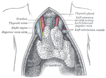
A juvenile thymus shrinks with age.
 
MG is an autoimmune synaptopathy. The disorder occurs when the immune system malfunctions and generates antibodies that attack the body's tissues. The antibodies in MG attack a normal human protein, the nicotinic acetylcholine receptor, or a related protein called MuSK a muscle-specific kinase. Other less frequent antibodies are found against LRP4, Agrin and titin proteins.

Human leukocyte antigen (HLA) haplotypes are associated with increased susceptibility to myasthenia gravis and other autoimmune disorders. Relatives of MG patients have a higher percentage of other immune disorders.

The thymus gland cells form part of the body's immune system. In those with myasthenia gravis, the thymus gland is large and abnormal. It sometimes contains clusters of immune cells which indicate lymphoid hyperplasia, and the thymus gland may give wrong instructions to immune cells.

In pregnancy

For women who are pregnant and already have MG, in a third of cases, they have been known to experience an exacerbation of their symptoms, and in those cases it usually occurs in the first trimester of pregnancy. Signs and symptoms in pregnant mothers tend to improve during the second and third trimesters. Complete remission can occur in some mothers. Immunosuppressive therapy should be maintained throughout pregnancy, as this reduces the chance of neonatal muscle weakness, and controls the mother's myasthenia.

About 10–20% of infants with mothers affected by the condition are born with transient neonatal myasthenia (TNM), which generally produces feeding and respiratory difficulties that develop about 12 hours to several days after birth. A child with TNM typically responds very well to acetylcholinesterase inhibitors, and the condition generally resolves over a period of three weeks as the antibodies diminish and generally does not result in any complications. Very rarely, an infant can be born with arthrogryposis multiplex congenita, secondary to profound intrauterine weakness. This is due to maternal antibodies that target an infant's acetylcholine receptors. In some cases, the mother remains asymptomatic.

Diagnosis

MG can be difficult to diagnose, as the symptoms can be subtle and hard to distinguish from both normal variants and other neurological disorders.

Three types of myasthenic symptoms in children can be distinguished:
  1. Transient neonatal myasthenia occurs in 10 to 15% of babies born to mothers afflicted with the disorder, and disappears after a few weeks.
  2. Congenitalmyasthenia, the rarest form, occurs when genes are present from both parents.
  3. Juvenile myasthenia gravis is most common in females.
Congenital myasthenias cause muscle weakness and fatigability similar to those of MG. The signs of congenital myasthenia usually are present in the first years of childhood, although they may not be recognized until adulthood.

Classification

When diagnosed with MG, a person is assessed for his or her neurological status and the level of illness is established. This is usually done using the accepted Myasthenia Gravis Foundation of America Clinical Classification scale, which is: 

Myasthenia Gravis Foundation of America Clinical Classification
Class Description
I Any eye muscle weakness, possible ptosis, no other evidence of muscle weakness elsewhere
II Eye muscle weakness of any severity, mild weakness of other muscles
IIa Predominantly limb or axial muscles
IIb Predominantly bulbar and/or respiratory muscles
III Eye muscle weakness of any severity, moderate weakness of other muscles
IIIa Predominantly limb or axial muscles
IIIb Predominantly bulbar and/or respiratory muscles
IV Eye muscle weakness of any severity, severe weakness of other muscles
IVa Predominantly limb or axial muscles
IVb Predominantly bulbar and/or respiratory muscles
V Intubation needed to maintain airway

Physical examination

During a physical examination to check for MG, a doctor might ask the person to perform repetitive movements. For instance, the doctor may ask one to look at a fixed point for 30 seconds and to relax the muscles of the forehead. This is done because a person with MG and ptosis of the eyes might be involuntarily using the forehead muscles to compensate for the weakness in the eyelids. The clinical examiner might also try to elicit the "curtain sign" in a patient by holding one of the person's eyes open, which in the case of MG will lead the other eye to close.

Blood tests

If the diagnosis is suspected, serology can be performed:
  • One test is for antibodies against the acetylcholine receptor; the test has a reasonable sensitivity of 80–96%, but in ocular myasthenia, the sensitivity falls to 50%.
  • A proportion of the patients without antibodies against the acetylcholine receptor have antibodies against the MuSK protein.
  • In specific situations, testing is performed for Lambert-Eaton syndrome.

Electrodiagnostics

A chest CT-scan showing a thymoma (red circle)
 
Photograph of a patient showing right partial ptosis (left picture), the left lid shows compensatory pseudo lid retraction because of equal innervation of the levator palpabrae superioris (Hering's law of equal innervation): Right picture: after an edrophonium test, note the improvement in ptosis.
 
Muscle fibers of people with MG are easily fatigued, which the repetitive nerve stimulation test can help diagnose. In single-fiber electromyography, which is considered to be the most sensitive (although not the most specific) test for MG, a thin needle electrode is inserted into different areas of a particular muscle to record the action potentials from several samplings of different individual muscle fibers. Two muscle fibers belonging to the same motor unit are identified, and the temporal variability in their firing patterns is measured. Frequency and proportion of particular abnormal action potential patterns, called "jitter" and "blocking", are diagnostic. Jitter refers to the abnormal variation in the time interval between action potentials of adjacent muscle fibers in the same motor unit. Blocking refers to the failure of nerve impulses to elicit action potentials in adjacent muscle fibers of the same motor unit.

Ice test

Applying ice for two to five minutes to the muscles reportedly has a sensitivity and specificity of 76.9% and 98.3%, respectively, for the identification of MG. Acetylcholinesterase is thought to be inhibited at the lower temperature, and this is the basis for this diagnostic test. This generally is performed on the eyelids when a ptosis is present, and is deemed positive if a ≥2 mm rise in the eyelid occurs after the ice is removed.

Edrophonium test

This test requires the intravenous administration of edrophonium chloride or neostigmine, drugs that block the breakdown of acetylcholine by cholinesterase (acetylcholinesterase inhibitors). This test is no longer typically performed, as its use can lead to life-threatening bradycardia (slow heart rate) which requires immediate emergency attention. Production of edrophonium was discontinued in 2008.

Imaging

A chest X-ray may identify widening of the mediastinum suggestive of thymoma, but computed tomography or magnetic resonance imaging (MRI) are more sensitive ways to identify thymomas and are generally done for this reason. MRI of the cranium and orbits may also be performed to exclude compressive and inflammatory lesions of the cranial nerves and ocular muscles.

Pulmonary function test

The forced vital capacity may be monitored at intervals to detect increasing muscular weakness. Acutely, negative inspiratory force may be used to determine adequacy of ventilation; it is performed on those individuals with MG.

Management

Treatment is by medication and/or surgery. Medication consists mainly of acetylcholinesterase inhibitors to directly improve muscle function and immunosuppressant drugs to reduce the autoimmune process. Thymectomy is a surgical method to treat MG.

Medication

Neostigmine, chemical structure
 
Azathioprine, chemical structure
 
Acetylcholinesterase inhibitors can provide symptomatic benefit and may not fully remove a person's weakness from MG. While they might not fully remove all symptoms of MG, they still may allow a person the ability to perform normal daily activities. Usually, acetylcholinesterase inhibitors are started at a low dose and increased until the desired result is achieved. If taken 30 minutes before a meal, symptoms will be mild during eating, which is helpful for those who have difficulty swallowing due to their illness. Another medication used for MG, atropine, can reduce the muscarinic side effects of acetylcholinesterase inhibitors. Pyridostigmine is a relatively long-acting drug (when compared to other cholinergic agonists), with a half-life around four hours with relatively few side effects. Generally, it is discontinued in those who are being mechanically ventilated as it is known to increase the amount of salivary secretions. A few high-quality studies have directly compared cholinesterase inhibitors with other treatments (or placebo); their practical benefit may be such that it would be difficult to conduct studies in which they would be withheld from some people. The steroid prednisone might also be used to achieve a better result, but it can lead to the worsening of symptoms for 14 days and takes 6–8 weeks to achieve its maximal effectiveness. Due to the myriad symptoms that steroid treatments can cause, it is not the preferred method of treatment. Other immune suppressing medications may also be used including rituximab. About 10% of people with generalized MG are considered treatment-refractory.

Autologous hematopoietic stem cell transplantation (HSCT) is sometimes used in severe, treatment-refractory MG. Available data provide preliminary evidence that HSCT can be an effective therapeutic option in carefully selected cases.

Plasmapheresis and IVIG

If the myasthenia is serious (myasthenic crisis), plasmapheresis can be used to remove the putative antibodies from the circulation. Also, intravenous immunoglobulins (IVIGs) can be used to bind the circulating antibodies. Both of these treatments have relatively short-lived benefits, typically measured in weeks, and often are associated with high costs which make them prohibitive; they are generally reserved for when MG requires hospitalization.

Surgery

As thymomas are seen in 10% of all people with the MG, people are often given a chest X-ray and CT scan to evaluate their need for surgical removal of their thymus and any cancerous tissue that may be present. Even if surgery is performed to remove a thymoma, it generally does not lead to the remission of MG. Surgery in the case of MG involves the removal of the thymus, although in 2013 there was no clear indication of any benefit except in the presence of a thymoma. A 2016 randomized controlled trial, however, found some benefits.

Physical measures

Patients with MG should be educated regarding the fluctuating nature of their symptoms, including weakness and exercise-induced fatigue. Exercise participation should be encouraged with frequent rest. In people with generalized MG, some evidence indicates a partial home program including training in diaphragmatic breathing, pursed lip breathing, and interval-based muscle therapy may improve respiratory muscle strength, chest wall mobility, respiratory pattern, and respiratory endurance.

Medical imaging

In people with myasthenia gravis, older forms of iodinated contrast used for medical imaging have caused an increased risk of exacerbation of the disease, but modern forms have no immediate increased risk.

Prognosis

The prognosis of MG patients is generally good, as is quality of life, given very good treatment. In the early 1900s, the mortality associated with MG was 70%; now, that number is estimated to be around 3–5%, which is attributed to increased awareness and medications to manage symptoms. Monitoring of a person with MG is very important, as at least 20% of people diagnosed with it will experience a myasthenic crisis within two years of their diagnosis, requiring rapid medical intervention. Generally, the most disabling period of MG might be years after the initial diagnosis.

Epidemiology

Myasthenia gravis occurs in all ethnic groups and both sexes. It most commonly affects women under 40 and people from 50 to 70 years old of either sex, but it has been known to occur at any age. Younger patients rarely have thymoma. The prevalence in the United States is estimated at between 0.5 and 20.4 cases per 100,000, with an estimated 60,000 Americans affected. Within the United Kingdom, an estimated 15 cases of MG occur per 100,000 people.

History

The first to write about MG were Thomas Willis, Samuel Wilks, Erb, and Goldflam. The term "myasthenia gravis pseudo-paralytica" was proposed in 1895 by Jolly, a German physician. Mary Walker treated a person with MG with physostigmine in 1934. Simpson and Nastuck detailed the autoimmune nature of the condition.[12] In 1973, Patrick and Lindstrom used rabbits to show that immunization with purified muscle-like acetylcholine receptors caused the development of MG-like symptoms.

Research

Immunomodulating substances, such as drugs that prevent acetylcholine receptor modulation by the immune system, are currently being researched. Some research recently has been on anti-c5 inhibitors for treatment research as they are safe and used in the treatment of other diseases. Ephedrine seems to benefit some people more than other medications, but it has not been properly studied as of 2014. In the laboratory MG is mostly studied in model organisms, such as rodents. In addition, in 2015, scientists developed an in vitro functional all-human, neuromuscular junction assay from human embryonic stem cells and somatic-muscle stem cells. After the addition of pathogenic antibodies against the acetylcholine receptor and activation of the complement system, the neuromuscular co-culture shows symptoms such as weaker muscle contractions.

Saturday, March 30, 2019

Functional neurological symptom disorder

From Wikipedia, the free encyclopedia

A functional neurological disorder (FND) is a condition in which patients experience neurological symptoms such as weakness, movement disorders, sensory symptoms and blackouts. The brain of a patient with functional neurological symptom disorder is structurally normal, but functions incorrectly. According to consensus from the literature and from physicians and psychologists practicing in the field, functional symptoms are also called 'medically unexplained'. Historically, other terms have been used to describe these symptoms. Symptoms of functional neurological disorders are clinically recognisable, but are not categorically associated with a definable organic disease. The intended contrast is with an organic brain syndrome, although the terms imply a level of certainty about causation that is often clinically unconfirmed. Subsets of functional neurological disorders include functional neurological symptom disorder (FNsD), conversion disorder, and psychogenic movement disorder/non-epileptic seizures. Functional neurological disorders are common in neurological services, accounting for up to one third of outpatient neurology clinic attendances, and associated with as much physical disability and distress as other neurological disorders. The diagnosis is made based on positive signs and symptoms in the history and examination during consultation of a neurologist (see below). Physiotherapy is particularly helpful for patients with motor symptoms (weakness, gait disorders, movement disorders) and tailored cognitive behavioural therapy has the best evidence in patients with dissociative (non-epileptic) attacks.

Signs and symptoms

There are a great number of symptoms experienced by those with a functional neurological disorder. It is important to note that the symptoms experienced by those with an FND are very real. At the same time, the origin of symptoms is complex since it can be associated with physical injury, severe psychological trauma (conversion disorder), and idiopathic neurological dysfunction. The core symptoms are those of motor or sensory function or episodes of altered awareness:
  • Limb weakness or paralysis
  • Blackouts (also called dissociative or non-epileptic seizures/attacks) – these may look like epileptic seizures or faints
  • Movement disorders including tremors, dystonia (spasms), myoclonus (jerky movements)
  • Visual symptoms including loss of vision or double vision
  • Speech symptoms including dysphonia (whispering speech), slurred or stuttering speech
  • Sensory disturbance including hemisensory syndrome (altered sensation down one side of the body)

Epidemiology and aetiology

Epidemiology

Functional neurological disorders are a common problem, and are the second most common reason for a neurological outpatient visit after headache/migraine. Dissociative (non-epileptic) seizures account for about 1 in 7 referrals to neurologists after an initial seizure, and functional weakness has a similar prevalence to multiple sclerosis.

Aetiology and mechanism

Epidemiological studies and meta-analysis have shown higher rates of depression and anxiety in patients with FND compared to the general population, but rates are similar to patients with other neurological disorders such as epilepsy or Parkinson's disease. This is often the case because of years of misdiagnosis and accusations of malingering.

Diagnostic criteria

A diagnosis of a functional neurological disorder is dependent on positive features from the history and examination. 

Positive features of functional weakness on examination include Hoover’s sign, when there is weakness of hip extension which normalises with contralateral hip flexion, and thigh abductor sign, weakness of thigh abduction which normalises with contralateral thigh abduction. Signs of functional tremor include entrainment and distractibility. The patient with tremor should be asked to copy rhythmical movements with one hand or foot. If the tremor of the other hand entrains to the same rhythm, stops, or if the patient has trouble copying a simple movement this may indicate a functional tremor. Functional dystonia usually presents with an inverted ankle posture or clenched fist. Positive features of dissociative or non-epileptic attacks include prolonged motionless unresponsiveness, long duration episodes (>2minutes) and symptoms of dissociation prior to the attack. These signs can be usefully discussed with patients when the diagnosis is being made. 

Patients with functional movement disorders and limb weakness may experience symptom onset triggered by an episode of acute pain, a physical injury or physical trauma. They may also experience symptoms when faced with a psychological stressor, but this isn't the case for most patients. Patients with functional neurological disorders are more likely to have a history of another illness such as irritable bowel syndrome, chronic pelvic pain or fibromyalgia but this cannot be used to make a diagnosis. FND does not show up on blood tests or structural brain imaging such as MRI or CT scanning. However, this is also the case for many other neurological conditions so negative investigations should not be used alone to make the diagnosis. FND can, however, occur alongside other neurological diseases and tests may show non-specific abnormalities which cause confusion for doctors and patients.

ICD-11 diagnostic criteria

The International Classification of Disease (ICD-11) which is due to be finalised in 2017 will have functional disorders within the neurology section for the first time.

Prevalence

Functional neurological disorder is a common problem, with estimates suggesting that up to a third of neurology outpatients having functional symptoms. In Scotland, around 5000 new cases of FND are diagnosed annually. Furthermore, non-epileptic seizures account for 1 in 7 referrals to neurologists after an initial seizure, and functional weakness has a similar prevalence to multiple sclerosis.

Misdiagnosis

Historically, misdiagnosis rates have been low.

Treatment

Treatment requires a firm and transparent diagnosis based on positive features which both health professionals and patients can feel confident about. It is essential that the health professional confirms that this is a common problem which is genuine, not imagined and not a diagnosis of exclusion.

Confidence in the diagnosis does not improve symptoms, but appears to improve the efficacy of treatments such as physiotherapy which require altering established abnormal patterns of movement.
A multi-disciplinary approach to treating functional neurological disorder is recommended. Treatment options can include:
  • Physiotherapy and occupational therapy
  • Medication such as sleeping tablets, painkillers, anti-epileptic medications and anti-depressants (for patients suffering with depression co-morbid or for pain relief)
Physiotherapy with someone who understands functional disorders may be the initial treatment of choice for patients with motor symptoms such as weakness, gait (walking) disorder and movement disorders. Nielsen et al. have reviewed the medical literature on physiotherapy for functional motor disorders up to 2012 and concluded that the available studies, although limited, mainly report positive results. Since then several studies have shown positive outcomes. In one study, up to 65% of patients were very much or much improved after five days of intensive physiotherapy even though 55% of patients were thought to have poor prognosis. In a randomised controlled trial of physiotherapy there was significant improvement in mobility which was sustained on one year follow up. In multidisciplinary settings 69% of patients markedly improved even with short rehabilitation programmes. Benefit from treatment continued even when patients were contacted up 25 months after treatment.

For patients with severe and chronic FND a combination of physiotherapy, occupational therapy and cognitive behavioural therapy may be the best combination with positive studies being published in patients who have had symptoms for up to three years before treatment.

Cognitive behavioural therapy (CBT) alone may be beneficial in treating patients with dissociative (non-epileptic) seizures. A randomised controlled trial of patients who undertook 12 sessions of CBT which taught patients how to interrupt warning signs before seizure onset, challenged unhelpful thoughts and helped patients start activities they had been avoiding found a reduction in the seizure frequency with positive outcomes sustained at six month follow up. A large multicentre trial of CBT for dissociative (non-epileptic) seizures started in 2015 in the UK.

For many patients with FND, accessing treatment can be difficult. Availability of expertise is limited and they may feel that they are being dismissed or told 'it's all in your head' especially if psychological input is part of the treatment plan. Some medical professionals are uncomfortable explaining and treating patients with functional symptoms. Changes in the diagnostic criteria, increasing evidence, literature about how to make the diagnosis and how to explain it and changes in medical training is slowly changing this.

After a diagnosis of functional neurological disorder has been made, it is important that the neurologist explains the illness fully to the patient to ensure the patient understands the diagnosis.

Some, but not all patients with FND may experience low moods or anxiety due to their condition. However, they will often not seek treatment due being worried that a doctor will blame their symptoms on their anxiety or depression.

It is recommended that the treatment of functional neurological disorder should be balanced and involve a whole-person approach. This means that it should include professionals from multiple departments, including neurologists, general practitioners (or primary health care providers), physiotherapists, occupational therapists. At the same time, ruling out secondary gain, malingering, conversion disorder and other factors, including the time and financial resources involved in assessing and treating patients who demand hospital resources but would be better served in psychological settings, must all be balanced.

Alternative diagnoses

Functional neurological symptom disorder can very rarely mimic many other conditions. Some alternative diagnoses for FND that are often considered include:
The main caveat however is that these conditions can co-exist and can be the trigger for functional neurological disorder.

History

From the 18th century, there is a move from the idea of FND being caused being caused by the nervous system. This led to an understanding that it could affect both sexes. Jean Martin Charcot argued that, what would be later called FND, was caused by "a hereditary degeneration of the nervous system, namely a neurological disorder".

In the 18th century, the illness was confirmed as being a neurological disorder but a small number of doctors still believed in the previous definition. However, as early as 1874, doctors including W. B. Carpenter and J. A. Omerod began to speak out against this other term due to there being no evidence of its existence.

Although the term "conversion disorder" has been in existence for many years, another term was still being used in the 20th century. However, by this point, it bore little resemblance to the original meaning, instead referring to symptoms which could not be explained by a recognised organic pathology, and was therefore believed to be the result of stress, anxiety, trauma or depression. The term fell out of favour of doctors over time due to the negative connotations this term held. Furthermore, critics pointed out that it can be challenging to find organic pathologies for all symptoms, and so the practice of diagnosing patients who suffered with such symptoms as imagining them led to the disorder being meaningless, vague and a sham-diagnosis, as it does not refer to any definable disease.

Throughout its history, many patients have been misdiagnosed with conversion disorder when they had organic disorders such as tumours or epilepsy or vascular diseases. This has led to patient deaths, a lack of appropriate care and suffering for the patients. Eliot Slater, after studying the condition in the 1950s, was outspoken against the condition, as there has never been any evidence to prove that it exists. He stated that "The diagnosis of 'hysteria' is a disguise for ignorance and a fertile source of clinical error. It is, in fact, not only a delusion but also a snare".

In 1980, the DSM III added 'conversion disorder' to its list of conditions. The diagnostic criteria for this condition are nearly identical to those used for hysteria. The diagnostic criteria were:

A. The predominant disturbance is a loss of or alteration in physical functioning suggesting a physical disorder. It is involuntary and medically unexplainable.

B. One of the following must also be present:
  1. A temporal relationship between symptom onset and some external event of psychological conflict.
  2. The symptom allows the individual to avoid unpleasant activity.
  3. The symptom provides opportunity for support which may not have been otherwise available.
Many illnesses and injuries can cause an individual to avoid unpleasant activities, and can provide the opportunity for support, particularly from a doctor. This makes Criteria B meaningless for the most part, and therefore any patient whose symptoms satisfy Criteria A by being medically unexplained, could be diagnosed with Conversion Disorder. 

Today, there is growing evidence that psychological stress does not cause FND. A recent study by the charity FNDHope found that psychological triggers affected only 30% of patients. Doctors are moving on to look at the role of the central nervous system in FND symptoms.

Controversy

There was historically much controversy surrounding the FND diagnosis. Many doctors continue to believe that all FND patients have unresolved traumatic events (often of a sexual nature) which are being expressed in a physical way. However, some doctors do not believe this to be the case. Wessely and White have argued that FND may merely be an unexplained somatic illness (like fibromyalgia, irritable bowel syndrome, or chronic fatigue syndrome) rather than a psychiatric condition.

Conversion disorder

From Wikipedia, the free encyclopedia

Conversion disorder
SpecialtyPsychiatry
CausesStress
TreatmentCBT, antidepressants, physical/occupational therapy

Conversion disorder (CD) is a diagnostic category used in some psychiatric classification systems. It is sometimes applied to patients who present with neurological symptoms, such as numbness, blindness, paralysis, or fits, which are not consistent with a well-established organic cause, which cause significant distress, and can be traced back to a psychological trigger. It is thought that these symptoms arise in response to stressful situations affecting a patient's mental health or an ongoing mental health condition such as depression. Conversion disorder was retained in DSM-5, but given the subtitle functional neurological symptom disorder. The new criteria cover the same range of symptoms, but remove the requirements for a psychological stressor to be present and for feigning to be disproved.

The theory of conversion disorder stems from ancient Egypt, and was formerly known as hysteria and hysterical blindness. The concept of conversion disorder came to prominence at the end of the 19th century, when the neurologists Jean-Martin Charcot and Sigmund Freud and psychologist Pierre Janet focused their studies on the subject. Before their studies, people with hysteria were often believed to be malingering. The term "conversion" has its origins in Freud's doctrine that anxiety is "converted" into physical symptoms. Though previously thought to have vanished from the West in the 20th century, some research has suggested that conversion disorder is as common as ever.

ICD-10 classifies conversion disorder as a dissociative disorder while DSM-IV classifies it as a somatoform disorder.

Signs and symptoms

Conversion disorder begins with some stressor, trauma, or psychological distress. Usually the physical symptoms of the syndrome affect the senses or movement. Common symptoms include blindness, partial or total paralysis, inability to speak, deafness, numbness, difficulty swallowing, incontinence, balance problems, seizures, tremors, and difficulty walking. These symptoms are attributed to conversion disorder when a medical explanation for the afflictions cannot be found. Symptoms of conversion disorder usually occur suddenly. Conversion disorder is typically seen in individuals aged 10 to 35, and affects between 0.011% and 0.5% of the general population.

Conversion disorder can present with motor or sensory symptoms including any of the following:
Motor symptoms or deficits:
  • Impaired coordination or balance
  • Weakness/paralysis of a limb or the entire body (hysterical paralysis or motor conversion disorders)
  • Impairment or loss of speech (hysterical aphonia)
  • Difficulty swallowing (dysphagia) or a sensation of a lump in the throat
  • Urinary retention
  • Psychogenic non-epileptic seizures or convulsions
  • Persistent dystonia
  • Tremor, myoclonus or other movement disorders
  • Gait problems (astasia-abasia)
  • Loss of consciousness (fainting)
Sensory symptoms or deficits:
  • Impaired vision (hysterical blindness), double vision
  • Impaired hearing (deafness)
  • Loss or disturbance of touch or pain sensation
Conversion symptoms typically do not conform to known anatomical pathways and physiological mechanisms. It has sometimes been stated that the presenting symptoms tend to reflect the patient's own understanding of anatomy and that the less medical knowledge a person has, the more implausible are the presenting symptoms. However, no systematic studies have yet been performed to substantiate this statement.

Diagnosis

Definition

Conversion disorder is now contained under the umbrella term functional neurological symptom disorder. In cases of conversion disorder, there is a psychological stressor. 

The diagnostic criteria for functional neurological symptom disorder, as set out in DSM-5, are:
  1. The patient has at least one symptom of altered voluntary motor or sensory function.
  2. Clinical findings provide evidence of incompatibility between the symptom and recognised neurological or medical conditions.
  3. The symptom or deficit is not better explained by another medical or mental disorder.
  4. The symptom or deficit causes clinically significant distress or impairment in social, occupational, or other important areas of functioning or warrants medical evaluation.
Specify type of symptom or deficit as:
  • With weakness or paralysis
  • With abnormal movement (e.g. tremor, dystonic movement, myoclonus, gait disorder)
  • With swallowing symptoms
  • With speech symptoms (e.g. dysphonia, slurred speech)
  • With attacks or seizures
  • With amnesia or memory loss
  • With special sensory loss symptoms (e.g. visual blindness, olfactory loss, or hearing disturbance)
  • With mixed symptoms.
Specify if:
  • Acute episode: symptoms present for less than six months
  • Persistent: symptoms present for six months or more.
Specify if:

Exclusion of neurological disease

Conversion disorder presents with symptoms that typically resemble a neurological disorder such as stroke, multiple sclerosis, epilepsy or hypokalemic periodic paralysis. The neurologist must carefully exclude neurological disease, through examination and appropriate investigations. However, it is not uncommon for patients with neurological disease to also have conversion disorder.

In excluding neurological disease, the neurologist has traditionally relied partly on the presence of positive signs of conversion disorder, i.e. certain aspects of the presentation that were thought to be rare in neurological disease but common in conversion. The validity of many of these signs has been questioned, however, by a study showing that they also occur in neurological disease. One such symptom, for example, is la belle indifférence, described in DSM-IV as "a relative lack of concern about the nature or implications of the symptoms". In a later study, no evidence was found that patients with functional symptoms are any more likely to exhibit this than patients with a confirmed organic disease. In DSM-V, la belle indifférence was removed as a diagnostic criteria.

Another feature thought to be important was that symptoms tended to be more severe on the non-dominant (usually left) side of the body. There have been a number of theories about this, such as the relative involvement of cerebral hemispheres in emotional processing, or more simply, that it was "easier" to live with a functional deficit on the non-dominant side. However, a literature review of 121 studies established that this was not true, with publication bias the most likely explanation for this commonly held view. Although agitation is often assumed to be a positive sign of conversion disorder, release of epinephrine is a well-demonstrated cause of paralysis from hypokalemic periodic paralysis.

Misdiagnosis does sometimes occur. In a highly influential study from the 1960s, Eliot Slater demonstrated that misdiagnoses had occurred in one third of his 112 patients with conversion disorder. Later authors have argued that the paper was flawed, however, and a meta-analysis has shown that misdiagnosis rates since that paper was published are around 4%, the same as for other neurological diseases.

Exclusion of feigning

Conversion disorder is unique in ICD-10 in explicitly requiring the exclusion of deliberate feigning. Unfortunately, this is likely to be demonstrable only where the patient confesses, or is "caught out" in a broader deception, such as a false identity. One neuroimaging study suggested that feigning may be distinguished from conversion by the pattern of frontal lobe activation; however, this was a piece of research, rather than a clinical technique. True rates of feigning in medicine remain unknown. However, it is believed that feigning of conversion disorder is no more likely than of other medical conditions.

Psychological mechanism

The psychological mechanism of conversion can be the most difficult aspect of a conversion diagnosis. Even if there is a clear antecedent trauma or other possible psychological trigger, it is still not clear exactly how this gives rise to the symptoms observed. Patients with medically unexplained neurological symptoms may not have any psychological stressor, hence the use of the term "functional neurological symptom disorder" in DSM-5 as opposed to "conversion disorder", and DSM-5's removal of the need for a psychological trigger.

Treatment

There are many number of different treatments that are available to treat and manage conversion syndrome. Treatments for conversion syndrome include hypnosis, psychotherapy, physical therapy, stress management, and transcranial magnetic stimulation. Treatment plans will consider duration and presentation of symptoms and may include one or multiple of the above treatments. This may include the following:
  1. Explanation. This must be clear and coherent as attributing physical symptoms to a psychological cause is not accepted by many educated people in Western cultures. It must emphasize the genuineness of the condition, that it is common, potentially reversible and does not mean that the sufferer is psychotic. Taking a neutral-cause-based stance by describing the symptoms as functional may be helpful, but further studies are required. Ideally, the patient should be followed up neurologically for a while to ensure that the diagnosis has been understood.
  2. Physiotherapy where appropriate;
  3. Occupational Therapy to maintain autonomy in activities of daily living;
  4. Treatment of comorbid depression or anxiety if present.
There is little evidence-based treatment of conversion disorder. Other treatments such as cognitive behavioral therapy, hypnosis, EMDR, and psychodynamic psychotherapy, EEG brain biofeedback need further trials. Psychoanalytic treatment may possibly be helpful. However, most studies assessing the efficacy of these treatments are of poor quality and larger, better controlled studies are urgently needed. Cognitive Behavioural Therapy is the most common treatment, however boasts a mere 13% improvement rate.

Prognosis

Empirical studies have found that the prognosis for conversion disorder varies widely, with some cases resolving in weeks, and others enduring for years or decades. There is also evidence that there is no cure for conversion disorder, and that although patients may go into remission, they can relapse at any point. Furthermore, many patients who are 'cured' continue to have some degree of symptoms indefinitely.

Epidemiology

Frequency

Information on the frequency of conversion disorder in the West is limited, in part due to the complexities of the diagnostic process. In neurology clinics, the reported prevalence of unexplained symptoms among new patients is very high (between 30 and 60%). However, diagnosis of conversion typically requires an additional psychiatric evaluation, and since few patients will see a psychiatrist it is unclear what proportion of the unexplained symptoms are actually due to conversion. Large scale psychiatric registers in the US and Iceland found incidence rates of 22 and 11 newly diagnosed cases per 100,000 person-years, respectively. Some estimates claim that in the general population, between 0.011% and 0.5% of the population have conversion disorder.

Culture

Although it is often thought that the frequency of conversion may be higher outside of the West, perhaps in relation to cultural and medical attitudes, evidence of this is limited. A community survey of urban Turkey found a prevalence of 5.6%. Many authors have found occurrence of conversion to be more frequent in rural, lower socio-economic groups, where technological investigation of patients is limited and individuals may be less knowledgeable about medical and psychological concepts.

Gender

Historically, the concept of 'hysteria' was originally understood to be a condition exclusively affecting women, though the concept was eventually extended to men. In recent surveys of conversion disorder (formerly classified as "hysterical neurosis, conversion type"), females predominate, with between 2 and 6 female patients for every male.

Age

Conversion disorder may present at any age but is rare in children younger than 10 years or in the elderly. Studies suggest a peak onset in the mid-to-late 30s.

History

The first evidence of functional neurological symptom disorder dates back to 1900 BC, when the symptoms were blamed on the uterus moving within the female body. The treatment varied "depending on the position of the uterus, which must be forced to return to its natural position. If the uterus had moved upwards, this could be done by placing malodorous and acrid substances near the woman’s mouth and nostrils, while scented ones were placed near her vagina; on the contrary, if the uterus had lowered, the document recommends placing the acrid substances near her vagina and the perfumed ones near her mouth and nostrils."

In Greek mythology, hysteria, the original name for functional neurological symptom disorder, was thought to be caused by a lack of orgasms, uterine melancholy and not procreating. Plato, Aristotle and Hippocrates believed that a lack of sex upset the uterus. The Greeks believed that it could be prevented and cured with wine and orgies. Hippocrates argued that a lack of regular sexual intercourse led to the uterus producing toxic fumes and caused it to move in the body, and that this meant that all women should be married and enjoy a satisfactory sexual life.

From the 13th century, women with hysteria were exorcised, as it was believed that they were possessed by the devil. It was believed that if doctors could not find the cause of a disease or illness, it must be caused by the devil.

At the beginning of the 16th century, women were sexually stimulated by midwives in order to relieve their symptoms. Gerolamo Cardano and Giambattista della Porta believed that polluted water and fumes caused the symptoms of hysteria. Towards the end of the century, however, the role of the uterus was no longer thought central to the disorder, with Thomas Willis discovering that the brain and central nervous system were the cause of the symptoms. Thomas Sydenham argued that the symptoms of hysteria may have an organic cause. He also proved that the uterus is not the cause of symptom.

In 1692, in the US town of Salem, Massachusetts, there was an outbreak of hysteria. This led to the Salem witch trials, where the women accused of being witches had symptoms such as sudden movements, staring eyes and uncontrollable jumping.

During the 18th century, there was a move from the idea of hysteria being caused by the uterus to it being caused by the brain. This led to an understanding that it could affect both sexes. Jean-Martin Charcot argued that hysteria was caused by "a hereditary degeneration of the nervous system, namely a neurological disorder".

In the 19th century, hysteria moved from being considered a neurological disorder to being considered a psychological disorder, when Pierre Janet argued that "dissociation appears autonomously for neurotic reasons, and in such a way as to adversely disturb the individual's everyday life". However, as early as 1874, doctors including W. B. Carpenter and J. A. Omerod began to speak out against the hysteria phenomenon as there was no evidence to prove its existence.

Sigmund Freud referred to the condition as both hysteria and conversion disorder throughout his career. He believed that those with the condition could not live in a mature relationship, and that those with the condition were unwell in order to achieve a "secondary gain", in that they are able to manipulate their situation to fit their needs or desires. He also found that both men and women could suffer from the disorder.

Freud's model suggested that the emotional charge deriving from painful experiences would be consciously repressed as a way of managing the pain, but that the emotional charge would be somehow "converted" into neurological symptoms. Freud later argued that the repressed experiences were of a sexual nature. As Peter Halligan comments, conversion has "the doubtful distinction among psychiatric diagnoses of still invoking Freudian mechanisms".

Pierre Janet, the other great theoretician of hysteria, argued that symptoms arose through the power of suggestion, acting on a personality vulnerable to dissociation. In this hypothetical process, the subject's experience of their leg, for example, is split off from the rest of their consciousness, resulting in paralysis or numbness in that leg. 

Later authors have attempted to combine elements of these various models, but none of them has a firm empirical basis. In 1908, Steyerthal predicted that: "Within a few years the concept of hysteria will belong to history ... there is no such disease and there never has been. What Charcot called hysteria is a tissue woven of a thousand threads, a cohort of the most varied diseases, with nothing in common but the so-called stigmata, which in fact may accompany any disease." However, the term "hysteria" was still being used well into the 20th century. 

Some support for the Freudian model comes from findings of high rates of childhood sexual abuse in conversion patients. Support for the dissociation model comes from studies showing heightened suggestibility in conversion patients. However, critics argue that it can be challenging to find organic pathologies for all symptoms, and so the practice of diagnosing patients who suffered with such symptoms as having hysteria led to the disorder being meaningless, vague and a sham diagnosis, as it does not refer to any definable disease. Furthermore, throughout its history, many patients have been misdiagnosed with hysteria or conversion disorder when they had organic disorders such as tumours or epilepsy or vascular diseases. This has led to patient deaths, a lack of appropriate care and suffering for the patients. Eliot Slater, after studying the condition in the 1950s, stated: "The diagnosis of 'hysteria' is all too often a way of avoiding a confrontation with our own ignorance. This is especially dangerous when there is an underlying organic pathology, not yet recognised. In this penumbra we find patients who know themselves to be ill but, coming up against the blank faces of doctors who refuse to believe in the reality of their illness, proceed by way of emotional lability, overstatement and demands for attention ... Here is an area where catastrophic errors can be made. In fact it is often possible to recognise the presence though not the nature of the unrecognisable, to know that a man must be ill or in pain when all the tests are negative. But it is only possible to those who come to their task in a spirit of humility. In the main the diagnosis of 'hysteria' applies to a disorder of the doctor–patient relationship. It is evidence of non-communication, of a mutual misunderstanding ... We are, often, unwilling to tell the full truth or to admit to ignorance ... Evasions, even untruths, on the doctor’s side are among the most powerful and frequently used methods he has for bringing about an efflorescence of 'hysteria'".

Much recent work has been done to identify the underlying causes of conversion and related disorders and to better understand why conversion disorder and hysteria appear more commonly in women. Current theoreticians tend to believe that there is no single cause for these disorders. Instead, the emphasis tends to be on the individual understanding of the patient and a variety of therapeutic techniques. In some cases, the onset of conversion disorder correlates to a traumatic or stressful event. There are also certain populations that are considered at risk for conversion disorder, including people suffering from a medical illness or condition, people with personality disorder, and individuals with dissociative identity disorder. However, no biomarkers have yet been found to support the idea that conversion disorder is caused by a psychiatric condition. 

There has been much recent interest in using functional neuroimaging to study conversion. As researchers identify the mechanisms which underlie conversion symptoms, it is hoped that they will enable the development of a neuropsychological model. A number of such studies have been performed, including some which suggest that the blood-flow in patients' brains may be abnormal while they are unwell. However, the studies have all been too small to be confident of the generalisability of their findings, so no neuropsychological model has been clearly established.

An evolutionary psychology explanation for conversion disorder is that the symptoms may have been evolutionarily advantageous during warfare. A non-combatant with these symptoms signals non-verbally, possibly to someone speaking a different language, that she or he is not dangerous as a combatant and also may be carrying some form of dangerous infectious disease. This can explain that conversion disorder may develop following a threatening situation, that there may be a group effect with many people simultaneously developing similar symptoms (as in mass psychogenic illness), and the gender difference in prevalence.

The Lacanian model accepts conversion disorder as a common phenomenon inherent in specific psychical structures. The higher prevalence of it among women is based on somewhat different intrapsychic relations to the body from those of typical males, which allows the formation of conversion symptoms.

Distance education

From Wikipedia, the free encyclopedia https://en.wikipedia.org/wiki/Distance_...