Search This Blog

Sunday, February 9, 2020

Plant secondary metabolism

From Wikipedia, the free encyclopedia

Secondary metabolism produces a large number of specialized compounds (estimated 200,000) that do not aid in the growth and development of plants but are required for the plant to survive in its environment. Secondary metabolism is connected to primary metabolism by using building blocks and biosynthetic enzymes derived from primary metabolism. Primary metabolism governs all basic physiological processes that allow a plant to grow and set seeds, by translating the genetic code into proteins, carbohydrates, and amino acids. Specialized compounds from secondary metabolism are essential for communicating with other organisms in mutualistic (e.g. attraction of beneficial organisms such as pollinators) or antagonistic interactions (e.g. deterrent against herbivores and pathogens). They further assist in coping with abiotic stress such as increased UV-radiation. The broad functional spectrum of specialized metabolism is still not fully understood. In any case, a good balance between products of primary and secondary metabolism is best for a plant’s optimal growth and development as well as for its effective coping with often changing environmental conditions. Well known specialized compounds include alkaloids, polyphenols including flavonoids, and terpenoids. Humans use quite a lot of these compounds, or the plants from which they originate, for culinary, medicinal and nutraceutical purposes.

History

Research into secondary plant metabolism primarily took off in the later half of the 19th century, however, there was still much confusion over what the exact function and usefulness of these compounds were. All that was known was that secondary plant metabolites were "by-products" of the primary metabolism and were not crucial to the plant's survival. Early research only succeeded as far as categorizing the secondary plant metabolites but did not give real insight into the actual function of the secondary plant metabolites. The study of plant metabolites is thought to have started in the early 1800s when Friedrich Willhelm Serturner isolated morphine from opium poppy, and after that new discoveries were made rapidly. In the early half of the 1900s, the main research around secondary plant metabolism was dedicated to the formation of secondary metabolites in plants, and this research was compounded by the use of tracer techniques which made deducing metabolic pathways much easier. However, there was still not much research being conducted into the functions of secondary plant metabolites until around the 1980s. Before then, secondary plant metabolites were thought of as simply waste products. In the 1970s, however, new research showed that secondary plant metabolites play an indispensable role in the survival of the plant in its environment. One of the most ground breaking ideas of this time argued that plant secondary metabolites evolved in relation to environmental conditions, and this indicated the high gene plasticity of secondary metabolites, but this theory was ignored for about half a century before gaining acceptance. Recently, the research around secondary plant metabolites is focused around the gene level and the genetic diversity of plant metabolites. Biologists are now trying to trace back genes to their origin and re-construct evolutionary pathways.

Primary vs. Secondary Plant Metabolism

Primary metabolism in a plant comprises all metabolic pathways that are essential to the plant's survival. Primary metabolites are compounds that are directly involved in the growth and development of a plant whereas secondary metabolites are compounds produced in other metabolic pathways that, although important, are not essential to the functioning of the plant. However, secondary plant metabolites are useful in the long term, often for defense purposes, and give plants characteristics such as color. Secondary plant metabolites are also used in signalling and regulation of primary metabolic pathways. Plant hormones, which are secondary metabolites, are often used to regulate the metabolic activity within cells and oversee the overall development of the plant. As mentioned above in the History tab, secondary plant metabolites help the plant maintain an intricate balance with the environment, often adapting to match the environmental needs. Plant metabolites that color the plant are a good example of this, as the coloring of a plant can attract pollinators and also defend against attack by animals.

Types of Secondary Metabolites in plants

There is no fixed, commonly agreed upon system for classifying secondary metabolites. Based on their biosynthetic origins, plant secondary metabolites can be divided into three major groups:
  1. Flavonoids and allied phenolic and polyphenolic compounds,
  2. Terpenoids and
  3. Nitrogen-containing alkaloids and sulphur-containing compounds.
Other researchers have classified secondary metabolites into following, more specific types
Class Type Number of known metabolites Examples
Alkaloids Nitrogen-containing 21000 Cocaine, Psilocin, Caffeine, Nicotine, Morphine, Berberine, Vincristine, Reserpine, Galantamine, Atropine, Vincamine, Quinidine, Ephedrine, Quinine
Non-protein amino acids (NPAAs) Nitrogen-containing 700 NPAAs are produced by specific plant families such as Leguminosae, Cucurbitaceae, Sapindaceae, Aceraceae and Hippocastanaceae. Examples: Azatyrosine, Canavanine
Amines Nitrogen-containing 100
Cyanogenic glycosides Nitrogen-containing 60 Amygdalin, Dhurrin, Linamarin, Lotaustralin, Prunasin
Glucosinolates Nitrogen-containing 100
Alkamides Nitrogen-containing 150
Lectins, peptides and polypeptides Nitrogen-containing 2000 Concanavalin A
Terpenes Without nitrogen >15,000 Azadirachtin, Artemisinin, Tetrahydrocannabinol
Steroids and saponins Without nitrogen NA These are terpenoids with a particular ring structure. Cycloartenol
Flavonoids and Tannins Without nitrogen 5000 Luteolin, tannic acid
Phenylpropanoids, lignins, coumarins and lignans Without nitrogen 2000 Resveratrol
Polyacetylenes, fatty acids and waxes Without nitrogen 1500
Polyketides Without nitrogen 750
Carbohydrates and organic acids Without nitrogen 200

Some of the secondary metabolites are discussed below:

Atropine

Atropine is a type of secondary metabolite called a tropane alkaloid. Alkaloids contain nitrogens, frequently in a ring structure, and are derived from amino acids. Tropane is an organic compound containing nitrogen and it is from tropane that atropine is derived. Atropine is synthesized by a reaction between tropine and tropate, catalyzed by atropinase. Both of the substrates involved in this reaction are derived from amino acids, tropine from pyridine (through several steps) and tropate directly from phenylalanine. Within Atropa belladonna atropine synthesis has been found to take place primarily in the root of the plant. The concentration of synthetic sites within the plant is indicative of the nature of secondary metabolites. Typically, secondary metabolites are not necessary for normal functioning of cells within the organism meaning the synthetic sites are not required throughout the organism. As atropine is not a primary metabolite, it does not interact specifically with any part of the organism, allowing it to travel throughout the plant. 

Flavonoids

Flavonoids are one class of secondary plant metabolites that are also known as Vitamin P or citrin. These metabolites are mostly used in plants to produce yellow and other pigments which play a big role in coloring the plants. In addition, Flavonoids are readily ingested by humans and they seem to display important anti-inflammatory, anti-allergic and anti-cancer activities. Flavonoids are also found to be powerful anti-oxidants and researchers are looking into their ability to prevent cancer and cardiovascular diseases. Flavonoids help prevent cancer by inducing certain mechanisms that may help to kill cancer cells, and researches believe that when the body processes extra flavonoid compounds, it triggers specific enzymes that fight carcinogens. Good dietary sources of Flavonoids are all citrus fruits, which contain the specific flavanoids hesperidins, quercitrin,and rutin, berries, tea, dark chocolate and red wine and many of the health benefits attributed to these foods come from the Flavonoids they contain. Flavonoids are synthesized by the phenylpropanoid metabolic pathway where the amino acid phenylalanine is used to produce 4-coumaryol-CoA, and this is then combined with malonyl-CoA to produce chalcones which are backbones of Flavonoids Chalcones are aromatic ketones with two phenyl rings that are important in many biological compounds. The closure of chalcones causes the formation of the flavonoid structure. Flavonoids are also closely related to flavones which are actually a sub class of flavonoids, and are the yellow pigments in plants. In addition to flavones, 11 other subclasses of Flavonoids including, isoflavones, flavans, flavanones, flavanols, flavanolols, anthocyanidins, catechins (including proanthocyanidins), leukoanthocyanidins, dihydrochalcones, and aurones. 

Cyanogenic glycoside

Many plants have adapted to iodine-deficient terrestrial environment by removing iodine from their metabolism, in fact iodine is essential only for animal cells. An important antiparasitic action is caused by the block of the transport of iodide of animal cells inhibiting sodium-iodide symporter (NIS). Many plant pesticides are cyanogenic glycoside which liberate cyanide, which, blocking cytochrome c oxidase and NIS, is poisonous only for a large part of parasites and herbivores and not for the plant cells in which it seems useful in seed dormancy phase. To get a better understanding of how secondary metabolites play a big role in plant defense mechanisms we can focus on the recognizable defense-related secondary metabolites, cyanogenic glycosides. The compounds of these secondary metabolites (As seen in Figure 1) are found in over 2000 plant species. Its structure allows the release of cyanide, a poison produced by certain bacteria, fungi, and algae that is found in numerous plants. Animals and humans possess the ability to detoxify cyanide from their systems naturally. Therefore, cyanogenic glycosides can be used for positive benefits in animal systems always. For example, the larvae of the southern armyworm consumes plants that contain this certain metabolite and have shown a better growth rate with this metabolite in their diet, as opposed to other secondary metabolite-containing plants. Although this example shows cyanogenic glycosides being beneficial to the larvae many still argue that this metabolite can do harm. To help in determining whether cyanogenic glycosides are harmful or helpful researchers look closer at its biosynthetic pathway (Figure 2). Past research suggests that cyanogenic glucosides stored in the seed of the plant are metabolized during germination to release nitrogen for seedling to grow. With this, it can be inferred that cyanogenic glycosides play various roles in plant metabolism. Though subject to change with future research, there is no evidence showing that cyanogenic glycosides are responsible for infections in plants. 

Phytic acid

Phytic acid is the main method of phosphorus storage in plant seeds, but is not readily absorbed by many animals (only absorbed by ruminant animals). Not only is phytic acid a phosphorus storage unit, but it also is a source of energy and cations, a natural antioxidant for plants, and can be a source of myoinositol which is one of the preliminary pieces for cell walls.

Phytic acid is also known to bond with many different minerals, and by doing so prevents those minerals from being absorbed; making phytic acid an anti-nutrient. There is a lot of concern with phytic acids in nuts and seeds because of its anti-nutrient characteristics. In preparing foods with high phytic acid concentrations, it is recommended they be soaked in after being ground to increase the surface area. Soaking allows the seed to undergo germination which increases the availability of vitamins and nutrient, while reducing phytic acid and protease inhibitors, ultimately increasing the nutritional value. Cooking can also reduce the amount of phytic acid in food but soaking is much more effective. 

Phytic acid is an antioxidant found in plant cells that most likely serves the purpose of preservation. This preservation is removed when soaked, reducing the phytic acid and allowing the germination and growth of the seed. When added to foods it can help prevent discoloration by inhibiting lipid peroxidation. There is also some belief that the chelating of phytic acid may have potential use in the treatment of cancer.

Gossypol

Gossypol has a yellow pigment and is found in cotton plants. It occurs mainly in the root and/or seeds of different species of cotton plants. Gossypol can have various chemical structures. It can exist in three forms: gossypol, gossypol acetic acid, and gossypol formic acid. All of these forms have very similar biological properties. Gossypol is a type of aldehyde, meaning that it has a formyl group. The formation of gossypol occurs through an isoprenoid pathway. Isoprenoid pathways are common among secondary metabolites. Gossypol's main function in the cotton plant is to act as an enzyme inhibitor. An example of gossypol's enzyme inhibition is its ability to inhibit nicotinamide adenine dinucleotide-linked enzymes of Trypanosoma cruzi. Trypanosoma cruzi is a parasite which causes Chaga's disease.

For some time it was believed that gossypol was merely a waste product produced during the processing of cottonseed products. Extensive studies have shown that gossypol has other functions. Many of the more popular studies on gossypol discuss how it can act as a male contraceptive. Gossypol has also been linked to causing hypokalemic paralysis. Hypokalemic paralysis is a disease characterized by muscle weakness or paralysis with a matching fall in potassium levels in the blood. Hypokalemic paralysis associated with gossypol in-take usually occurs in March, when vegetables are in short supply, and in September, when people are sweating a lot. This side effect of gossypol in-take is very rare however. Gossypol induced hypokalemic paralysis is easily treatable with potassium repletion. 

Phytoestrogens

Plants synthesize certain compounds called secondary metabolites which are not naturally produced by humans but can play vital roles in protection or destruction of human health. One such group of metabolites is phytoestrogens, found in nuts, oilseeds, soy, and other foods. Phytoestrogens are chemicals which act like the hormone estrogen. Estrogen is important for women's bone and heart health, but high amounts of it has been linked to breast cancer. In the plant, the phytoestrogens are involved in the defense system against fungi. Phytoestrogens can do two different things in a human body. At low doses it mimics estrogen, but at high doses it actually blocks the body's natural estrogen. The estrogen receptors in the body which are stimulated by estrogen will acknowledge the phytoestrogen, thus the body may reduce its own production of the hormone. This has a negative result, because there are various abilities of the phytoestrogen which estrogen does not do. Its effects the communication pathways between cells and has effects on other parts of the body where estrogen normally does not play a role. It has also been found to induce tumor growth of the estrogen receptor cells in the breast. But, one role of estrogens which phytoestrogens mimic is its protective behavior for the heart. So, an intake of phytoestrogens has also been seen to reduce the risk of cardiovascular disease. Resveratrol, a phytoestrogen found in grapes is responsible for this. For example, the French suffer relatively little heart disease despite the average French diet being relatively high in fat. One proposed reason for this is the resveratrol found in red wine, which has been linked to decreased risk of cardiovascular disease.

Carotenoids

Carotenoids are organic pigments found in the chloroplasts and chromoplasts of plants. They are also found in some organisms such as algae, fungi, some bacteria, and certain species of aphids. There are over 600 known carotenoids. They are split into two classes, xanthophylls and carotenes. Xanthophylls are carotenoids with molecules containing oxygen, such as lutein and zeaxanthin. Carotenes are carotenoids with molecules that are unoxygenated, such as α-carotene, β-carotene and lycopene. In plants, carotenoids can occur in roots, stems, leaves, flowers, and fruits. Carotenoids have two important functions in plants. First, they can contribute to photosynthesis. They do this by transferring some of the light energy they absorb to chlorophylls, which then uses this energy for photosynthesis. Second, they can protect plants which are over-exposed to sunlight. They do this by harmlessly dissipating excess light energy which they absorb as heat. In the absence of carotenoids, this excess light energy could destroy proteins, membranes, and other molecules. Some plant physiologists believe that carotenoids may have an additional function as regulators of certain developmental responses in plants. Tetraterpenes are synthesized from DOXP precursors in plants and some bacteria. Carotenoids involved in photosynthesis are formed in chloroplasts; Others are formed in plastids. Carotenoids formed in fungi are presumably formed from mevalonic acid precursors. Carotenoids are formed by a head-to-head condensation of geranylgeranyl pyrophosphate or diphosphate (GGPP) and there is no NADPH requirement.

Dormancy

From Wikipedia, the free encyclopedia
 
During winter dormancy, plant metabolism virtually comes to a standstill due, in part, to low temperatures that slow chemical activity.
 
Dormancy is a period in an organism's life cycle when growth, development, and (in animals) physical activity are temporarily stopped. This minimizes metabolic activity and therefore helps an organism to conserve energy. Dormancy tends to be closely associated with environmental conditions. Organisms can synchronize entry to a dormant phase with their environment through predictive or consequential means. Predictive dormancy occurs when an organism enters a dormant phase before the onset of adverse conditions. For example, photoperiod and decreasing temperature are used by many plants to predict the onset of winter. Consequential dormancy occurs when organisms enter a dormant phase after adverse conditions have arisen. This is commonly found in areas with an unpredictable climate. While very sudden changes in conditions may lead to a high mortality rate among animals relying on consequential dormancy, its use can be advantageous, as organisms remain active longer and are therefore able to make greater use of available resources. 

Animals


Hibernation

Hibernation is a mechanism used by many mammals to reduce energy expenditure and survive food shortage over the winter. Hibernation may be predictive or consequential. An animal prepares for hibernation by building up a thick layer of body fat during late summer and autumn that will provide it with energy during the dormant period. During hibernation, the animal undergoes many physiological changes, including decreased heart rate (by as much as 95%) and decreased body temperature. In addition to shivering, some hibernating animals also produce body heat by non-shivering thermogenesis to avoid freezing. Non-shivering thermogenesis is a regulated process in which the proton gradient generated by electron transport in mitochondria is used to produce heat instead of ATP in brown adipose tissue.[3] Animals that hibernate include bats, ground squirrels and other rodents, mouse lemurs, the European hedgehog and other insectivores, monotremes and marsupials. Although hibernation is almost exclusively seen in mammals, some birds, such as the common poorwill, may hibernate.

Diapause

Diapause is a predictive strategy that is predetermined by an animal's genotype. Diapause is common in insects, allowing them to suspend development between autumn and spring, and in mammals such as the roe deer (Capreolus capreolus, the only ungulate with embryonic diapause), in which a delay in attachment of the embryo to the uterine lining ensures that offspring are born in spring, when conditions are most favorable.

Aestivation

Aestivation, also spelled estivation, is an example of consequential dormancy in response to very hot or dry conditions. It is common in invertebrates such as the garden snail and worm but also occurs in other animals such as lungfish, salamanders, desert tortoises, and crocodiles

Brumation

While endotherms and other heterotherms are described scientifically as hibernating, the way ectotherms like lizards become dormant in cold is very different, and a separate name was invented for it in the 1920s, brumation. It differs from hibernation in the metabolic processes involved.

Reptiles generally begin brumation in late autumn (more specific times depend on the species). They often wake up to drink water and return to "sleep". They can go for months without food. Reptiles may eat more than usual before the brumation time but eat less or refuse food as the temperature drops. However, they do need to drink water. The brumation period is anywhere from one to eight months depending on the air temperature and the size, age, and health of the reptile. During the first year of life, many small reptiles do not fully brumate, but rather slow down and eat less often. Brumation is triggered by lack of heat and the decrease in the hours of daylight in winter, similar to hibernation. 

Plants

In plant physiology, dormancy is a period of arrested plant growth. It is a survival strategy exhibited by many plant species, which enables them to survive in climates where part of the year is unsuitable for growth, such as winter or dry seasons.

Many plant species that exhibit dormancy have a biological clock that tells them when to slow activity and to prepare soft tissues for a period of freezing temperatures or water shortage. On the other hand, dormancy can be triggered after a normal growing season by decreasing temperatures, shortened day length, and/or a reduction in rainfall. Chemical treatment on dormant plants has been proven to be an effective method to break dormancy, particularly in woody plants such as grapes, berries, apples, peaches and kiwis. Specifically, hydrogen cyanamide stimulates cell division and growth in dormant plants, causing budbreak when the plant is on the edge of breaking dormancy.[citation needed] Slight injury of cells may play a role in the mechanism of action. The injury is thought to result in increased permeability of cellular membranes. The injury is associated with the inhibition of catalase, which in turn stimulates the pentose phosphate cycle. Hydrogen cyanamide interacts with the cytokinin metabolic cycle, which results in triggering a new growth cycle. The images below show two particularly widespread dormancy patterns amongst sympodially growing orchids:

Annual life cycle of sympodially growing orchids with dormancy after completion of new growth/pseudobulb, e.g., Miltonia, or Odontoglossum
 
Annual life cycle of sympodially growing orchids with dormancy after blooming, e.g., Cycnoches ventricosum, Dendrobium nobile, or Laelia
 

Seeds

When a mature and viable seed under a favorable condition fails to germinate, it is said to be dormant. Seed dormancy is referred to as embryo dormancy or internal dormancy and is caused by endogenous characteristics of the embryo that prevent germination (Black M, Butler J, Hughes M. 1987). Dormancy should not be confused with seed coat dormancy, external dormancy, or hardseededness, which is caused by the presence of a hard seed covering or seed coat that prevents water and oxygen from reaching and activating the embryo. It is a physical barrier to germination, not a true form of dormancy (Quinliven, 1971; Quinliven and Nichol, 1971).

Seed dormancy is desired in nature, but the opposite in agriculture field. This is due to agricultural practice desires rapid germination and growth for food while as in nature, most plants are only capable of germinating once every year, making it favorable for plants to pick a specific time to reproduce. For many plants, it is preferable to reproduce in spring as opposed to fall even when there are similar conditions in terms of light and temperature due to the ensuing winter that follows fall. Many plants and seeds do recognize this and enters a dormant period in the fall to stop growing. Grain is a popular example in this aspect, where they would die above ground during the winter, so dormancy is favorable to its seedlings but extensive domestication and crossbreeding has removed most dormancy mechanisms that their ancestors had.

While seed dormancy is linked to many genes, Abscisic Acid (ABA), a plant hormone, has been linked as a major influencer to seed dormancy. In a study on rice and tobacco plants, plants defective in zeaxanthin epoxidase gene, which are linked to ABA-synthesis pathway. Seeds with higher ABA content, from over expressing zeaxanthin epoxidase, led to an increased dormancy period while plants with lower numbers of zeaxanthin epoxidase shown to have shorter period of dormancy. A simple diagram can be drawn of ABA inhibits seed germination, while Gibberellin (GA, also plant hormone), inhibits ABA production and promotes seed germination.

Trees

Typically, temperate woody perennial plants require chilling temperatures to overcome winter dormancy (rest). The effect of chilling temperatures depends on species and growth stage (Fuchigami et al. 1987). In some species, rest can be broken within hours at any stage of dormancy, with either chemicals, heat, or freezing temperatures, effective dosages of which would seem to be a function of sublethal stress, which results in stimulation of ethylene production and increased cell membrane permeability. 

Dormancy is a general term applicable to any instance in which a tissue predisposed to elongate or grow in some other manner does not do so (Nienstaedt 1966). Quiescence is dormancy imposed by the external environment. Correlated inhibition is a kind of physiological dormancy maintained by agents or conditions originating within the plant, but not within the dormant tissue itself. Rest (winter dormancy) is a kind of physiological dormancy maintained by agents or conditions within the organ itself. However, physiological subdivisions of dormancy do not coincide with the morphological dormancy found in white spruce (Picea glauca) and other conifers (Owens et al. 1977). Physiological dormancy often includes early stages of bud-scale initiation before measurable shoot elongation or before flushing. It may also include late leaf initiation after shoot elongation has been completed. In either of those cases, buds that appear to be dormant are nevertheless very active morphologically and physiologically. 

Dormancy of various kinds is expressed in white spruce (Romberger 1963). White spruce, like many woody plants in temperate and cooler regions, requires exposure to low temperature for a period of weeks before it can resume normal growth and development. This “chilling requirement” for white spruce is satisfied by uninterrupted exposure to temperatures below 7 °C for 4 to 8 weeks, depending on physiological condition (Nienstaedt 1966, 1967).

Tree species that have well-developed dormancy needs may be tricked to some degree, but not completely. For instance, if a Japanese Maple (Acer palmatum) is given an "eternal summer" through exposure to additional daylight, it grows continuously for as long as two years. Eventually, however, a temperate-climate plant automatically goes dormant, no matter what environmental conditions it experiences. Deciduous plants lose their leaves; evergreens curtail all new growth. Going through an "eternal summer" and the resultant automatic dormancy is stressful to the plant and usually fatal. The fatality rate increases to 100% if the plant does not receive the necessary period of cold temperatures required to break the dormancy. Most plants require a certain number of hours of "chilling" at temperatures between about 0 °C and 10 °C to be able to break dormancy (Bewley, Black, K.D 1994). 

Short photoperiods induce dormancy and permit the formation of needle primordia. Primordia formation requires 8 to 10 weeks and must be followed by 6 weeks of chilling at 2 °C. Bud break occurs promptly if seedlings are then exposed to 16-hour photoperiods at the 25 °C/20 °C temperature regime. The free growth mode, a juvenile characteristic that is lost after 5 years or so, ceases in seedlings experiencing environmental stress (Logan and Pollard 1976, Logan 1977).

Bacteria

Many bacteria can survive adverse conditions such as temperature, desiccation, and antibiotics by endospores, cysts, conidia or states of reduced metabolic activity lacking specialized cellular structures. Up to 80% of the bacteria in samples from the wild appear to be metabolically inactive—many of which can be resuscitated. Such dormancy is responsible for the high diversity levels of most natural ecosystems.

Recent research has characterized the bacterial cytoplasm as a glass forming fluid approaching the liquid-glass transition, such that large cytoplasmic components require the aid of metabolic activity to fluidize the surrounding cytoplasm, allowing them to move through a viscous, glass-like cytoplasm. During dormancy, when such metabolic activities are put on hold, the cytoplasm behaves like a solid glass, 'freezing' subcellular structures in place and perhaps protecting them, while allowing small molecules like metabolites to move freely through the cell, which may be helpful in cells transitioning out of dormancy.

Viruses

Dormancy in its rigid definition doesn't apply to viruses, as they are not metabolically active. However, some viruses such as poxviruses and picornaviruses after entering the host can become latent for long periods of time, or even indefinitely until they are externally activated. Herpesviruses for example can become latent after infecting the host and after years activate again if the host is under stress or exposed to ultraviolet radiation.

Sleep in non-human animals

From Wikipedia, the free encyclopedia
 
A sleeping cat

Sleep in non-human animals refers to a behavioral and physiological state characterized by altered consciousness, reduced responsiveness to external stimuli, and homeostatic regulation. Sleep is observed in mammals, birds, reptiles, amphibians, and some fish, and, in some form, in insects and even in simpler animals such as nematodes. The internal circadian clock promotes sleep at night for diurnal organisms (such as humans) and in the day for nocturnal organisms (such as rodents). Sleep patterns vary widely among species. It appears to be a requirement for all mammals and most other animals.

Definition

Sleep can follow a physiological or behavioral definition. In the physiological sense, sleep is a state characterized by reversible unconsciousness, special brainwave patterns, sporadic eye movement, loss of muscle tone (possibly with some exceptions; see below regarding the sleep of birds and of aquatic mammals), and a compensatory increase following deprivation of the state. In the behavioral sense, sleep is characterized by minimal movement, non-responsiveness to external stimuli (i.e. increased sensory threshold), the adoption of a typical posture, and the occupation of a sheltered site, all of which is usually repeated on a 24-hour basis. The physiological definition applies well to birds and mammals, but in other animals (whose brain is not as complex), the behavioral definition is more often used. In very simple animals, behavioral definitions of sleep are the only ones possible, and even then the behavioral repertoire of the animal may not be extensive enough to allow distinction between sleep and wakefulness. Sleep is quickly reversible, as opposed to hibernation or coma, and sleep deprivation is followed by longer or deeper rebound sleep. 

Necessity

If sleep were not essential, one would expect to find:
  • Animal species that do not sleep at all
  • Animals that do not need recovery sleep after staying awake longer than usual
  • Animals that suffer no serious consequences as a result of lack of sleep
Outside of a few basal animals that have no brain or a very simple one, no animals have been found to date that satisfy any of these criteria. While some varieties of shark, such as great whites and hammerheads, must remain in motion at all times to move oxygenated water over their gills, it is possible they still sleep one cerebral hemisphere at a time as marine mammals do. However it remains to be shown definitively whether any fish is capable of unihemispheric sleep.

Invertebrates

Caenorhabditis elegans is among the most primitive organisms in which sleep-like states have been observed.
 
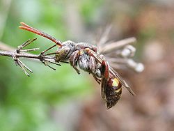
A cuckoo bee from the genus Nomada, sleeping. Note the characteristic position anchored by the mandibles. Bees have some of the most complex sleep states amongst insects.
 
Sleep as a phenomenon appears to have very old evolutionary roots. Unicellular organisms do not necessarily "sleep", although many of them have pronounced circadian rhythms. The jellyfish Cassiopea is among the most primitive organism in which sleep-like states have been observed. The nematode C. elegans is another primitive organism that appears to require sleep. Here, a lethargus phase occurs in short periods preceding each moult, a fact which may indicate that sleep primitively is connected to developmental processes. Raizen et al.'s results furthermore suggest that sleep is necessary for changes in the neural system.

The electrophysiological study of sleep in small invertebrates is complicated. Insects go through circadian rhythms of activity and passivity but some do not seem to have a homeostatic sleep need. Insects do not seem to exhibit REM sleep. However, fruit flies appear to sleep, and systematic disturbance of that state leads to cognitive disabilities. There are several methods of measuring cognitive functions in fruit flies. A common method is to let the flies choose whether they want to fly through a tunnel that leads to a light source, or through a dark tunnel. Normally, flies are attracted to light. But if sugar is placed in the end of the dark tunnel, and something the flies dislike is placed in the end of the light tunnel, the flies will eventually learn to fly towards darkness rather than light. Flies deprived of sleep require a longer time to learn this and also forget it more quickly. If an arthropod is experimentally kept awake longer than it is used to, then its coming rest period will be prolonged. In cockroaches that rest period is characterized by the antennae being folded down and by a decreased sensitivity to external stimuli. Sleep has been described in crayfish, too, characterized by passivity and increased thresholds for sensory stimuli as well as changes in the EEG pattern, markedly differing from the patterns found in crayfish when they are awake. In honeybees, it has been suggested they could be able to dream.

Fish

Sleep in fish is subject of current scientific research. Typically fish exhibit periods of inactivity but show no significant reactions to deprivation of this condition. Some species that always live in shoals or that swim continuously (because of a need for ram ventilation of the gills, for example) are suspected never to sleep. There is also doubt about certain blind species that live in caves. Other fish seem to sleep, however. For example, zebrafish, tilapia, tench, brown bullhead, and swell shark become motionless and unresponsive at night (or by day, in the case of the swell shark); Spanish hogfish and blue-headed wrasse can even be lifted by hand all the way to the surface without evoking a response. A 1961 observational study of approximately 200 species in European public aquaria reported many cases of apparent sleep. On the other hand, sleep patterns are easily disrupted and may even disappear during periods of migration, spawning, and parental care.

Land vertebrates

Mammals, birds and reptiles evolved from amniotic ancestors, the first vertebrates with life cycles independent of water. The fact that birds and mammals are the only known animals to exhibit REM and NREM sleep indicates a common trait before divergence. However, recent evidence of REM-like sleep in fish suggests this divergence may have occurred much earlier than previously thought. Up to this point, reptiles were considered the most logical group to investigate the origins of sleep. Daytime activity in reptiles alternates between basking and short bouts of active behavior, which has significant neurological and physiological similarities to sleep states in mammals. It is proposed that REM sleep evolved from short bouts of motor activity in reptiles while Slow-Wave Sleep (SWS) evolved from their basking state which shows similar slow wave EEG patterns.

Reptiles and amphibians

Sleeping African dwarf Fischer's chameleon
 
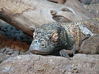
A Komodo dragon sleeping.

Reptiles have quiescent periods similar to mammalian sleep, and a decrease in electrical activity in the brain has been registered when the animals have been asleep. However, the EEG pattern in reptilian sleep differs from what is seen in mammals and other animals. In reptiles, sleep time increases following sleep deprivation, and stronger stimuli are needed to awaken the animals when they have been deprived of sleep as compared to when they have slept normally. This suggests that the sleep which follows deprivation is compensatorily deeper.

In 2016, a study report the existence of REM- and NREM-like sleep stages in the Australian dragon Pogona vitticeps. Amphibians have periods of inactivity but show high vigilance (receptivity to potentially threatening stimuli) in this state.

Birds

There are significant similarities between sleep in birds and sleep in mammals, which is one of the reasons for the idea that sleep in higher animals with its division into REM and NREM sleep has evolved together with warm-bloodedness. Birds compensate for sleep loss in a manner similar to mammals, by deeper or more intense SWS (slow-wave sleep).

Birds have both REM and NREM sleep, and the EEG patterns of both have similarities to those of mammals. Different birds sleep different amounts, but the associations seen in mammals between sleep and variables such as body mass, brain mass, relative brain mass, basal metabolism and other factors (see below) are not found in birds. The only clear explanatory factor for the variations in sleep amounts for birds of different species is that birds who sleep in environments where they are exposed to predators have less deep sleep than birds sleeping in more protected environments.

A sleeping cockatiel
 
A flamingo with at least one cerebral hemisphere awake

Birds do not necessarily exhibit sleep debt, but a peculiarity that birds share with aquatic mammals, and possibly also with certain species of lizards (opinions differ about that last point), is the ability for unihemispheric sleep. That is the ability to sleep with one cerebral hemisphere at a time, while the other hemisphere is awake (Unihemispheric slow-wave sleep). When only one hemisphere is sleeping, only the contralateral eye will be shut; that is, when the right hemisphere is asleep the left eye will be shut, and vice versa. The distribution of sleep between the two hemispheres and the amount of unihemispheric sleep are determined both by which part of the brain has been the most active during the previous period of wake—that part will sleep the deepest—and it is also determined by the risk of attacks from predators. Ducks near the perimeter of the flock are likely to be the ones that first will detect predator attacks. These ducks have significantly more unihemispheric sleep than those who sleep in the middle of the flock, and they react to threatening stimuli seen by the open eye.

Opinions partly differ about sleep in migratory birds. The controversy is mainly about whether they can sleep while flying or not. Theoretically, certain types of sleep could be possible while flying, but technical difficulties preclude the recording of brain activity in birds while they are flying.

Mammals

 
Mammals have wide diversity in sleep phenomena. Generally, they go through periods of alternating non-REM and REM sleep, but these manifest differently. Horses and other herbivorous ungulates can sleep while standing, but must necessarily lie down for REM sleep (which causes muscular atony) for short periods. Giraffes, for example, only need to lie down for REM sleep for a few minutes at a time. Bats sleep while hanging upside down. Male armadillos get erections during non-REM sleep, and the inverse is true in rats. Early mammals engaged in polyphasic sleep, dividing sleep into multiple bouts per day. Higher daily sleep quotas and shorter sleep cycles in polyphasic species as compared to monophasic species, suggest that polyphasic sleep may be a less efficient means of attaining sleep’s benefits. Small species with higher BMR may therefore have less efficient sleep patterns. It follows that the evolution of monophasic sleep may hitherto be an unknown advantage of evolving larger mammalian body sizes and therefore lower BMR.

Sleep is sometimes thought to help conserve energy, though this theory is not fully adequate as it only decreases metabolism by about 5–10%. Additionally it is observed that mammals require sleep even during the hypometabolic state of hibernation, in which circumstance it is actually a net loss of energy as the animal returns from hypothermia to euthermia in order to sleep.

Nocturnal animals have higher body temperatures, greater activity, rising serotonin, and diminishing cortisol during the night—the inverse of diurnal animals. Nocturnal and diurnal animals both have increased electrical activity in the suprachiasmatic nucleus, and corresponding secretion of melatonin from the pineal gland, at night. Nocturnal mammals, which tend to stay awake at night, have higher melatonin at night just like diurnal mammals do. And, although removing the pineal gland in many animals abolishes melatonin rhythms, it does not stop circadian rhythms altogether—though it may alter them and weaken their responsiveness to light cues. Cortisol levels in diurnal animals typically rise throughout the night, peak in the awakening hours, and diminish during the day. In diurnal animals, sleepiness increases during the night. 

Duration

Flying foxes, asleep

Different mammals sleep different amounts. Some, such as bats, sleep 18–20 hours per day, while others, including giraffes, sleep only 3–4 hours per day. There can be big differences even between closely related species. There can also be differences between laboratory and field studies: for example, researchers in 1983 reported that captive sloths slept nearly 16 hours a day, but in 2008, when miniature neurophysiological recorders were developed that could be affixed to wild animals, sloths in nature were found to sleep only 9.6 hours a day.

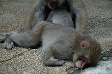
Sleeping polar bears

As with birds, the main rule for mammals (with certain exceptions, see below) is that they have two essentially different stages of sleep: REM and NREM sleep (see above). Mammals' feeding habits are associated with their sleep length. The daily need for sleep is highest in carnivores, lower in omnivores and lowest in herbivores. Humans sleep less than many other omnivores but otherwise not unusually much or unusually little in comparison with other mammals.

Many herbivores, like Ruminantia (such as cattle), spend much of their wake time in a state of drowsiness, which perhaps could partly explain their relatively low need for sleep. In herbivores, an inverse correlation is apparent between body mass and sleep length; big mammals sleep less than smaller ones. This correlation is thought to explain about 25% of the difference in sleep amount between different mammals. Also, the length of a particular sleep cycle is associated with the size of the animal; on average, bigger animals will have sleep cycles of longer durations than smaller animals. Sleep amount is also coupled to factors like basal metabolism, brain mass, and relative brain mass. The duration of sleep among species is also directly related to basal metabolic rate (BMR). Rats, which have a high BMR, sleep for up to 14 hours a day, whereas elephants and giraffes, which have lower BMRs, sleep only 3–4 hours per day.

It has been suggested that mammalian species which invest in longer sleep times are investing in the immune system, as species with the longer sleep times have higher white blood cell counts. Mammals born with well-developed regulatory systems, such as the horse and giraffe, tend to have less REM sleep than the species which are less developed at birth, such as cats and rats. This appears to echo the greater need for REM sleep among newborns than among adults in most mammal species. Many mammals sleep for a large proportion of each 24-hour period when they are very young. The giraffe only sleeps 2 hours a day in about 5–15 minute sessions. Koalas are the longest sleeping-mammals, about 20–22 hours a day. However, killer whales and some other dolphins do not sleep during the first month of life. Instead, young dolphins and whales frequently take rests by pressing their body next to their mother’s while she swims. As the mother swims she is keeping her offspring afloat to prevent them from drowning. This allows young dolphins and whales to rest, which will help keep their immune system healthy; in turn, protecting them from illnesses. During this period, mothers often sacrifice sleep for the protection of their young from predators. However, unlike other mammals, adult dolphins and whales are able to go without sleep for a month.
Comparative average sleep periods for various mammals (in captivity) over 24 hours.
A sleeping dog
Reasons given for the wide variations include the fact that mammals "that nap in hiding, like bats or rodents tend to have longer, deeper snoozes than those on constant alert." Lions, which have little fear of predators also have relatively long sleep periods, while elephants have to eat most of the time to support their huge bodies. Little brown bats conserve their energy except for the few hours each night when their insect prey are available, and platypuses eat a high energy crustacean diet and, therefore, probably do not need to spend as much time awake as many other mammals.

Rodents

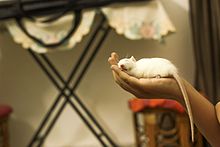
A sleeping rat

A study conducted by Datta indirectly supports the idea that memory benefits from sleep.[57] A box was constructed wherein a single rat could move freely from one end to the other. The bottom of the box was made of a steel grate. A light would shine in the box accompanied by a sound. After a five-second delay, an electrical shock would be applied. Once the shock commenced, the rat could move to the other end of the box, ending the shock immediately. The rat could also use the five-second delay to move to the other end of the box and avoid the shock entirely. The length of the shock never exceeded five seconds. This was repeated 30 times for half the rats. The other half, the control group, was placed in the same trial, but the rats were shocked regardless of their reaction. After each of the training sessions, the rat would be placed in a recording cage for six hours of polygraphic recordings. This process was repeated for three consecutive days. During the posttrial sleep recording session, rats spent 25.47% more time in REM sleep after learning trials than after control trials.

An observation of the Datta study is that the learning group spent 180% more time in SWS than did the control group during the post-trial sleep-recording session. This study shows that after spatial exploration activity, patterns of hippocampal place cells are reactivated during SWS following the experiment. Rats were run through a linear track using rewards on either end. The rats would then be placed in the track for 30 minutes to allow them to adjust (PRE), then they ran the track with reward-based training for 30 minutes (RUN), and then they were allowed to rest for 30 minutes.

During each of these three periods, EEG data were collected for information on the rats' sleep stages. The mean firing rates of hippocampal place cells during prebehavior SWS (PRE) and three ten-minute intervals in postbehavior SWS (POST) were calculated by averaging across 22 track-running sessions from seven rats. The results showed that ten minutes after the trial RUN session, there was a 12% increase in the mean firing rate of hippocampal place cells from the PRE level. After 20 minutes, the mean firing rate returned rapidly toward the PRE level. The elevated firing of hippocampal place cells during SWS after spatial exploration could explain why there were elevated levels of slow-wave sleep in Datta's study, as it also dealt with a form of spatial exploration.

In rats, sleep deprivation causes weight loss and reduced body temperature. Rats kept awake indefinitely develop skin lesions, hyperphagia, loss of body mass, hypothermia, and, eventually, fatal sepsis. Sleep deprivation also hinders the healing of burns on rats. When compared with a control group, sleep-deprived rats' blood tests indicated a 20% decrease in white blood cell count, a significant change in the immune system.

A 2014 study found that depriving mice of sleep increased cancer growth and dampened the immune system's ability to control cancers. The researchers found higher levels of M2 tumor-associated macrophages and TLR4 molecules in the sleep deprived mice and proposed this as the mechanism for increased susceptibility of the mice to cancer growth. M2 cells suppress the immune system and encourage tumour growth. TRL4 molecules are signalling molecules in the activation of the immune system.

Monotremes

Since monotremes (egg-laying mammals) are considered to represent one of the evolutionarily oldest groups of mammals, they have been subject to special interest in the study of mammalian sleep. As early studies of these animals could not find clear evidence for REM sleep, it was initially assumed that such sleep did not exist in monotremes, but developed after the monotremes branched off from the rest of the mammalian evolutionary line, and became a separate, distinct group. However, EEG recordings of the brain stem in monotremes show a firing pattern that is quite similar to the patterns seen in REM sleep in higher mammals. In fact, the largest amount of REM sleep known in any animal is found in the platypus. REM electrical activation does not extend at all to the forebrain in platypods, suggesting that they do not dream. The average sleep time of the platypus in a 24-hour period is said to be as long as 14 hours, though this may be because of their high-calorie crustacean diet.

Aquatic mammals

Northern sea lion pup with adult female and male, the largest of the eared seals. Habitat: the northern Pacific.
 
The consequences of falling into a deep sleep for marine mammalian species can be suffocation and drowning, or becoming easy prey for predators. Thus, dolphins, whales, and pinnipeds (seals) engage in unihemispheric sleep while swimming, which allows one brain hemisphere to remain fully functional, while the other goes to sleep. The hemisphere that is asleep alternates, so that both hemispheres can be fully rested. Just like terrestrial mammals, pinnipeds that sleep on land fall into a deep sleep and both hemispheres of their brain shut down and are in full sleep mode. Aquatic mammal infants do not have REM sleep in infancy; REM sleep increases as they age. 

Among others, seals and whales belong to the aquatic mammals. Earless seals and eared seals have solved the problem of sleeping in water via two different methods. Eared seals, like whales, show unihemispheric sleep. The sleeping half of the brain does not awaken when they surface to breathe. When one half of a seal's brain shows slow-wave sleep, the flippers and whiskers on its opposite side are immobile. While in the water, these seals have almost no REM sleep and may go a week or two without it. As soon as they move onto land they switch to bilateral REM sleep and NREM sleep comparable to land mammals, surprising researchers with their lack of "recovery sleep" after missing so much REM.

Cape fur seal, asleep in a zoo

Earless seals sleep bihemispherically like most mammals, under water, hanging at the water surface or on land. They hold their breath while sleeping under water, and wake up regularly to surface and breathe. They can also hang with their nostrils above water and in that position have REM sleep, but they do not have REM sleep underwater.

REM sleep has been observed in the pilot whale, a species of dolphin. Whales do not seem to have REM sleep, nor do they seem to have any problems because of this. One reason REM sleep might be difficult in marine settings is the fact that REM sleep causes muscular atony; that is to say, a functional paralysis of skeletal muscles that can be difficult to combine with the need to breathe regularly.

Conscious breathing cetaceans sleep but cannot afford to be unconscious for long, because they may drown. While knowledge of sleep in wild cetaceans is limited, toothed cetaceans in captivity have been recorded to exhibit unihemispheric slow-wave sleep (USWS), which means they sleep with one side of their brain at a time, so that they may swim, breathe consciously and avoid both predators and social contact during their period of rest.

A 2008 study found that sperm whales sleep in vertical postures just under the surface in passive shallow 'drift-dives', generally during the day, during which whales do not respond to passing vessels unless they are in contact, leading to the suggestion that whales possibly sleep during such dives.


Unihemispherism

Unihemispheric sleep refers to sleeping with only a single cerebral hemisphere. The phenomenon has been observed in birds and aquatic mammals, as well as in several reptilian species (the latter being disputed: many reptiles behave in a way which could be construed as unihemispheric sleeping, but EEG studies have given contradictory results). Reasons for the development of unihemispheric sleep are likely that it enables the sleeping animal to receive stimuli—threats, for instance—from its environment, and that it enables the animal to fly or periodically surface to breathe when immersed in water. Only NREM sleep exists unihemispherically, and there seems to exist a continuum in unihemispheric sleep regarding the differences in the hemispheres: in animals exhibiting unihemispheric sleep, conditions range from one hemisphere being in deep sleep with the other hemisphere being awake to one hemisphere sleeping lightly with the other hemisphere being awake. If one hemisphere is selectively deprived of sleep in an animal exhibiting unihemispheric sleep (one hemisphere is allowed to sleep freely but the other is awoken whenever it falls asleep), the amount of deep sleep will selectively increase in the hemisphere that was deprived of sleep when both hemispheres are allowed to sleep freely. 

The neurobiological background for unihemispheric sleep is still unclear. In experiments on cats in which the connection between the left and the right halves of the brain stem has been severed, the brain hemispheres show periods of a desynchronized EEG, during which the two hemispheres can sleep independently of each other. In these cats, the state where one hemisphere slept NREM and the other was awake, as well as one hemisphere sleeping NREM with the other state sleeping REM were observed. The cats were never seen to sleep REM sleep with one hemisphere while the other hemisphere was awake. This is in accordance with the fact that REM sleep, as far as is currently known, does not occur unihemispherically.

The fact that unihemispheric sleep exists has been used as an argument for the necessity of sleep. It appears that no animal has developed an ability to go without sleep altogether. 

Hibernation

Animals that hibernate are in a state of torpor, differing from sleep. Hibernation markedly reduces the need for sleep, but does not remove it. Some hibernating animals end their hibernation a couple of times during the winter so that they can sleep. Hibernating animals waking up from hibernation often go into rebound sleep because of lack of sleep during the hibernation period. They are definitely well-rested and are conserving energy during hibernation, but need sleep for something else.

Operator (computer programming)

From Wikipedia, the free encyclopedia https://en.wikipedia.org/wiki/Operator_(computer_programmin...