Search This Blog

Saturday, September 18, 2021

Biomedical engineering

From Wikipedia, the free encyclopedia

Ultrasound representation of urinary bladder (black butterfly-like shape) a hyperplastic prostate. An example of practical science and medical science working together.
 
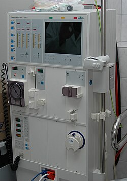
Hemodialysis a process of purifying the blood of a person whose kidneys are not working normally.

Biomedical engineering (BME) or medical engineering is the application of engineering principles and design concepts to medicine and biology for healthcare purposes (e.g., diagnostic or therapeutic). BME is also traditionally known as "bioengineering", but this term has come to also refer to biological engineering. This field seeks to close the gap between engineering and medicine, combining the design and problem-solving skills of engineering with medical biological sciences to advance health care treatment, including diagnosis, monitoring, and therapy. Also included under the scope of a biomedical engineer is the management of current medical equipment in hospitals while adhering to relevant industry standards. This involves making equipment recommendations, procurement, routine testing, and preventive maintenance, a role also known as a Biomedical Equipment Technician (BMET) or as clinical engineering.

Biomedical engineering has recently emerged as its own study, as compared to many other engineering fields. Such an evolution is common as a new field transition from being an interdisciplinary specialization among already-established fields to being considered a field in itself. Much of the work in biomedical engineering consists of research and development, spanning a broad array of subfields (see below). Prominent biomedical engineering applications include the development of biocompatible prostheses, various diagnostic and therapeutic medical devices ranging from clinical equipment to micro-implants, common imaging equipment such as MRIs and EKG/ECGs, regenerative tissue growth, pharmaceutical drugs and therapeutic biologicals.

Bioinformatics

Example of an approximately 40,000 probe spotted oligo microarray with enlarged inset to show detail.

Bioinformatics is an interdisciplinary field that develops methods and software tools for understanding biological data. As an interdisciplinary field of science, bioinformatics combines computer science, statistics, mathematics, and engineering to analyze and interpret biological data.

Bioinformatics is considered both an umbrella term for the body of biological studies that use computer programming as part of their methodology, as well as a reference to specific analysis "pipelines" that are repeatedly used, particularly in the field of genomics. Common uses of bioinformatics include the identification of candidate genes and nucleotides (SNPs). Often, such identification is made with the aim of better understanding the genetic basis of disease, unique adaptations, desirable properties (esp. in agricultural species), or differences between populations. In a less formal way, bioinformatics also tries to understand the organizational principles within nucleic acid and protein sequences.

Biomechanics

Biomechanics is the study of the structure and function of the mechanical aspects of biological systems, at any level from whole organisms to organs, cells and cell organelles, using the methods of mechanics.

Biomaterial

A biomaterial is any matter, surface, or construct that interacts with living systems. As a science, biomaterials is about fifty years old. The study of biomaterials is called biomaterials science or biomaterials engineering. It has experienced steady and strong growth over its history, with many companies investing large amounts of money into the development of new products. Biomaterials science encompasses elements of medicine, biology, chemistry, tissue engineering and materials science.

Biomedical optics

Biomedical optics refers to the interaction of biological tissue and light, and how this can be exploited for sensing, imaging, and treatment.

Tissue engineering

Tissue engineering, like genetic engineering (see below), is a major segment of biotechnology – which overlaps significantly with BME.

One of the goals of tissue engineering is to create artificial organs (via biological material) for patients that need organ transplants. Biomedical engineers are currently researching methods of creating such organs. Researchers have grown solid jawbones and tracheas from human stem cells towards this end. Several artificial urinary bladders have been grown in laboratories and transplanted successfully into human patients. Bioartificial organs, which use both synthetic and biological component, are also a focus area in research, such as with hepatic assist devices that use liver cells within an artificial bioreactor construct.

Micromass cultures of C3H-10T1/2 cells at varied oxygen tensions stained with Alcian blue.

Genetic engineering

Genetic engineering, recombinant DNA technology, genetic modification/manipulation (GM) and gene splicing are terms that apply to the direct manipulation of an organism's genes. Unlike traditional breeding, an indirect method of genetic manipulation, genetic engineering utilizes modern tools such as molecular cloning and transformation to directly alter the structure and characteristics of target genes. Genetic engineering techniques have found success in numerous applications. Some examples include the improvement of crop technology (not a medical application, but see biological systems engineering), the manufacture of synthetic human insulin through the use of modified bacteria, the manufacture of erythropoietin in hamster ovary cells, and the production of new types of experimental mice such as the oncomouse (cancer mouse) for research.

Neural engineering

Neural engineering (also known as neuroengineering) is a discipline that uses engineering techniques to understand, repair, replace, or enhance neural systems. Neural engineers are uniquely qualified to solve design problems at the interface of living neural tissue and non-living constructs.

Pharmaceutical engineering

Pharmaceutical engineering is an interdisciplinary science that includes drug engineering, novel drug delivery and targeting, pharmaceutical technology, unit operations of Chemical Engineering, and Pharmaceutical Analysis. It may be deemed as a part of pharmacy due to its focus on the use of technology on chemical agents in providing better medicinal treatment.

Medical devices

Schematic of silicone membrane oxygenator

This is an extremely broad category—essentially covering all health care products that do not achieve their intended results through predominantly chemical (e.g., pharmaceuticals) or biological (e.g., vaccines) means, and do not involve metabolism.

A medical device is intended for use in:

  • the diagnosis of disease or other conditions
  • in the cure, mitigation, treatment, or prevention of disease.

Some examples include pacemakers, infusion pumps, the heart-lung machine, dialysis machines, artificial organs, implants, artificial limbs, corrective lenses, cochlear implants, ocular prosthetics, facial prosthetics, somato prosthetics, and dental implants.

Biomedical instrumentation amplifier schematic used in monitoring low voltage biological signals, an example of a biomedical engineering application of electronic engineering to electrophysiology.

Stereolithography is a practical example of medical modeling being used to create physical objects. Beyond modeling organs and the human body, emerging engineering techniques are also currently used in the research and development of new devices for innovative therapies, treatments, patient monitoring, of complex diseases.

Medical devices are regulated and classified (in the US) as follows (see also Regulation):

  • Class I devices present minimal potential for harm to the user and are often simpler in design than Class II or Class III devices. Devices in this category include tongue depressors, bedpans, elastic bandages, examination gloves, and hand-held surgical instruments, and other similar types of common equipment.
  • Class II devices are subject to special controls in addition to the general controls of Class I devices. Special controls may include special labeling requirements, mandatory performance standards, and postmarket surveillance. Devices in this class are typically non-invasive and include X-ray machines, PACS, powered wheelchairs, infusion pumps, and surgical drapes.
  • Class III devices generally require premarket approval (PMA) or premarket notification (510k), a scientific review to ensure the device's safety and effectiveness, in addition to the general controls of Class I. Examples include replacement heart valves, hip and knee joint implants, silicone gel-filled breast implants, implanted cerebellar stimulators, implantable pacemaker pulse generators and endosseous (intra-bone) implants.

Medical imaging

Medical/biomedical imaging is a major segment of medical devices. This area deals with enabling clinicians to directly or indirectly "view" things not visible in plain sight (such as due to their size, and/or location). This can involve utilizing ultrasound, magnetism, UV, radiology, and other means.

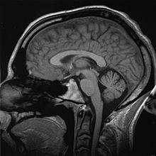
An MRI scan of a human head, an example of a biomedical engineering application of electrical engineering to diagnostic imaging. Click here to view an animated sequence of slices.

Imaging technologies are often essential to medical diagnosis, and are typically the most complex equipment found in a hospital including: fluoroscopy, magnetic resonance imaging (MRI), nuclear medicine, positron emission tomography (PET), PET-CT scans, projection radiography such as X-rays and CT scans, tomography, ultrasound, optical microscopy, and electron microscopy.

Implants

An implant is a kind of medical device made to replace and act as a missing biological structure (as compared with a transplant, which indicates transplanted biomedical tissue). The surface of implants that contact the body might be made of a biomedical material such as titanium, silicone or apatite depending on what is the most functional. In some cases, implants contain electronics, e.g. artificial pacemakers and cochlear implants. Some implants are bioactive, such as subcutaneous drug delivery devices in the form of implantable pills or drug-eluting stents.

Artificial limbs: The right arm is an example of a prosthesis, and the left arm is an example of myoelectric control.
 
A prosthetic eye, an example of a biomedical engineering application of mechanical engineering and biocompatible materials to ophthalmology.

Bionics

Artificial body part replacements are one of the many applications of bionics. Concerned with the intricate and thorough study of the properties and function of human body systems, bionics may be applied to solve some engineering problems. Careful study of the different functions and processes of the eyes, ears, and other organs paved the way for improved cameras, television, radio transmitters and receivers, and many other tools.

Biomedical sensors

In recent years biomedical sensors based in microwave technology have gained more attention. Different sensors can be manufactured for specific uses in both diagnosing and monitoring disease conditions, for example microwave sensors can be used as a complementary technique to X-ray to monitor lower extremity trauma. The sensor monitor the dielectric properties and can thus notice change in tissue (bone, muscle, fat etc.) under the skin so when measuring at different times during the healing process the response from the sensor will change as the trauma heals.

Clinical engineering

Clinical engineering is the branch of biomedical engineering dealing with the actual implementation of medical equipment and technologies in hospitals or other clinical settings. Major roles of clinical engineers include training and supervising biomedical equipment technicians (BMETs), selecting technological products/services and logistically managing their implementation, working with governmental regulators on inspections/audits, and serving as technological consultants for other hospital staff (e.g. physicians, administrators, I.T., etc.). Clinical engineers also advise and collaborate with medical device producers regarding prospective design improvements based on clinical experiences, as well as monitor the progression of the state of the art so as to redirect procurement patterns accordingly.

Their inherent focus on practical implementation of technology has tended to keep them oriented more towards incremental-level redesigns and reconfigurations, as opposed to revolutionary research & development or ideas that would be many years from clinical adoption; however, there is a growing effort to expand this time-horizon over which clinical engineers can influence the trajectory of biomedical innovation. In their various roles, they form a "bridge" between the primary designers and the end-users, by combining the perspectives of being both close to the point-of-use, while also trained in product and process engineering. Clinical engineering departments will sometimes hire not just biomedical engineers, but also industrial/systems engineers to help address operations research/optimization, human factors, cost analysis, etc. Also, see safety engineering for a discussion of the procedures used to design safe systems. The clinical engineering department is constructed with a manager, supervisor, engineer, and technician. One engineer per eighty beds in the hospital is the ratio. Clinical engineers are also authorized to audit pharmaceutical and associated stores to monitor FDA recalls of invasive items.

Rehabilitation engineering

Rehabilitation engineering is the systematic application of engineering sciences to design, develop, adapt, test, evaluate, apply, and distribute technological solutions to problems confronted by individuals with disabilities. Functional areas addressed through rehabilitation engineering may include mobility, communications, hearing, vision, and cognition, and activities associated with employment, independent living, education, and integration into the community.

While some rehabilitation engineers have master's degrees in rehabilitation engineering, usually a subspecialty of Biomedical engineering, most rehabilitation engineers have an undergraduate or graduate degrees in biomedical engineering, mechanical engineering, or electrical engineering. A Portuguese university provides an undergraduate degree and a master's degree in Rehabilitation Engineering and Accessibility. Qualification to become a Rehab' Engineer in the UK is possible via a University BSc Honours Degree course such as Health Design & Technology Institute, Coventry University.

The rehabilitation process for people with disabilities often entails the design of assistive devices such as Walking aids intended to promote the inclusion of their users into the mainstream of society, commerce, and recreation.

Schematic representation of a normal ECG trace showing sinus rhythm; an example of widely used clinical medical equipment (operates by applying electronic engineering to electrophysiology and medical diagnosis).

Regulatory issues

Regulatory issues have been constantly increased in the last decades to respond to the many incidents caused by devices to patients. For example, from 2008 to 2011, in US, there were 119 FDA recalls of medical devices classified as class I. According to U.S. Food and Drug Administration (FDA), Class I recall is associated to "a situation in which there is a reasonable probability that the use of, or exposure to, a product will cause serious adverse health consequences or death"

Regardless of the country-specific legislation, the main regulatory objectives coincide worldwide. For example, in the medical device regulations, a product must be: 1) safe and 2) effective and 3) for all the manufactured devices

A product is safe if patients, users, and third parties do not run unacceptable risks of physical hazards (death, injuries, ...) in its intended use. Protective measures have to be introduced on the devices to reduce residual risks at an acceptable level if compared with the benefit derived from the use of it.

A product is effective if it performs as specified by the manufacturer in the intended use. Effectiveness is achieved through clinical evaluation, compliance to performance standards or demonstrations of substantial equivalence with an already marketed device.

The previous features have to be ensured for all the manufactured items of the medical device. This requires that a quality system shall be in place for all the relevant entities and processes that may impact safety and effectiveness over the whole medical device lifecycle.

The medical device engineering area is among the most heavily regulated fields of engineering, and practicing biomedical engineers must routinely consult and cooperate with regulatory law attorneys and other experts. The Food and Drug Administration (FDA) is the principal healthcare regulatory authority in the United States, having jurisdiction over medical devices, drugs, biologics, and combination products. The paramount objectives driving policy decisions by the FDA are safety and effectiveness of healthcare products that have to be assured through a quality system in place as specified under 21 CFR 829 regulation. In addition, because biomedical engineers often develop devices and technologies for "consumer" use, such as physical therapy devices (which are also "medical" devices), these may also be governed in some respects by the Consumer Product Safety Commission. The greatest hurdles tend to be 510K "clearance" (typically for Class 2 devices) or pre-market "approval" (typically for drugs and class 3 devices).

In the European context, safety effectiveness and quality is ensured through the "Conformity Assessment" which is defined as "the method by which a manufacturer demonstrates that its device complies with the requirements of the European Medical Device Directive". The directive specifies different procedures according to the class of the device ranging from the simple Declaration of Conformity (Annex VII) for Class I devices to EC verification (Annex IV), Production quality assurance (Annex V), Product quality assurance (Annex VI) and Full quality assurance (Annex II). The Medical Device Directive specifies detailed procedures for Certification. In general terms, these procedures include tests and verifications that are to be contained in specific deliveries such as the risk management file, the technical file, and the quality system deliveries. The risk management file is the first deliverable that conditions the following design and manufacturing steps. The risk management stage shall drive the product so that product risks are reduced at an acceptable level with respect to the benefits expected for the patients for the use of the device. The technical file contains all the documentation data and records supporting medical device certification. FDA technical file has similar content although organized in a different structure. The Quality System deliverables usually include procedures that ensure quality throughout all product life cycles. The same standard (ISO EN 13485) is usually applied for quality management systems in the US and worldwide.

Implants, such as artificial hip joints, are generally extensively regulated due to the invasive nature of such devices.

In the European Union, there are certifying entities named "Notified Bodies", accredited by the European Member States. The Notified Bodies must ensure the effectiveness of the certification process for all medical devices apart from the class I devices where a declaration of conformity produced by the manufacturer is sufficient for marketing. Once a product has passed all the steps required by the Medical Device Directive, the device is entitled to bear a CE marking, indicating that the device is believed to be safe and effective when used as intended, and, therefore, it can be marketed within the European Union area.

The different regulatory arrangements sometimes result in particular technologies being developed first for either the U.S. or in Europe depending on the more favorable form of regulation. While nations often strive for substantive harmony to facilitate cross-national distribution, philosophical differences about the optimal extent of regulation can be a hindrance; more restrictive regulations seem appealing on an intuitive level, but critics decry the tradeoff cost in terms of slowing access to life-saving developments.

RoHS II

Directive 2011/65/EU, better known as RoHS 2 is a recast of legislation originally introduced in 2002. The original EU legislation "Restrictions of Certain Hazardous Substances in Electrical and Electronics Devices" (RoHS Directive 2002/95/EC) was replaced and superseded by 2011/65/EU published in July 2011 and commonly known as RoHS 2. RoHS seeks to limit the dangerous substances in circulation in electronics products, in particular toxins and heavy metals, which are subsequently released into the environment when such devices are recycled.

The scope of RoHS 2 is widened to include products previously excluded, such as medical devices and industrial equipment. In addition, manufacturers are now obliged to provide conformity risk assessments and test reports – or explain why they are lacking. For the first time, not only manufacturers but also importers and distributors share a responsibility to ensure Electrical and Electronic Equipment within the scope of RoHS complies with the hazardous substances limits and have a CE mark on their products.

IEC 60601

The new International Standard IEC 60601 for home healthcare electro-medical devices defining the requirements for devices used in the home healthcare environment. IEC 60601-1-11 (2010) must now be incorporated into the design and verification of a wide range of home use and point of care medical devices along with other applicable standards in the IEC 60601 3rd edition series.

The mandatory date for implementation of the EN European version of the standard is June 1, 2013. The US FDA requires the use of the standard on June 30, 2013, while Health Canada recently extended the required date from June 2012 to April 2013. The North American agencies will only require these standards for new device submissions, while the EU will take the more severe approach of requiring all applicable devices being placed on the market to consider the home healthcare standard.

AS/NZS 3551:2012

AS/ANS 3551:2012 is the Australian and New Zealand standards for the management of medical devices. The standard specifies the procedures required to maintain a wide range of medical assets in a clinical setting (e.g. Hospital). The standards are based on the IEC 606101 standards.

The standard covers a wide range of medical equipment management elements including, procurement, acceptance testing, maintenance (electrical safety and preventive maintenance testing) and decommissioning.

Training and certification

Education

Biomedical engineers require considerable knowledge of both engineering and biology, and typically have a Bachelor's (B.Sc., B.S., B.Eng. or B.S.E.) or Master's (M.S., M.Sc., M.S.E., or M.Eng.) or a doctoral (Ph.D.) degree in BME (Biomedical Engineering) or another branch of engineering with considerable potential for BME overlap. As interest in BME increases, many engineering colleges now have a Biomedical Engineering Department or Program, with offerings ranging from the undergraduate (B.Sc., B.S., B.Eng. or B.S.E.) to doctoral levels. Biomedical engineering has only recently been emerging as its own discipline rather than a cross-disciplinary hybrid specialization of other disciplines; and BME programs at all levels are becoming more widespread, including the Bachelor of Science in Biomedical Engineering which actually includes so much biological science content that many students use it as a "pre-med" major in preparation for medical school. The number of biomedical engineers is expected to rise as both a cause and effect of improvements in medical technology.

In the U.S., an increasing number of undergraduate programs are also becoming recognized by ABET as accredited bioengineering/biomedical engineering programs. Over 65 programs are currently accredited by ABET.

In Canada and Australia, accredited graduate programs in biomedical engineering are common. For example, McMaster University offers an M.A.Sc, an MD/PhD, and a PhD in Biomedical engineering. The first Canadian undergraduate BME program was offered at Ryerson University as a four-year B.Eng. program. The Polytechnique in Montreal is also offering a bachelors's degree in biomedical engineering as is Flinders University.

As with many degrees, the reputation and ranking of a program may factor into the desirability of a degree holder for either employment or graduate admission. The reputation of many undergraduate degrees is also linked to the institution's graduate or research programs, which have some tangible factors for rating, such as research funding and volume, publications and citations. With BME specifically, the ranking of a university's hospital and medical school can also be a significant factor in the perceived prestige of its BME department/program.

Graduate education is a particularly important aspect in BME. While many engineering fields (such as mechanical or electrical engineering) do not need graduate-level training to obtain an entry-level job in their field, the majority of BME positions do prefer or even require them. Since most BME-related professions involve scientific research, such as in pharmaceutical and medical device development, graduate education is almost a requirement (as undergraduate degrees typically do not involve sufficient research training and experience). This can be either a Masters or Doctoral level degree; while in certain specialties a Ph.D. is notably more common than in others, it is hardly ever the majority (except in academia). In fact, the perceived need for some kind of graduate credential is so strong that some undergraduate BME programs will actively discourage students from majoring in BME without an expressed intention to also obtain a master's degree or apply to medical school afterwards.

Graduate programs in BME, like in other scientific fields, are highly varied, and particular programs may emphasize certain aspects within the field. They may also feature extensive collaborative efforts with programs in other fields (such as the university's Medical School or other engineering divisions), owing again to the interdisciplinary nature of BME. M.S. and Ph.D. programs will typically require applicants to have an undergraduate degree in BME, or another engineering discipline (plus certain life science coursework), or life science (plus certain engineering coursework).

Education in BME also varies greatly around the world. By virtue of its extensive biotechnology sector, its numerous major universities, and relatively few internal barriers, the U.S. has progressed a great deal in its development of BME education and training opportunities. Europe, which also has a large biotechnology sector and an impressive education system, has encountered trouble in creating uniform standards as the European community attempts to supplant some of the national jurisdictional barriers that still exist. Recently, initiatives such as BIOMEDEA have sprung up to develop BME-related education and professional standards. Other countries, such as Australia, are recognizing and moving to correct deficiencies in their BME education. Also, as high technology endeavors are usually marks of developed nations, some areas of the world are prone to slower development in education, including in BME.

Licensure/certification

As with other learned professions, each state has certain (fairly similar) requirements for becoming licensed as a registered Professional Engineer (PE), but, in US, in industry such a license is not required to be an employee as an engineer in the majority of situations (due to an exception known as the industrial exemption, which effectively applies to the vast majority of American engineers). The US model has generally been only to require the practicing engineers offering engineering services that impact the public welfare, safety, safeguarding of life, health, or property to be licensed, while engineers working in private industry without a direct offering of engineering services to the public or other businesses, education, and government need not be licensed. This is notably not the case in many other countries, where a license is as legally necessary to practice engineering as it is for law or medicine.

Biomedical engineering is regulated in some countries, such as Australia, but registration is typically only recommended and not required.

In the UK, mechanical engineers working in the areas of Medical Engineering, Bioengineering or Biomedical engineering can gain Chartered Engineer status through the Institution of Mechanical Engineers. The Institution also runs the Engineering in Medicine and Health Division. The Institute of Physics and Engineering in Medicine (IPEM) has a panel for the accreditation of MSc courses in Biomedical Engineering and Chartered Engineering status can also be sought through IPEM.

The Fundamentals of Engineering exam – the first (and more general) of two licensure examinations for most U.S. jurisdictions—does now cover biology (although technically not BME). For the second exam, called the Principles and Practices, Part 2, or the Professional Engineering exam, candidates may select a particular engineering discipline's content to be tested on; there is currently not an option for BME with this, meaning that any biomedical engineers seeking a license must prepare to take this examination in another category (which does not affect the actual license, since most jurisdictions do not recognize discipline specialties anyway). However, the Biomedical Engineering Society (BMES) is, as of 2009, exploring the possibility of seeking to implement a BME-specific version of this exam to facilitate biomedical engineers pursuing licensure.

Beyond governmental registration, certain private-sector professional/industrial organizations also offer certifications with varying degrees of prominence. One such example is the Certified Clinical Engineer (CCE) certification for Clinical engineers.

Career prospects

In 2012 there were about 19,400 biomedical engineers employed in the US, and the field was predicted to grow by 5% (faster than average) from 2012 to 2022. Biomedical engineering has the highest percentage of female engineers compared to other common engineering professions.

Notable figures

See also


Deep brain stimulation

From Wikipedia, the free encyclopedia
 
Deep brain stimulation
Tiefe Hirnstimulation - Sonden RoeSchaedel ap.jpg
DBS-probes shown in X-ray of the skull (white areas around maxilla and mandible represent metal dentures and are unrelated to DBS devices)
MeSHD046690
MedlinePlus007453

Deep brain stimulation (DBS) is a neurosurgical procedure involving the placement of a medical device called a neurostimulator (sometimes referred to as a "brain pacemaker"), which sends electrical impulses, through implanted electrodes, to specific targets in the brain (brain nuclei) for the treatment of movement disorders, including Parkinson's disease, essential tremor, and dystonia. and other conditions such as obsessive-compulsive disorder and epilepsy. While its underlying principles and mechanisms are not fully understood, DBS directly changes brain activity in a controlled manner.

DBS has been approved by the Food and Drug Administration as a treatment for essential tremor and Parkinson's disease (PD) since 1997. DBS was approved for dystonia in 2003, obsessive–compulsive disorder (OCD) in 2009, and epilepsy in 2018. DBS has been studied in clinical trials as a potential treatment for chronic pain for various affective disorders, including major depression. It is one of few neurosurgical procedures that allow blinded studies.

Medical use

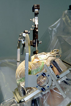
Insertion of electrode during surgery using a stereotactic frame

Parkinson's disease

DBS is used to manage some of the symptoms of Parkinson's disease that cannot be adequately controlled with medications. PD is treated by applying high-frequency (> 100 Hz) stimulation to three target structures namely to entrolateral thalamus, internal pallidum, and subthalamic nucleus (STN) to mimic the clinical effects of lesioning. It is recommended for people who have PD with motor fluctuations and tremor inadequately controlled by medication, or to those who are intolerant to medication, as long as they do not have severe neuropsychiatric problems. Four areas of the brain have been treated with neural stimulators in PD. These are the globus pallidus internus, thalamus, subthalamic nucleus and the pedunculopontine nucleus. However, most DBS surgeries in routine practice target either the globus pallidus internus, or the Subthalamic nucleus.

  • DBS of the globus pallidus internus reduces uncontrollable shaking movements called dyskinesias. This enables a patient to take adequate quantities of medications (especially levodopa), thus leading to better control of symptoms.
  • DBS of the subthalamic nucleus directly reduces symptoms of Parkinson's. This enables a decrease in the dose of anti-parkinsonian medications.
  • DBS of the PPN may help with freezing of gait, while DBS of the thalamus may help with tremor. These targets are not routinely utilized.

Selection of the correct DBS target is a complicated process. Multiple clinical characteristics are used to select the target including – identifying the most troublesome symptoms, the dose of levodopa that the patient is currently taking, the effects and side-effects of current medications and concurrent problems. For example, subthalamic nucleus DBS may worsen depression and hence is not preferred in patients with uncontrolled depression.

Generally DBS is associated with 30–60% improvement in motor score evaluations.

Tourette syndrome

DBS has been used experimentally in treating adults with severe Tourette syndrome that does not respond to conventional treatment. Despite widely publicized early successes, DBS remains a highly experimental procedure for the treatment of Tourette's, and more study is needed to determine whether long-term benefits outweigh the risks. The procedure is well tolerated, but complications include "short battery life, abrupt symptom worsening upon cessation of stimulation, hypomanic or manic conversion, and the significant time and effort involved in optimizing stimulation parameters". As of 2006, five people with TS had been reported on; all experienced reduction in tics and the disappearance of obsessive-compulsive behaviors.

The procedure is invasive and expensive, and requires long-term expert care. Benefits for severe Tourette's are not conclusive, considering less robust effects of this surgery seen in the Netherlands. Tourette's is more common in pediatric populations, tending to remit in adulthood, so in general this would not be a recommended procedure for use on children. Because diagnosis of Tourette's is made based on a history of symptoms rather than analysis of neurological activity, it may not always be clear how to apply DBS for a particular person. Due to concern over the use of DBS in Tourette syndrome treatment, the Tourette Association of America convened a group of experts to develop recommendations guiding the use and potential clinical trials of DBS for TS.

Robertson reported that DBS had been used on 55 adults by 2011, remained an experimental treatment at that time, and recommended that the procedure "should only be conducted by experienced functional neurosurgeons operating in centres which also have a dedicated Tourette syndrome clinic". According to Malone et al. (2006), "Only patients with severe, debilitating, and treatment-refractory illness should be considered; while those with severe personality disorders and substance-abuse problems should be excluded." Du et al. (2010) say, "As an invasive therapy, DBS is currently only advisable for severely affected, treatment-refractory TS adults". Singer (2011) says, "pending determination of patient selection criteria and the outcome of carefully controlled clinical trials, a cautious approach is recommended". Viswanathan et al. (2012) say DBS should be used for people with "severe functional impairment that cannot be managed medically".

Adverse effects

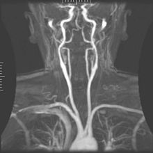
Arteriogram of the arterial supply that can hemorrhage during DBS implantation.

DBS carries the risks of major surgery, with a complication rate related to the experience of the surgical team. The major complications include hemorrhage (1–2%) and infection (3–5%).

The potential exists for neuropsychiatric side effects after DBS, including apathy, hallucinations, hypersexuality, cognitive dysfunction, depression, and euphoria. However, these effects may be temporary and related to (1) correct placement of electrodes, (2) open-loop VS closed loop stimulation , meaning a constant stimulation or an A.I. monitoring delivery system and (3) calibration of the stimulator, so these side effects are potentially reversible.

Because the brain can shift slightly during surgery, the electrodes can become displaced or dislodged from the specific location. This may cause more profound complications such as personality changes, but electrode misplacement is relatively easy to identify using CT scan. Also, complications of surgery may occur, such as bleeding within the brain. After surgery, swelling of the brain tissue, mild disorientation, and sleepiness are normal. After 2–4 weeks, a follow-up visit is used to remove sutures, turn on the neurostimulator, and program it.

Impaired swimming skills surfaced as an unexpected risk of the procedure; several Parkinson's disease patients lost their ability to swim after receiving deep brain stimulation.

Mechanisms

The exact mechanism of action of DBS is not known. A variety of hypotheses try to explain the mechanisms of DBS:

  1. Depolarization blockade: Electrical currents block the neuronal output at or near the electrode site.
  2. Synaptic inhibition: This causes an indirect regulation of the neuronal output by activating axon terminals with synaptic connections to neurons near the stimulating electrode.
  3. Desynchronization of abnormal oscillatory activity of neurons
  4. Antidromic activation either activating/blockading distant neurons or blockading slow axons

DBS represents an advance on previous treatments which involved pallidotomy (i.e., surgical ablation of the globus pallidus) or thalamotomy (i.e., surgical ablation of the thalamus). Instead, a thin lead with multiple electrodes is implanted in the globus pallidus, nucleus ventralis intermedius thalami, or subthalamic nucleus, and electric pulses are used therapeutically. The lead from the implant is extended to the neurostimulator under the skin in the chest area.

Its direct effect on the physiology of brain cells and neurotransmitters is currently debated, but by sending high-frequency electrical impulses into specific areas of the brain, it can mitigate symptoms and directly diminish the side effects induced by PD medications, allowing a decrease in medications, or making a medication regimen more tolerable.

Components and placement

The DBS system consists of three components: the implanted pulse generator (IPG), the lead, and an extension. The IPG is a battery-powered neurostimulator encased in a titanium housing, which sends electrical pulses to the brain that interfere with neural activity at the target site. The lead is a coiled wire insulated in polyurethane with four platinum-iridium electrodes and is placed in one or two different nuclei of the brain. The lead is connected to the IPG by an extension, an insulated wire that runs below the skin, from the head, down the side of the neck, behind the ear, to the IPG, which is placed subcutaneously below the clavicle, or in some cases, the abdomen. The IPG can be calibrated by a neurologist, nurse, or trained technician to optimize symptom suppression and control side effects.

DBS leads are placed in the brain according to the type of symptoms to be addressed. For non-Parkinsonian essential tremor, the lead is placed in either the ventrointermediate nucleus of the thalamus or the zona incerta; for dystonia and symptoms associated with PD (rigidity, bradykinesia/akinesia, and tremor), the lead may be placed in either the globus pallidus internus or the subthalamic nucleus; for OCD and depression to the nucleus accumbens; for incessant pain to the posterior thalamic region or periaqueductal gray; and for epilepsy treatment to the anterior thalamic nucleus.

All three components are surgically implanted inside the body. Lead implantation may take place under local anesthesia or under general anesthesia ("asleep DBS") such as for dystonia. A hole about 14 mm in diameter is drilled in the skull and the probe electrode is inserted stereotactically, using either frame-based or frameless stereotaxis. During the awake procedure with local anesthesia, feedback from the person is used to determine the optimal placement of the permanent electrode. During the asleep procedure, intraoperative MRI guidance is used for direct visualization of brain tissue and device. The installation of the IPG and extension leads occurs under general anesthesia. The right side of the brain is stimulated to address symptoms on the left side of the body and vice versa.

Research

Chronic pain

Stimulation of the periaqueductal gray and periventricular gray for nociceptive pain, and the internal capsule, ventral posterolateral nucleus, and ventral posteromedial nucleus for neuropathic pain has produced impressive results with some people, but results vary. One study of 17 people with intractable cancer pain found that 13 were virtually pain free and only four required opioid analgesics on release from hospital after the intervention. Most ultimately did resort to opioids, usually in the last few weeks of life. DBS has also been applied for phantom limb pain.

Major depression and obsessive-compulsive disorder

Lateral X-ray of the head: Deep brain stimulation in obsessive–compulsive disorder (OCD). 42 year old man, surgery in 2013.

DBS has been used in a small number of clinical trials to treat people with severe treatment-resistant depression (TRD). A number of neuroanatomical targets have been used for DBS for TRD including the subgenual cingulate gyrus, posterior gyrus rectus, nucleus accumbens, ventral capsule/ventral striatum, inferior thalamic peduncle, and the lateral habenula. A recently proposed target of DBS intervention in depression is the superolateral branch of the medial forebrain bundle; its stimulation lead to surprisingly rapid antidepressant effects.

The small numbers in the early trials of DBS for TRD currently limit the selection of an optimal neuroanatomical target. Evidence is insufficient to support DBS as a therapeutic modality for depression; however, the procedure may be an effective treatment modality in the future. In fact, beneficial results have been documented in the neurosurgical literature, including a few instances in which people who were deeply depressed were provided with portable stimulators for self treatment.

A systematic review of DBS for TRD and OCD identified 23 cases, nine for OCD, seven for TRD, and one for both. "[A]bout half the patients did show dramatic improvement" and adverse events were "generally trivial" given the younger age of the psychiatric population relative to the age of people with movement disorders. The first randomized, controlled study of DBS for the treatment of TRD targeting the ventral capsule/ventral striatum area did not demonstrate a significant difference in response rates between the active and sham groups at the end of a 16-week study. However, a second randomized controlled study of ventral capsule DBS for TRD did demonstrate a significant difference in response rates between active DBS (44% responders) and sham DBS (0% responders). Efficacy of DBS is established for OCD, with on average 60% responders in severely ill and treatment-resistant patients. Based on these results the FDA has approved DBS for treatment-resistant OCD under a Humanitarian Device Exemption (HDE), requiring that the procedure be performed only in a hospital with specialist qualifications to do so.

DBS for TRD can be as effective as antidepressants and can have good response and remission rates, but adverse effects and safety must be more fully evaluated. Common side effects include "wound infection, perioperative headache, and worsening/irritable mood [and] increased suicidality".

Other clinical applications

Results of DBS in people with dystonia, where positive effects often appear gradually over a period of weeks to months, indicate a role of functional reorganization in at least some cases. The procedure has been tested for effectiveness in people with epilepsy that is resistant to medication. DBS may reduce or eliminate epileptic seizures with programmed or responsive stimulation.

DBS of the septal areas of persons with schizophrenia have resulted in enhanced alertness, cooperation, and euphoria. Persons with narcolepsy and complex-partial seizures also reported euphoria and sexual thoughts from self-elicited DBS of the septal nuclei.

Orgasmic ecstasy was reported with the electrical stimulation of the brain with depth electrodes in the left hippocampus at 3mA, and the right hippocampus at 1 mA.

In 2015, a group of Brazilian researchers led by neurosurgeon Erich Fonoff [pt] described a new technique that allows for simultaneous implants of electrodes called bilateral stereotactic procedure for DBS. The main benefits are less time spent on the procedure and greater accuracy.

In 2016, DBS was found to improve learning and memory in a mouse model of Rett syndrome. More recent (2018) work showed, that forniceal DBS upregulates genes involved in synaptic function, cell survival, and neurogenesis, making some first steps at explaining the restoration of hippocampal circuit function.

See also


Neural engineering

From Wikipedia, the free encyclopedia
https://en.wikipedia.org/wiki/Neural_engineering

Neural engineering (also known as neuroengineering) is a discipline within biomedical engineering that uses engineering techniques to understand, repair, replace, or enhance neural systems. Neural engineers are uniquely qualified to solve design problems at the interface of living neural tissue and non-living constructs (Hetling, 2008).

Overview

The field of neural engineering draws on the fields of computational neuroscience, experimental neuroscience, clinical neurology, electrical engineering and signal processing of living neural tissue, and encompasses elements from robotics, cybernetics, computer engineering, neural tissue engineering, materials science, and nanotechnology.

Prominent goals in the field include restoration and augmentation of human function via direct interactions between the nervous system and artificial devices.

Much current research is focused on understanding the coding and processing of information in the sensory and motor systems, quantifying how this processing is altered in the pathological state, and how it can be manipulated through interactions with artificial devices including brain–computer interfaces and neuroprosthetics.

Other research concentrates more on investigation by experimentation, including the use of neural implants connected with external technology.

Neurohydrodynamics is a division of neural engineering that focuses on hydrodynamics of the neurological system.

History

As neural engineering is a relatively new field, information and research relating to it is comparatively limited, although this is changing rapidly. The first journals specifically devoted to neural engineering, The Journal of Neural Engineering and The Journal of NeuroEngineering and Rehabilitation both emerged in 2004. International conferences on neural engineering have been held by the IEEE since 2003, from 29 April until 2 May 2009 in Antalya, Turkey 4th Conference on Neural Engineering, the 5th International IEEE EMBS Conference on Neural Engineering in April/May 2011 in Cancún, Mexico, and the 6th conference in San Diego, California in November 2013. The 7th conference was held in April 2015 in Montpellier. The 8th conference was held in May 2017 in Shanghai.

Fundamentals

The fundamentals behind neuroengineering involve the relationship of neurons, neural networks, and nervous system functions to quantifiable models to aid the development of devices that could interpret and control signals and produce purposeful responses.

Neuroscience

Messages that the body uses to influence thoughts, senses, movements, and survival are directed by nerve impulses transmitted across brain tissue and to the rest of the body. Neurons are the basic functional unit of the nervous system and are highly specialized cells that are capable of sending these signals that operate high and low level functions needed for survival and quality of life. Neurons have special electro-chemical properties that allow them to process information and then transmit that information to other cells. Neuronal activity is dependent upon neural membrane potential and the changes that occur along and across it. A constant voltage, known as the membrane potential, is normally maintained by certain concentrations of specific ions across neuronal membranes. Disruptions or variations in this voltage create an imbalance, or polarization, across the membrane. Depolarization of the membrane past its threshold potential generates an action potential, which is the main source of signal transmission, known as neurotransmission of the nervous system. An action potential results in a cascade of ion flux down and across an axonal membrane, creating an effective voltage spike train or "electrical signal" which can transmit further electrical changes in other cells. Signals can be generated by electrical, chemical, magnetic, optical, and other forms of stimuli that influence the flow of charges, and thus voltage levels across neural membranes (He 2005).

Engineering

Engineers employ quantitative tools that can be used for understanding and interacting with complex neural systems. Methods of studying and generating chemical, electrical, magnetic, and optical signals responsible for extracellular field potentials and synaptic transmission in neural tissue aid researchers in the modulation of neural system activity (Babb et al. 2008). To understand properties of neural system activity, engineers use signal processing techniques and computational modeling (Eliasmith & Anderson 2003). To process these signals, neural engineers must translate the voltages across neural membranes into corresponding code, a process known as neural coding. Neural coding studies on how the brain encodes simple commands in the form of central pattern generators (CPGs), movement vectors, the cerebellar internal model, and somatotopic maps to understand movement and sensory phenomena. Decoding of these signals in the realm of neuroscience is the process by which neurons understand the voltages that have been transmitted to them. Transformations involve the mechanisms that signals of a certain form get interpreted and then translated into another form. Engineers look to mathematically model these transformations (Eliasmith & Anderson 2003). There are a variety of methods being used to record these voltage signals. These can be intracellular or extracellular. Extracellular methods involve single-unit recordings, extracellular field potentials, and amperometry; more recently, multielectrode arrays have been used to record and mimic signals.

Scope

Neuromechanics

Neuromechanics is the coupling of neurobiology, biomechanics, sensation and perception, and robotics (Edwards 2010). Researchers are using advanced techniques and models to study the mechanical properties of neural tissues and their effects on the tissues' ability to withstand and generate force and movements as well as their vulnerability to traumatic loading (Laplaca & Prado 2010). This area of research focuses on translating the transformations of information among the neuromuscular and skeletal systems to develop functions and governing rules relating to operation and organization of these systems (Nishikawa et al. 2007). Neuromechanics can be simulated by connecting computational models of neural circuits to models of animal bodies situated in virtual physical worlds (Edwards 2010). Experimental analysis of biomechanics including the kinematics and dynamics of movements, the process and patterns of motor and sensory feedback during movement processes, and the circuit and synaptic organization of the brain responsible for motor control are all currently being researched to understand the complexity of animal movement. Dr. Michelle LaPlaca's lab at Georgia Institute of Technology is involved in the study of mechanical stretch of cell cultures, shear deformation of planar cell cultures, and shear deformation of 3D cell containing matrices. Understanding of these processes is followed by development of functioning models capable of characterizing these systems under closed loop conditions with specially defined parameters. The study of neuromechanics is aimed at improving treatments for physiological health problems which includes optimization of prostheses design, restoration of movement post injury, and design and control of mobile robots. By studying structures in 3D hydrogels, researchers can identify new models of nerve cell mechanoproperties. For example, LaPlaca et al. developed a new model showing that strain may play a role in cell culture (LaPlaca et al. 2005).

Neuromodulation

Neuromodulation aims to treat disease or injury by employing medical device technologies that would enhance or suppress activity of the nervous system with the delivery of pharmaceutical agents, electrical signals, or other forms of energy stimulus to re-establish balance in impaired regions of the brain. Researchers in this field face the challenge of linking advances in understanding neural signals to advancements in technologies delivering and analyzing these signals with increased sensitivity, biocompatibility, and viability in closed loops schemes in the brain such that new treatments and clinical applications can be created to treat those suffering from neural damage of various kinds. Neuromodulator devices can correct nervous system dysfunction related to Parkinson's disease, dystonia, tremor, Tourette's, chronic pain, OCD, severe depression, and eventually epilepsy. Neuromodulation is appealing as treatment for varying defects because it focuses in on treating highly specific regions of the brain only, contrasting that of systemic treatments that can have side effects on the body. Neuromodulator stimulators such as microelectrode arrays can stimulate and record brain function and with further improvements are meant to become adjustable and responsive delivery devices for drugs and other stimuli.

Neural regrowth and repair

Neural engineering and rehabilitation applies neuroscience and engineering to investigating peripheral and central nervous system function and to finding clinical solutions to problems created by brain damage or malfunction. Engineering applied to neuroregeneration focuses on engineering devices and materials that facilitate the growth of neurons for specific applications such as the regeneration of peripheral nerve injury, the regeneration of the spinal cord tissue for spinal cord injury, and the regeneration of retinal tissue. Genetic engineering and tissue engineering are areas developing scaffolds for spinal cord to regrow across thus helping neurological problems (Schmidt & Leach 2003).

Research and applications

Research focused on neural engineering utilizes devices to study how the nervous system functions and malfunctions (Schmidt & Leach 2003).

Neural imaging

Neuroimaging techniques are used to investigate the activity of neural networks, as well as the structure and function of the brain. Neuroimaging technologies include functional magnetic resonance imaging (fMRI), magnetic resonance imaging (MRI), positron emission tomography (PET) and computed axial tomography (CAT) scans. Functional neuroimaging studies are interested in which areas of the brain perform specific tasks. fMRI measures hemodynamic activity that is closely linked to neural activity. It is used to map metabolic responses in specific regions of the brain to a given task or stimulus. PET, CT scanners, and electroencephalography (EEG) are currently being improved and used for similar purposes.

Neural networks

Scientists can use experimental observations of neuronal systems and theoretical and computational models of these systems to create Neural networks with the hopes of modeling neural systems in as realistic a manner as possible. Neural networks can be used for analyses to help design further neurotechnological devices. Specifically, researchers handle analytical or finite element modeling to determine nervous system control of movements and apply these techniques to help patients with brain injuries or disorders. Artificial neural networks can be built from theoretical and computational models and implemented on computers from theoretically devices equations or experimental results of observed behavior of neuronal systems. Models might represent ion concentration dynamics, channel kinetics, synaptic transmission, single neuron computation, oxygen metabolism, or application of dynamic system theory (LaPlaca et al. 2005). Liquid-based template assembly was used to engineer 3D neural networks from neuron-seeded microcarrier beads.

Neural interfaces

Neural interfaces are a major element used for studying neural systems and enhancing or replacing neuronal function with engineered devices. Engineers are challenged with developing electrodes that can selectively record from associated electronic circuits to collect information about the nervous system activity and to stimulate specified regions of neural tissue to restore function or sensation of that tissue (Cullen et al. 2011). The materials used for these devices must match the mechanical properties of neural tissue in which they are placed and the damage must be assessed. Neural interfacing involves temporary regeneration of biomaterial scaffolds or chronic electrodes and must manage the body's response to foreign materials. Microelectrode arrays are recent advances that can be used to study neural networks (Cullen & Pfister 2011). Optical neural interfaces involve optical recordings and optogenetics stimulation that makes brain cells light sensitive. Fiber optics can be implanted in the brain to stimulate and record this photon activity instead of electrodes. Two-photon excitation microscopy can study living neuronal networks and the communicatory events among neurons.

Brain–computer interfaces

Brain–computer interfaces seek to directly communicate with human nervous system to monitor and stimulate neural circuits as well as diagnose and treat intrinsic neurological dysfunction. Deep brain stimulation is a significant advance in this field that is especially effective in treating movement disorders such as Parkinson's disease with high frequency stimulation of neural tissue to suppress tremors (Lega et al. 2011).

Microsystems

Neural microsystems can be developed to interpret and deliver electrical, chemical, magnetic, and optical signals to neural tissue. They can detect variations in membrane potential and measure electrical properties such as spike population, amplitude, or rate by using electrodes, or by assessment of chemical concentrations, fluorescence light intensity, or magnetic field potential. The goal of these systems is to deliver signals that would influence neuronal tissue potential and thus stimulate the brain tissue to evoke a desired response (He 2005).

Microelectrode arrays

Microelectrode arrays are specific tools used to detect the sharp changes in voltage in the extracellular environments that occur from propagation of an action potential down an axon. Dr. Mark Allen and Dr. LaPlaca have microfabricated 3D electrodes out of cytocompatible materials such as SU-8 and SLA polymers which have led to the development of in vitro and in vivo microelectrode systems with the characteristics of high compliance and flexibility to minimize tissue disruption.

Neural prostheses

Neuroprosthetics are devices capable of supplementing or replacing missing functions of the nervous system by stimulating the nervous system and recording its activity. Electrodes that measure firing of nerves can integrate with prosthetic devices and signal them to perform the function intended by the transmitted signal. Sensory prostheses use artificial sensors to replace neural input that might be missing from biological sources (He 2005). Engineers researching these devices are charged with providing a chronic, safe, artificial interface with neuronal tissue. Perhaps the most successful of these sensory prostheses is the cochlear implant which has restored hearing abilities to the deaf. Visual prosthesis for restoring visual capabilities of blind persons is still in more elementary stages of development. Motor prosthesics are devices involved with electrical stimulation of biological neural muscular system that can substitute for control mechanisms of the brain or spinal cord. Smart prostheses can be designed to replace missing limbs controlled by neural signals by transplanting nerves from the stump of an amputee to muscles. Sensory prosthetics provide sensory feedback by transforming mechanical stimuli from the periphery into encoded information accessible by the nervous system. Electrodes placed on the skin can interpret signals and then control the prosthetic limb. These prosthetics have been very successful. Functional electrical stimulation (FES) is a system aimed at restoring motor processes such as standing, walking, and hand grasp.

Neurorobotics

Neurorobotics is the study of how neural systems can be embodied and movements emulated in mechanical machines. Neurorobots are typically used to study motor control and locomotion, learning and memory selection, and value systems and action selection. By studying neurorobots in real-world environments, they are more easily observed and assessed to describe heuristics of robot function in terms of its embedded neural systems and the reactions of these systems to its environment (Krichmar 2008). For instance, making use of a computational model of epilectic spike-wave dynamics, it has been already proven the effectiveness of a method to simulate seizure abatement through a pseudospectral protocol. The computational model emulates the brain connectivity by using a magnetic imaging resonance from a patient suffering of idiopathic generalized epilepsy. The method was able to generate stimuli able to lessen the seizures.

Neural tissue regeneration

Neural tissue regeneration, or neuroregeneration looks to restore function to those neurons that have been damaged in small injuries and larger injuries like those caused by traumatic brain injury. Functional restoration of damaged nerves involves re-establishment of a continuous pathway for regenerating axons to the site of innervation. Researchers like Dr. LaPlaca at Georgia Institute of Technology are looking to help find treatment for repair and regeneration after traumatic brain injury and spinal cord injuries by applying tissue engineering strategies. Dr. LaPlaca is looking into methods combining neural stem cells with an extracellular matrix protein based scaffold for minimally invasive delivery into the irregular shaped lesions that form after a traumatic insult. By studying the neural stem cells in vitro and exploring alternative cell sources, engineering novel biopolymers that could be utilized in a scaffold, and investigating cell or tissue engineered construct transplants in vivo in models of traumatic brain and spinal cord injury, Dr. LaPlaca's lab aims to identify optimal strategies for nerve regeneration post injury.

Current approaches to clinical treatment

End to end surgical suture of damaged nerve ends can repair small gaps with autologous nerve grafts. For larger injuries, an autologous nerve graft that has been harvested from another site in the body might be used, though this process is time consuming, costly and requires two surgeries (Schmidt & Leach 2003). Clinical treatment for CNS is minimally available and focuses mostly on reducing collateral damage caused by bone fragments near the site of injury or inflammation. After swelling surrounding injury lessens, patients undergo rehabilitation so that remaining nerves can be trained to compensate for the lack of nerve function in injured nerves. No treatment currently exists to restore nerve function of CNS nerves that have been damaged (Schmidt & Leach 2003).

Engineering strategies for repair

Engineering strategies for the repair of spinal cord injury are focused on creating a friendly environment for nerve regeneration. Only PNS nerve damage has been clinically possible so far, but advances in research of genetic techniques and biomaterials demonstrate the potential for SC nerves to regenerate in permissible environments.

Grafts

Advantages of autologous tissue grafts are that they come from natural materials which have a high likelihood of biocompatibility while providing structural support to nerves that encourage cell adhesion and migration (Schmidt & Leach 2003). Nonautologous tissue, acellular grafts, and extracellular matrix based materials are all options that may also provide ideal scaffolding for nerve regeneration. Some come from allogenic or xenogenic tissues that must be combined with immunosuppressants. while others include small intestinal submucosa and amniotic tissue grafts (Schmidt & Leach 2003). Synthetic materials are attractive options because their physical and chemical properties can typically be controlled. A challenge that remains with synthetic materials is biocompatibility (Schmidt & Leach 2003). Methylcellulose-based constructs have been shown to be a biocompatible option serving this purpose (Tate et al. 2001). AxoGen uses a cell graft technology AVANCE to mimic a human nerve. It has been shown to achieve meaningful recovery in 87 percent of patients with peripheral nerve injuries.

Nerve guidance channels

Nerve guidance channels, Nerve guidance conduit are innovative strategies focusing on larger defects that provide a conduit for sprouting axons directing growth and reducing growth inhibition from scar tissue. Nerve guidance channels must be readily formed into a conduit with the desired dimensions, sterilizable, tear resistant, and easy to handle and suture (Schmidt & Leach 2003). Ideally they would degrade over time with nerve regeneration, be pliable, semipermeable, maintain their shape, and have a smooth inner wall that mimics that of a real nerve (Schmidt & Leach 2003).

Biomolecular therapies

Highly controlled delivery systems are needed to promote neural regeneration. Neurotrophic factors can influence development, survival, outgrowth, and branching. Neurotrophins include nerve growth factor (NGF), brain derived neurotrophic factor (BDNF), neurotrophin-3 (NT-3) and neurotrophin-4/5 (NT-4/5). Other factors are ciliary neurotrophic factor (CNTF), glial cell line-derived growth factor (GDNF) and acidic and basic fibroblast growth factor (aFGF, bFGF) that promote a range of neural responses.(Schmidt & Leach 2003) Fibronectin has also been shown to support nerve regeneration following TBI in rats (Tate et al. 2002). Other therapies are looking into regeneration of nerves by upregulating regeneration associated genes (RAGs), neuronal cytoskeletal components, and antiapoptosis factors. RAGs include GAP-43 and Cap-23, adhesion molecules such as L1 family, NCAM, and N-cadherin (Schmidt & Leach 2003). There is also the potential for blocking inhibitory biomolecules in the CNS due to glial scarring. Some currently being studied are treatments with chondroitinase ABC and blocking NgR, ADP-ribose (Schmidt & Leach 2003).

Delivery techniques

Delivery devices must be biocompatible and stable in vivo. Some examples include osmotic pumps, silicone reservoirs, polymer matrices, and microspheres. Gene therapy techniques have also been studied to provide long-term production of growth factors and could be delivered with viral or non-viral vectors such as lipoplexes. Cells are also effective delivery vehicles for ECM components, neurotrophic factors and cell adhesion molecules. Olfactory ensheathing cells (OECs) and stem cells as well as genetically modified cells have been used as transplants to support nerve regeneration (LaPlaca et al. 2005, Schmidt & Leach 2003, Tate et al. 2002).

Advanced therapies

Advanced therapies combine complex guidance channels and multiple stimuli that focus on internal structures that mimic the nerve architecture containing internal matrices of longitudinally aligned fibers or channels. Fabrication of these structures can use a number of technologies: magnetic polymer fiber alignment, injection molding, phase separation, solid free-form fabrication, and ink jet polymer printing (Schmidt & Leach 2003).

Neural enhancement

Augmentation of human neural systems, or human enhancement using engineering techniques is another possible application of neuroengineering. Deep brain stimulation has already been shown to enhance memory recall as noted by patients currently using this treatment for neurological disorders. Brain stimulation techniques are postulated to be able to sculpt emotions and personalities as well as enhance motivation, reduce inhibitions, etc. as requested by the individual. Ethical issues with this sort of human augmentation are a new set of questions that neural engineers have to grapple with as these studies develop.

See also


Representation of a Lie group

From Wikipedia, the free encyclopedia https://en.wikipedia.org/wiki/Representation_of_a_Lie_group...