From Wikipedia, the free encyclopedia
Omega−3 fatty acids, also called Omega-3 oils, ω−3 fatty acids or n−3 fatty acids, are polyunsaturated fatty acids
(PUFAs) characterized by the presence of a double bond, three atoms
away from the terminal methyl group in their chemical structure. They are widely distributed in nature, being important constituents of animal lipid metabolism, and they play an important role in the human diet and in human physiology. The three types of omega−3 fatty acids involved in human physiology are α-linolenic acid (ALA), found in plant oils, and eicosapentaenoic acid (EPA) and docosahexaenoic acid (DHA), both commonly found in oils of marine fish. Marine algae and phytoplankton are primary sources of omega−3 fatty acids (which also accumulate in fish). Common sources of plant oils containing ALA include walnuts, edible seeds, and flaxseeds, while sources of EPA and DHA include fish and fish oils.
Mammals are unable to synthesize the essential
omega−3 fatty acid ALA and can only obtain it through diet. However,
they can use ALA, when available, to form EPA and DHA, by creating
additional double bonds along its carbon chain (desaturation) and extending it (elongation).
Namely, ALA (18 carbons and 3 double bonds) is used to make EPA (20
carbons and 5 double bonds), which is then used to make DHA (22 carbons
and 6 double bonds). The ability to make the longer-chain omega−3 fatty acids from ALA may be impaired in aging. In foods exposed to air, unsaturated fatty acids are vulnerable to oxidation and rancidity.
Dietary supplementation with omega−3 fatty acids does not appear to affect the risk of cancer or heart disease. Furthermore, fish oil supplement studies have failed to support claims of preventing heart attacks or strokes or any vascular disease outcomes.
Nomenclature
Chemical structure of α-linolenic acid (ALA), a fatty acid with a chain of 18 carbons with three
double bonds
on carbons numbered 9, 12, and 15. Note that the omega (ω) end of the
chain is at carbon 18, and the double bond closest to the omega carbon
begins at carbon 15 = 18−3. Hence, ALA is a
ω−3 fatty acid with
ω = 18.
The terms ω–3 ("omega–3") fatty acid and n–3 fatty acid are derived from organic nomenclature. One way in which an unsaturated fatty acid is named is determined by the location, in its carbon chain, of the double bond which is closest to the methyl end of the molecule. In general terminology, n (or ω) represents the locant of the methyl end of the molecule, while the number n–x (or ω–x) refers to the locant of its nearest double bond. Thus, in omega–3
fatty acids in particular, there is a double bond located at the carbon
numbered 3, starting from the methyl end of the fatty acid chain. This
classification scheme is useful since most chemical changes occur at the
carboxyl end of the molecule, while the methyl group and its nearest double bond are unchanged in most chemical or enzymatic reactions.
In the expressions n–x or ω–x, the dash is actually meant to be a minus sign, although it is never read as such. Also, the symbol n (or ω) represents the locant of the methyl end, counted from the carboxyl
end of the fatty acid carbon chain. For instance, in an omega-3 fatty
acid with 18 carbon atoms, where the methyl end is at
location 18 from the carboxyl end, n (or ω) represents the
number 18, and the notation n–3 (or ω–3) represents the subtraction 18–3
= 15, where 15 is the locant of the double bond which is closest to the
methyl end, counted from the carboxyl end of the chain.
Although n and ω (omega) are synonymous, the IUPAC recommends that n be used to identify the highest carbon number of a fatty acid. Nevertheless, the more common name – omega–3 fatty acid – is used in both the lay media and scientific literature.
Example
For
example, α-linolenic acid (ALA; illustration) is an 18-carbon chain
having three double bonds, the first being located at the third carbon
from the methyl end of the fatty acid chain. Hence, it is an omega–3 fatty acid. Counting from the other end of the chain, that is the carboxyl
end, the three double bonds are located at carbons 9, 12, and 15. These
three locants are typically indicated as Δ9c ,Δ12c, Δ15c, or cisΔ9, cisΔ12, cisΔ15, or cis-cis-cis-Δ9,12,15, where c or cis means that the double bonds have a cis configuration.
α-Linolenic acid is polyunsaturated (containing more than one double bond) and is also described by a lipid number, 18:3, meaning that there are 18 carbon atoms and 3 double bonds.
Health effects
The association between supplementation and a lower risk of all-cause mortality appears inconclusive.
Cancer
The evidence linking the consumption of marine omega−3 fats to a lower risk of cancer is poor. With the possible exception of breast cancer, there is insufficient evidence that supplementation with omega−3 fatty acids has an effect on different cancers. The effect of consumption on prostate cancer is not conclusive. There is a decreased risk with higher blood levels of DPA, but possibly an increased risk of more aggressive prostate cancer was shown with higher blood levels of combined EPA and DHA. In people with advanced cancer and cachexia, omega−3 fatty acids supplements may be of benefit, improving appetite, weight, and quality of life.
Cardiovascular disease
Moderate
and high quality evidence from a 2020 review showed that EPA and DHA,
such as that found in omega‐3 polyunsaturated fatty acid supplements,
does not appear to improve mortality or cardiovascular health.
There is weak evidence indicating that α-linolenic acid may be
associated with a small reduction in the risk of a cardiovascular event
or the risk of arrhythmia.
A 2018 meta-analysis found no support that daily intake of one
gram of omega-3 fatty acid in individuals with a history of coronary
heart disease prevents fatal coronary heart disease, nonfatal myocardial
infarction or any other vascular event.
However, omega−3 fatty acid supplementation greater than one gram daily
for at least a year may be protective against cardiac death, sudden
death, and myocardial infarction in people who have a history of
cardiovascular disease. No protective effect against the development of stroke or all-cause mortality was seen in this population.
A 2018 study found that omega-3 supplementation was helpful in
protecting cardiac health in those who did not regularly eat fish,
particularly in the African American population. Eating a diet high in fish that contain long chain omega−3 fatty acids does appear to decrease the risk of stroke. Fish oil supplementation has not been shown to benefit revascularization or abnormal heart rhythms and has no effect on heart failure hospital admission rates. Furthermore, fish oil supplement studies have failed to support claims of preventing heart attacks or strokes. In the EU, a review by the European Medicines Agency
of omega-3 fatty acid medicines containing a combination of an ethyl
ester of eicosapentaenoic acid and docosahexaenoic acid at a dose of 1 g
per day concluded that these medicines are not effective in secondary
prevention of heart problems in patients who have had a myocardial
infarction.
Evidence suggests that omega−3 fatty acids modestly lower blood pressure (systolic and diastolic) in people with hypertension and in people with normal blood pressure. Omega-3 fatty acids can also reduce heart rate - an emerging risk factor. Some evidence suggests that people with certain circulatory problems, such as varicose veins, may benefit from the consumption of EPA and DHA, which may stimulate blood circulation and increase the breakdown of fibrin, a protein involved in blood clotting and scar formation.
Omega−3 fatty acids reduce blood triglyceride levels but do not significantly change the level of LDL cholesterol or HDL cholesterol in the blood.
The American Heart Association position (2011) is that borderline
elevated triglycerides, defined as 150–199 mg/dL, can be lowered by
0.5-1.0 grams of EPA and DHA per day; high triglycerides 200–499 mg/dL
benefit from 1-2 g/day; and >500 mg/dL be treated under a physician's
supervision with 2-4 g/day using a prescription product. In this population omega-3 fatty acid supplementation decreases the risk of heart disease by about 25%.
ALA does not confer the cardiovascular health benefits of EPA and DHAs.
The effect of omega−3 polyunsaturated fatty acids on stroke is unclear, with a possible benefit in women.
Inflammation
A
2013 systematic review found tentative evidence of benefit for lowering
inflammation levels in healthy adults and in people with one or more biomarkers of metabolic syndrome. Consumption of omega−3 fatty acids from marine sources lowers blood markers of inflammation such as C-reactive protein, interleukin 6, and TNF alpha.
For rheumatoid arthritis,
one systematic review found consistent but modest, evidence for the
effect of marine n−3 PUFAs on symptoms such as "joint swelling and pain,
duration of morning stiffness, global assessments of pain and disease
activity" as well as the use of non-steroidal anti-inflammatory drugs. The American College of Rheumatology
has stated that there may be modest benefit from the use of fish oils,
but that it may take months for effects to be seen, and cautions for
possible gastrointestinal side effects and the possibility of the
supplements containing mercury or vitamin A at toxic levels. The National Center for Complementary and Integrative Health has concluded that "supplements containing omega-3 fatty acids ...
may help relieve rheumatoid arthritis symptoms" and warns that such
supplements "may interact with drugs that affect blood clotting".
Developmental disabilities
Although not supported by current scientific evidence as a primary treatment for attention deficit hyperactivity disorder (ADHD), autism, and other developmental disabilities, omega−3 fatty acid supplements are being given to children with these conditions.
One meta-analysis concluded that omega−3 fatty acid supplementation demonstrated a modest effect for improving ADHD symptoms. A Cochrane review
of PUFA (not necessarily omega−3) supplementation found "there is
little evidence that PUFA supplementation provides any benefit for the
symptoms of ADHD in children and adolescents",
while a different review found "insufficient evidence to draw any
conclusion about the use of PUFAs for children with specific learning
disorders".
Another review concluded that the evidence is inconclusive for the use
of omega−3 fatty acids in behavior and non-neurodegenerative
neuropsychiatric disorders such as ADHD and depression.
Fish oil has only a small benefit on the risk of premature birth.
A 2015 meta-analysis of the effect of omega−3 supplementation during
pregnancy did not demonstrate a decrease in the rate of preterm birth or
improve outcomes in women with singleton pregnancies with no prior
preterm births.
A 2018 Cochrane systematic review with moderate to high quality of
evidence suggested that omega−3 fatty acids may reduce risk of perinatal
death, risk of low body weight babies; and possibly mildly increased LGA babies.
However, a 2019 clinical trial in Australia showed no significant
reduction on rate of preterm delivery, and no higher incidence of
interventions in post-term deliveries than control.
Mental health
There is evidence that omega−3 fatty acids are related to mental health, particularly for depression where there are now large meta-analyses showing treatment efficacy compared to placebo.
There has been research showing positive changes in brain chemistry in
mice under duress by omega-3's combined with polyphenols.
These data have also recently resulted in international clinical
guidelines regarding the use omega-3 fatty acids in the treatment of
depression.
The link between omega−3 and depression has been attributed to the fact
that many of the products of the omega−3 synthesis pathway play key
roles in regulating inflammation (such as prostaglandin E3) which have been linked to depression. This link to inflammation regulation has been supported in both in vivo studies and in a meta-analysis. Omega-3 fatty acids have also been investigated as an add-on for the treatment of depression associated with bipolar disorder.
Significant benefits due to EPA supplementation were only seen,
however, when treating depressive symptoms and not manic symptoms
suggesting a link between omega−3 and depressive mood.
In contrast to dietary supplementation studies, there is
significant difficulty in interpreting the literature regarding dietary
intake of omega-3 fatty acids (e.g. from fish) due to participant recall
and systematic differences in diets.
There is also controversy as to the efficacy of omega−3, with many
meta-analysis papers finding heterogeneity among results which can be
explained mostly by publication bias.
A significant correlation between shorter treatment trials was
associated with increased omega−3 efficacy for treating depressed
symptoms further implicating bias in publication.
One review found that "Although evidence of benefits for any specific
intervention is not conclusive, these findings suggest that it might be
possible to delay or prevent transition to psychosis."
Non alcoholic fatty liver disease (NAFLD)
Omega‐3 fatty acids were reported to have beneficial effect on NAFLD through ameliorating associated endoplasmic reticulum
stress and hepatic lipogenesis in an NAFLD rat model. Omega‐3 fatty
acids decreased blood glucose, triglycerides, total cholesterol and
hepatic fat accumulation. It also decreased NAFLD associated ER stress markers CHOP, XBP-1, GRP78 besides the hepatic lipogenic gene ChREBP.
Cognitive aging
Epidemiological studies are inconclusive about an effect of omega−3 fatty acids on the mechanisms of Alzheimer's disease. There is preliminary evidence of effect on mild cognitive problems, but none supporting an effect in healthy people or those with dementia.
Brain and visual functions
Brain function and vision rely on dietary intake of DHA to support a broad range of cell membrane properties, particularly in grey matter, which is rich in membranes. A major structural component of the mammalian brain, DHA is the most abundant omega−3 fatty acid in the brain.
Atopic diseases
Results of studies investigating the role of LCPUFA supplementation and LCPUFA status in the prevention and therapy of atopic
diseases (allergic rhinoconjunctivitis, atopic dermatitis, and allergic
asthma) are controversial; therefore, at the present stage of our
knowledge (as of 2013) we cannot state either that the nutritional
intake of n−3 fatty acids has a clear preventive or therapeutic role, or
that the intake of n-6 fatty acids has a promoting role in the context
of atopic diseases.
Risk of deficiency
People with PKU
often have low intake of omega−3 fatty acids, because nutrients rich in
omega−3 fatty acids are excluded from their diet due to high protein
content.
Asthma
As of 2015, there was no evidence that taking omega−3 supplements can prevent asthma attacks in children.
Chemistry
An omega−3 fatty acid is a fatty acid with multiple double bonds,
where the first double bond is between the third and fourth carbon
atoms from the end of the carbon atom chain. "Short-chain" omega−3 fatty
acids have a chain of 18 carbon atoms or less, while "long-chain"
omega−3 fatty acids have a chain of 20 or more.
Three omega−3 fatty acids are important in human physiology, α-linolenic acid (18:3, n-3; ALA), eicosapentaenoic acid (20:5, n-3; EPA), and docosahexaenoic acid (22:6, n-3; DHA). These three polyunsaturates
have either 3, 5, or 6 double bonds in a carbon chain of 18, 20, or 22
carbon atoms, respectively. As with most naturally-produced fatty acids,
all double bonds are in the cis-configuration,
in other words, the two hydrogen atoms are on the same side of the
double bond; and the double bonds are interrupted by methylene bridges (-CH
2-), so that there are two single bonds between each pair of adjacent double bonds.
List of omega−3 fatty acids
This table lists several different names for the most common omega−3 fatty acids found in nature.
| Common name
|
Lipid number
|
Chemical name
|
| Hexadecatrienoic acid (HTA)
|
16:3 (n-3)
|
all-cis-7,10,13-hexadecatrienoic acid
|
| α-Linolenic acid (ALA)
|
18:3 (n-3)
|
all-cis-9,12,15-octadecatrienoic acid
|
| Stearidonic acid (SDA)
|
18:4 (n-3)
|
all-cis-6,9,12,15-octadecatetraenoic acid
|
| Eicosatrienoic acid (ETE)
|
20:3 (n-3)
|
all-cis-11,14,17-eicosatrienoic acid
|
| Eicosatetraenoic acid (ETA)
|
20:4 (n-3)
|
all-cis-8,11,14,17-eicosatetraenoic acid
|
| Eicosapentaenoic acid (EPA)
|
20:5 (n-3)
|
all-cis-5,8,11,14,17-eicosapentaenoic acid
|
| Heneicosapentaenoic acid (HPA)
|
21:5 (n-3)
|
all-cis-6,9,12,15,18-heneicosapentaenoic acid
|
Docosapentaenoic acid (DPA),
Clupanodonic acid
|
22:5 (n-3)
|
all-cis-7,10,13,16,19-docosapentaenoic acid
|
| Docosahexaenoic acid (DHA)
|
22:6 (n-3)
|
all-cis-4,7,10,13,16,19-docosahexaenoic acid
|
| Tetracosapentaenoic acid
|
24:5 (n-3)
|
all-cis-9,12,15,18,21-tetracosapentaenoic acid
|
| Tetracosahexaenoic acid (Nisinic acid)
|
24:6 (n-3)
|
all-cis-6,9,12,15,18,21-tetracosahexaenoic acid
|
Forms
Omega−3 fatty acids occur naturally in two forms, triglycerides and phospholipids.
In the triglycerides, they, together with other fatty acids, are bonded
to glycerol; three fatty acids are attached to glycerol. Phospholipid
omega−3 is composed of two fatty acids attached to a phosphate group via
glycerol.
The triglycerides can be converted to the free fatty acid or to
methyl or ethyl esters, and the individual esters of omega−3 fatty acids
are available.
Biochemistry
Transporters
DHA in the form of lysophosphatidylcholine is transported into the brain by a membrane transport protein, MFSD2A, which is exclusively expressed in the endothelium of the blood–brain barrier.
Mechanism of action
The
'essential' fatty acids were given their name when researchers found
that they are essential to normal growth in young children and animals.
The omega−3 fatty acid DHA, also known as docosahexaenoic acid, is found
in high abundance in the human brain. It is produced by a desaturation process, but humans lack the desaturase enzyme, which acts to insert double bonds at the ω6 and ω3 position. Therefore, the ω6 and ω3
polyunsaturated fatty acids cannot be synthesized, are appropriately
called essential fatty acids, and must be obtained from the diet.
In 1964, it was discovered that enzymes found in sheep tissues convert omega−6 arachidonic acid into the inflammatory agent, prostaglandin E2, which is involved in the immune response of traumatized and infected tissues. By 1979, eicosanoids were further identified, including thromboxanes, prostacyclins, and leukotrienes.
The eicosanoids typically have a short period of activity in the body,
starting with synthesis from fatty acids and ending with metabolism by enzymes. If the rate of synthesis exceeds the rate of metabolism, the excess eicosanoids may have deleterious effects. Researchers found that certain omega−3 fatty acids are also converted into eicosanoids and docosanoids, but at a slower rate. If both omega−3 and omega−6 fatty acids are present, they will "compete" to be transformed, so the ratio of long-chain omega−3:omega−6 fatty acids directly affects the type of eicosanoids that are produced.
Interconversion
Conversion efficiency of ALA to EPA and DHA
Humans can convert short-chain omega−3 fatty acids to long-chain forms (EPA, DHA) with an efficiency below 5%. The omega−3 conversion efficiency is greater in women than in men, but less studied.
Higher ALA and DHA values found in plasma phospholipids of women may be
due to the higher activity of desaturases, especially that of
delta-6-desaturase.
These conversions occur competitively with omega−6 fatty acids,
which are essential closely related chemical analogues that are derived
from linoleic acid. They both utilize the same desaturase and elongase
proteins in order to synthesize inflammatory regulatory proteins.
The products of both pathways are vital for growth making a balanced
diet of omega−3 and omega−6 important to an individual's health.
A balanced intake ratio of 1:1 was believed to be ideal in order for
proteins to be able to synthesize both pathways sufficiently, but this
has been controversial as of recent research.
The conversion of ALA to EPA and further to DHA in humans has been reported to be limited, but varies with individuals. Women have higher ALA-to-DHA conversion efficiency than men, which is presumed
to be due to the lower rate of use of dietary ALA for beta-oxidation.
One preliminary study showed that EPA can be increased by lowering the
amount of dietary linoleic acid, and DHA can be increased by elevating
intake of dietary ALA.
Omega−6 to omega−3 ratio
Human diet has changed rapidly in recent centuries resulting in a reported increased diet of omega−6 in comparison to omega−3. The rapid evolution of human diet away from a 1:1 omega−3 and omega−6 ratio, such as during the Neolithic Agricultural Revolution,
has presumably been too fast for humans to have adapted to biological
profiles adept at balancing omega−3 and omega−6 ratios of 1:1. This is commonly believed to be the reason why modern diets are correlated with many inflammatory disorders.
While omega−3 polyunsaturated fatty acids may be beneficial in
preventing heart disease in humans, the level of omega−6 polyunsaturated
fatty acids (and, therefore, the ratio) does not matter.
Both omega−6 and omega−3 fatty acids are essential: humans must
consume them in their diet. Omega−6 and omega−3 eighteen-carbon
polyunsaturated fatty acids compete for the same metabolic enzymes, thus
the omega−6:omega−3 ratio of ingested fatty acids has significant
influence on the ratio and rate of production of eicosanoids, a group of
hormones intimately involved in the body's inflammatory and homeostatic
processes, which include the prostaglandins, leukotrienes, and thromboxanes, among others. Altering this ratio can change the body's metabolic and inflammatory state. In general, grass-fed animals accumulate more omega−3 than do grain-fed animals, which accumulate relatively more omega−6. Metabolites
of omega−6 are more inflammatory (esp. arachidonic acid) than those of
omega−3. This necessitates that omega−6 and omega−3 be consumed in a
balanced proportion; healthy ratios of omega−6:omega−3, according to
some authors, range from 1:1 to 1:4. Other authors believe that a ratio of 4:1 (4 times as much omega−6 as omega−3) is already healthy.
Studies suggest the evolutionary human diet, rich in game animals,
seafood, and other sources of omega−3, may have provided such a ratio.
Typical Western diets provide ratios of between 10:1 and 30:1 (i.e., dramatically higher levels of omega−6 than omega−3). The ratios of omega−6 to omega−3 fatty acids in some common vegetable oils are: canola 2:1, hemp 2–3:1, soybean 7:1, olive 3–13:1, sunflower (no omega−3), flax 1:3, cottonseed (almost no omega−3), peanut (no omega−3), grapeseed oil (almost no omega−3) and corn oil 46:1.
History
Although
omega−3 fatty acids have been known as essential to normal growth and
health since the 1930s, awareness of their health benefits has
dramatically increased since the 1980s.
On September 8, 2004, the U.S. Food and Drug Administration gave
"qualified health claim" status to EPA and DHA omega−3 fatty acids,
stating, "supportive but not conclusive research shows that consumption
of EPA and DHA [omega−3] fatty acids may reduce the risk of coronary
heart disease". This updated and modified their health risk advice letter of 2001 (see below).
The Canadian Food Inspection Agency has recognized the importance
of DHA omega−3 and permits the following claim for DHA: "DHA, an
omega−3 fatty acid, supports the normal physical development of the
brain, eyes, and nerves primarily in children under two years of age."
Historically, whole food diets contained sufficient amounts of omega−3, but because omega−3 is readily oxidized, the trend to shelf-stable, processed foods has led to a deficiency in omega−3 in manufactured foods.
Dietary sources
Dietary recommendations
In the United States, the Institute of Medicine publishes a system of Dietary Reference Intakes,
which includes Recommended Dietary Allowances (RDAs) for individual
nutrients, and Acceptable Macronutrient Distribution Ranges (AMDRs) for
certain groups of nutrients, such as fats. When there is insufficient
evidence to determine an RDA, the institute may publish an Adequate Intake (AI) instead, which has a similar meaning but is less certain. The AI for α-linolenic acid
is 1.6 grams/day for men and 1.1 grams/day for women, while the AMDR is
0.6% to 1.2% of total energy. Because the physiological potency of EPA
and DHA is much greater than that of ALA, it is not possible to estimate
one AMDR for all omega−3 fatty acids. Approximately 10 percent of the
AMDR can be consumed as EPA and/or DHA.
The Institute of Medicine has not established a RDA or AI for EPA, DHA
or the combination, so there is no Daily Value (DVs are derived from
RDAs), no labeling of foods or supplements as providing a DV percentage
of these fatty acids per serving, and no labeling a food or supplement
as an excellent source, or "High in..." As for safety, there was insufficient evidence as of 2005 to set an upper tolerable limit for omega−3 fatty acids,
although the FDA has advised that adults can safely consume up to a
total of 3 grams per day of combined DHA and EPA, with no more than 2 g
from dietary supplements.
The American Heart Association
(AHA) has made recommendations for EPA and DHA due to their
cardiovascular benefits: individuals with no history of coronary heart
disease or myocardial infarction should consume oily fish two times per
week; and "Treatment is reasonable" for those having been diagnosed with
coronary heart disease. For the latter the AHA does not recommend a
specific amount of EPA + DHA, although it notes that most trials were at
or close to 1000 mg/day. The benefit appears to be on the order of a 9%
decrease in relative risk. The European Food Safety Authority
(EFSA) approved a claim "EPA and DHA contributes to the normal function
of the heart" for products that contain at least 250 mg EPA + DHA. The
report did not address the issue of people with pre-existing heart
disease. The World Health Organization
recommends regular fish consumption (1-2 servings per week, equivalent
to 200 to 500 mg/day EPA + DHA) as protective against coronary heart
disease and ischaemic stroke.
Contamination
Heavy metal poisoning from consuming fish oil supplements is highly unlikely, because heavy metals (mercury, lead, nickel, arsenic, and cadmium) selectively bind with protein in the fish flesh rather than accumulate in the oil.
However, other contaminants (PCBs, furans, dioxins, and PBDEs) might be found, especially in less-refined fish oil supplements.
Throughout their history, the Council for Responsible Nutrition and the World Health Organization
have published acceptability standards regarding contaminants in fish
oil. The most stringent current standard is the International Fish Oils
Standard. Fish oils that are molecularly distilled under vacuum typically make this highest-grade; levels of contaminants are stated in parts per billion per trillion.
Fish
The most widely available dietary source of EPA and DHA is oily fish, such as salmon, herring, mackerel, anchovies, and sardines. Oils from these fishes have around seven times as much omega−3 as omega−6. Other oily fish, such as tuna, also contain n-3 in somewhat lesser amounts.
Although fish are a dietary source of omega−3 fatty acids, fish do not
synthesize omega-3 fatty acids, but rather obtain them via their food
supply, including algae or plankton.
In order for farmed marine fish to have amounts of EPA and DHA
comparable to those of wild-caught fish, their feed must be supplemented
with EPA and DHA, most commonly in the form of fish oil. For this
reason, 81% of the global fish oil supply in 2009 was consumed by
aquaculture.
Fish oil
Marine and freshwater fish oil vary in content of arachidonic acid, EPA and DHA. They also differ in their effects on organ lipids.
Not all forms of fish oil may be equally digestible. Of four
studies that compare bioavailability of the glyceryl ester form of fish
oil vs. the ethyl ester
form, two have concluded the natural glyceryl ester form is better, and
the other two studies did not find a significant difference. No studies
have shown the ethyl ester form to be superior, although it is cheaper
to manufacture.
Krill
Krill oil is a source of omega−3 fatty acids.
The effect of krill oil, at a lower dose of EPA + DHA (62.8%), was
demonstrated to be similar to that of fish oil on blood lipid levels and
markers of inflammation in healthy humans. While not an endangered species,
krill are a mainstay of the diets of many ocean-based species including
whales, causing environmental and scientific concerns about their
sustainability.
Preliminary studies appear to indicate that the DHA and EPA omega-3 fatty acids found in krill oil may be more bio-available than in fish oil. Additionally, krill oil contains astaxanthin, a marine-source keto-carotenoid antioxidant that may act synergistically with EPA and DHA.
Plant sources
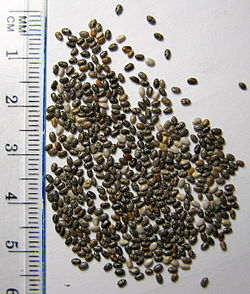
Chia is grown commercially for its seeds rich in ALA.
Table 1. ALA content as the percentage of the seed oil.
Table 2. ALA content as the percentage of the whole food.
Linseed (or flaxseed) (Linum usitatissimum) and its oil are perhaps the most widely available botanical source of the omega−3 fatty acid ALA. Flaxseed oil consists of approximately 55% ALA, which makes it six times richer than most fish oils in omega−3 fatty acids.
A portion of this is converted by the body to EPA and DHA, though the
actual converted percentage may differ between men and women.
In 2013 Rothamsted Research in the UK reported they had developed a genetically modified form of the plant Camelina
that produced EPA and DHA. Oil from the seeds of this plant contained
on average 11% EPA and 8% DHA in one development and 24% EPA in another.
Eggs
Eggs produced
by hens fed a diet of greens and insects contain higher levels of
omega−3 fatty acids than those produced by chickens fed corn or
soybeans. In addition to feeding chickens insects and greens, fish oils may be added to their diets to increase the omega−3 fatty acid concentrations in eggs.
The addition of flax and canola seeds to the diets of chickens,
both good sources of alpha-linolenic acid, increases the omega−3 content
of the eggs, predominantly DHA.
The addition of green algae or seaweed to the diets boosts the
content of DHA and EPA, which are the forms of omega−3 approved by the
FDA for medical claims. A common consumer complaint is "Omega−3 eggs can
sometimes have a fishy taste if the hens are fed marine oils".
Meat
Omega−3 fatty
acids are formed in the chloroplasts of green leaves and algae. While
seaweeds and algae are the sources of omega−3 fatty acids present in
fish, grass is the source of omega−3 fatty acids present in grass-fed
animals.
When cattle are taken off omega−3 fatty acid-rich grass and shipped to a
feedlot to be fattened on omega−3 fatty acid deficient grain, they
begin losing their store of this beneficial fat. Each day that an animal
spends in the feedlot, the amount of omega−3 fatty acids in its meat is
diminished.
The omega−6:omega−3 ratio of grass-fed beef is about 2:1, making it a more useful source of omega−3 than grain-fed beef, which usually has a ratio of 4:1.
In a 2009 joint study by the USDA and researchers at Clemson
University in South Carolina, grass-fed beef was compared with
grain-finished beef. The researchers found that grass-finished beef is
higher in moisture content, 42.5% lower total lipid content, 54% lower
in total fatty acids, 54% higher in beta-carotene, 288% higher in
vitamin E (alpha-tocopherol), higher in the B-vitamins thiamin and
riboflavin, higher in the minerals calcium, magnesium, and potassium,
193% higher in total omega−3s, 117% higher in CLA (cis-9, trans-11
octadecenoic acid, a conjugated linoleic acid, which is a potential
cancer fighter), 90% higher in vaccenic acid (which can be transformed
into CLA), lower in the saturated fats, and has a healthier ratio of
omega−6 to omega−3 fatty acids (1.65 vs 4.84). Protein and cholesterol
content were equal.
The omega−3 content of chicken meat may be enhanced by increasing the animals' dietary intake of grains high in omega−3, such as flax, chia, and canola.
Kangaroo meat is also a source of omega−3, with fillet and steak containing 74 mg per 100 g of raw meat.
Seal oil
Seal oil is a source of EPA, DPA, and DHA. According to Health Canada, it helps to support the development of the brain, eyes, and nerves in children up to 12 years of age. Like all seal products, it is not allowed to be imported into the European Union.
Other sources
A trend in the early 21st century was to fortify food with omega−3 fatty acids. The microalgae Crypthecodinium cohnii and Schizochytrium are rich sources of DHA, but not EPA, and can be produced commercially in bioreactors for use as food additives. Oil from brown algae (kelp) is a source of EPA. The alga Nannochloropsis also has high levels of EPA.
See also


