Search This Blog

Wednesday, April 1, 2026

Embryology

From Wikipedia, the free encyclopedia
1 - morula, 2 - blastula
1 - blastula, 2 - gastrula with blastopore; orange - ectoderm, red - endoderm

Embryology (from Greek ἔμβρυον, embryon, 'the unborn, embryo'; and -λογία, -logia) is the branch of zoology that studies the prenatal development of gametes sex cells, fertilization and development of embryos and fetuses. Embryology includes teratology, the study of congenital disorders that occur before birth.

Early embryology, put forward by Marcello Malpighi, was preformationist in concept: based on the idea that organisms develop from pre-existing miniature versions of themselves. The theory now accepted, epigenesis, is the idea that organisms develop from seed or egg in a sequence of steps. This concept was proposed in antiquity by Aristotle. Modern embryology developed from the work of Karl Ernst von Baer, though accurate observations had been made in Italy by anatomists such as Aldrovandi and Leonardo da Vinci in the Renaissance.

Comparative embryology

Preformationism and epigenesis

A tiny person (a homunculus) inside a sperm, as drawn by Nicolaas Hartsoeker in 1695

As recently as the 18th century, the prevailing notion in western human embryology was preformation: the idea that a sperm cell itself contains an embryo—a preformed, miniature infant, or homunculus—which simply becomes larger as it develops.

The competing explanation of embryonic development was epigenesis, originally proposed 2,000 years earlier by Aristotle. Much early embryology came from the work of the Italian anatomists Aldrovandi, Aranzio, Leonardo da Vinci, Marcello Malpighi, Gabriele Falloppio, Girolamo Cardano, Emilio Parisano, Fortunio Liceti, Stefano Lorenzini, Spallanzani, Enrico Sertoli, and Mauro Ruscóni. According to epigenesis, the form of an animal emerges gradually from a relatively formless egg. As microscopy improved during the 19th century, biologists could see that embryos took shape in a series of progressive steps, and epigenesis displaced preformation as the favored explanation among embryologists.

Cleavage

The cleavage phase of embryonic development is the series of several mitotic cell divisions that occurs immediately after the egg is fertilized by the sperm, producing the blastula (in mammals the blastocyst). The blastula is a single sheet of cells; in most phyla it then undergoes gastrulation. The resulting gastrula has in some species two, in most three, cell layers. The distinctive feature of cleavage, as a type of cell division, is that the cell divides without increase of cytoplasmic mass. The daughter cells share it, each having roughly half.

Overall, the cleavage phase of any species takes one of several forms. These forms are characteristic of different types of bilateral animal. (In the basal phyla cleavage is radial.)

Holoblastic

Holoblastic cleavage is cleavage of all the cells derived from the original zygote. The division furrow crosses the entire cell cluster; the whole cell cluster eventually becomes the embryo. (In meroblastic cleavage some cells will become the yolk sac.) Different types of animal differ in the geometry of the division furrow: cleavage is radial, spiral, bilateral or rotational.

Meroblastic

Meroblastic cleavage is the division of some but not all cells, as the division furrow does not protrude into the yolky region. The cells there impede formation of the associated membrane and only the other cells separate. Meroblastic cleavage is bilateral, discoidal or centrolecithal.

Basal phyla

Animals that belong to the basal phyla have holoblastic radial cleavage which results in radial symmetry (see: Symmetry in biology). During cleavage, there is a central axis that all divisions rotate about. The basal phyla also have only one to two embryonic cell layers, compared to the three in bilateral animals.

Bilaterians

In the bilateral animals cleavage can be either holoblastic or meroblastic. The subsequent gastrulation occurs in one of two ways, and this contrast divides the whole animal kingdom into two major groups (see: Embryological origins of the mouth and anus). In protostomes the first pore of the blastula (the blastopore) becomes the mouth of the animal; in deuterostomes the mouth derives from a later pore and the blastopore becomes the anus. The protostomes include most invertebrate animals, such as insects, worms and molluscs, while the deuterostomes include a few invertebrates such as the echinoderms (starfish and relatives) and all the vertebrates.

The bilaterian gastrula then develops three distinct layers of cells (the germ layers, endoderm, mesoderm and ectoderm); from them all the bodily organs and tissues subsequently arise.

Germ layers

  • The innermost layer, or endoderm, gives rise to the digestive organs, the gills, lungs or swim bladder if present, and kidneys or nephrites.
  • The middle layer, or mesoderm, gives rise to the muscles, skeleton if any, and blood system.
  • The outer layer of cells, or ectoderm, gives rise to the nervous system, including the brain, and skin or carapace, and hair, bristles or scales.

Drosophila melanogaster (fruit fly)

Drosophila have been used as a developmental model for many years. These studies have discovered many useful aspects of development that apply to other species. Outlined below is the process that leads to cell and tissue differentiation.

  1. Maternal-effect genes help to define the anterior-posterior axis using the bicoid and nanos genes.
  2. Gap genes establish three 'broad segments' of the embryo.
  3. Pair-rule genes define seven segments of the embryo within the second of those 'broad' segments.
  4. Segment-polarity genes divide each of those pre-existing seven segments into anterior and posterior halves (thus defining another seven segments), using a gradient of Hedgehog and Wnt signal proteins.
  5. Homeotic (Hox) genes use the 14 segments as pinpoints for specific types of cell differentiation and the histological developments that correspond to each cell type.

Humans

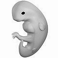
Humans are bilateral animals that have holoblastic rotational cleavage. Humans are also deuterostomes. In regard to humans, the term embryo refers to the ball of dividing cells from the moment the zygote implants itself in the uterus wall until the end of the eighth week after conception. Beyond the eighth week after conception (tenth week of pregnancy), the developing human is then called a fetus.

Evolutionary embryology

Evolutionary embryology is the expansion of comparative embryology by the ideas of Charles Darwin. Similarly to Karl Ernst von Baer's principles that explained why many species often appear similar to one another in early developmental stages, Darwin argued that the relationship between groups can be determined based upon common embryonic and larval structures.

Von Baer's principles

  1. The general features appear earlier in development than do the specialized features.
  2. More specialized characters develop from the more general ones.
  3. The embryo of a given species never resembles the adult form of a lower one.
  4. The embryo of a given species does resemble the embryonic form of a lower one.

Using Darwin's theory, evolutionary embryologists have since been able to distinguish between homologous and analogous structures appearing in different species. Homologous structures are those whose similarities derive from a common ancestor, such as the human arm and bat wings. Analogous structures are those that seem similar despite lacking common ancestral derivation.

Origins of modern embryology

Until the birth of modern embryology through observation of the mammalian ovum by Karl Ernst von Baer in 1827, there was no clear scientific understanding of embryology, although later discussions in this article show that some cultures had a fairly refined understanding of some of the principles. Only in the late 1950s when ultrasound was first used for uterine scanning, was the true developmental chronology of human fetus available. Karl Ernst von Baer along with Heinz Christian Pander, also proposed the germ layer theory of development which helped to explain how the embryo developed in progressive steps. Part of this explanation explored why embryos in many species often appear similar to one another in early developmental stages using his four principles.

Modern embryology research

Embryology is central to evolutionary developmental biology ("evo-devo"), which studies the genetic control of the development process (e.g. morphogens), its link to cell signalling, its roles in certain diseases and mutations, and its links to stem cell research. Embryology is the key to gestational surrogacy, which is when the sperm of the intended father and egg of intended mother are fused in a lab forming an embryo. This embryo is then put into the surrogate who carries the child to term.

Medical embryology

Medical embryology is used widely to detect abnormalities before birth. 2–5% of babies are born with an observable abnormality, and medical embryology explores the different ways and stages that these abnormalities appear. Genetically derived abnormalities are referred to as malformations. When there are multiple malformations, this is considered a syndrome. When abnormalities appear due to outside contributors, these are disruptions. The outside contributors causing disruptions are known as teratogens. Common teratogens are alcohol, retinoic acid, ionizing radiation or hyperthermic stress.

Vertebrate and invertebrate embryology

Many principles of embryology apply to invertebrates as well as to vertebrates. Therefore, the study of invertebrate embryology has advanced the study of vertebrate embryology. However, there are many differences as well. For example, numerous invertebrate species release a larva before development is complete; at the end of the larval period, an animal for the first time comes to resemble an adult similar to its parent or parents. Although invertebrate embryology is similar in some ways for different invertebrate animals, there are also countless variations. For instance, while spiders proceed directly from egg to adult form, many insects develop through at least one larval stage. For decades, a number of so-called normal staging tables were produced for the embryology of particular species, mainly focussing on external developmental characters. As variation in developmental progress makes comparison among species difficult, a character-based Standard Event System was developed, which documents these differences and allows for phylogenetic comparisons among species.

Origin of developmental biology

After the 1950s, with the DNA helical structure being unraveled and the increasing knowledge in the field of molecular biology, developmental biology emerged as a field of study which attempts to correlate the genes with morphological change, and so tries to determine which genes are responsible for each morphological change that takes place in an embryo, and how these genes are regulated.

As of today, human embryology is taught as a cornerstone subject in medical schools, as well as in biology and zoology programs at both an undergraduate and graduate level.

History

Ancient Egypt

Knowledge of the placenta goes back at least to ancient Egypt, where it was viewed as the seat of the soul. There was an Egyptian official with the title Opener of the Kings Placenta. An Egyptian text from the time of Akhenaten said that a human originates from the egg that grows in women.

Ancient Asia

Various interpretations of embryology have existed in Asia throughout history. Included in the ancient Indian tradition of Ayurveda is garbhasharir or the study of embryology, which refers to conceptions of embryology from antiquity. Descriptions of the amniotic sac appear in the Bhagavad Gita, Bhagavata Purana, and the Sushruta Samhita. One of the Upanishads known as the Garbhopanisaḍ states that the embryo is "like water in the first night, in seven nights it is like a bubble, at the end of half a month it becomes a ball. At the end of a month it is hardened, in two months the head is formed". In Indian literature, the start of consciousness in an embryo is not clearly defined. Some scriptures state that it is active at conception, while others suggest that consciousness begins in the seventh to ninth month of fetal development. Many South Asian traditions, including some Tibetan traditions, believe that the fetus has conscious experiences towards the end of its development.

The development of the human embryo is mentioned in the ancient Buddhist text of Garbhāvakrāntisūtra (1st–4th century CE). It mentions the human gestation period of 38 days. The text describes embryonic development in first three weeks as a liquid part of yogurt and the differentiation of body parts such as arms, leg, feet and head in the third month.

Ancient Greece

Pre-Socratic philosophers

Many pre-Socratic philosophers are recorded as having opinions on different aspects of embryology, although there is some bias in the description of their views in later authors such as Aristotle. According to Empedocles (whose views are described by Plutarch in the 1st century AD), who lived in the 5th century BC, the embryo derives and receives its blood from four vessels in all; two arteries and two veins. He also held sinews as originating from equal mixtures of earth and air. He further said men begin to form within the first month and are finished within fifty days. Asclepiades agreed that men are formed within fifty days, but he believed that women took a full two months to be fully knit. One observation, variously attributed to either Anaxagoras of Clazomenae or Alcmaeon of Croton, says that the milk produced by mammals is analogous to the white of fowl egg. Diogenes of Apollonia said that a mass of flesh forms first, only then followed by the development of bone and nerves. Diogenes recognized that the placenta was a nutritional source for the growing fetus. He also said that the development of males took four months, but that the development of females took five months. He did not think the embryo was alive. Alcmaeon also made some contributions, and is the first person reported to have practiced dissection. One idea, first stated by Parmenides, was that there was a connection between the right side of the body and the male embryo, and between the left side of the body and the female embryo. According to Democritus and Epicurus, the fetus is nourished at the mouth inside the mother and there are comparable teats that supply this nourishment within the mother's body to the fetus. Discussion on various views regarding how long it takes for specific parts of the embryo to form appear in an anonymous document known as the Nutriment.

Ancient Greeks discussed whether only the male had a seed which developed into the embryo within the female womb, or both the male and the female each had a seed that made a contribution to the developing embryo. The difficulty that one-seed theorists confronted was to explain the maternal resemblance of the progeny. One issue that two-seed theorists confronted was why the female seed was needed if the male already had a seed. One common solution to this problem was to assert that the female seed was either inferior or inactive. Another question was the origin of the seed. The encephalomyelogenic theory stated that the seed originated from the brain or and/or bone marrow. Later came pangenesis, which asserted the seed was drawn from the whole body in order to explain the general resemblance in the body of the offspring. Later on, hematogenous theory developed, which asserted that the seed was drawn from the blood. A third question was how or in what form the progeny existed in the seed prior to developing into an embryo and a fetus. According to preformationists, the body of the progeny already existed in a pre-existing but undeveloped form in the seed. Three variants of preformationism were homoiomerous preformationism, anhomoiomerous preformationism, and homuncular preformationism. According to the first, the homoiomerous parts of the body (e.g. humors, bone) already exist pre-formed in the seed. The second held that it was the anhomoiomerous parts that were pre-formed. Finally, the third view held that the whole was already a unified organic thing. Preformationism was not the only view. According to epigenesists, parts of the embryo successively form after conception takes place.

Hippocrates

Some of the most well-known early ideas on embryology come from Hippocrates and the Hippocratic Corpus, where discussion on the embryo is usually given in the context of discussing obstetrics (pregnancy and childbirth). Some of the most relevant Hippocratic texts on embryology include the Regimen on Acute Diseases, On Semen, and On the Development of the Child. Hippocrates claimed that the development of the embryo is put into motion by fire and that nourishment comes from food and breath introduced into the mother. An outer layer of the embryo solidifies, and the fire within consumes humidity which makes way for development of bone and nerve. The fire in the innermost part becomes the belly and air channels are developed in order to route nourishment to it. The enclosed fire also helps form veins and allows for circulation. In this description, Hippocrates aims at describing the causes of development rather than describing what develops. Hippocrates also develops views similar to preformationism, where he claims that all parts of the embryo simultaneously develop. Hippocrates also believed that maternal blood nourishes the embryo. This blood flows and coagulates to help form the flesh of the fetus. This idea was derived from the observation that menstrual blood ceases during pregnancy, which Hippocrates took to imply that it was being redirected to fetal development. Hippocrates also claimed that the flesh differentiates into different organs of the body, and Hippocrates saw as analogous an experiment where a mixture of substances placed into water will differentiate into different layers. Comparing the seed to the embryo, Hippocrates further compared the stalk to the umbilical cord.

Aristotle

Some embryological discussion appears in the writings of Aristotle's predecessor Plato, especially in his Timaeus. One of his views were that the bone marrow acted as the seedbed, and that the soul itself was the seed out of which the embryo developed, though he did not explain how this development proceeded. Scholars also continue to debate the views he held on various other aspects of embryology. However, a much more voluminous discussion on the subject comes from the writings of Aristotle, especially as appears in his On the Generation of Animals. Some ideas related to embryology also appear in his History of Animals, On the Parts of Animals, On Respiration, and On the Motion of Animals. Means by which we know Aristotle studied embryology, and most likely his predecessors as well, was through studying developing embryos taken out from animals as well as aborted and miscarried human embryos. Aristotle believed that the female supplied the matter for the development of the embryo formed from the menstrual blood, whereas the semen that comes from the male shapes that matter. Aristotle's belief that both the male and female made a contribution to the actual fetus goes against some prior beliefs. According to Aeschylus and some Egyptian traditions, the fetus solely develops from the male contribution and that the female womb simply nourishes this growing fetus. On the other hand, the Melanesians held that the fetus is solely a product of the female contribution. Aristotle did not believe there were any external influences on the development of the embryo. Against Hippocrates, Aristotle believed that new parts of the body developed over time rather than all forming immediately and developing from then on. He also considered whether each new part derives from a previously formed part or develops independently of any previously formed part. On the basis that different parts of the body do not resemble each other, he decided in favor of the latter view. He also described development of fetal parts in terms of mechanical and automatic processes. In terms of the development of the embryo, he says it begins in a liquid-like state as the material secreted by the female combines with the semen of the male, and then the surface begins to solidify as it interacts with processes of heating and cooling. The first part of the body to differentiate is the heart, which Aristotle and many of his contemporaries believed was the location of reason and thinking. Aristotle claimed that vessels join to the uterus in order to supply nourishment to the developing fetus. Some of the most solid parts of the fetus cool and, as they lose moisture to heat, turn into nails, horns, hoofs, beaks, etc. Internal heat dries away moisture and forms sinews and bones and the skin results from drying of the flesh. Aristotle also describes the development of birds in eggs at length. He further described embryonic development in dolphins, some sharks, and many other animals. Aristotle singularly wrote more on embryology than any other pre-modern author, and his influence on the subsequent discussion on the subject for many centuries was immense, introducing into the subject forms of classification, a comparative method from various animals, discussion of the development of sexual characteristics, compared the development of the embryo to mechanistic processes, and so forth.

Later Greek embryology

Reportedly, some Stoics claimed that most parts of the body formed at once during embryological development. Some Epicureans claimed that the fetus is nourished by either the amniotic fluid or the blood, and that both male and female supply material to the development of the fetus. According to the writings of Tertullian, Herophilus in the 5th century BC described the ovaries and fallopian tubes (but not past what was already described by Aristotle) and also dissected some embryos. One advance Herophilus made, against the conceptions of other individuals such as Aristotle, was that the brain was the center of intellect rather than the heart. Though not a part of Greek tradition, in Job 10, the formation of the embryo is likened to the curdling of milk into cheese, as described by Aristotle. Whereas Needham sees this statement in Job as part of the Aristotelian tradition, others see it as evidence that the milk analogy predates the Aristotelian Greek tradition and originates in Jewish circles. In addition, the Wisdom of Solomon (7:2) also has the embryo formed from menstrual blood. Soranus of Ephesus also wrote texts on embryology which went into use for a long time. Some rabbinic texts discuss the embryology of a female Greek writer named Cleopatra, a contemporary of Galen and Soranus, who was said to have claimed that the male fetus is complete in 41 days whereas the female fetus is complete in 81 days. Various other texts of less importance also appear and describe various aspects of embryology, though without making much progress from Aristotle. Plutarch has a chapter in one of his works titled "Whether was before, the hen or egg?" Discussion on embryological tradition also appears in many Neoplatonic traditions.

Next to Aristotle, the most impactful and important Greek writer on biology was Galen of Pergamum, and his works were transmitted throughout the Middle Ages. Galen discusses his understanding of embryology in two of his texts, those being his On the Natural Faculties and his On the Formation of the Foetus. There is an additional text spuriously attributed to Galen known as On the Question of whether the Embryo is an Animal. Galen described embryological development in four stages. In the first stage, the semen predominates. In the second stage, the embryo is filled with blood. In the third stage, the main outlines of the organs have developed but various other parts remain undeveloped. In the fourth stage, formation is complete and has reached a stage where we can call it a child. Galen described processes that played a role in furthering development of the embryo such as warming, drying, cooling, and combinations thereof. As this development plays out, the form of life of the embryo also moves from that like a plant to that of an animal (where the analogy between the root and umbilical cord is made). Galen claimed that the embryo forms from menstrual blood, by which his experimental analogy was that when you cut the vein of an animal and allow blood to flow out and into some mildly heated water, a sort of coagulation can be observed. He gave detailed descriptions of the position of the umbilical cord relative to other veins.

Patristics

The question of embryology is discussed among a number of early Christian writers, largely in terms of theological questions such as whether the fetus has value and/or when it begins to have value. (Although a number of Christian authors continued the classical discussions on the description of the development of the embryo, such as Jacob of Serugh. Passing reference to the embryo also appears in the eighth hymn of Ephrem the Syrian's Paradise Hymns.) Many patristic treatments of embryology continued in the stream of Greek tradition. The earlier Greek and Roman view that it was not was reversed and all pre-natal infanticide was condemned. Tertullian held that the soul was present from the moment of conception. The Quinisext Council concluded that "we pay no attention to the subtle division as to whether the foetus is formed or unformed". In this time, then, the Roman practice of child exposure came to an end, where unwanted yet birthed children, usually females, were discarded by the parents to die. Other more liberal traditions followed Augustine, who instead viewed that the animation of life began on the 40th day in males and the 80th day in females but not prior. Before the 40th day for men and 80th day for women, the embryo was referred to as the embryo informatus, and after this period was reached, it was referred to as the embryo formatus. The notion originating from the Greeks that the male embryo developed faster remained in various authors until it was experimentally disproven by Andreas Ottomar Goelicke in 1723.

Various patristic literature from backgrounds ranging from Nestorian, Miaphysite and Chalcedonian discuss and choose between three different conceptions on the relation between the soul and the embryo. According to one view, the soul pre-exists and enters the embryo at the moment of conception (prohyparxis). According to a second view, the soul enters into existence at the moment of conception (synhyparxis). In a third view, the soul enters into the body after it has been formed (methyparxis). The first option was proposed by Origen, but was increasingly rejected after the fourth century. On the other hand, the other two options were equally accepted after this point. The second position appears to have been proposed as a response to Origen's notion of a pre-existing soul. After the sixth century, the second position was also increasingly seen as Origenist and so rejected on those grounds. The writings of Origen were condemned during the Second Origenist Crises in 553. Those defending prohyparxis usually appealed to the Platonic notion of an eternally moving soul. Those defending the second position also appealed to Plato but rejected his notion on the eternality of the soul. Finally, those appealing to the third position appealed both to Aristotle and scripture. Aristotelian notions included the progression of the development of the soul, from an initial plant-like soul, to a sensitive soul found in animals and allows for movement and perception, and finally the formation of a rational soul which can only be found in the fully-formed human. Furthermore, some scriptural texts were seen as implying the formation of the soul temporally after the formation of the body (namely Genesis 2:7; Exodus 21:22–23; Zachariah 12:1). In the De hominis opificio of Gregory of Nyssa, Aristotle's triparitate notion of the soul was accepted. Gregory also held that the rational soul was present at conception. Theodoret argued based on Genesis 2:7 and Exodus 21:22 that the embryo is only ensouled after the body is fully formed. Based on Exodus 21:22 and Zachariah 12:1, Philoxenus of Mabbug claimed that the soul was created in the body forty days after conception. In his De opificio mundi, the Christian philosopher John Philoponus claimed that the soul is formed after the body. Later still, the author Leontius held that the body and soul were created simultaneously, though it is also possible he held that the soul pre-existed the body.[27]

Some Miaphysites and Chalcedonians seemed to have been compelled into accepting synhyparxis in the case of Jesus because of their view that the incarnation of Christ resulted in both one hypostasis and one nature, whereas some Nestorians claimed that Christ, like us, must have had his soul formed after the formation of his body because, per Hebrews 4:15, Christ was like us in all ways but sin. (On the other hand, Leontinus dismissed the relevance of Hebrews 4:15 on the basis that Christ differed from us not only in sinfulness but also conception without semen, making synhyparxis another of Christ's supernatural feats.) They felt comfortable holding this view, under their belief that the human nature of Jesus was separate from the divine hypostasis. Some Nestorians still wondered, however, if the body united with the soul in the moment the soul was created or whether it came with it only later. The Syriac author Babai argued for the former on the basis that the latter was hardly better than adoptionism. Maximus the Confessor ridiculed the Aristotelian notion of the development of the soul on the basis that it would make humans parents of both plants and animals. He held to synhyparxis and regarded the other two positions both as incorrect extremes. After the 7th century, Chalcedonian discussion on embryology is slight and the few works that touch on the topic support synhyparxis. But debate among other groups remains lively, still divided on similar sectarian grounds. The patriarch Timothy I argued that the Word first united with the body, and only later with the soul. He cited John 1:1, claiming on its basis that the Word became flesh first, not a human being first. Then, Jacob of Edessa rejected prohyparxis because Origen had defended it and methyparxis because he believed that it made the soul ontologically inferior and as only being made for the body. Then, Moses Bar Kepha claimed, for Christological reasons as a Miaphysite, that only synhyparxis was acceptable. He claimed that Genesis 2:7 has no temporal sequence and that Exodus 21:22 regards the formation of the body and not the soul and so is not relevant. To argue against methyparxis, he reasoned that body and soul are both present at death and, because what is at the end must correspond to what is also at the beginning, conception must also have body and soul together.

Embryology in Jewish tradition

Many Jewish authors also discussed notions of embryology, especially as they appear in the Talmud. Much of the embryological data in the Talmud is part of discussions related to the impurity of the mother after childbirth. The embryo was described as the peri habbetten (fruit of the body) and it developed through various stages: (1) golem (formless and rolled-up) (2) shefir meruqqam (embroidered foetus) (3) ubbar (something carried) (4) walad (child) (5) walad shel qayama (viable child) (6) ben she-kallu khadashaw (child whose months have been completed).

Some mystical notions regarding embryology appear in the Sefer Yetzirah. The text in the Book of Job relating to the fetus forming by analogy to the curdling of milk into cheese was cited in the Babylonian Talmud and in even greater detail in the Midrash: "When the womb of the woman is full of retained blood which then comes forth to the area of her menstruation, by the will of the Lord comes a drop of white-matter which falls into it: at once the embryo is created. [This can be] compared to milk being put in a vessel: if you add to it some lab-ferment [drug or herb], it coagulates and stands still; if not, the milk remains liquid." The Talmud sages held that there were two seeds that participated in the formation of the embryo, one from the male and one from the female, and that their relative proportions determine whether that develops into a male or a female.

In the Tractate Nidda, the mother was said to provide a "red-seed" which allows for the development of skin, flesh, hair, and the black part of the eye (pupil), whereas the father provides the "white-seed" which forms the bones, nerves, brain, and the white part of the eye. And finally, God himself was thought to provide the spirit and soul, facial expressions, capacity for hearing and vision, movement, comprehension, and intelligence. Not all strands of Jewish tradition accepted that both the male and female contributed parts to the formation of the fetus.

The 13th century medieval commentator Nachmanides, for example, rejected the female contribution. In Tractate Hullin in the Talmud, whether the organs of the child resemble more closely those of the mother or father is said to depend on which one contribute more matter to the embryo depending on the child. Rabbi Ishmael and other sages are said to have disagreed on one matter: they agreed that the male embryo developed on the 41st day, but disagreed on whether this was the case for the female embryo. Some believed that the female embryo was complete later, whereas others held that they were finished at the same time. The only ancient Jewish authors who associated abortion with homicide were Josephus and Philo of Alexandria in the 1st century. In the Talmud, a child is granted humanness at birth, while other rabbinical texts place it at the 13th postnatal day.

Some Talmudic texts discuss magical influences on the development of the embryo, such as one text which claims that if one sleeps on a bed that is pointed to the north–south will have a male child. According to Nachmanides, a child born of a cold drop of semen will be foolish, one born from a warm drop of semen will be passionate and irascible, and one born from a semen drop of medium temperature will be clever and level-headed. Some Talmudic discussions follow from Hippocratic claims that a child born on the eighth month could not survive, whereas others follow Aristotle in claiming that they sometimes could survive. One text even says that survival is possible on the seventh month, but not the eighth. Talmudic embryology, in various aspects, follows Greek discourses especially from Hippocrates and Aristotle, but in other areas, makes novel statements on the subject.

Judaism allows assisted reproduction, such as in vitro fertilization (IVF) embryo transfer and maternal surrogacy, when the spermatozoon and oocyte originate from the respective husband and wife.

Embryology in the Islamic tradition

A passing reference to embryological notions also appears in the Qur'an (22:5), where the development of the embryo proceeds in four stages from drop, to a clinging clot, to a partially developed stage, to a fully developed child. The notion of clay turning into flesh is seen by some as analogous to a text by Theodoret that describes the same process. The four stages of development in the Qur'an are similar to the four stages of embryological development as described by Galen. In the early 6th century, Sergius of Reshaina devoted himself to the translation of Greek medical texts into Syriac and became the most important figure in this process. Included in his translations were the relevant embryological texts of Galen. Anurshirvan founded a medical school in the southern Mesopotamian city of Gundeshapur, known as the Academy of Gondishapur, which also acted as a medium for the transmission, reception, and development of notions from Greek medicine. These factors helped the transmission of Greek notions on embryology, such as found in Galen, to enter into the Arabian milieu. Very similar embryonic descriptions also appear in the Syriac Jacob of Serugh's letter to the Archdeacon Mar Julian.

Embryological discussions also appear in the Islamic legal tradition.

Teleportation

From Wikipedia, the free encyclopedia

Teleportation is the hypothetical transfer of matter or energy from one point to another without traversing the physical space between them. It is a common subject in science fiction and fantasy literature. Teleportation is often paired with time travel, being that the traveling between the two points takes an unknown period of time, sometimes being immediate. An apport is a similar phenomenon featured in parapsychology and spiritualism.

There is no known physical mechanism that would allow for teleportation. Some scientific papers and media articles describe "quantum teleportation", a scheme for quantum information transfer, which does not allow for faster-than-light communication.

Etymology

The use of the term teleport to describe the hypothetical movement of material objects between one place and another without physically traversing the distance between them has been documented as early as 1878.

American writer Charles Fort is credited with having coined the word teleportation in 1931 to describe the strange disappearances and appearances of anomalies, which he suggested may be connected. As in the earlier usage, he joined the Greek prefix tele- (meaning "remote") to the root of the Latin verb portare (meaning "to carry"). Fort's first formal use of the word occurred in the second chapter of his 1931 book Lo!:

Mostly in this book I shall specialize upon indications that there exists a transportory force that I shall call Teleportation. I shall be accused of having assembled lies, yarns, hoaxes, and superstitions. To some degree I think so, myself. To some degree, I do not. I offer the data.

Cultural references

Fiction

A mockup of the transporter room from Star Trek: The Original Series
McCoy, Kirk and Spock in the Star Trek transporter room

Teleportation is a common subject in science fiction literature, film, video games, and television. The use of matter transmitters in science fiction originated at least as early as the 19th century. An early example of scientific teleportation (as opposed to magical or spiritual teleportation) is found in the 1897 novel To Venus in Five Seconds by Fred T. Jane. Jane's protagonist is transported from a strange-machinery-containing gazebo on Earth to planet Venus – hence the title.

The earliest recorded story of a "matter transmitter" was Edward Page Mitchell's "The Man Without a Body" in 1877.

Live performance

Teleportation illusions have featured in live performances throughout history, often under the fiction of miracles, psychic phenomenon, or magic. The cups and balls trick has been performed since 3 BC and can involve balls vanishing, reappearing, teleporting and transposing (objects in two locations interchanging places). A common trick of close-up magic is the apparent teleportation of a small object, such as a marked playing card, which can involve sleight-of-hand, misdirection, and pickpocketing. Magic shows were popular entertainments at fairs in the 18th century and moved into permanent theatres in the mid-19th century. Theatres provided greater control of the environment and viewing angles for more elaborate illusions, and teleportation tricks grew in scale and ambition. To increase audience excitement, the teleportation illusion could be conducted under the theme of a predicament escape. Magic shows achieved widespread success during the Golden Age of Magic in the late 19th and early 20th centuries.

Quantum teleportation

Quantum teleportation is distinct from regular teleportation, as it does not transfer matter from one place to another, but rather transmits the quantum information necessary to prepare a (microscopic) target system in the same quantum state as the source system. The scheme was named quantum "teleportation", because certain properties of the source system are recreated in the target system without any apparent quantum information carrier propagating between the two.

In 1993, Bennett et al proposed that a quantum state of a particle could be transferred to another distant particle, without moving the two particles at all. This is called quantum state teleportation. There are many following theoretical and experimental papers published.

In 2008, M. Hotta proposed that it may be possible to teleport energy by exploiting quantum energy fluctuations of an entangled vacuum state of a quantum field. In 2023, quantum energy teleportation was observed and recorded by Kazuki Ikeda for the first-time across microscopic distances using IBM superconducting computers that are used for quantum computing.

In 2014, researcher Ronald Hanson and colleagues from the Technical University Delft in the Netherlands, demonstrated the teleportation of information between two entangled quantumbits three metres apart.

A generalization of quantum mechanics suggests particles could be teleported from one place to another. This is called particle teleportation. With this concept, superconductivity can be viewed as the teleportation of some electrons in the superconductor and superfluidity as the teleportation of some of the atoms in the cellular tube. Further analysis shows that the teleportation time increases with the square root of mass and longer teleportation times require sustained quantum coherence. While particle teleportation may be feasible for an electron, a proton may not be feasible.

Evolution of the brain

From Wikipedia, the free encyclopedia
Evolution of the brain from ape to man

The evolution of the brain is the progressive development and complexity of neural structures over millions of years, resulting in the diverse range of brain sizes and functions observed across different species today, particularly in vertebrates.

The evolution of the brain has exhibited diverging adaptations within taxonomic classes, such as Mammalia, and even more diverse adaptations across other taxonomic classes. Brain-to-body size scales allometrically. This means that as body size changes, so do other physiological, anatomical, and biochemical connections between the brain and body. Small-bodied mammals tend to have relatively large brains compared to their bodies, while larger mammals (such as whales) have smaller brain-to-body ratios. When brain weight is plotted against body weight for primates, the regression line of the sample points can indicate the brain power of a species. For example, lemurs fall below this line, suggesting that for a primate of their size, a larger brain would be expected. In contrast, humans lie well above this line, indicating they are more encephalized than lemurs and, in fact, more encephalized than any other primate. This suggests that human brains have undergone a larger evolutionary increase in complexity relative to size. Some of these changes have been linked to multiple genetic factors, including proteins and other organelles.

Early history

Unsolved problem in biology
How and why did the brain evolve?

One approach to understanding overall brain evolution is to use a paleoarchaeological timeline to trace the necessity for ever-increasing complexity in structures that allow for chemical and electrical signaling. Because brains and other soft tissues do not fossilize as readily as mineralized tissues, scientists often look to other structures as evidence in the fossil record to get an understanding of brain evolution. This, however, leads to a dilemma as the emergence of organisms with more complex nervous systems with protective bone or other protective tissues that can then readily fossilize occur in the fossil record before evidence for chemical and electrical signaling. Evidence from 2008 showed that the ability to transmit electrical and chemical signals existed even before more complex multicellular lifeforms.

Fossilization of brain tissue, as well as other soft tissue, is nonetheless possible, and scientists can infer that the first brain structure appeared at least 521 million years ago, with fossil brain tissue present in sites of exceptional preservation.

Another approach to understanding brain evolution is to look at extant organisms that do not possess complex nervous systems, comparing anatomical features that allow for chemical or electrical messaging. For example, choanoflagellates are organisms that possess various membrane channels that are crucial to electrical signaling. The membrane channels of choanoflagellates' are homologous to the ones found in animal cells, and this is supported by the evolutionary connection between early choanoflagellates and the ancestors of animals. Another example of extant organisms with the capacity to transmit electrical signals would be the glass sponge, a multicellular organism, which is capable of propagating electrical impulses without the presence of a nervous system.

Before the evolutionary development of the brain, nerve nets, the simplest form of a nervous system developed. These nerve nets were a sort of precursor for the more evolutionarily advanced brains. They were first observed in Cnidaria and consist of a number of neurons spread apart that allow the organism to respond to physical contact. They are able to rudimentarily detect food and other chemicals, but these nerve nets do not allow them to detect the source of the stimulus.

Ctenophores also demonstrate this crude precursor to a brain or centralized nervous system, however they phylogenetically diverged before the phylum Porifera (the Sponges) and Cnidaria. There are two current theories on the emergence of nerve nets. One theory is that nerve nets may have developed independently in Ctenophores and Cnidarians. The other theory states that a common ancestor may have developed nerve nets, but they were lost in Porifera. While comparing the average neuron size and the packing density the difference between primate and mammal brains is shown.

A trend in brain evolution according to a study done with mice, chickens, monkeys and apes concluded that more evolved species tend to preserve the structures responsible for basic behaviors. A long term human study comparing the human brain to the primitive brain found that the modern human brain contains the primitive hindbrain region – what most neuroscientists call the protoreptilian brain. The purpose of this part of the brain is to sustain fundamental homeostatic functions, which are self regulating processes organisms use to help their bodies adapt. The pons and medulla are major structures found there. A new region of the brain developed in mammals about 250 million years after the appearance of the hindbrain. This region is known as the paleomammalian brain, the major parts of which are the hippocampi and amygdalas, often referred to as the limbic system. The limbic system deals with more complex functions including emotional, sexual and fighting behaviors. Of course, animals that are not vertebrates also have brains, and their brains have undergone separate evolutionary histories.

The brainstem and limbic system are largely based on nuclei, which are essentially balled-up clusters of tightly packed neurons and the axon fibers that connect them to each other, as well as to neurons in other locations. The other two major brain areas (the cerebrum and cerebellum) are based on a cortical architecture. At the outer periphery of the cortex, the neurons are arranged into layers (the number of which vary according to species and function) a few millimeters thick. There are axons that travel between the layers, but the majority of axon mass is below the neurons themselves. Since cortical neurons and most of their axon fiber tracts do not have to compete for space, cortical structures can scale more easily than nuclear ones. A key feature of cortex is that because it scales with surface area, more of it can be fit inside a skull by introducing convolutions, in much the same way that a dinner napkin can be stuffed into a glass by wadding it up. The degree of convolution is generally greater in species with more complex behavior, which benefits from the increased surface area.

The cerebellum, or "little brain," is behind the brainstem and below the occipital lobe of the cerebrum in humans. Its purposes include the coordination of fine sensorimotor tasks, and it may be involved in some cognitive functions, such as language and different motor skills that may involve hands and feet. The cerebellum helps keep equilibrium. Damage to the cerebellum would result in all physical roles in life to be affected. Human cerebellar cortex is finely convoluted, much more so than cerebral cortex. Its interior axon fiber tracts are called the arbor vitae, or Tree of Life.

The area of the brain with the greatest amount of recent evolutionary change is called the neocortex. In reptiles and fish, this area is called the pallium and is smaller and simpler relative to body mass than what is found in mammals. According to research, the cerebrum first developed about 200 million years ago. It is responsible for higher cognitive functions—for example, language, thinking, and related forms of information processing. It is also responsible for processing sensory input (together with the thalamus, a part of the limbic system that acts as an information router). The thalamus receives the different sensations before the information is then passed onto the cerebral cortex. Most of its function is subconscious, that is, not available for inspection or intervention by the conscious mind. The neocortex is an elaboration, or outgrowth, of structures in the limbic system, with which it is tightly integrated. The neocortex is the main part controlling many brain functions as it covers half of the whole brain in volume. The development of these recent evolutionary changes in the neocortex likely occurred as a result of new neural network formations and positive selections of certain genetic components.

Role of embryology

In addition to studying the fossil record, evolutionary history can be investigated via embryology. An embryo is an unborn/unhatched animal and evolutionary history can be studied by observing how processes in embryonic development are conserved (or not conserved) across species. Similarities between different species may indicate evolutionary connection. One way anthropologists study evolutionary connection between species is by observing orthologs. An ortholog is defined as two or more homologous genes between species that are evolutionarily related by linear descent. By using embryology the evolution of the brain can be tracked between various species.

Bone morphogenetic protein (BMP), a growth factor that plays a significant role in embryonic neural development, is highly conserved amongst vertebrates, as is sonic hedgehog (SHH), a morphogen that inhibits BMP to allow neural crest development. Tracking these growth factors with the use of embryology provides a deeper understanding of what areas of the brain diverged in their evolution. Varying levels of these growth factors lead to differing embryonic neural development which then in turn affects the complexity of future neural systems. Studying the brain's development at various embryonic stages across differing species provides additional insight into what evolutionary changes may have historically occurred. This then allows scientists to look into what factors may have caused such changes, such as links to neural network diversity, growth factor production, protein- coding selections, and other genetic factors.

Randomizing access and increasing size

Some animal phyla have gone through major brain enlargement through evolution (e.g. vertebrates and cephalopods both contain many lineages in which brains have grown through evolution) but most animal groups are composed only of species with extremely small brains. Some scientists argue that this difference is due to vertebrate and cephalopod neurons having evolved ways of communicating that overcome the scalability problem of neural networks while most animal groups have not. They argue that traditional neural networks fail to improve their function when scaled up because filtering based on previously known probabilities creates self-fulfilling prophecy-like biases. These biases generate false statistical evidence, producing a completely inaccurate worldview. In contrast, randomized access can overcome this problem, allowing brains to scale to more discriminating conditioned reflexes. This, in turn, can lead to new worldview-forming abilities once certain thresholds are reached. This means when neurons scale in a non randomized fashion that their functionality becomes more limited due to their neural networks being unable to process more complex systems without the exposure to new formations. This is explained by randomization allowing the entire brain to eventually get access to all information over the course of many shifts even though instant privileged access is physically impossible. They cite that vertebrate neurons transmit virus-like capsules containing RNA that are sometimes read in the neuron to which it is transmitted and sometimes passed further on unread which creates randomized access, and that cephalopod neurons make different proteins from the same gene which suggests another mechanism for randomization of concentrated information in neurons, both making it evolutionarily worth scaling up brains.

Brain re-organization

With the use of in vivo Magnetic resonance imaging (MRI) and tissue sampling, different cortical samples from members of each hominoid species were analyzed. In each species, specific areas were either relatively enlarged or shrunken, which can detail neural organizations. Different sizes in the cortical areas can show specific adaptations, functional specializations and evolutionary events that were changes in how the hominoid brain is organized. In early prediction it was thought that the frontal lobe, a large part of the brain that is generally devoted to behavior and social interaction, predicted the differences in behavior between hominoid and humans. Discrediting this theory was evidence supporting that damage to the frontal lobe in both humans and hominoids show atypical social and emotional behavior; thus, this similarity means that the frontal lobe was not very likely to be selected for reorganization. Instead, it is now believed that evolution occurred in other parts of the brain that are strictly associated with certain behaviors. The reorganization that took place is thought to have been more organizational than volumetric; whereas the brain volumes were relatively the same but specific landmark position of surface anatomical features, for example, the lunate sulcus suggest that the brains had been through a neurological reorganization. There is also evidence that the early hominin lineage also underwent a quiescent period, or a period of dormancy, which supports the idea of neural reorganization.

Dental fossil records for early humans and hominins show that immature hominins, including australopithecines and members of Homo, have a quiescent period (Bown et al. 1987). A quiescent period is a period in which there are no dental eruptions of adult teeth; at this time the child becomes more accustomed to social structure, and development of culture. During this time the child is given an extra advantage over other hominoids, devoting several years into developing speech and learning to cooperate within a community. This period is also discussed in relation to encephalization. It was discovered that chimpanzees do not have this neutral dental period, which suggests that a quiescent period occurred in very early hominin evolution. Using the models for neurological reorganization it can be suggested the cause for this period, dubbed middle childhood, is most likely for enhanced foraging abilities in varying seasonal environments.

Genetic factors in recent evolution

Genes involved in the neuro-development and in neuron physiology are extremely conserved between mammalian species (94% of genes expressed in common between humans and chimpanzees, 75% between humans and mice), compared to other organs. Therefore, few genes account for species differences in the human brain development and function.

Development of the human cerebral cortex

Main differences rely on the evolution of non-coding genomic regions, involved in the regulation of gene expression. This leads to differential expression of genes during the development of the human brain compared to other species, including chimpanzees. Some of these regions evolved fast in the human genome (human accelerated regions). The new genes expressed during human neurogenesis are notably associated with the NOTCH, WNT and mTOR pathways, but are also involved ZEB2, PDGFD and its receptor PDGFRβ. The human cerebral cortex is also characterized by a higher gradient of retinoic acid in the prefrontal cortex, leading to higher prefrontal cortex volume. All these differential gene expression lead to higher proliferation of the neural progenitors leading to more neurons in the human cerebral cortex. Some genes are lost in their expression during the development of the human cerebral cortex like GADD45G and FLRT2/FLRT3.

Another source of molecular novelty rely on new genes in the human or hominid genomes through segmental duplication. Around 30 new genes in the hominid genomes are dynamically expressed during human corticogenesis. Some were linked to higher proliferation of neural progenitors: NOTCH2NLA/B/C, ARHGAP11B, CROCCP2, TBC1D3, TMEM14B. Patients with deletions with NOTCH2NL genes display microcephaly, showing the necessity of such duplicated genes, acquired in the human genomes, in the proper corticogenesis.

MCPH1 and ASPM

Bruce Lahn, the senior author at the Howard Hughes Medical Center at the University of Chicago and colleagues have suggested that there are specific genes that control the size of the human brain. These genes continue to play a role in brain evolution, implying that the brain is continuing to evolve. The study began with the researchers assessing 214 genes that are involved in brain development. These genes were obtained from humans, macaques, rats and mice. Lahn and the other researchers noted points in the DNA sequences that caused protein alterations. These DNA changes were then scaled to the evolutionary time that it took for those changes to occur. The data showed the genes in the human brain evolved much faster than those of the other species. Once this genomic evidence was acquired, Lahn and his team decided to find the specific gene or genes that allowed for or even controlled this rapid evolution. Two genes were found to control the size of the human brain as it develops. These genes are Microcephalin (MCPH1) and Abnormal Spindle-like Microcephaly (ASPM). The researchers at the University of Chicago were able to determine that under the pressures of selection, both of these genes showed significant DNA sequence changes. Lahn's earlier studies displayed that Microcephalin experienced rapid evolution along the primate lineage which eventually led to the emergence of Homo sapiens. After the emergence of humans, Microcephalin seems to have shown a slower evolution rate. On the contrary, ASPM showed its most rapid evolution in the later years of human evolution once the divergence between chimpanzees and humans had already occurred.

Each of the gene sequences went through specific changes that led to the evolution of humans from ancestral relatives. In order to determine these alterations, Lahn and his colleagues used DNA sequences from multiple primates then compared and contrasted the sequences with those of humans. Following this step, the researchers statistically analyzed the key differences between the primate and human DNA to come to the conclusion, that the differences were due to natural selection. The changes in DNA sequences of these genes accumulated to bring about a competitive advantage and higher fitness that humans possess in relation to other primates. This comparative advantage is coupled with a larger brain size which ultimately allows the human mind to have a higher cognitive awareness.

ZEB2 protein

ZEB2

ZEB2 is a protein- coding gene in the Homo sapien species. A 2021 study found that a delayed change in the shape of early brain cells causes the distinctly large human forebrain compared to other apes and identify ZEB2 as a genetic regulator of it, whose manipulation lead to acquisition of nonhuman ape cortical architecture in brain organoids.

NOVA1

In 2021, researchers reported that brain organoids created with stem cells into which they reintroduced the archaic gene variant NOVA1 present in Neanderthals and Denisovans via CRISPR-Cas9 shows that it has a major impact on neurodevelopment and that such genetic mutations during the evolution of the human brain underlie traits that separate modern humans from extinct Homo species. They found that expression of the archaic NOVA1 in cortical organoids leads to "modified synaptic protein interactions, affects glutamatergic signaling, underlies differences in neuronal connectivity, and promotes higher heterogeneity of neurons regarding their electrophysiological profiles". This research suggests positive selection of the modern NOVA1 gene, which may have promoted the randomization of neural scaling. A subsequent study failed to replicate the differences in organoid morphology between the modern human and the archaic NOVA1 variant, consistent with suspected unwanted side effects of CRISPR editing in the original study.

SRGAP2C and neuronal maturation

Less is known about neuronal maturation. Synaptic gene and protein expression are protracted, in line with the protracted synaptic maturation of human cortical neurons so called neoteny. This probably relies on the evolution of non-coding genomic regions. The consequence of the neoteny could be an extension of the period of synaptic plasticity and therefore of learning. A human-specific duplicated gene, SRGAP2C accounts for this synaptic neoteny and acts by regulating molecular pathways linked to neurodevelopmental disorders. Other genes are deferentially expressed in human neurons during their development such as osteocrin or cerebelin-2 .

LRRC37B and neuronal electrical properties

Even less is known about molecular specificities linked to the physiology of the human neurons. Human neurons are more divergent in the genes they express compared to chimpanzees than chimpanzees to gorilla, which suggests an acceleration of non-coding genomic regions associated with genes involved in neuronal physiology, in particular linked to the synapses. A hominid-specific duplicated gene, LRRC37B, codes for a transmembrane receptor that is selectively localized at the axon initial segment of human cortical pyramidal neurons. It inhibits their voltage-gated sodium channels that generate the action potentials leading to a lower neuronal excitability. Human cortical pyramidal neurons display a lower excitability compared to other mammalian species (including macaques and marmosets) which could lead to different circuit functions in the human species. Therefore, LRRC37B whose expression has been acquired in the human lineage after the separation from the chimpanzees could be a key gene in the function of the human cerebral cortex. LRRC37B binds to secreted FGF13A and SCN1B and modulate indirectly the activity of SCN8A, all involved in neural disorders such as epilepsy and autism. Therefore, LRRC37B may contribute to human-specific sensitivities to such disorders, both involved defects in neuronal excitability.

Genome repair

The genomic DNA of postmitotic neurons ordinarily does not replicate. Protection strategies have evolved to ensure the distinctive longevity of the neuronal genome. Human neurons are reliant on DNA repair processes to maintain function during an individual's life-time. DNA repair tends to occur preferentially at evolutionarily conserved sites that are specifically involved with the regulation of expression of genes essential for neuronal identity and function.

Other factors

Many other genetics may also be involved in recent evolution of the brain.

  • For instance, scientists showed experimentally, with brain organoids grown from stem cells, how differences between humans and chimpanzees are also substantially caused by non-coding DNA (often discarded as relatively meaningless "junk DNA") – in particular via CRE-regulated expression of the ZNF558 gene for a transcription factor that regulates the SPATA18 gene.SPATA18 gene encodes a protein and is able to influence lysosome-like organelles that are found within mitochondria that eradicate oxidized mitochondrial proteins. This helps monitor the quality of the mitochondria as the disregulation of its quality control has been linked to cancer and degenerative diseases. This example may contribute to illustrations of the complexity and scope of relatively recent evolution to Homo sapiens.
  • A change in gene TKTL1 could be a key factor of recent brain evolution and difference of modern humans to (other) apes and Neanderthals, related to neocortex-neurogenesis. However, the "archaic" allele attributed to Neanderthals is present in 0.03% of Homo sapiens, but no resultant phenotypic differences have been reported in these people. Additionally, as Herai et al. contend, more is not always better. In fact, enhanced neuron production "can lead to an abnormally enlarged cortex and layer-specific imbalances in glia/neuron ratios and neuronal subpopulations during neurodevelopment." Even the original study's authors agree that "any attempt to discuss prefrontal cortex and cognitive advantage of modern humans over Neandertals based on TKTL1 alone is problematic".
  • Some of the prior study's authors reported a similar ARHGAP11B mutation in 2016.
  • Epigenetics also play a major role in the brain evolution in and to humans.

Recently evolved traits

Language

A genome-wide association study meta-analysis reported genetic factors of, the so far uniquely human, language-related capacities, in particular factors of differences in skill-levels of five tested traits. It e.g. identified association with neuroanatomy of a language-related brain area via neuroimaging correlation. The data contributes to identifying or understanding the biological basis of this recently evolved characteristic capability.

Human brain

One of the prominent ways of tracking the evolution of the human brain is through direct evidence in the form of fossils. The evolutionary history of the human brain shows primarily a gradually bigger brain relative to body size during the evolutionary path from early primates to hominids and finally to Homo sapiens. Because fossilized brain tissue is rare, a more reliable approach is to observe anatomical characteristics of the skull that offer insight into brain characteristics. One such method is to observe the endocranial cast (also referred to as endocasts). Endocasts occur when, during the fossilization process, the brain deteriorates away, leaving a space that is filled by surrounding sedimentary material over time. These casts, give an imprint of the lining of the brain cavity, which allows a visualization of what was there. This approach, however, is limited in regard to what information can be gathered. Information gleaned from endocasts is primarily limited to the size of the brain (cranial capacity or endocranial volume), prominent sulci and gyri, and size of dominant lobes or regions of the brain. While endocasts are extremely helpful in revealing superficial brain anatomy, they cannot reveal brain structure, particularly of deeper brain areas. By determining scaling metrics of cranial capacity as it relates to total number of neurons present in primates, it is also possible to estimate the number of neurons through fossil evidence.

Facial reconstruction of a Homo georgicus from over 1.5 Mya

Despite the limitations to endocasts, they can and do provide a basis for understanding human brain evolution, which shows primarily a gradually bigger brain. The evolutionary history of the human brain shows primarily a gradually bigger brain relative to body size during the evolutionary path from early primates to hominins and finally to Homo sapiens. This trend that has led to the present day human brain size indicates that there has been a 2-3 factor increase in size over the past 3 million years. This can be visualized with current data on hominin evolution, starting with Australopithecus, a group of hominins from which humans are likely descended. After all of the data, all observations concluded that the main development that occurred during evolution was the increase of brain size.

However, recent research has called into question the hypothesis of a threefold increase in brain size when comparing Homo sapiens with Australopithecus and chimpanzees. For example, in an article published in 2022 compiled a large data set of contemporary humans and found that the smallest human brains are less than twice that of large brained chimpanzees. As the authors write '...the upper limit of chimpanzee brain size is 500g/ml yet numerous modern humans have brain size below 900 g/ml.'[53] (Note that in this quote, the unit g/ml is to be understood not in the usual way as gram per millilitre but rather as gram or millilitre. This is consistent because brain density is close to 1 g/ml.) Consequently, the authors argue that the notion of an increase in brain size being related to advances in cognition needs to be re-thought in light of global variation in brain size, as the brains of many modern humans with normal cognitive capacities are only 400g/ml larger than chimpanzees. Additionally, much of the increase in brain size - which occurs to a much greater degree in specific modern populations - can be explained by increases in correlated body size related to diet and climatic factors.

Australopiths lived from 3.85 to 2.95 million years ago with the general cranial capacity somewhere near that of the extant chimpanzee—around 300–500 cm3. Considering that the volume of the modern human brain is around 1,352 cm3 on average this represents a substantial amount of brain mass evolved. Australopiths are estimated to have a total neuron count of ~30-35 billion.

Progressing along the human ancestral timeline, brain size continues to steadily increase (see Homininae) when moving into the era of Homo. For example, Homo habilis, living 2.4 million to 1.4 million years ago and argued to be the first Homo species based on a host of characteristics, had a cranial capacity of around 600 cm3Homo habilis is estimated to have had ~40 billion neurons.

A little closer to present day, Homo heidelbergensis lived from around 700,000 to 200,000 years ago and had a cranial capacity of around 1290 cm3 and having around 76 billion neurons.

Homo neaderthalensis, living 400,000 to 40,000 years ago, had a cranial capacity comparable to that of modern humans at around 1500–1600 cm3on average, with some specimens of Neanderthal having even greater cranial capacity. Neanderthals are estimated to have had around 85 billion neurons. The increase in brain size topped with Neanderthals, possibly due to their larger visual systems.

It is also important to note that the measure of brain mass or volume, seen as cranial capacity, or even relative brain size, which is brain mass that is expressed as a percentage of body mass, are not a measure of intelligence, use, or function of regions of the brain. Total neurons, however, also do not indicate a higher ranking in cognitive abilities. Elephants have a higher number of total neurons (257 billion) compared to humans (100 billion). Relative brain size, overall mass, and total number of neurons are only a few metrics that help scientists follow the evolutionary trend of increased brain to body ratio through the hominin phylogeny.

In 2021, scientists suggested that the brains of early Homo from Africa and Dmanisi, Georgia, Western Asia "retained a great ape-like structure of the frontal lobe" for far longer than previously thought – until about 1.5 million years ago. Their findings imply that Homo first dispersed out of Africa before human brains evolved to roughly their modern anatomical structure in terms of the location and organization of individual brain regions. It also suggests that this evolution occurred – not during – but only long after the Homo lineage evolved ~2.5 million years ago and after they – Homo erectus in particular – evolved to walk upright. What is the least controversial is that the brain expansion started about 2.6 Ma (about the same as the start of the Pleistocene), and ended around 0.2 Ma.

Evolution of the neocortex

In addition to just the size of the brain, scientists have observed changes in the folding of the brain, as well as in the thickness of the cortex. The more convoluted the surface of the brain is, the greater the surface area of the cortex which allows for an expansion of cortex. It is the most evolutionarily advanced part of the brain. Greater surface area of the brain is linked to higher intelligence as is the thicker cortex but there is an inverse relationship—the thicker the cortex, the more difficult it is for it to fold. In adult humans, thicker cerebral cortex has been linked to higher intelligence.

The neocortex is the most advanced and most evolutionarily young part of the human brain. It is six layers thick and is only present in mammals. It is especially prominent in humans and is the location of most higher level functioning and cognitive ability. The six-layered neocortex found in mammals is evolutionarily derived from a three-layer cortex present in all modern reptiles. This three-layer cortex is still conserved in some parts of the human brain such as the hippocampus and is believed to have evolved in mammals to the neocortex during the transition between the Triassic and Jurassic periods. After looking at history, the mammals had little neocortex compared to the primates as they had more cortex. The three layers of this reptilian cortex correlate strongly to the first, fifth and sixth layers of the mammalian neocortex. Across species of mammals, primates have greater neuronal density compared to rodents of similar brain mass and this may account for increased intelligence.

Theories of human brain evolution

Explanations of the rapid evolution and exceptional size of the human brain can be classified into five groups: instrumental, social, environmental, dietary, and anatomo-physiological. The instrumental hypotheses are based on the logic that evolutionary selection for larger brains is beneficial for species survival, dominance, and spread, because larger brains facilitate food-finding and mating success. The social hypotheses suggest that social behavior stimulates evolutionary expansion of brain size. Similarly, the environmental hypotheses suppose that encephalization is promoted by environmental factors such as stress, variability, and consistency. The dietary hypotheses maintain that food quality and certain nutritional components directly contributed to the brain growth in the Homo genus. The anatomo-physiologic concepts, such as cranio-cerebral vascular hypertension due to head-down posture of the anthropoid fetus during pregnancy, are primarily focused on anatomic-functional changes that predispose to brain enlargement.

No single theory can completely account for human brain evolution. Multiple selective pressures in combination seems to have been involved. Synthetic theories have been proposed, but have not clearly explained reasons for the uniqueness of the human brain. Puzzlingly, brain enlargement has been found to have occurred independently in different primate lineages, but only human lineage ended up with an exceptional brain capacity. Fetal head-down posture may be an explanation of this conundrum  because Homo sapiens is the only primate obligatory biped with upright posture.

Brain–computer interface

From Wikipedia, the free encyclopedia https://en.wikipedia.org/wiki/Brain%E2...