Search This Blog

Wednesday, February 26, 2020

Blunt trauma

From Wikipedia, the free encyclopedia

Blunt trauma
Other namesBlunt injury, non-penetrating trauma, blunt force trauma
Black eye 2.jpg

Blunt trauma is physical trauma to a body part, either by impact, injury or physical attack. The latter is often referred to as blunt force trauma, though it can also result from high-velocity impact. Blunt trauma is the initial trauma, from which develops more specific types such as contusions, abrasions, lacerations, and/or bone fractures. Blunt trauma is contrasted with penetrating trauma, in which an object such as a projectile or knife enters the body, though either can prove fatal.

Classification

Blunt abdominal trauma

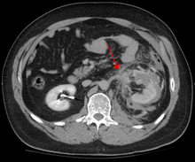
Abdominal CT showing left renal artery injury

Blunt abdominal trauma (BAT) represents 75% of all blunt trauma and is the most common example of this injury. The majority occurs in motor vehicle accidents, in which rapid deceleration may propel the driver into the steering wheel, dashboard, or seatbelt causing contusions in less serious cases, or rupture of internal organs from briefly increased intraluminal pressure in the more serious, depending on the force applied. Initially, there may be few indications that serious internal abdominal injury has occurred, making assessment more challenging and requiring a high degree of clinical suspicion.

There are two basic physical mechanisms at play with the potential of injury to intra-abdominal organs: compression and deceleration. The former occurs from a direct blow, such as a punch, or compression against a non-yielding object such as a seat belt or steering column.
This force may deform a hollow organ, increasing its intraluminal or internal pressure and possibly lead to rupture. Deceleration, on the other hand, causes stretching and shearing at the points where mobile contents in the abdomen, like bowel, are anchored. This can cause tearing of the mesentery of the bowel and injury to the blood vessels that travel within the mesentery. Classic examples of these mechanisms are a hepatic tear along the ligamentum teres and injuries to the renal arteries.

When blunt abdominal trauma is complicated by 'internal injury,' the liver and spleen (see blunt splenic trauma) are most frequently involved, followed by the small intestine.

In rare cases, this injury has been attributed to medical techniques such as the Heimlich Maneuver, attempts at CPR and manual thrusts to clear an airway. Although these are rare examples, it has been suggested that they are caused by applying excessive pressure when performing these life-saving techniques. Finally, the occurrence of splenic rupture with mild blunt abdominal trauma in those recovering from infectious mononucleosis or ‘mono’ is well reported.

Blunt abdominal trauma in sports

The supervised environment in which most sports injuries occur allows for mild deviations from the traditional trauma treatment algorithms, such as ATLS, due to the greater precision in identifying the mechanism of injury. The priority in assessing blunt trauma in sports injuries is separating contusions and musculo-tendinous injuries from injuries to solid organs and the gut and recognizing potential for developing blood loss, and reacting accordingly. Blunt injuries to the kidney from helmets, shoulder pads, and knees are described in American football, association football, martial arts, and all-terrain vehicle accidents.

A depiction of flail chest, a very serious blunt chest injury

Blunt thoracic trauma

The term blunt thoracic trauma or, put in a more familiar way, blunt chest injury, encompasses a variety of injuries to the chest. Broadly, this also includes damage caused by direct blunt force (such as a fist or a bat in an assault), acceleration or deceleration (such as that from a rear-end automotive accident), shear force (a combination of acceleration and deceleration), compression (such as a heavy object falling on a person), and blasts (such as an explosion of some sort). Common signs and symptoms include something as simple as bruising, but occasionally as complicated as hypoxia, ventilation-perfusion mismatch, hypovolemia, and reduced cardiac output due to the way the thoracic organs may have been affected. Blunt thoracic trauma is not always visible from the outside and such internal injuries may not show signs or symptoms at the time the trauma initially occurs or even until hours after. A high degree of clinical suspicion may sometimes be required to identify such injuries, a CT scan may prove useful in such instances. Those experiencing more obvious complications from a blunt chest injury will likely undergo a focused assessment with sonography for trauma (FAST) which can reliably detect a significant amount of blood around the heart or in the lung by using a special machine that visualizes sound waves sent through the body. Only 10-15% of thoracic traumas require surgery, but they can have serious impacts on the heart, lungs, and great vessels.

This table depicts mechanisms of blunt thoracic trauma and the most common injuries from each mechanism
 
The most immediate life-threatening injuries that may occur include tension pneumothorax, open pneumothorax, hemothorax, flail chest, cardiac tamponade, airway obstruction/rupture.

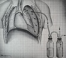
An example of a chest tube

The injuries may necessitate a procedure, with the most common being the insertion of an intercostal drain, more commonly referred to as a chest tube. This tube is typically placed because it helps restore a certain balance in pressures (usually due to misplaced air or surrounding blood) that are impeding the lungs ability to inflate and thus exchange vital gases that allow the body to function. A less common procedure that may be employed is a pericardiocentesis which by removing blood surrounding the heart, permits the heart to regain some ability to appropriately pump blood. In certain dire circumstances an emergent thoracotomy may be employed.

Blunt cranial trauma

The primary clinical concern when blunt trauma to the head occurs is damage to the brain, although other structures, including the skull, face, orbits, and neck are also at risk. Following assessment of the patient's airway, circulation, and breathing, a cervical collar may be placed if there is suspicion of trauma to the neck. Evaluation of blunt trauma to the head continues with the secondary survey in which evidence of cranial trauma, including bruises, contusions, lacerations, and abrasions are noted. In addition to noting external injury, a comprehensive neurologic exam is typically performed to assess for damage to the brain. Depending on the mechanism of injury and examination, a CT scan of the skull and brain may be ordered. This is typically done to assess for blood within the skull, or fracture of the skull bones.

A CT-scan showing an epidural hematoma, a variety of intracranial bleeding commonly associated with blunt trauma to the temple region

Traumatic brain injury

Traumatic brain injury (TBI) is a significant cause of morbidity and mortality and is most commonly caused by falls, motor vehicle accidents, sports- and work-related injuries, and assaults. It is the most common cause of death in patients under the age of 25. TBI is graded from mild to severe, with greater severity correlating with increased morbidity and mortality.

Most patients with more severe traumatic brain injury have of a combination of intracranial injuries, which can include diffuse axonal injury, cerebral contusions, as well as intracranial bleeding, including subarachnoid hemorrhage, subdural hematoma, epidural hematoma, and intraparenchymal hemorrhage. The recovery of brain function following a traumatic accident is highly variable and depends upon the specific intracranial injuries that occur, however there is significant correlation between the severity of the initial insult as well as the level of neurologic function during the initial assessment and the level of lasting neurologic deficits. Initial treatment may be targeted at reducing the intracranial pressure if there is concern for swelling or bleeding within this skull, which may require surgery such as a hemicraniectomy in which part of the skull is removed. 

A fracture, an injury to the skeletal component of the upper extremity.

Blunt trauma to extremities

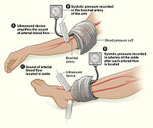
The Ankle-Brachial Index is depicted here. Note: ultrasound enhancement of pulses is not required but may be helpful.

Injury to extremities (like arms, legs, hands, feet) is extremely common. Falls are the most common etiology, making up as much as 30% of upper & 60% of lower extremity injuries. The most common mechanism for solely upper extremity injuries is machine operation or tool use. Work related accidents and vehicle crashes are also common causes. The injured extremity is examined for four major functional components which include soft tissues, nerves, vessels, and bones. Vessels are examined for expanding hematoma, bruit, distal pulse exam, and signs/symptoms of ischemia. Essentially asking the question, “Does blood seem to be getting through the injured area in a way that enough is getting to the parts past the injury?” When it is not obvious that the answer to this question is, “yes,” an injured extremity index or ankle-brachial index may be used to help guide whether further evaluation with computed tomography arteriography. This uses a special scanner and a substance that makes it easier to examine the vessels in finer detail than what the human hand can feel or the human eye can see.  Soft tissue damage can lead to rhabdomyolysis (a rapid breakdown of injured muscle that can overwhelm the kidneys) or may potentially develop compartment syndrome (when pressure builds up in muscle compartments damages the nerves and vessels in the same compartment). Bones are evaluated with plain film x-ray or computed tomography if deformity (misshapen), bruising, or joint laxity (looser or more flexible than usual) are observed. Neurologic evaluation involves testing of the major nerve functions of the axillary, radial, and median nerves in the upper extremity as well as the femoral, sciatic, deep peroneal, and tibial nerves in the lower extremity. Surgical treatment may be necessary depending on the extent of injury and involved structures, but many are managed nonoperatively.

Blunt pelvic trauma

The most common causes of blunt pelvic trauma are motor vehicle accidents and multiple-story falls, and thus pelvic injuries are commonly associated with additional traumatic injuries in other locations. In the pelvis specifically, the structures at risk include the pelvic bones, the proximal femur, major blood vessels such as the iliac arteries, the urinary tract, reproductive organs, and the rectum.

FractureRtSandIRami(Sin2).png
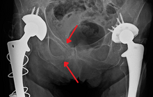
An X-ray showing a fracture of the inferior and superior pubic rami in a patient with previous hip replacements
 
One of the primary concerns is the risk of pelvic fracture, which itself is associated with a myriad of complications including bleeding, damage to the urethra and bladder, and nerve damage.  If pelvic trauma is suspected, emergency medical services personnel may place a pelvic binder on patients to stabilize the patient's pelvis and prevent further damage to these structures while patients are transported to a hospital. During the evaluation of trauma patients in an emergency department, the stability of the pelvis is typically assessed by the healthcare provider to determine whether fracture may have occurred. Providers may then decide to order imaging such as an X-ray or CT scan to detect fractures; however, if there is concern for life-threatening bleeding, patients should receive an X-ray of the pelvis. Following initial treatment of the patient, fractures may be need to be treated surgically if significant, while some minor fractures may heal without requiring surgery.

A life-threatening concern is hemorrhage, which may result from damage to the aorta, iliac arteries or veins in the pelvis. The majority of bleeding due to pelvic trauma is due to injury to the veins. Fluid (often blood) may be detected in the pelvis via ultrasound during the FAST scan that is often performed following traumatic accidents. Should a patient appear hemodynamically unstable in the absence of obvious blood on the FAST scan, there may be concern for bleeding into the retroperitoneal space, known as retroperitoneal hematoma. Stopping the bleeding may require endovascular intervention or surgery, depending on the location and severity. 

Diagnosis

In most settings, the initial evaluation and stabilization of traumatic injury follows the same general principles of identifying and treating immediately life-threatening injuries. In the US, the American College of Surgeons publishes the Advanced Trauma Life Support guidelines, which provide a step-by-step approach to the initial assessment, stabilization, diagnostic reasoning, and treatment of traumatic injuries that codifies this general principle. The assessment typically begins by ensuring that the subject's airway is open and competent, that breathing is unlabored, and that circulation—i.e. pulses that can be felt—is present. This is sometimes described as the "A, B, C's"—Airway, Breathing, and Circulation—and is the first step in any resuscitation or triage. Then, the history of the accident or injury is amplified with any medical, dietary (timing of last oral intake) and past history, from whatever sources such as family, friends, previous treating physicians that might be available. This method is sometimes given the mnemonic "SAMPLE". The amount of time spent on diagnosis should be minimized and expedited by a combination of clinical assessment and appropriate use of technology, such as diagnostic peritoneal lavage (DPL), or bedside ultrasound examination (FAST) before proceeding to laparotomy if required. If time and the patient's stability permits, CT examination may be carried out if available. Its advantages include superior definition of the injury, leading to grading of the injury and sometimes the confidence to avoid or postpone surgery. Its disadvantages include the time taken to acquire images, although this gets shorter with each generation of scanners, and the removal of the patient from the immediate view of the emergency or surgical staff. Many providers use the aid of a algorithm such as the ATLS guidelines to determine which images to obtain following the initial assessment. These algorithms take into account the mechanism of injury, physical examination, and patient's vital signs to determine whether patients should have imaging or proceed directly to surgery.
 
Recently, criteria have been defined that might allow patients with blunt abdominal trauma to be discharged safely without further evaluation. The characteristics of such patients would include:
  • absence of intoxication
  • no evidence of lowered blood pressure or raised pulse rate
  • no abdominal pain or tenderness
  • no blood in the urine.
To be considered low risk, patients would need to meet all low-risk criteria.

Treatment

When blunt trauma is significant enough to require evaluation by a healthcare provider, treatment is typically aimed at treating life-threatening injuries, which requires ensuring the patient is able to breathe and preventing ongoing blood loss. If there is evidence that the patient has lost blood, one or more intravenous lines may be placed and crystalloid solutions and/or blood will be administered at rates sufficient to maintain the circulation.

In the United States, surgical treatment of trauma typically follows the advanced trauma life support guidelines, which are developed by the American College of Surgeons. These guidelines use evidence-based algorithms to determine whether immediate surgery is warranted based on the patient's vital signs and whether or not there is evidence of ongoing internal or external bleeding. Further treatment depends on the severity of organ damage estimated by the exam and any diagnostic studies. Ultimately treatment will vary from close observation with the ability to intervene quickly, to surgery, which may be open or laparoscopic. In the case of blunt abdominal trauma, there is no shown benefit from surgery unless bleeding or peritonitis is present.
Bruise

From Wikipedia, the free encyclopedia

Bruise
Other namesContusion, ecchymosis
Pferdekuss.jpg
Bruise on upper leg caused by a blunt object
SpecialtyEmergency medicine

A bruise, also known as a contusion or ecchymosis, is a type of hematoma of tissue, the most common cause being capillaries damaged by trauma, causing localized bleeding that extravasate into the surrounding interstitial tissues. Most bruises are not very deep under the skin so that the bleeding causes a visible discoloration. The bruise then remains visible until the blood is either absorbed by tissues or cleared by immune system action. Bruises, which do not blanch under pressure, can involve capillaries at the level of skin, subcutaneous tissue, muscle, or bone. Bruises are not to be confused with other similar-looking lesions. These lesions include petechia (< 3 mm result from numerous and diverse etiologies such as adverse reactions from medications such as warfarin, straining, asphyxiation, platelet disorders and diseases such as cytomegalovirus), purpura (3 mm to 1 cm, classified as palpable purpura or non-palpable purpura and indicates various pathologic conditions such as thrombocytopenia), The term ecchymosis (defined as an area of >1 cm) is synonymous.

As a type of hematoma, a bruise is always caused by internal bleeding into the interstitial tissues which does not break through the skin, usually initiated by blunt trauma, which causes damage through physical compression and deceleration forces. Trauma sufficient to cause bruising can occur from a wide variety of situations including accidents, falls, and surgeries. Disease states such as insufficient or malfunctioning platelets, other coagulation deficiencies, or vascular disorders, such as venous blockage associated with severe allergies can lead to the formation of purpura which is not to be confused with trauma-related bruising/contusion. If the trauma is sufficient to break the skin and allow blood to escape the interstitial tissues, the injury is not a bruise but bleeding, a different variety of hemorrhage. Such injuries may be accompanied by bruising elsewhere.

Signs and symptoms

A woman's bruising after a severe fall.

Bruises often induce pain immediately after the trauma that results in their formation, but small bruises are not normally dangerous alone. Sometimes bruises can be serious, leading to other more life-threatening forms of hematoma, such as when associated with serious injuries, including fractures and more severe internal bleeding. The likelihood and severity of bruising depends on many factors, including type and healthiness of affected tissues. Minor bruises may be easily recognized in people with light skin color by characteristic blue or purple appearance (idiomatically described as "black and blue") in the days following the injury.

Hematomas can be subdivided by size. By definition, ecchymoses are 1 centimetres in size or larger, and are therefore larger than petechiae (less than 3 millimetres in diameter) or purpura (3 to 10 millimetres in diameter). Ecchymoses also have a more diffuse border than other purpura. A broader definition of ecchymosis is the escape of blood into the tissues from ruptured blood vessels. The term also applies to the subcutaneous discoloration resulting from seepage of blood within the contused tissue.

Cause

There are many causes of subcutaneous hematomas including ecchymoses. Coagulopathies such as Hemophilia A may cause ecchymosis formation in children. The medication betamethasone can have the adverse effect of causing ecchymosis.

The presence of bruises may be seen in patients with platelet or coagulation disorders, or those who are being treated with an anticoagulant. Unexplained bruising may be a warning sign of child abuse, domestic abuse, or serious medical problems such as leukemia or meningoccocal infection. Unexplained bruising can also indicate internal bleeding or certain types of cancer. Long-term glucocorticoid therapy can cause easy bruising. Bruising present around the navel (belly button) with severe abdominal pain suggests acute pancreatitis. Connective tissue disorders such as Ehlers-Danlos syndrome may cause relatively easy or spontaneous bruising depending on the severity. Spontaneous bruising or bruising with minimal trauma in the absence of other explanations and together with other minor or major criteria suggestive of Vascular Ehlers-Danlos Syndrome (vEDS) suggests genetic testing for the condition. 

Bruising can also occur during or after venipuncture.

During an autopsy, bruises accompanying abrasions indicate the abrasions occurred while the individual was alive, as opposed to damage incurred post mortem.

Size and shape

Bruise caused by a handrail, typical of extreme sports
 
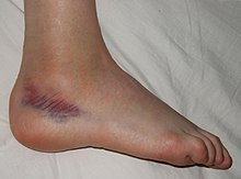
Bruise caused by a sprained ankle
 
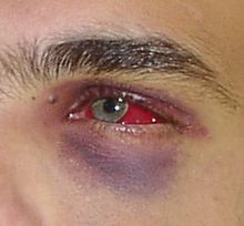
Black eye and subconjunctival hemorrhage after a punch to the face
 
Bruise shapes may correspond directly to the instrument of injury or be modified by additional factors. Bruises often become more prominent as time lapses, resulting in additional size and swelling, and may grow to a large size over the course of the hours after the injury that caused the bruise was inflicted.
  • Condition and type of tissue: In soft tissues, a larger area is bruised than would be in firmer tissue due to ease of blood to invade tissue.
  • Age: elderly skin and other tissues are often thinner and less elastic and thus more prone to bruising.
  • Gender: More bruising occurs in females due to increased subcutaneous fat.
  • Skin tone: Discoloration caused by bruises is more prominent in lighter complexions.
  • Diseases: Coagulation, platelet and blood vessel diseases or deficiencies can increase bruising due to more bleeding.
  • Location: More extensive vascularity causes more bleeding. Areas such as the arms, knees, shins and the facial area are especially common bruise sites.
  • Forces: Greater striking forces cause greater bruising.
  • Genes: Despite having completely normal coagulation factors, natural redheads have been shown to bruise more, although this may just be due to greater visibility on commonly associated lighter complexion.

Severity

Bruises can be scored on a scale from 0–5 to categorize the severity and danger of the injury.

Bruise harm score
Harm score Severity level Notes
0 Light bruise No damage
1 Mild bruise Little damage
2 Moderate bruise Some damage
3 Serious bruise Dangerous
4 Extremely serious bruise Very dangerous
5 Critical bruise Risk of death

The harm score is determined by the extent and severity of the injuries to the organs and tissues causing the bruising, in turn depending on multiple factors. For example, a contracted muscle will bruise more severely, as will tissues crushed against underlying bone. Capillaries vary in strength, stiffness and toughness, which can also vary by age and medical conditions.

Low levels of damaging forces produce small bruises and generally cause the individual to feel minor pain straight away. Repeated impacts worsen bruises, increasing the harm level. Normally, light bruises heal nearly completely within two weeks, although duration is affected by variation in severity and individual healing processes; generally, more severe or deeper bruises take somewhat longer. 

Severe bruising (harm score 2–3) may be dangerous or cause serious complications. Further bleeding and excess fluid may accumulate causing a hard, fluctuating lump or swelling hematoma. This has the potential to cause compartment syndrome in which the swelling cuts off blood flow to the tissues. The trauma that induced the bruise may also have caused other severe and potentially fatal harm to internal organs. For example, impacts to the head can cause traumatic brain injury: bleeding, bruising and massive swelling of the brain with the potential to cause concussion, coma and death. Treatment for brain bruising may involve emergency surgery to relieve the pressure on the brain.

Damage that causes bruising can also cause bones to be broken, tendons or muscles to be strained, ligaments to be sprained, or other tissue to be damaged. The symptoms and signs of these injuries may initially appear to be those of simple bruising. Abdominal bruising or severe injuries that cause difficulty in moving a limb or the feeling of liquid under the skin may indicate life-threatening injury and require the attention of a physician.

Mechanism

Severe bruising resulting from yard work injury
 
Increased distress to tissue causes capillaries to break under the skin, allowing blood to escape and build up. As time progresses, blood seeps into the surrounding tissues, causing the bruise to darken and spread. Nerve endings within the affected tissue detect the increased pressure, which, depending on severity and location, may be perceived as pain or pressure or be asymptomatic. The damaged capillary endothelium releases endothelin, a hormone that causes narrowing of the blood vessel to minimize bleeding. As the endothelium is destroyed, the underlying von Willebrand factor is exposed and initiates coagulation, which creates a temporary clot to plug the wound and eventually leads to restoration of normal tissue.

During this time, larger bruises may change color due to the breakdown of hemoglobin from within escaped red blood cells in the extracellular space. The striking colors of a bruise are caused by the phagocytosis and sequential degradation of hemoglobin to biliverdin to bilirubin to hemosiderin, with hemoglobin itself producing a red-blue color, biliverdin producing a green color, bilirubin producing a yellow color, and hemosiderin producing a golden-brown color. As these products are cleared from the area, the bruise disappears. Often the underlying tissue damage has been repaired long before this process is complete.

Treatment

Remarkable healing of a black eye over a 9-day period caused by a wisdom tooth extraction.
 
Treatment for light bruises is minimal and may include RICE (rest, immobilize, cold, elevate), painkillers (particularly NSAIDs) and, later in recovery, light stretching exercises. Particularly, immediate application of ice while elevating the area may reduce or completely prevent swelling by restricting blood flow to the area and preventing internal bleeding. Rest and preventing re-injury is essential for rapid recovery.

Very gently massaging the area and applying heat may encourage blood flow and relieve pain according to the gate control theory of pain, although causing additional pain may indicate the massage is exacerbating the injury. As for most injuries, these techniques should not be applied until at least three days following the initial damage to ensure all internal bleeding has stopped, because although increasing blood flow will allow more healing factors into the area and encourage drainage, if the injury is still bleeding this will allow more blood to seep out of the wound and cause the bruise to become worse. 

In most cases hematomas spontaneously revert, but in cases of large hematomas or those localized in certain organs (e.g., the brain), the physician may perform a puncture of the hematoma to allow blood to exit.

History

Folk medicine, including ancient medicine of Egyptians, Greeks, Celts, Turks, Slavs, Mayans, Aztecs and Chinese, has used bruising as a treatment for some health problems. The methods vary widely and include cupping, scraping, and slapping. Fire cupping uses suction which causes bruising in patients. Scraping (Gua Sha) uses a small hand device with a rounded edge to gently scrape the scalp or the skin. Another ancient device that creates mild bruising is a strigil, used by Greeks and Romans in the bath. Archaeologically there is no precedent for scraping tools before Greek archaeological evidence, not Chinese or Egyptian.

Etymology and pronunciation

The word ecchymosis (/ˌɛkɪˈmsɪs/; plural ecchymoses, /ˌɛkɪˈmss/), comes to English from New Latin, based on Greek ἐκχύμωσις ekchymōsis, from ἐκχυμοῦσθαι ekchymousthai "to extravasate blood", from ἐκ- ek- (elided to ἐ- e-) and χυμός chymos "juice". Compare enchyma, "tissue infused with organic juice"; elaboration from chyme, the formative juice of tissues.

Chemical polarity

From Wikipedia, the free encyclopedia

A water molecule, a commonly used example of polarity. Two charges are present with a negative charge in the middle (red shade), and a positive charge at the ends (blue shade).

In chemistry, polarity is a separation of electric charge leading to a molecule or its chemical groups having an electric dipole moment, with a negatively charged end and a positively charged end.

Polar molecules must contain polar bonds due to a difference in electronegativity between the bonded atoms. A polar molecule with two or more polar bonds must have a geometry which is asymmetric in at least one direction, so that the bond dipoles do not cancel each other.

Polar molecules interact through dipole–dipole intermolecular forces and hydrogen bonds. Polarity underlies a number of physical properties including surface tension, solubility, and melting and boiling points.

Polarity of bonds

In a molecule of hydrogen fluoride (HF), the more electronegative atom (fluorine) is shown in yellow. Because the electrons spend more time by the fluorine atom in the H−F bond, the red represents partially negatively charged regions, while blue represents partially positively charged regions.

Not all atoms attract electrons with the same force. The amount of "pull" an atom exerts on its electrons is called its electronegativity. Atoms with high electronegativities – such as fluorine, oxygen, and nitrogen – exert a greater pull on electrons than atoms with lower electronegativities such as alkali metals and alkaline earth metals. In a bond, this leads to unequal sharing of electrons between the atoms, as electrons will be drawn closer to the atom with the higher electronegativity.
Because electrons have a negative charge, the unequal sharing of electrons within a bond leads to the formation of an electric dipole: a separation of positive and negative electric charge. Because the amount of charge separated in such dipoles is usually smaller than a fundamental charge, they are called partial charges, denoted as δ+ (delta plus) and δ− (delta minus). These symbols were introduced by Sir Christopher Ingold and Dr. Edith Hilda (Usherwood) Ingold in 1926. The bond dipole moment is calculated by multiplying the amount of charge separated and the distance between the charges. 

These dipoles within molecules can interact with dipoles in other molecules, creating dipole-dipole intermolecular forces.

Classification

Bonds can fall between one of two extremes – being completely nonpolar or completely polar. A completely nonpolar bond occurs when the electronegativities are identical and therefore possess a difference of zero. A completely polar bond is more correctly called an ionic bond, and occurs when the difference between electronegativities is large enough that one atom actually takes an electron from the other. The terms "polar" and "nonpolar" are usually applied to covalent bonds, that is, bonds where the polarity is not complete. To determine the polarity of a covalent bond using numerical means, the difference between the electronegativity of the atoms is used.

Bond polarity is typically divided into three groups that are loosely based on the difference in electronegativity between the two bonded atoms. According to the Pauling scale:
  • Nonpolar bonds generally occur when the difference in electronegativity between the two atoms is less than 0.5
  • Polar bonds generally occur when the difference in electronegativity between the two atoms is roughly between 0.5 and 2.0
  • Ionic bonds generally occur when the difference in electronegativity between the two atoms is greater than 2.0
Pauling based this classification scheme on the partial ionic character of a bond, which is an approximate function of the difference in electronegativity between the two bonded atoms. He estimated that a difference of 1.7 corresponds to 50% ionic character, so that a greater difference corresponds to a bond which is predominantly ionic.

As a quantum-mechanical description, Pauling proposed that the wave function for a polar molecule AB is a linear combination of wave functions for covalent and ionic molecules: ψ = aψ(A:B) + bψ(A+B). The amount of covalent and ionic character depends on the values of the squared coefficients a2 and b2.

Polarity of molecules

While the molecules can be described as "polar covalent", "nonpolar covalent", or "ionic", this is often a relative term, with one molecule simply being more polar or more nonpolar than another. However, the following properties are typical of such molecules.

A molecule is composed of one or more chemical bonds between molecular orbitals of different atoms. A molecule may be polar either as a result of polar bonds due to differences in electronegativity as described above, or as a result of an asymmetric arrangement of nonpolar covalent bonds and non-bonding pairs of electrons known as a full molecular orbital.

Polar molecules

The water molecule is made up of oxygen and hydrogen, with respective electronegativities of 3.44 and 2.20. The electronegativity difference polarizes each H–O bond, shifting its electrons towards the oxygen (illustrated by red arrows). These effects add as vectors to make the overall molecule polar.

A polar molecule has a net dipole as a result of the opposing charges (i.e. having partial positive and partial negative charges) from polar bonds arranged asymmetrically. Water (H2O) is an example of a polar molecule since it has a slight positive charge on one side and a slight negative charge on the other. The dipoles do not cancel out, resulting in a net dipole. Due to the polar nature of the water molecule itself, other polar molecules are generally able to dissolve in water. In liquid water, molecules possess a distribution of dipole moments (range ≈ 1.9 - 3.1 D (Debye)) due to the variety of hydrogen-bonded environments. Other examples include sugars (like sucrose), which have many polar oxygen–hydrogen (−OH) groups and are overall highly polar.

If the bond dipole moments of the molecule do not cancel, the molecule is polar. For example, the water molecule (H2O) contains two polar O−H bonds in a bent (nonlinear) geometry. The bond dipole moments do not cancel, so that the molecule forms a molecular dipole with its negative pole at the oxygen and its positive pole midway between the two hydrogen atoms. In the figure each bond joins the central O atom with a negative charge (red) to an H atom with a positive charge (blue).

The hydrogen fluoride, HF, molecule is polar by virtue of polar covalent bonds – in the covalent bond electrons are displaced toward the more electronegative fluorine atom.

The ammonia molecule, NH3, is polar as a result of its molecular geometry. The red represents partially negatively charged regions.

Ammonia, NH3, molecule the three N−H bonds have only a slight polarity (toward the more electronegative nitrogen atom). The molecule has two lone electrons in an orbital, that points towards the fourth apex of the approximate tetrahedron, (VSEPR). This orbital is not participating in covalent bonding; it is electron-rich, which results in a powerful dipole across the whole ammonia molecule.

Resonance Lewis structures of the ozone molecule

In ozone (O3) molecules, the two O−O bonds are nonpolar (there is no electronegativity difference between atoms of the same element). However, the distribution of other electrons is uneven – since the central atom has to share electrons with two other atoms, but each of the outer atoms has to share electrons with only one other atom, the central atom is more deprived of electrons than the others (the central atom has a formal charge of +1, while the outer atoms each have a formal charge of −​12). Since the molecule has a bent geometry, the result is a dipole across the whole ozone molecule.

When comparing a polar and nonpolar molecule with similar molar masses, the polar molecule in general has a higher boiling point, because the dipole–dipole interaction between polar molecules results in stronger intermolecular attractions. One common form of polar interaction is the hydrogen bond, which is also known as the H-bond. For example, water forms H-bonds and has a molar mass M = 18 and a boiling point of +100 °C, compared to nonpolar methane with M = 16 and a boiling point of –161 °C.

Nonpolar molecules

A molecule may be nonpolar either when there is an equal sharing of electrons between the two atoms of a diatomic molecule or because of the symmetrical arrangement of polar bonds in a more complex molecule. For example, boron trifluoride (BF3) has a trigonal planar arrangement of three polar bonds at 120°. This results in no overall dipole in the molecule.

In a molecule of boron trifluoride, the trigonal planar arrangement of three polar bonds results in no overall dipole.
 
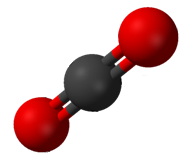
Carbon dioxide has two polar C-O bonds in a linear geometry.
 
Carbon dioxide (CO2) has two polar C=O bonds, but the geometry of CO2 is linear so that the two bond dipole moments cancel and there is no net molecular dipole moment; the molecule is nonpolar.

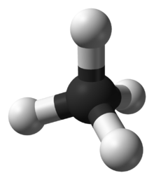
In methane, the bonds are arranged symmetrically (in a tetrahedral arrangement) so there is no overall dipole.

Examples of household nonpolar compounds include fats, oil, and petrol/gasoline. Most nonpolar molecules are water-insoluble (hydrophobic) at room temperature. Many nonpolar organic solvents, such as turpentine, are able to dissolve non-polar substances.

In the methane molecule (CH4) the four C−H bonds are arranged tetrahedrally around the carbon atom. Each bond has polarity (though not very strong). The bonds are arranged symmetrically so there is no overall dipole in the molecule. The diatomic oxygen molecule (O2) does not have polarity in the covalent bond because of equal electronegativity, hence there is no polarity in the molecule.

Amphiphilic molecules

Large molecules that have one end with polar groups attached and another end with nonpolar groups are described as amphiphiles or amphiphilic molecules. They are good surfactants and can aid in the formation of stable emulsions, or blends, of water and fats. Surfactants reduce the interfacial tension between oil and water by adsorbing at the liquid–liquid interface.

Predicting molecule polarity


Formula Description Example Name Dipole moment
Polar AB Linear molecules CO Carbon monoxide 0.112
HAx Molecules with a single H HF Hydrogen fluoride 1.86
AxOH Molecules with an OH at one end C2H5OH Ethanol 1.69
OxAy Molecules with an O at one end H2O Water 1.85
NxAy Molecules with an N at one end NH3 Ammonia 1.42
Nonpolar A2 Diatomic molecules of the same element O2 Dioxygen 0.0
CxAy Most hydrocarbon compounds C3H8 Propane 0.083
CxAy Hydrocarbon with center of inversion C4H10 Butane 0.0

Determining the point group is a useful way to predict polarity of a molecule. In general, a molecule will not possess dipole moment if the individual bond dipole moments of the molecule cancel each other out. This is because dipole moments are euclidean vector quantities with magnitude and direction, and a two equal vectors who oppose each other will cancel out.

Any molecule with a centre of inversion ("i") or a horizontal mirror plane ("σh") will not possess dipole moments. Likewise, a molecule with more than one Cn axis of rotation will not possess a dipole moment because dipole moments cannot lie in more than one dimension. As a consequence of that constraint, all molecules with dihedral symmetry (Dn) will not have a dipole moment because, by definition, D point groups have two or multiple Cn axes.

Since C1, Cs,C∞h Cn and Cnv point groups do not have a centre of inversion, horizontal mirror planes or multiple Cn axis, molecules in one of those point groups will have dipole moment.

Electrical deflection of water

Contrary to popular misconception, the electrical deflection of a stream of water from a charged object is not based on polarity. The deflection occurs because of electrically charged droplets in the stream, which the charged object induces. A stream of water can also be deflected in a uniform electrical field, which cannot exert force on polar molecules. Additionally, after a stream of water is grounded, it can no longer be deflected. Weak deflection is even possible for nonpolar liquids.

Lie group

From Wikipedia, the free encyclopedia https://en.wikipedia.org/wiki/Lie_group In mathematics , a Lie gro...